
Back to Journals » OncoTargets and Therapy » Volume 19
Cervical Lymphadenopathy as Initial Presentation of Foamy Gland Adenocarcinoma of the Prostate: A Case Report
Authors Wan Z  , Wang J, Wang C, Kong J, Zhao J, Liang W, Peng H, Li M
Received 19 October 2025
Accepted for publication 14 January 2026
Published 24 January 2026 Volume 2026:19 570801
DOI https://doi.org/10.2147/OTT.S570801
Checked for plagiarism Yes
Review by Single anonymous peer review
Peer reviewer comments 3
Editor who approved publication: Dr John Maher
Zhicheng Wan,1,* Junren Wang,2,* Cuicui Wang,1 Jing Kong,3 Jin Zhao,1 Weihua Liang,1 Hao Peng,1,* Man Li1,*
1Department of Pathology and Key Laboratory for Xinjiang Endemic and Ethnic Diseases, Shihezi University School of Medicine, Shihezi, Xinjiang, People’s Republic of China; 2Department of Pathology, The 948th Hospital of the People’s Liberation Army, Wusu, Xinjiang, People’s Republic of China; 3Department of Oncology, Pingyi County Hospital of Traditional Chinese Medicine, Pingyi, Shandong, People’s Republic of China
*These authors contributed equally to this work
Correspondence: Hao Peng, Department of Pathology and Key Laboratory for Xinjiang Endemic and Ethnic Diseases, Shihezi University School of Medicine, Shihezi, Xinjiang, People’s Republic of China, Email [email protected] Man Li, Department of Pathology and Key Laboratory for Xinjiang Endemic and Ethnic Diseases, Shihezi University School of Medicine, Shihezi, Xinjiang, People’s Republic of China, Email [email protected]
Abstract: Prostate cancer (PCa) is a common urogenital malignancy in elderly males. The typical clinical presentation of advanced PCa includes symptoms of the lower urinary tract. The most common sites of metastasis are the bone and regional lymph nodes. However, PCa with cervical lymphadenopathy as the initial symptom is extremely rare, and one of its variants, foamy gland adenocarcinoma, is even rarer. We present a case of a patient with foamy gland adenocarcinoma of the prostate having enlarged lymph nodes in the neck as the initial symptom. An 81-year-old male was admitted to the hospital with a chief complaint of an enlarged left cervical lymph node without any urological history. Cervical ultrasound showed multiple lymphadenopathies in the left supraclavicular region. The patient underwent a lymph node biopsy, and the pathology results revealed metastatic foamy gland adenocarcinoma of the prostate. Distant lymph node metastasis in PCa is a rare form of metastasis. The initial presentation and tests led to the diagnosis of foamy gland adenocarcinoma of the prostate with cervical lymph node metastasis. Following the diagnosis, the patient received conservative management with androgen deprivation therapy (bicalutamide and goserelin) and zoledronic acid. Unfortunately, he passed away 9 months later due to a COVID-19 infection. We present this rare variant of PCa metastasis to attract the attention of clinicians and pathologists.
Keywords: prostate cancer, foamy gland adenocarcinoma, metastases, cervical lymphadenopathy, primary symptom
Introduction
Prostate cancer (PCa) is a leading cause of cancer-related morbidity and mortality in men worldwide.1 While early-stage disease is often asymptomatic, progression leads to lower urinary tract symptoms.2 Advanced PCa commonly metastasizes to bone (65–75% of cases)3 and regional pelvic lymph nodes.4 However, PCa rarely metastasizes to distant lymph nodes, such as the axilla and cervical regions. Once primary PCa progresses to metastatic PCa, the outcome is poor. Most PCa are conventional acinar adenocarcinomas; however, a rare variant known as foamy gland adenocarcinoma was recognized in the 2016 WHO classification.5 A review of reported foamy gland adenocarcinoma cases reveals that initial presentation is typically urinary (Table 1).6–8 In contrast, we report a unique case of foamy gland adenocarcinoma presenting with left cervical lymph node metastasis as the initial symptom to highlight that this rare variant can present with distant lymphadenopathy, emphasizing the need for clinicians and pathologists to consider metastatic prostate cancer in the differential diagnosis of cervical masses, even in the absence of urinary symptoms.
 |
Table 1 Case Reports of Foamy Gland Adenocarcinoma of the Prostate |
Case Presentation
In early April 2021, an 81-year-old male presented to the outpatient clinic with a chief complaint of an enlarged left cervical lymph node. A cervical ultrasound performed on April 1 showed a normal thyroid gland and multiple left supraclavicular lymph node enlargements. On April 11, he was admitted to the surgical ward of The 948th Hospital of the People’s Liberation Army in Wusu City, Xinjiang Uygur Autonomous Region, China (specific time points are illustrated in Figure 1). He had a history of hypertension, coronary artery disease, and pulmonary tuberculosis. Physical examination revealed multiple firm, non-tender, and movable lymph nodes in the left supraclavicular fossa (level Vb), measuring approximately 1.5×1.0 × 1.0 cm.
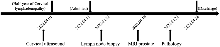 |
Figure 1 Specific time points corresponding to the diagnostic process of this case. |
Based on these findings, an excisional biopsy of the nine left supraclavicular lymph nodes was performed on April 12; the largest measured 3.0×3.0 × 2.0 cm. Concurrently, laboratory tests revealed a markedly elevated total PSA (33.26 ng/mL), while other tumor markers were within normal limits. The preliminary pathological report from the primary hospital, issued on April 14, indicated “suspected metastatic carcinoma.” To identify the primary tumor, further imaging was obtained. A prostate MRI (April 18) revealed a PI-RADS 5 lesion in the transition zone, with suspected local invasion and pelvic lymph node metastasis, Vertebral MRI showed bone metastases (Figure 2). Chest CT with intravenous contrast indicated chronic bronchitis. A whole-body PET/CT scan was not performed for comprehensive staging, as this modality was unavailable at the treating hospital.
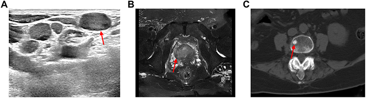 |
Figure 2 Diagnostic images of the case. (A) Cervical ultrasonography showing enlarged left supraclavicular lymph nodes (indicated by arrows) that were secondarily involved by metastatic carcinoma. (B) Prostate magnetic resonance imaging (MRI) shows a PI-RADS 5 lesion (indicated by arrows) in the transition zone. (C) Vertebral MRI demonstrating abnormal signals consistent with bone metastases in the spine (indicated by arrows). |
For a definitive diagnosis, the biopsy specimen was referred to our pathology department for consultation on April 22. Histology showed a poorly differentiated carcinoma replacing nodal architecture, with cells featuring abundant clear cytoplasm (Figure 3A), which was negative for periodic acid–Schiff (PAS) (Figure 3B). Immunohistochemistry (IHC) was positive for AE1/3, CAM5.2, PSA, and P504S (Figure 3C–F), and negative for CK7 (Figure 3H) and other markers (CK34βE12, CD56, Syn, S-100, Vimentin, P63, SMA). Ki-67 was 60%+ (Figure 3G). The IHC profile was highly suggestive of a prostatic origin. Notably, this histological assessment was initially made without the clinical context of the elevated PSA or imaging findings, as the consultation request provided only basic information (age, sex, and biopsy site). This suspicion prompted an active review of the patient’s full clinical history and correlation with the subsequently available PSA and MRI results, culminating in the definitive diagnosis of metastatic prostate foamy gland adenocarcinoma.
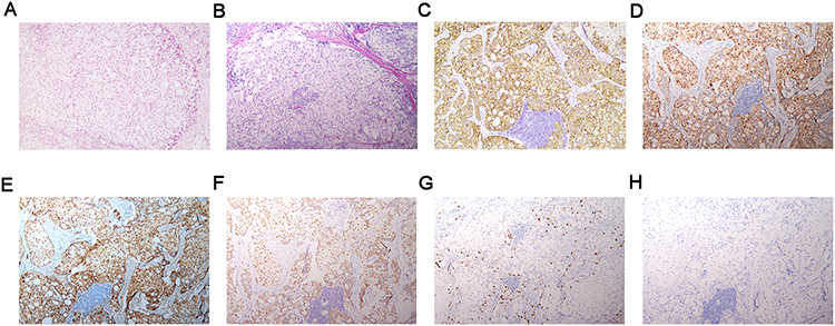 |
Figure 3 Histopathological results of the lesion. (A) Hematoxylin and eosin staining showed metastatic, poorly differentiated adenocarcinoma in lymph nodes. (B) Foam cells were negative for PAS. IHC staining was positive for (C) AE1/3, (D) CAM5.2, (E) PSA, and (F) P504S. IHC staining (G) negative for CK7; (H) positive for Ki-67. (A–H) (200× magnification). |
The final stage was T4N1M1b (clinical stage IVA). Given the patient’s advanced age, multiple comorbidities, and advanced disease, he was not considered a surgical candidate. He opted for conservative management with androgen deprivation therapy, consisting of subcutaneous goserelin (3.6 mg implant every 28 days) and oral bicalutamide (50 mg once daily), along with zoledronic acid (4 mg via intravenous infusion every 28 days) for bone protection. Following treatment initiation, the patient’s condition remained clinically stable for the subsequent months. He reported manageable symptoms and maintained a reasonable performance status during follow-up. Unfortunately, in January 2023, he contracted COVID-19 and died from complications of the infection, with an overall survival of 9 months from diagnosis.
Discussion
Foamy gland adenocarcinoma is a rare subtype of prostate cancer (PCa), predominantly affecting elderly men and typically presenting with urinary symptoms. In contrast, our case was remarkable for its initial presentation as distant lymph node metastasis, highlighting a unique diagnostic challenge for clinicians and pathologists.
The diagnosis in this case integrated histomorphology, IHC, and clinical correlation. Evaluation of the left cervical lymph node biopsy revealed replacement by epithelioid cells expressing prostatic markers (eg, PSA, P504S), while serology showed elevated PSA and imaging identified a primary prostate lesion.
The first diagnostic challenge was the rarity of the initial manifestation. Prostate cancer typically presents with local urinary symptoms and commonly metastasizes to regional pelvic lymph nodes.2 In contrast, distant metastasis to cervical or axillary nodes is rare. To our knowledge, there have been 17 reports describing 50 cases of PCa with peripheral lymph node metastasis as the initial clinical presentation involving cervical, supraclavicular, and axillary lymph nodes. Among these cases, 41 cases occurred in the left cervical or supraclavicular regions,9–19 4 occurred in the right cervical or supraclavicular regions,10,20–22 3 occurred in the bilateral cervical regions,17,18,23 1 occurred in the left axilla region,24 and 1 case did not specify the location of the cervical region.25 It is important to note that adenocarcinomas originating from internal abdominal organs, including those of the genitourinary tract, can metastasize to a broader range of cervical lymph nodes beyond the supraclavicular region, potentially involving levels II through IV along the internal jugular chain.26,27 In our case, the enlargement of lymph nodes was noted in the left supraclavicular region. After a thorough search, we found that cervical lymph node metastasis occurs in only about 0.4% of patients with PCa.28,29 This low incidence rate for initial manifestation makes our diagnosis difficult.
The second challenge was the microscopically atypical cell morphology. The most common pathological type is acinar adenocarcinoma in PCa. The 2022 WHO classification of acinar adenocarcinoma in the prostate includes four variants, namely atrophic pattern adenocarcinoma, pseudohyperplastic adenocarcinoma, microcystic adenocarcinoma, and foamy gland adenocarcinoma.30 The foamy gland adenocarcinoma is a rare type of prostatic acinar adenocarcinoma and shows fused glandular structures with a focal cribriform pattern with cells having abundant foamy cytoplasm.31 Due to the atypical and rare histological morphology of PCa, the pathological diagnosis is difficult.
Lastly, this is a consultation case. The absence of clinical information contributed to the challenge in diagnosis. When the cervical lymph nodes are enlarged, especially when a large number of tumor cells appear in the lymph nodes, it is easy to diagnose it as metastatic carcinoma. However, most cancers that metastasize to cervical lymph nodes originate from primary cancers of the head and neck.32 The most common primary sites include the oral cavity, salivary glands with non-mucosal neoplasms, and thyroid glands. The rarity of such metastases from distant sites is exemplified by a documented case in which cervical lymph node metastasis was ultimately attributed to a metachronous salivary gland carcinoma, not to the patient’s concurrent prostate cancer.33 Therefore, metastatic cancers of the cervical chain originating from non-head and neck origin are rare. The absence of clinical imaging, serological data, and other information makes it easy for pathologists to initially assume this is a case of head and neck carcinoma. In this case, the initial pathological evaluation was performed without knowledge of the elevated PSA or the prostate MRI results, exemplifying this exact challenge.
IHC plays a principal role in differential diagnosis. In this case, a broad panel of markers, including 5 positive and 8 negative markers were used to confirm the diagnosis of metastatic PCa (PSA and P504S positive, CK7 negative) and to rule out other diseases, such as metastatic myoepithelial carcinoma (P63 and SMA negative), neuroendocrine carcinoma (CD56 and Syn negative), and mucoepidermoid carcinoma (special staining of PAS showed negative). The diagnosis was greatly aided by the CK7-negative profile, which, in conjunction with the morphology, strongly suggested a prostatic origin. The diagnostic value of CK7 in this context stems from its low expression in prostate cancers among other epithelial tumors.34–36 This underscores the indispensability of a comprehensive IHC panel in resolving such diagnostic dilemmas.
The management of foamy gland adenocarcinoma follows standard prostate cancer protocols (eg, radical prostatectomy).37,38 Its prognosis is comparable to conventional acinar adenocarcinoma and depends on typical prognostic factors such as PSA level, stage, and Gleason score.39 For patients with isolated or symptomatic cervical metastases, local control options including surgical excision (cervical lymph node dissection) followed by radiotherapy and/or chemotherapy may be considered, in addition to systemic therapy.40
Conclusion
In conclusion, we present a rare case of foamy gland adenocarcinoma of the prostate that manifested initially with left supraclavicular lymphadenopathy. This case highlights significant diagnostic pitfalls, including its atypical presentation mimicking a head and neck primary, its uncommon histological variant, and the challenges posed by incomplete clinical information during initial pathological consultation. Therefore, prostate carcinoma should be included in the differential diagnosis of metastatic carcinoma in cervical lymph nodes, even in the absence of urological symptoms. A comprehensive immunohistochemical panel, including prostate-specific markers, is essential for accurate diagnosis in such challenging scenarios.
Data Sharing Statement
The original contributions presented in the study are included in the article. Further inquiries can be directed to the corresponding author.
Ethics Statement
The publication of this case report was approved by the Ethics Committee of The First Affiliated Hospital of Shihezi University.
Consent to Publish Declaration
Written informed consent for publication was obtained from the patient’s next of kin. All information and images have been anonymized to protect the patient’s privacy.
Patient Informed Consent
Consent to publish was obtained from the patients family due to the patient passed away in January 2023. To help more pathologists recognize this rare morphology and uncommon pattern of metastasis in prostate cancer, which may enable earlier and more accurate diagnoses for other patients, the patient’s family has given consent for publication.
Acknowledgments
We thank the patient for providing permission to share his information.
Author Contributions
All authors made a significant contribution to the work reported, whether that is in the conception, study design, execution, acquisition of data, analysis and interpretation, or in all these areas; took part in drafting, revising or critically reviewing the article; gave final approval of the version to be published; have agreed on the journal to which the article has been submitted; and agree to be accountable for all aspects of the work.
Funding
The work was supported by the Science and Technology Program Project of Xinjiang Production and Construction Corps (2024ZD017).
Disclosure
The authors declare no conflicts of interest in this work.
References
1. Bray F, Laversanne M, Sung H, et al. Global cancer statistics 2022: GLOBOCAN estimates of incidence and mortality worldwide for 36 cancers in 185 countries. CA Cancer J Clin. 2024;74(3):229–6. doi:10.3322/caac.21834
2. Groeben C, Wirth MP. Prostate cancer: basics on clinical appearance, diagnostics and treatment. Med Monatsschr Pharm. 2017;40(5):192–201.
3. Dong B, Gu Y, Sun X, et al. Targeting TUBB3 suppresses anoikis resistance and bone metastasis in prostate cancer. Adv Healthc Mater. 2024;13(28):e2400673. doi:10.1002/adhm.202400673
4. Plata Bello A, Apatov SE, Benfante NE, et al. Prevalence of high-risk prostate cancer metastasis to Cloquet’s ilioinguinal lymph node. J Urol. 2022;207(6):1222–1226. doi:10.1097/JU.0000000000002439
5. Humphrey PA, Moch H, Cubilla AL, et al. The 2016 WHO classification of tumours of the urinary system and male genital organs-part B: prostate and bladder tumours. Eur Urol. 2016;70(1):106–119. doi:10.1016/j.eururo.2016.02.028
6. Zhang T, Feng W, Yin S, et al. Foamy gland adenocarcinoma of the prostate: a case report. Asian J Surg. 2023;46(5):2027–2028. doi:10.1016/j.asjsur.2022.11.008
7. Arora A, Jaiswal R, Anand N, et al. Foamy gland variant of adenocarcinoma of prostate: a rare pathological variant. BMJ Case Rep. 2017;2017:bcr2016218384. doi:10.1136/bcr-2016-218384
8. Madakshira MG. Foamy gland variant of prostate adenocarcinoma. Med J Armed Forces India. 2023;79(2):241–243. doi:10.1016/j.mjafi.2020.12.011
9. Sehrawat A, Gupta S, Alok T. Prostatic adenocarcinoma incognito manifestation as generalized lymphadenopathy: a rare case report. Indian J Surg Oncol. 2017;8(1):91–93. doi:10.1007/s13193-016-0558-5
10. Butler JJ, Howe CD, Johnson DE. Enlargement of the supraclavicular lymph nodes as the initial sign of prostatic carcinoma. Cancer. 1971;27(5):1055–1063. doi:10.1002/1097-0142(197105)27:5<1055::AID-CNCR2820270509>3.0.CO;2-T
11. Chan G, Domes T. Supraclavicular lymphadenopathy as the initial presentation of metastatic prostate cancer: a case report and review of literature. Can Urol Assoc J. 2013;7(5–6):433. doi:10.5489/cuaj.1385
12. Davarci M, Gokce A, Guven EO, et al. Metastatic prostate adenocarcinoma presenting as supraclavicular lymphadenopathy: a report of two cases. Contemp Oncol. 2012;1:53–55. doi:10.5114/wo.2012.27337
13. Dubhashi SP, Kumar H, Nath SR. Prostate cancer presenting as cervical lymphadenopathy. Am J Case Rep. 2012;13:206–208. doi:10.12659/AJCR.883334
14. Jones H, Anthony PP. Metastatic prostatic carcinoma presenting as left-sided cervical lymphadenopathy: a series of 11 cases. Histopathology. 1992;21(2):149–154. doi:10.1111/j.1365-2559.1992.tb00363.x
15. Lin YY, Lin DS, Kang BH, et al. Neck mass as the first presentation of metastatic prostatic adenocarcinoma. J Chin Med Assoc. 2011;74(12):570–573. doi:10.1016/j.jcma.2011.09.012
16. Liu Y, Dai Z, Hao J, et al. Cervical lymphadenopathy as initial presentation of metastatic prostate cancer: a retrospective study of five cases and literature review. Front Surg. 2023;10:1081951. doi:10.3389/fsurg.2023.1081951
17. Sepúlveda L, Gorgal T, Pires V, et al. Prostate cancer metastatic to the cervical lymph nodes. Case Rep Urol. 2015;2015:263978. doi:10.1155/2015/263978
18. Wang HJ, Chiang PH, Peng JP, et al. Presentation of prostate carcinoma with cervical lymphadenopathy: report of three cases. Chang Gung Med J. 2004;27(11):840–844.
19. Zhu L, Luo C, Wu W, et al. Prostate adenocarcinoma with negative immunohistochemical stain of prostate-specific antigen presenting with cervical mass: a case report. J Res Med Sci. 2013;18(9):814–817.
20. Elabbady A, Kotb AF. Unusual presentations of prostate cancer: a review and case reports. Arab J Urol. 2013;11(1):48–53. doi:10.1016/j.aju.2012.10.002
21. Hsieh KH, Tan HCG, Chong PYA, et al. Metastatic prostate adenocarcinoma presenting as a large right supraclavicular and anterior chest wall mass. BMJ Case Rep. 2016;2016:bcr2016214797. doi:10.1136/bcr-2016-214797
22. Ozgür A, Ilker Y, Türkeri LN. Cervical lymph node enlargement on the right side as the initial manifestation of metastatic prostate cancer. Arch Esp Urol. 2003;56(7):859–861.
23. Ahamed S, Agarwal A, Raju P. Metastatic prostate carcinoma presenting as supraclavicular lymphadenopathy – is it unusual? Ann R Coll Surg Engl. 2006;88(6):4–5. doi:10.1308/147870806X129278
24. Mremi A, Mbwambo OJ, Bright F, et al. Left axillary lymphadenopathy as initial presentation of metastatic prostate cancer: a rare case report. Int J Surg Case Rep. 2021;82:105889. doi:10.1016/j.ijscr.2021.105889
25. Albadri ST, Salomão D. Metastatic prostate adenocarcinoma to cervical lymph nodes: an unusual diagnosis on fine-needle aspiration biopsy. J Am Soc Cytopathol. 2021;10(2):231–238. doi:10.1016/j.jasc.2020.08.009
26. Nikolaev N, Yankov Y. Lymph nodes of the head and neck: in normal and pathological conditions. vmf. 2023;12(1):69. doi:10.14748/vmf.v12i1.9034
27. Yankov Y, Parvanov B, Tuneva K. Lymphadenopathy—general data and its significance for maxillofacial surgery. A review article. SSM. 2024;56(1):16. doi:10.14748/ssm.v56i1.10427
28. Hematpour K, Bennett CJ, Rogers D, et al. Supraclavicular lymph node: incidence of unsuspected metastatic prostate cancer. Eur Arch Otorhinolaryngol. 2006;263(9):872–874. doi:10.1007/s00405-006-0066-2
29. Saitoh H, Hida M, Shimbo T, et al. Metastatic patterns of prostatic cancer. Correlation between sites and number of organs involved. Cancer. 1984;54(12):3078–3084. doi:10.1002/1097-0142(19841215)54:12<3078::AID-CNCR2820541245>3.0.CO;2-U
30. Kench JG, Amin MB, Berney DM, et al. WHO classification of tumours fifth edition: evolving issues in the classification, diagnosis, and prognostication of prostate cancer. Histopathology. 2022;81(4):447–458. doi:10.1111/his.14711
31. Tran TT, Sengupta E, Yang XJ. Prostatic foamy gland carcinoma with aggressive behavior: clinicopathologic, immunohistochemical, and ultrastructural analysis. Am J Surg Pathol. 2001;25(5):618–623. doi:10.1097/00000478-200105000-00008
32. Eskander A, Dziegielewski PT, Patel MR, et al. Oral cavity cancer surgical and nodal management: a review from the American Head and Neck Society. JAMA Otolaryngol Head Neck Surg. 2024;150(2):172–178. doi:10.1001/jamaoto.2023.4049
33. Yankov YG, Stoev L, Stanislavova K, et al. An unusual case of metachronous tumors of prostate and parotid gland: a diagnostic dilemma. Cureus. 2023;15(11). doi:10.7759/cureus.48477
34. Dum D, Menz A, Völkel C, et al. Cytokeratin 7 and cytokeratin 20 expression in cancer: a tissue microarray study on 15,424 cancers. Exp Mol Pathol. 2022;126:104762. doi:10.1016/j.yexmp.2022.104762
35. Durnez A, Verslype C, Nevens F, et al. The clinicopathological and prognostic relevance of cytokeratin 7 and 19 expression in hepatocellular carcinoma. A possible progenitor cell origin. Histopathology. 2006;49(2):138–151. doi:10.1111/j.1365-2559.2006.02468.x
36. Pan CC, Chen PCH, Tsay SH, et al. Differential immunoprofiles of hepatocellular carcinoma, renal cell carcinoma, and adrenocortical carcinoma: a systemic immunohistochemical survey using tissue array technique. Appl Immunohistochem Mol Morphol. 2005;13(4):347–352. doi:10.1097/01.pai.0000146525.72531.19
37. Lowrance W, Dreicer R, Jarrard DF, et al. Updates to advanced prostate cancer: AUA/SUO guideline (2023). J Urol. 2023;209(6):1082–1090. doi:10.1097/JU.0000000000003452
38. Raychaudhuri R, Lin DW, Montgomery RB. Prostate Cancer: a Review. JAMA. 2025;333(16):1433–1446. doi:10.1001/jama.2025.0228
39. Hudson J, Cao D, Vollmer R, et al. Foamy gland adenocarcinoma of the prostate: incidence, Gleason grade, and early clinical outcome. Hum Pathol. 2012;43(7):974–979. doi:10.1016/j.humpath.2011.09.009
40. Yankov Y, Stoev L, Yotsova R, et al. Types of cervical lymph node dissection in the maxillofacial region. A review article. Scr Sci Med. 2025;11(1):39–44.
 © 2026 The Author(s). This work is published and licensed by Dove Medical Press Limited. The
full terms of this license are available at https://www.dovepress.com/terms
and incorporate the Creative Commons Attribution
- Non Commercial (unported, 4.0) License.
By accessing the work you hereby accept the Terms. Non-commercial uses of the work are permitted
without any further permission from Dove Medical Press Limited, provided the work is properly
attributed. For permission for commercial use of this work, please see paragraphs 4.2 and 5 of our Terms.
© 2026 The Author(s). This work is published and licensed by Dove Medical Press Limited. The
full terms of this license are available at https://www.dovepress.com/terms
and incorporate the Creative Commons Attribution
- Non Commercial (unported, 4.0) License.
By accessing the work you hereby accept the Terms. Non-commercial uses of the work are permitted
without any further permission from Dove Medical Press Limited, provided the work is properly
attributed. For permission for commercial use of this work, please see paragraphs 4.2 and 5 of our Terms.