Back to Journals » Drug Design, Development and Therapy » Volume 14
Cationic/Anionic Polyelectrolyte (PLL/PGA) Coated Vesicular Phospholipid Gels (VPGs) Loaded with Cytarabine for Sustained Release and Anti-glioma Effects
Authors Qi N ![]() , Zhang Y, Tang X, Li A
, Zhang Y, Tang X, Li A ![]()
Received 4 February 2020
Accepted for publication 9 April 2020
Published 12 May 2020 Volume 2020:14 Pages 1825—1836
DOI https://doi.org/10.2147/DDDT.S248362
Checked for plagiarism Yes
Review by Single anonymous peer review
Peer reviewer comments 2
Editor who approved publication: Dr Tuo Deng
Na Qi,1,2 Yu Zhang,3 Xing Tang,3 Aimin Li1
1Integrated Hospital of Traditional Chinese Medicine, Southern Medical University, Guangzhou 510315, People’s Republic of China; 2Department of Pharmacy, Guilin Medical University, Guilin 541004, People’s Republic of China; 3Department of Pharmaceutics, Shenyang Pharmaceutical University, Shenyang 110016, People’s Republic of China
Correspondence: Aimin Li; Xing Tang Email [email protected]; [email protected]
Background: Cationic and anionic polymer-modified nanoparticles offer promising properties for the drug and gene delivery. Our study uses cationic/anionic polyelectrolyte coated vesicular phospholipid gels (VPGs) loaded with cytarabine (Ara-C) that enhance in vitro and in vivo anti-glioma effects.
Methods: Sodium cholesteryl sulfate (SCS) or octadecylamine (ODA) incorporated in a phospholipids phase were used to prepare charged VPGs, and cationic ϵ-polylysine (PLL) coated VPGs (PLL-SCS VPGs) and anionic γ-polyglutamic acid (PGA) coated VPGs (PGA-ODA VPGs) were prepared via electrostatic interactions, respectively. The morphology, particle size, zeta potential, rheology properties, and in vitro release were then characterized. The in vitro cytotoxicity and cellular uptake were evaluated on U87-MG glioma cells. The in vivo antitumor effects were studied on BALB/c nude mice bearing a right flank U87-MG glioma model.
Results: The TEM images and physicochemical properties of cationic/anionic polyelectrolyte coated VPGs exhibited that polymers covered on the vesicular surface. The results of rheologic property analysis showed that cationic/anionic polyelectrolyte coated VPGs enhanced the viscosity of uncoated VPGs. The in vitro release experiments revealed that cationic/anionic polyelectrolyte coated VPGs kept Ara-C sustained release up to 18 days. Specially, compared with PLL-SCS VPGs, PGA-ODA VPGs demonstrated higher in vitro cytotoxicity and cellular uptake efficiency in U87-MG glioma cells, and enhanced in vivo antitumor effects when subcutaneously injected around the tumor. No severe toxicity appeared in the right flank U87-MG glioma model of BALB/c nude mice.
Conclusion: Anionic γ-PGA coated VPGs were superior to cationic PLL coated VPGs in terms of improving the anti-glioma effect for local delivery.
Keywords: biodegradable depots, polyelectrolyte coating, sustained release, glioma, local therapy
Introduction
Gliomas represent 75% of all malignant brain tumors in humans.1 Due to the barrier obstructing effects of the blood-brain barrier (BBB), it is difficult to achieve effective treatment of neoplasms in the brain with systemic delivery of chemotherapeutic drugs.2 To overcome the BBB blockage, researchers placed a biodegradable delivery system into the tumor bed at the time of tumorectomy.3 Novel injectable depots, as a sustained release drug delivery system, were designed for local chemotherapy, and which can be injected or implanted into tissue to maintain high drug concentrations around the site of implantation.4 Thus, novel injectable depots can maintain effective drug concentration levels for a long time, and systemic toxicity can be avoided. VPGs are composed of phospholipids that offer good biocompatibility and nontoxicity,5 and VPGs have been used as a novel biodegradable depots for cancer chemotherapy.6,7
In our previous study, we explored the characteristics of Ara-C loaded VPGs as a local delivery depots, and a nude mice right flank U87-MG glioma model was used to study the antitumor effect of Ara-C loaded VPGs in vivo, and Ara-C-loaded VPGs, with sustained release, good stability, and no shape restriction of the surgical cavity, offer the superior effectiveness against glioma.8,9 Recent studies have shown that polyelectrolytes increase the permeability of cell membranes, improving siRNA delivery and enhancing the efficacy of gene transfer.10–13 Some studies show that the surface properties of polymer-modified tumor targeting carriers enhance anti-tumor effects.14
Usually, polyelectrolyte modification can change the original properties of carriers, and polyelectrolyte modified carriers with cationic/anionic changes produce specific electrostatic interactions with the surface charge of cancer cells.15,16 These polyelectrolyte modified carriers can deliver chemotherapeutic drugs into the cancer cells via endosomal pathways mediated by electrostatic interaction.16,17 As a result, the cellular uptake increased, and the in vitro or in vivo antitumor effects were enhanced. ε-Polylysine (PLL), a commercially available and biodegradable cationic polyelectrolyte, has been widely used in modification and coating of carriers. Several studies reported that PLL coated complexes can increase their stability avoiding aggregation and enhance the permeability of complexes into cells.18,19 Compared with PLL, γ-polyglutamic acid (PGA) is an anionic polyelectrolyte with excellent biocompatibility, nontoxicity, and biodegradability; PGA has been utilized for drug and gene delivery, and has been shown to improve the intracellular uptake.20–22 Recently, we have reported that γ-PGA-coated Doxorubicin lipoplexes exhibited higher cellular uptake, significant in vitro cytotoxicity in HepG2 cells, and improved in vivo antitumor efficacy.23
Based on the mentioned above promising properties of cationic and anionic polymers modified nanoparticles used for the drug and gene delivery. In our present study, cationic PLL coated VPGs and anionic PGA coated VPGs as local delivery depots were prepared, respectively, for the study of in vitro and in vivo antitumor efficacy against glioma. We investigated physicochemical characterization, rheological properties, in vitro release, cellular uptake and cytotoxicity in U87-MG glioma cells of polyelectrolyte coated VPGs loaded with Ara-C. And we also studied the in vivo antitumor efficacy of polyelectrolyte coated VPGs loaded with Ara-C in a nude mice right flank U87-MG glioma model.
Materials and Methods
Materials
Cytarabine (purity 99.6%, MW 279.68) was purchased from Peking University Pharmaceutical Co., Ltd. (Beijing, China). Egg lecithin (E80) was obtained from Lipoid GmbH (Ludwigshafen, Germany). Sodium Cholesteryl sulfate (SCS, MW 488.7) was obtained from Hubei Saibo Medical Chemistry Goods Limited Company; Octadecylamine (ODA, MW 269.51) was supplied by Sigma International Inc. ε-polylysine (PLL, molecular weight 4000kDa) was purchased from Lanzhou Weiri Bioengineering Limited Company. γ-Polyglutamic acid (PGA, molecular weight 1200kDa) was obtained from Zhejiang Haining City Biotechnology Violet Gold Harbour Limited Company. Sodium sulfite was supplied by Nanjing Longyan Chemical Co., Ltd. Methylthiazolyldiphenyl-tetrazolium bromide (MTT) was purchased from Beyotime Institute of Biotechnology. Fluorescein isothiocyanate (FITC) was obtained from Sigma International Inc. Paraformaldehyde was purchased from Guangzhou Chemical Reagent Factory. Chromatographic grade methanol was purchased from Concord Technology (Tianjin) Co., Inc. Water was purified by a Barnstead EASYpure® II RF/UV ultrapure water system (Dubuque, Lowa, USA). All other materials were of analytical grade.
Cell and Animals
The U87-MG glioma cell line was purchased from Shanghai Guandao Biological Engineering Co., Ltd (Shanghai, China). The antitumor efficacy was evaluated using U87-MG glioma cells bearing male BALB/c nude mice. The experimental animals protocol was approved by the University Ethics Committee. All studies using experimental animals were carried out according to the eighth edition of the Guide for Care and Use of Laboratory Animals.
Preparation of Polyelectrolyte Coated VPGs
Phospholipids and SCS (or ODA) with a charge molar ratio 9:1 were dissolved in the solution of chloroform and methanol. This mixture was dried by slowly reducing the pressure at 50°C using the rotary evaporator (Buchi, R-210, Switzerland). The obtained film was kept under vacuum drying baker (Nanjing Shenwei Pharmaceutical Equipment Co., Ltd.) for 6 h at 40°C. We hydrated 400 mg lipid complex/g with 50 mM pH7.4 PBS for 2 h, and added the hydrated complex to a PVC container, and then further added 10 mg Ara-C/g VPGs and 0.2 mg sodium sulfite/g VPGs. This mixture was homogenized in a dual asymmetric centrifuge (DAC 150 FVZ, Hauschild GmbH&CoKG, Hamm, Germany) at the speed of 3540 rpm in 5 min runs a total of two times.24 PLL or PGA solutions were added to complexes at a charge molar ratio of SCS or ODA (1:2, 1:1, 2:1) respectively, and then the mixtures were homogenized as above. Two different polyelectrolyte coated VPGs—PGA-ODA VPGs and PLL-SCS VPGs—were obtained, and they were then sealed in glass vials under nitrogen gas and autoclaved for 15 min at 121°C in an autoclave (Nanjing Ascent Technology Development Co., Ltd.), after which it was stored at 4–8°C for in vitro and in vivo research. FITC loaded polyelectrolyte coated VPGs were prepared with the same method, but instead using FITC in place of Ara-C. The final FITC concentration in polyelectrolyte coated VPGs was 1 mg/g. Empty polyelectrolyte coated VPGs lacked drugs.
Physicochemical Characterization of Polyelectrolyte Coated VPGs
The morphology of VPGs was examined by freeze-fracture electron microscopy (FF-TEM) as described in the literature.8 The morphology of redispersed polyelectrolyte coated VPGs was examined with a Transmission Electron Microscope (TEM, Hitachi H600, Hitachi, Japan) by negative-staining method at an acceleration voltage of 100kV.
The particle size and zeta potential were measured with a NicompTM 380 Particle Sizer/Zeta Potential (Particle Sizing System, Santa Barbara, USA) at 25°C. The entrapment efficiency (EE) of redispersed polyelectrolyte coated VPGs was evaluated by the ultrafiltration (UF) method, as described in the literature.8 All the experiments were performed in triplicate.
Rheology
Rheological properties of polyelectrolyte coated VPGs were measured by Rheometer (AR-2000, TA Instruments, New Castle, USA) in the oscillation mode with a Cone-Plate geometry of 40mm in diameter with 2° an angle. In the rheology measurement, a 2.0 ml sample was used to fill the slit, and stress-sweeps, strain-sweeps, temperature-sweeps and angular frequency-sweeps were performed individually. The angular frequency was scanned from 0.1 to 100 1/s at 37°C. The rheological property indexes of elasticity modulus G′ (storage modulus) and the viscosity modulus G″ (loss modulus) were recorded. Samples were measured in triplicate.
In vitro Release
In vitro release of polyelectrolyte coated VPGs loaded with Ara-C was determined by dialysis method. Polyelectrolyte coated VPGs (1g) were enveloped in dialysis bags (Sigma, 12,000~14,000MW cutoff) and immersed in 10mL phosphate buffer solution (PBS, pH7.4) in 15mL glass vials. The samples were held in a Water bath shaker (ZHWY-110X30, Shanghai Zhicheng Machinery Equipment Co., Ltd.) at 37°C with shake speed of 100 rpm. Full release media were taken out at predetermined time intervals and replaced with the same volume of PBS (pH7.4). The concentration of Ara-C in the release medium was monitored by HPLC.9 All experiments were carried out in triplicate.
Cellular Uptake Study
The U87-MG cells were maintained in Dulbecco’s Modified Eagle’s Medium (DMEM; GIBCO Inc.) containing 10% fetal bovine serum, 100 U/ml penicillin and 100 μg/ml streptomycin. Cells were kept in a minimum relative humidity of 95% air with 5% CO2 at 37°C. Cellular uptake of FITC loaded formulations was assessed in U87-MG glioma cells using Fluorescence Microscopy. U87-MG glioma cells were seeded into 96-well plate at a density of 1×10⁴cells per well and incubated for 24 h at 37°C. Water solution, VPGs and two kinds of polyelectrolyte coated VPGs loaded with FITC at a concentration of 10 μg/mL were added to each well, followed by 4 h of incubation, after which the medium was removed, and the cells were rinsed with cold PBS three times. After cells were fixed with 4% paraformaldehyde in PBS at 25°C for 15 min, the cell nuclei were stained with Hoechst 33258 for 15 min. Each well was mounted with 50% glycerol and was then observed under Olympus IX71 fluorescence microscope (Olympus America Inc., Japan).
To research the influence of PLL or PGA on the cellular uptake of polyelectrolyte coated VPGs, U87-MG cells were pre-incubated with PLL (2.5mg/L) or PGA (2.5mg/L) for 30 minutes at 37°C. Then, the medium containing the PLL or PGA was removed, and was replaced with the medium containing the polyelectrolyte coated VPGs loaded C6, and further incubated at 37°C for 4 hours. The cells were then washed 3 times with PBS, and then lysed by 1% Triton X-100 at 4°C for more than 0.5 h in darkness. The fluorescence intensity was tested using a filter type multi-function microplate reader (infinite F500, Tecan Austria GmbH). Cellular uptake of PGA-ODA VPGs and PLL-SCS VPGs was also evaluated at a temperature of 4°C.
Cell Viability Assay
Cell viability was evaluated by the MTT method, with 5×10³ U87-MG glioma cells seeded into 96-well plates. After reaching semi-confluence, the cultures were treated with Ara-C solution, Ara-C loaded VPGs, PLL-SCS VPGs, PGA-ODA VPGs, and control formulations. After 24 h of exposure to the formulations, and culture medium was removed. MTT was then added to the culture wells and incubated for 3 h, and then the supernatant was removed and the precipitate was retained. Then, 100 μL DMSO was added to the wells. After vortex mixing, the optical density was measured by ELISA at 490 nm. The optical density was linearly proportional to the number of live cells with active mitochondria.

where ODT is the optical density of cells treated with different formulations and ODC is the optical density of control cells (incubated with cell culture medium only).
Therapeutic Efficacy Study
Initially, 6-week-old BALB/c nude mice (18–20 g) were inoculated with 1×10⁷ U87-MG glioma cells subcutaneously via injection to the right flanks. When the tumor volume was allowed to grow to 100 mm3, experimental treatment commenced. Animals were randomized into five groups (n=6) as follows: a single dose Placebo Control (Normal saline), Ara-C solution, Ara-C loaded VPGs, PLL-SCS VPGs and PGA-ODA VPGs were injected around the tumor subcutaneously (containing 50mg Ara-C/kg body weight). The antitumor activity was evaluated according to tumor volume, with Tumor Volume =A×B2/2.25 In the formula, A and B are major axis (A) and minor axis (B, perpendicular to the major axis) of the tumor that were determined by a vernier caliper. Body weight of the animals, along with tumor volume was measured every two days for up to 32 days total.
Statistical Analysis
Comparisons among groups were evaluated using the two-tailed Student’s t-test. The data were presented as mean ± standard deviation (SD). * p < 0.05 was considered as statistical significance and ** p < 0.01 as extreme statistical significance.
Results
Characterization of Polyelectrolyte Coated VPGs
Polyelectrolyte coated VPGs were prepared by dual asymmetric centrifugation (DAC), and then stored in glass vials (Figure 1A). As shown schematically in Figure 1C and D, SCS (or ODA) incorporated in phospholipids phase were prepared to charged VPGs, then PLL or PGA was coated with oppositely charged vesicles in VPGs by electrostatic interaction (Figure 1B). Polyelectrolyte coated VPGs were gel-like semisolid dispersions (Figure 1A), and their morphology was observed by FF-TEM, shown in Figure 2A and B, with no significant difference between PGA-ODA VPGs and PLL-SCS VPGs. While, the TEM images of redispersed PGA-ODA VPGs in Figure 2C clearly showed that the vesicular surface was covered a thick surface layer, the vesicular border was visible. Figure 2D shows that a layer of a light cloudy structure was covering the vesicular surface of redispersed PLL-SCS VPGs.
 |
Figure 1 Schematic illustration of polyelectrolyte coated VPGs: (A) Polyelectrolyte coated VPGs stored in glass vials, (B) Polyelectrolyte coated on the surface of VPGs by electrostatic interaction, (C) PLL-SCS VPGs, (D) PGA-ODA VPGs. |
 |
Figure 2 FF-TEM photographs of polyelectrolyte coated VPGs: (A) PGA-ODA VPGs, (B) PLL-SCS VPGs. TEM photographs of redispersed polyelectrolyte coated VPGs: (C) PGA-ODA VPGs, (D) PLL-SCS VPGs; The bar represents 100 nm. |
Particle sizes and zeta potentials of redispersed polyelectrolyte coated VPGs are shown in Table 1. The particle size of redispersed PGA-ODA VPGs or PLL-SCS VPGs increased with an increasing concentration of PGA or PLL, and the zeta potential of redispersed PGA-ODA VPGs shifted from a positive to a strong negative with increasing PGA concentration. On the contrary, the zeta potential of redispersed PLL-SCS VPGs shifted from negative to positive with increasing PLL concentration. When the zeta potential of redispersed polyelectrolyte coated VPGs was relatively small, its polydispersity index (PI) was high. In this case, the results suggested that aggregation of vesicles occurred.
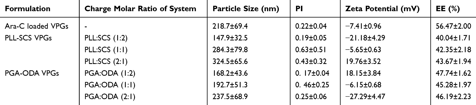 |
Table 1 Particle Size, Zeta Potential and EE of Redispersed Polyelectrolyte Complexed VPGs (n=3) |
The EE of redispersed PLL-SCS VPGs and PGA-ODA VPGs showed no significant change with increasing PLL or PGA concentration (Table 1). Yet, the EE of redispersed PLL-SCS VPGs and PGA-ODA VPGs were lower than that of Ara-C loaded VPGs.
Properties of Rheology
Rheological properties of polyelectrolyte coated VPGs were measured with oscillation mode. As the angular frequency was scanned from 0.1 to 100 1/s at 37°C, the elasticity modulus (G′) and the viscosity modulus (G″) were recorded in Figure 3A and B, G′ increased rapidly with its angular frequency from 0.1 to 0.5 1/s, and G′ increased slowly with its angular frequency from 0.5 to 100 1/s, G′ was higher than G″ on the whole.
 |
Figure 3 (A) The elasticity modulus G′ and the viscosity modulus G″ of PLL-SCS VPGs with different charge molar ratio of PLL and SCS at 37°C as a function of angular frequency. Solid symbol represents G′ and hollow symbol represents G″. (B) The elasticity modulus G′ and the viscosity modulus G″ of polyelectrolyte complexed VPGs (charge molar ratio of system is 1:2) and Ara-C loaded VPGs at 37°C as a function of angular frequency. Solid symbol represents G′ and hollow symbol represents G″. |
As shown in Figure 3A, with increasing concentration of PGA, G′ and G″ of PGA-ODA VPGs increased. Rheological properties of PLL-SCS VPGs were similar to PGA-ODA VPGs with increasing concentration of PLL (data not shown). From Figure 3B, when the charge molar ratio of polyelectrolyte coated VPGs is 1:2, the G′ and G″ of PGA-ODA VPGs and PLL-SCS VPGs are higher than Ara-C loaded VPGs.
In vitro Release
Figure 4 shows the in vitro release of polyelectrolyte coated VPGs loaded with Ara-C. When compared to Ara-C solutions, polyelectrolyte coated VPGs with 400 mg/g lipid concentration showed obvious sustained release properties, its cumulative release rate was near to 90% up to 18 days in vitro. However, compared to Ara-C loaded VPGs with the same lipid concentration, polyelectrolyte coated VPGs showed a rapid release rate slightly. The in vitro release properties of PLL-SCS VPGs and PGA-ODA VPGs showed no significant difference.
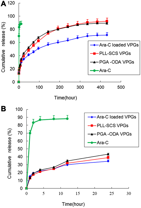 |
Figure 4 (A) In vitro drug release profiles of Ara-C, Ara-C loaded VPGs, PLL-SCS VPGs, and PGA-ODA VPGs in pH 7.4 PBS (n=3). The error bars represent the standard deviation. (B) In vitro drug release profiles of Ara-C, Ara-C loaded VPGs, PLL-SCS VPGs, and PGA-ODA VPGs in pH 7.4 PBS at the first 24 hours (n=3). The error bars represent the standard deviation. |
Cellular Uptake
The micrographs in Figure 5A showed that the cellular uptake of U87-MG glioma cells after 4 h incubation with FITC solution, FITC loaded VPGs and FITC loaded polyelectrolyte coated VPGs, respectively. In Figure 5A, the uptake of FITC loaded PGA-ODA VPGs was the highest in four formulations, with the uptake of FITC loaded VPGs slightly lower than that of the FITC loaded PGA-ODA VPGs, and the uptake of FITC loaded PLL-SCS VPGs slightly lower than that of the FITC loaded VPGs, while the uptake of free FITC solution was the lowest.
 |
Figure 5 (A) Fluorescent microscopic images of FITC uptake by U87-MG glioma cells in vitro incubated with FITC solution (a), FITC loaded VPGs (b), FITC loaded PGA-ODA VPGs (c) and FITC loaded PLL-SCS VPGs (d). Different VPGs containing FITC (green), and Hoechst 33258 (blue) for nucleus. (B) Cellular uptake of PGA-ODA VPGs at 37°C no PGA treatment, 4°C no PGA treatment, 37°C with PGA treatment. (a) 37°C, no PGA treatment; (b) 4°C, no PGA treatment; (c) 37°C, with PGA treatment. **p<0.01 vs cellular uptake of PGA-ODA VPGs at 37°C no PGA treatment. (C) Cellular uptake of PLL-SCS VPGs at 37°C no PLL treatment, 4°C no PLL treatment, 37°C with PLL treatment. (a) 37°C, no PLL treatment; (b) 4°C, no PLL treatment; (c) 37°C, with PLL treatment. **p<0.01 vs cellular uptake of PLL-SCS VPGs at 37°C no PLL treatment. |
From Figure 5B and C, uptake of PLL-SCS VPGs and PGA-ODA VPGs was significantly reduced at 4°C incubation at 4h (p< 0.01). And in Figure 5B, γ-PGA reduced the uptake of PGA-ODA VPGs at 37°C incubation at 4h (p< 0.01). Yet, in Figure 5C, ε-PLL did not affect the uptake of the PLL-SCS VPGs at 37°C incubation at 4h.
In vitro Cytotoxicity
MTT assay was used to investigate the effect of the developed formulations on cell viability. As Figure 6A shown, after 72h incubation with these empty formulations containing the lipid concentration from 0.01 to 10 mg/g, cell viability was not significantly different from the change of lipid concentration on U87-MG glioma cells. The results suggested that there was no cytotoxic effect on U87-MG glioma cell line in all control formulations. From Figure 6B, all formulations loaded with Ara-C concentration from 0.1 to 100 μM inhibited the growth of U87-MG glioma cells in a dose-dependent manner. For U87-MG glioma cells, PGA-ODA VPGs loaded with Ara-C exhibited more obvious cytotoxicity than other formulations, and the cytotoxicity of PLL-SCS VPGs loaded with Ara-C was lower than that of Ara-C loaded VPGs.
 |
Figure 6 (A) Cell viability of U87-MG glioma cells treated with empty VPGs, empty PLL-SCS VPGs, empty PGA-ODA VPGs at the lipid concentration from 0.01 to 10 mg/g (n=5). (B) Cell viability of U87-MG glioma cells treated with Ara-C solution, Ara-C loaded VPGs, PLL-SCS VPGs, and PGA-ODA VPGs at Ara-C concentrations ranging from 0.1 to 100 μM (n=5). *p<0.05, **p<0.01, vs Ara-C loaded VPGs; ΔΔp<0.01, vs PLL-SCS VPGs. |
In vivo Antitumor Activity
The in vivo antitumor activity of formulations was evaluated in the U87-MG glioma on the right flank of BALB/c nude mice. Figure 7A and B showed the change in tumor volume of all Ara-C formulation groups, compared to the Normal saline control group, the PGA-ODA VPGs, the Ara-C loaded VPGs and the PLL-SCS VPGs groups exhibited significantly more tumor growth suppression. In particular, the PGA-ODA VPGs group showed more significant antitumor effect than the Ara-C loaded VPGs (p<0.01) and PLL-SCS VPGs groups (p<0.01). However, compared to PLL-SCS VPGs group, the Ara-C loaded VPGs group exhibited slightly more tumor growth suppression (p>0.05). In addition, there was no significant body weight loss in all test groups (Figure 7C).
 |
Figure 7 (A) In vivo tumor volume profiles of U87-MG glioma subcutaneous bearing nude mice treated with different formulation groups for 32 days (n=6). (◆) Normal saline; (■) Ara-C solution; (▲) Ara-C loaded VPGs; (●) PLL-SCS VPGs; (※) PGA-ODA VPGs. Error bars represent the standard deviation. **p<0.01, vs Ara-C loaded VPGs; ΔΔp<0.01, vs PLL-SCS VPGs. (B) Photographs of excised tumors on U87-MG glioma subcutaneous bearing nude mice from the tested groups (n=6). (a) Normal saline; (b) Ara-C solution; (c) Ara-C loaded VPGs; d: PLL-SCS VPGs; e: PGA-ODA VPGs. Animals were sacrificed after 32 days. (C) Changes in the body weight of U87-MG glioma subcutaneous bearing nude mice (n=6). (◆) Normal saline; (■) Ara-C solution; (▲) Ara-C loaded VPGs; (×) PLL-SCS VPGs; (※) PGA-ODA VPGs. Error bars represent the standard deviation. |
Discussion
Recently, several studies have reported that polyelectrolytes enhanced the cells membrane permeability, and also increased the efficacy of gene transfer and siRNA delivery.10–13 So polyelectrolytes have been widely used as a coating material for carriers. In the present study, cationic/anionic polyelectrolyte coated VPGs loaded with Ara-C were prepared to enhance both in vitro and in vivo anti-glioma effects.
The TEM images of redispersed polyelectrolyte coated VPGs showed a layer structure covering the vesicular surface. The appearance was probably due to the effects of electrostatic interaction between polyelectrolyte and charged groups at the vesicular surface. It was presumed that the polyelectrolyte chains are in a random coil conformation around the vesicular surface.26
However, we found that aggregation of polyelectrolyte coated VPGs appeared in the process of redispersion, which was consistent with earlier results reported by others.27 We think that the aggregation is due to the fact that a lower surface charge, which was not enough to stabilize the polyelectrolyte coated VPGs, and led to aggregations of vesicles during the redispersion course.27 Another possible cause is electrostatic interaction between charged polyelectrolyte and opposite charge on the vesicular surface, which resulted in vesicular aggregation and fusion. Yet the reason might be a non-homogeneous overcompensation of vesicular surface charge via adsorption of polyelectrolyte molecules.26,27 However, the appearance has no effect on the local delivery of polyelectrolyte coated VPGs.
The EE of redispersed polyelectrolyte coated VPGs was lower than that of Ara-C loaded VPGs. This result might be due to SCS and ODA are incorporating in the vesicular lipid phase; during the redispersed course, the nonhomogeneous lipid membrane can increase drug leakage.28 In addition, the effects of electrostatic interaction between polyelectrolyte and charged groups at the vesicular surface could lead to drug leakage. At the charge molar ratio of PLL:SCS (1:2) and PGA:ODA (1:2), redispersed PLL-SCS VPGs and PGA-ODA VPGs exhibit lower particle sizes than that of Ara-C loaded VPGs, respectively. This result may be due to the insertion of SCS and ODA in vesicular lipid phase, which may lead to the upward curvature of the lipid vesicular membrane. However, unlike liposomes, each small vesicle was tightly packed among neighboring vesicles in polyelectrolyte coated VPGs, when free drug was entrapped in semisolid polyelectrolyte coated VPGs, unencapsulated drug was no need to remove, and it was equivalent to 100% loading of drug into it.5,29,30
We have previously reported that VPGs display gel-like characteristics[9]. Here, the rheological curves of polyelectrolyte coated VPGs were similar to VPGs, but polyelectrolyte coated VPGs showed much higher viscoelasticity than Ara-C loaded VPGs. With increasing concentration of polyelectrolytes, the viscoelasticity of polyelectrolyte coated VPGs was strengthened. Yet, polyelectrolyte coated VPGs still showed good syringability. Given syringability and system stability considerations, we have chosen polyelectrolyte coated VPGs at system charge molar ratio of 1:2 for further studies, because the 1:2 proportion prescription showed, no aggregation of vesicles with redispersed polyelectrolyte coated VPGs.
Usually, drug release is controlled by polymer degradation via carrier and drug diffusion on the polymer surface. Macromolecular polymer degradation is relatively slow, and polymer systems with high viscosities display slow diffusion. So, we selected high molecular weights of PLL and PGA in this study, and we expected macromolecules polymers to slow down the release rate of the gel system. According to the rheological properties of polyelectrolyte coated VPGs, we know that the addition of PLL and PGA as the coating material can increase the viscoelasticity of VPGs, which should slow drug release from polyelectrolyte coated VPGs. On the contrary, as a result, the release rate of polyelectrolyte coated VPGs was faster than that of Ara-C loaded VPGs. The result was explained that polyelectrolyte can increase membrane permeability,31 in addition, the incorporation of charged ODA or SCS into the vesicular lipid membrane led to the decrease of membrane homogeneity, which could accelerate drug release.28 The possible mechanisms involved in the drug release may involve permeation through the membrane of polyelectrolyte coated VPGs via the style of pore channels and partition diffusion.32 As for Ara-C loaded formulations, nearly 40% of the drug was released in the first 24 hours. We think that the drug inter-vesicles space rapidly diffuses across the dialysis bags, leading to the initial burst release effect.9
In our present study, FITC loaded PGA-ODA VPGs group showed the most intense intracellular fluorescence, and the fluorescence intensity of FITC loaded VPGs, FITC loaded PLL-SCS VPGs and free FITC solution groups were weakened sequentially (Figure 5A). In fact, FITC encapsulated into lipid vesicles of VPGs can facilitate the transportation of FITC into cells by endocytosis.25 Therefore, the cellular uptake efficacy of free FITC solution group was the lowest. Generally, polyelectrolytes with positive charge can promote drug permeation into negatively charged cancer cells and increase the cellular uptake by electrostatic interaction.28,33 However, compared with the FITC loaded PGA-ODA VPGs and the FITC loaded VPGs groups, the fluorescence intensity of FITC loaded PLL-SCS VPGs in U87-MG glioma cells were lower. We speculate that the charge molar ratio of lysine residue and SCS (1:2) of PLL-SCS VPGs might ensure that almost all lysine residues of PLL electrostatically interacted with SCS on the vesicular surface. In the prescription composition of PLL-SCS VPGs, SCS was excessive, and the whole system was negatively charged. The lack of positively charged, residual lysine residue on the surface of PLL-SCS VPGs, weakened the electrostatic interaction between the PLL-SCS VPGs and the glioma cells with negative charge. Therefore, the cellular uptake efficacy of PLL-SCS VPGs was lower than that of Ara-C loaded VPGs.
On the contrary, anionic complexes are generally not taken up by negatively charged cancer cells because the cellular membrane electrostatically repels the complex. Recent studies have reported that PGA complexes showed high gene expression without cytotoxicity.28,34 However, in our study, PGA-ODA VPGs loaded with Ara-C showed higher cellular uptake efficacy and cytotoxicity than that of other formations (Figures 5A and 6B). We speculated that γ-PGA with anionic charges might have a different uptake mechanism than cationic polyelectrolytes.
In order to explore the uptake mechanism, the inhibition experiment for the uptake of PLL-SCS VPGs and PGA-ODA VPGs was carried out at 37°C with no free polyelectrolyte treatment, 4°C with no free polyelectrolyte treatment, and 37°C with free polyelectrolyte treatment. From Figure 5B, compared with the uptake of PGA-ODA VPGs at 37°C with no PGA treatment, uptake of PGA-ODA VPGs was significantly reduced at 4°C with no PGA treatment and was inhibited by PGA at 37°C with PGA treatment. Our results indicated that the PGA-ODA VPGs were taken up by the U87-MG cells through an energy-dependent process and the PGA-specific receptor-mediated pathway, which is consistent with the results published by other scientists.20,35,36 In Figure 5C, compared with the uptake of PLL-SCS VPGs at 37°C no PLL treatment, uptake of PLL-SCS VPGs was significantly reduced at 4°C no PLL treatment, yet, uptake of PLL-SCS VPGs was not affected by PLL at 37°C with PLL treatment, which suggests that uptake of PLL-SCS VPGs was just an energy-dependent process.
For in vivo antitumor effects, from Figure 7A and B, the PGA-ODA VPGs group showed more significant antitumor effects than the Ara-C loaded VPGs group (p<0.01), and the PLL-SCS VPGs group (p<0.01). However, compared to the PLL-SCS VPGs group, the Ara-C loaded VPGs group exhibited slightly stronger tumor growth suppressive effect (p>0.05). In addition, there was no significant body weight loss in all test groups (Figure 7C). This suggests that no severe toxicity appeared in the treated animals. The results on in vivo antitumor effect of Ara-loaded formulations were consistent with that of the cellular uptake (Figure 5A), and in vitro cytotoxicity (Figure 6B). In short, the cellular uptake efficacy, in vitro cytotoxicity, and in vivo antitumor effects of the PGA-ODA VPGs group were superior to PLL-SCS VPGs. Here, our findings have confirmed the PGA-ODA VPGs were taken up by the U87-MG cells through the γ-PGA-specific receptor-mediated pathway (Figure 5B). Which is in agreement with available literatures on the subject.20,35 This uptake mechanism was different from that of cationic polymeric carriers by cancer cells.
Based upon the upper results, we surmise that PGA-ODA VPGs have the advantageous for local delivery to tumor tissue, and improve the anti-glioma effect both in vitro and in vivo.
Conclusion
This work describes cationic/anionic polyelectrolytes coated with oppositely charged vesicles in VPGs via electrostatic interactions. The TEM images and physicochemical properties verified the formation of polyelectrolyte coated VPGs. Our study has shown that polyelectrolyte coated VPGs offer distinct surface properties of VPGs, and altered the interaction between the complexes and the tumor cells. Notably, when compared with PLL coated VPGs, PGA coated VPGs significantly increased cellular uptake efficacy and in vitro cytotoxicity. Furthermore, PGA coated VPGs enhanced in vivo antitumor effects when subcutaneously injected around the tumor. Our results indicate that anionic PGA coated VPGs are superior to cationic PLL coated VPGs, and improve the anti-glioma effect for local delivery.
Acknowledgments
This project is financially supported by the National Natural Science Foundation of China (NO. 81560655, NO. 81860707, NO. 81572797), the Natural Science Foundation of Guangdong Province (NO. 2016A030311015), the Natural Science Foundation of Guangxi Province (NO. 2017GXNSFAA198061) and the Cultivation Plan of One Thousand Young and Middle-aged Key Teachers in Colleges and Universities of Guangxi. We are grateful to Danielle for English editing.
Disclosure
The authors report no conflicts of interest in this work.
References
1. Lapointe S, Perry A, Butowski NA. Primary brain tumours in adults. Lancet. 2018;392(10145):432–446. doi:10.1016/S0140-6736(18)30990-5
2. Cho C-F. The blood-brain barrier: brain cancer therapy hits a wall. Oncol Times. 2018;40(2):1,6–7. doi:10.1097/01.COT.0000530114.97923.aa
3. Hasegawa Y, Iuchi T, Sakaida T, et al. The influence of carmustine wafer implantation on tumor bed cysts and peritumoral brain edema. J Clin Neurosci. 2016;31:67–71. doi:10.1016/j.jocn.2015.12.033
4. Manaspon C, Nasongkla N, Chaimongkolnukul K, et al. Injectable SN-38-loaded polymeric depots for cancer chemotherapy of glioblastoma multiforme. Pharm Res. 2016;33(12):1–13. doi:10.1007/s11095-016-2011-4
5. Brandl M. Vesicular phospholipid gels: a technology platform. J Liposome Res. 2007;17(1):15–26. doi:10.1080/08982100601186490
6. Güthlein F, Burger AM, Brandl M, et al. Pharmacokinetics and antitumor activity of vincristine entrapped in vesicular phospholipid gels. Anticancer Drugs. 2002;13(8):797–805. doi:10.1097/00001813-200209000-00003
7. Moog R, Burger AM, Brandl M, et al. Change in pharmacokinetic and pharmacodynamic behavior of gemcitabine in human tumor xenografts upon entrapment in vesicular phospholipid gels. Cancer Chemother Pharmacol. 2002;49(5):356–366. doi:10.1016/S0140-6736(18)30990-5
8. Qi N, Cai C, Zhang W, et al. Sustained delivery of cytarabine-loaded vesicular phospholipid gels for treatment of xenografted glioma. Int J Pharm. 2014;472(1–2):48–55. doi:10.1016/j.ijpharm.2014.06.005
9. Qi N, Tang X, Lin X, et al. Sterilization stability of vesicular phospholipid gels loaded with cytarabine for brain implant. Int J Pharm. 2012;427(2):234–241. doi:10.1016/j.ijpharm.2012.02.008
10. Pötzinger Y, Rabel M, Ahrem H, et al. Polyelectrolyte layer assembly of bacterial nanocellulose whiskers with plasmid DNA as biocompatible non-viral gene delivery system. Cellulose. 2018;25(3):1–22. doi:10.1007/s10570-018-1664-z
11. Veleva-Kostadinova E, Dimitrov I, Toncheva-Moncheva N, et al. Nanoparticulate polyelectrolyte complexes of thermally sensitive poly(L-lysine)-based copolymers and DNA. Eur Polym J. 2018;102:219–230. doi:10.1016/j.eurpolymj.2018.03.028
12. Chen Y, Li J, Oupický D. Conjugate polyplexes with anti-invasive properties and improved siRNA delivery in vivo. Bioconjug Chem. 2018;29(2):296–305. doi:10.1021/acs.bioconjchem.7b00622
13. Hussein WM, Cheong YS, Liu C, et al. Peptide-based targeted polymeric nanoparticles for siRNA delivery. Nanotechnology. 2019;30(41):415604. doi:10.1088/1361-6528/ab313d
14. Yoshizaki Y, Yuba E, Sakaguchi N, et al. Potentiation of pH-sensitive polymer-modified liposomes with cationic lipid inclusion as antigen delivery carriers for cancer immunotherapy. Biomaterials. 2014;35(28):8186–8196. doi:10.1016/j.biomaterials.2014.05.077
15. Ge L, Möhwald H, Li J. Phospholipid liposomes stabilized by the coverage of polyelectrolyte. Colloids Surf A Physicochem Eng Asp. 2003;221(1–3):49–53. doi:10.1016/S0927-7757(03)00106-7
16. Yaroslavov A, Efimova A, Lobyshev V, et al. Reversibility of structural rearrangements in the negative vesicular membrane upon electrostatic adsorption/desorption of the polycation. Biochim Biophys Acta Biomembr. 2002;1560(1–2):14–24. doi:10.1016/S0005-2736(01)00453-9
17. Ranaldi G, Marigliano I, Vespignani I, et al. The effect of chitosan and other polycations on tight junction permeability in the human intestinal Caco-2 cell line. J Nutr Biochem. 2002;13(3):157–167. doi:10.1016/S0955-2863(01)00208-X
18. Yaroslavov A, Kuchenkova OY, Okuneva I, et al. Effect of polylysine on transformations and permeability of negative vesicular membranes. Biochim Biophys Acta Biomembr. 2003;1611(1–2):44–54. doi:10.1016/S0005-2736(02)00701-0
19. Shen W-C, Ryser H. Poly (L-lysine) has different membrane transport and drug-carrier properties when complexed with heparin. Proc Natl Acad Sci U S A. 1981;78(12):7589–7593. doi:10.1073/pnas.78.12.7589
20. Kurosaki T, Kitahara T, Fumoto S, et al. Ternary complexes of pDNA, polyethylenimine, and γ-polyglutamic acid for gene delivery systems. Biomaterials. 2009;30(14):2846–2853. doi:10.1016/j.biomaterials.2009.01.055
21. Auzenne E, Donato NJ, Li C, et al. Superior therapeutic profile of poly-L-glutamic acid-paclitaxel copolymer compared with taxol in xenogeneic compartmental models of human ovarian carcinoma. Clin Cancer Res. 2002;8(2):573–581. doi:10.1093/carcin/23.2.373-A
22. Manocha B, Margaritis A. Production and characterization of γ-polyglutamic acid nanoparticles for controlled anticancer drug release. Crit Rev Biotechnol. 2008;28(2):83–99. doi:10.1080/07388550802107483
23. Qi N, Tang B, Liu G, Liang X. Poly (γ-glutamic acid)-coated lipoplexes loaded with doxorubicin for enhancing the antitumor activity against liver tumors. Nanoscale Res Lett. 2017;12(1):361. doi:10.1186/s11671-017-2081-1
24. Massing U, Cicko S, Ziroli V. Dual asymmetric centrifugation (DAC)–a new technique for liposome preparation. J Control Release. 2008;125(1):16–24. doi:10.1016/j.jconrel.2007.09.010
25. Zhao P, Wang H, Yu M, et al. Paclitaxel loaded folic acid targeted nanoparticles of mixed lipid-shell and polymer-core: in vitro and in vivo evaluation. Eur J Pharm Biopharm. 2012;81(2):248–256. doi:10.1016/j.ejpb.2012.03.004
26. Sennato S, Bordi F, Cametti C, et al. Charge patch attraction and reentrant condensation in DNA–liposome complexes. Biochim Biophys Acta Biomembr. 2005;1714(1):11–24. doi:10.1016/j.bbamem.2005.06.004
27. Volodkin D, Ball V, Schaaf P, et al. Complexation of phosphocholine liposomes with polylysine. Stabilization by surface coverage versus aggregation. Biochim Biophys Acta Biomembr. 2007;1768(2):280–290. doi:10.1016/j.bbamem.2006.09.015
28. Dragicevic-Curic N, Gräfe S, Gitter B, et al. Surface charged temoporfin-loaded flexible vesicles: in vitro skin penetration studies and stability. Int J Pharm. 2010;384:100–108. doi:10.1016/j.ijpharm.2009.10.006
29. Tardi C, Drechsler M, Bauer KH, et al. Steam sterilisation of vesicular phospholipid gels. Int J Pharm. 2001;217(1–2):161–172. doi:10.1016/S0378-5173(01)00605-6
30. Tardi C, Brandl M, Schubert R. Erosion and controlled release properties of semisolid vesicular phospholipid dispersions. J Control Release. 1998;55(2–3):261–270. doi:10.1016/s0168-3659(98)00058-3
31. Yaroslavov A, Kiseliova E, Udalykh OY, et al. Integrity of mixed liposomes contacting a polycation depends on the negatively charged lipid content. Langmuir. 1998;14(18):5160–5163. doi:10.1021/la970510f
32. Volodkin D, Mohwald H, Voegel J-C, et al. Coating of negatively charged liposomes by polylysine: drug release study. J Control Release. 2007;117(1):111–120. doi:10.1016/j.jconrel.2006.10.021
33. Yang R, Shim W-S, Cui F-D, et al. Enhanced electrostatic interaction between chitosan-modified PLGA nanoparticle and tumor. Int J Pharm. 2009;371(1–2):142–147. doi:10.1016/j.ijpharm.2008.12.007
34. Wang C, Feng M, Deng J, et al. Poly (α-glutamic acid) combined with polycation as serum-resistant carriers for gene delivery. Int J Pharm. 2010;398(1–2):237–245. doi:10.1016/j.ijpharm.2010.07.048
35. Sano K, Iwamiya Y, Kurosaki T, et al. Radiolabeled γ-polyglutamic acid complex as a nano-platform for sentinel lymph node imaging. J Control Release. 2014;194:310–315. doi:10.1016/j.jconrel.2014.08.025
36. Kurosaki T, Kitahara T, Kawakami S, et al. γ-Polyglutamic acid-coated vectors for effective and safe gene therapy. J Control Release. 2010;142(3):404–410. doi:10.1016/j.jconrel.2009.11.010
 © 2020 The Author(s). This work is published and licensed by Dove Medical Press Limited. The
full terms of this license are available at https://www.dovepress.com/terms
and incorporate the Creative Commons Attribution
- Non Commercial (unported, 3.0) License.
By accessing the work you hereby accept the Terms. Non-commercial uses of the work are permitted
without any further permission from Dove Medical Press Limited, provided the work is properly
attributed. For permission for commercial use of this work, please see paragraphs 4.2 and 5 of our Terms.
© 2020 The Author(s). This work is published and licensed by Dove Medical Press Limited. The
full terms of this license are available at https://www.dovepress.com/terms
and incorporate the Creative Commons Attribution
- Non Commercial (unported, 3.0) License.
By accessing the work you hereby accept the Terms. Non-commercial uses of the work are permitted
without any further permission from Dove Medical Press Limited, provided the work is properly
attributed. For permission for commercial use of this work, please see paragraphs 4.2 and 5 of our Terms.