")
Back to Journals » Clinical, Cosmetic and Investigational Dermatology » Volume 16
Case Series of Phthirus pubis Infestation in Non-Perineal Regions
Authors Nie J, Shen X, Li Y, Wang W
Received 22 May 2023
Accepted for publication 1 August 2023
Published 22 August 2023 Volume 2023:16 Pages 2277—2281
DOI https://doi.org/10.2147/CCID.S422318
Checked for plagiarism Yes
Review by Single anonymous peer review
Peer reviewer comments 3
Editor who approved publication: Dr Anne-Claire Fougerousse
Jing Nie, Xue Shen, Yan Li, Wenju Wang
Department of Dermatology, Chengdu Second People’s Hospital, Chengdu, Sichuan, 610000, People’s Republic of China
Correspondence: Jing Nie, Department of dermatology, Chengdu Second People’s Hospital, 165 Caoshi Street, Qingyang District, Chengdu, Sichuan, 610000, People’s Republic of China, Tel +86-15902687253, Email [email protected]
Abstract: Pediculosis has been reported in all countries and socio-economic classes. Due to the small size and translucent nature of their nits, patients with Pthirus pubis may be misdiagnosed. Here, we report three cases of Pthirus pubis infection involving different body sites, including the eyelashes, axillary region and head, respectively. This case series have discussed their clinical features and corresponding responses. A large number of eggs and adult pubic lice were observed under dermoscopic observation, which can be considered an effective tool for diagnosing Pthirus pubis and reducing the misdiagnosis rate.
Keywords: Phthirus pubis, eyelashes, axillary region, head, dermoscopy
Introduction
Pediculosis infection has been reported in almost all countries, particularly in developing countries. The infection caused by Pthirus pubis typically occurs in the pubis area, but it can also affect the eyelashes, eyebrows, axillary region and head.1–3 Phthiriasis in eyelashes, known as Phthiriasis palpebrarum, is primarily seen in children but can also occur in adults.1,2,4 Phthiriasis palpebrarum is a common type of eyelid infestation, and approximately 30% of cases are categorized as sexually transmitted infections.5 Till now, about 20 cases with Pthirus pubis involved in the scalp have been reported, particularly in individuals with widely spaced head hair.6 The main clinical manifestation of Pthirus pubis infection is itching, and persistent scratching and mechanical irritation can lead to diffuse inflammatory responses.7
The pubic louse can swiftly migrate from the genital areas to other sites, such as eyelashes, axillary hair, and scalp.8,9 Due to the small size and translucent nature of its nits, patients with Pthirus pubis infection may sometimes be misdiagnosed as having conjunctivitis, blepharitis, dermatosis, or other conditions. This is especially the case when the symptoms are not typical such as when there is no hyperemia, redness, burning sensation or reddish-brown crusts.10,11 In this report, we describe three cases of Pthirus pubis infection involving the eyelashes, axillary region and scalp, respectively. We also discuss the optimal strategies for diagnosis and treatment.
Case Presentation
The first case is a 6-year-old girl who was admitted to the Department of Ophthalmology due to experiencing “itching and foreign body sensation in both eyes for 8 days”. Her family reported that the child had been frequently scratching her eyes for the past 6 days. Upon examination, her best corrected visual acuity of both eyes was normal, and silver-gray foreign bodies were observed on her eyelashes. Additionally, there were red-brown scabs at the base of her eyelids and eyelashes. The child complained of intense itching in both eyes. Further examination using a slit-lamp microscope showed the presence of silver granular foreign bodies and parasites measuring less than 1 mm around the eyelashes (Figure 1a). Dermoscopy examination showed numerous empty transparent eggs, brown eggs containing larvae, and adult lice (Figure 1b). No abnormalities were found in the patient’s family members. The child has recently stayed in the countryside where the local hygiene conditions are poor. It is suspected that the child may have been infected by the external environment. Based on the clinical symptoms and examination results, a diagnosis of Pthirus pubis i infestation in her eyelashes was made. Considering the large number of insect eggs attached to the eyelashes of the child, Vaseline gauze was applied to gently wipe the eyelashes from the root outward, in an attempt to remove some of the eggs and mechanically clean the lice on the eyelashes.
 |
Figure 1 Examinations of three clinical cases with the Pthirus pubis infection involved in eyelashes (a and b), axillary region (c and d) and head (e and f), respectively. (a) Slit-lamp microscope examination. (b) Dermoscopy examination. (c) Skin examination. (d) Dermoscopy examination. (e) Scalp examination. (f) Dermoscopy examination. Red circles, pubic lice bodies; White arrows, lice eggs. |
The second case is a 37-year-old man who had been diagnosed with Pthirus pubis due to perineal itching that had persisted for 1 month. He had been treated with sulfur ointment. However, in recent days, he started experiencing itching in the axillary region and sought medical attention once again. Upon examination by a specialist, brown granular foreign bodies and brown parasites were found in the right axilla (Figure 1c). Dermoscopy revealed a significant number of brown eggs and adult lice attached to the hair shafts and roots in the right axilla (Figure 1d). No obvious changes were observed on the left side of the axilla. The diagnosis was Pthirus pubis infestation in the axilla region. Treatment involved the application of topical sulfur ointment for 3 days after removing the axillary hair.
The third case is a 52-year-old female patient who complained of “scattered scales with itching in the scalp for 1 week”, along with bleeding due to repeated scratching. She had recently stayed in a hostel with poor sanitary conditions. Upon examination by a specialist, silver-white scabs were observed on the hair of her head (Figure 1e). Dermoscopy revealed reddish-brown scabs on the scalp, as well as silvery-white empty insect eggs and pale brown insect eggs attached to the hair shafts. Adult insects could also be seen at the root of the hair (Figure 1f). The diagnosis was Pthirus pubis infestation in the scalp. Treatment involved using a head lice comb with fine teeth to remove the eggs, followed by washing the hair with sulfur soap.
Discussion
Pubic lice are highly mobile when the host is at rest, such as during sleep, and they can easily move from one infested site to another if conditions allow. They have been associated with sexually transmitted diseases and are typically transmitted through sexual contact.12 The gold standard for diagnosing Pthirus pubis is to identify live adult lice and viable eggs on the hair shafts in the affected sites.6 In the present study, we reported three cases of Pthirus pubis infection occurring in different body sites, including eyelashes, axillary region and head, respectively. Through dermoscopic examination, we observed a large number of eggs and adult pubic lice. When magnified, the adults were found to have a body size ranging from 1 mm to 3 mm (Figure 2a and b). Pubic lice do not have wings, which means their primary mode of transmission is through close contact.13 The morphology of pubic lice is clearly distinct from that of head lice, thus considering the presence of pubic lice in these three reported cases. Pthirus pubis is primarily acquired through sexual contact or close contact with infected patients.14
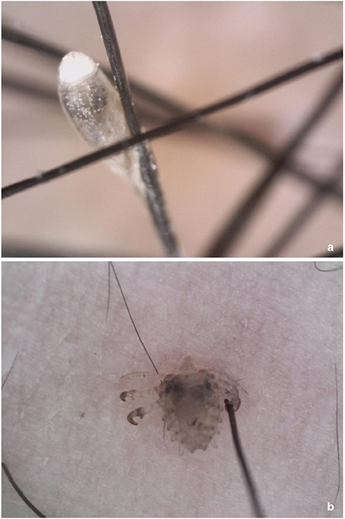 |
Figure 2 The morphology of pubic lice under dermoscopy. (a) Eggs of pubic lice. (b) Adult pubic lice. |
Phthiriasis in the eyelashes is commonly observed in children, and physicians should inquire about the medical history to explore the possibility of child abuse, given its sexual transmission feature.15–17 Differentiating Phthiriasis palpebrarum from common ocular diseases, such as seborrheic blepharitis, conjunctivitis, chalazion, dry eye disease, hordeolum, and eyelid eczema, requires careful consideration.18 When dealing with Pthirus pubis infections in other body sites such as the axilla and head, it is important to distinguish them from dermatologic conditions such as seborrheic dermatitis, capillary hair, eczema, superficial fungal infection, etc.19,20 Dermoscopic examination proves effective in clearly distinguishing Pthirus pubis from other diseases, aiding in clinical diagnosis and reducing misdiagnosis rates.21,22
The body of the lice is flat and resembles a crab-like shape, with a dark brown area in the middle of the abdomen. A considerable number of unhatched oval-shaped brown lice eggs, as well as transparent empty eggshells, are attached to the dry hair. During the treatment process for head lice in patients with long hair, it is recommended to cut the hair short. However, due to concerns about appearance, some patients have poor compliance, leading to a prolonged treatment period and unsatisfactory treatment results. Multiple therapeutic approaches have been developed for Pthirus pubis infection. Topical drug therapies, such as pyrethroids, malathion, lindane, topical and oral ivermectin, and other topical insecticides, are considered first-line treatments for Pthirus pubis.22 When treating Phthiriasis palpebrarum, the initial choice is to manually remove the lice and eggs or apply ophthalmic-grade Vaseline ointment externally.23
Conclusion
Collectively, Pthirus pubis infection primarily occurs in the pubic and inguinal regions, as well as other body sites, inkling the perineal site, eyelashes, axillary region and head. In this report, we have described three patients with Pthirus pubis infections in different anatomical locations, highlighting their clinical characteristics and respective treatments. Dermoscopy has been recognized as a valuable and effective tool for diagnosing Pthirus pubis, aiding in reducing the misdiagnosis rate.
Ethics Approval and Informed Consent
This study was designed in accordance with the Declaration of Helsinki and approved by the ethics committee of Chengdu Second People’s Hospital.
Data Sharing Statement
The data that support the findings of this study are available from the corresponding author upon request.
Consent for Publication
Informed consent was obtained from 6 years old patient’s guardians and others patient involved in the study.
Acknowledgments
We would like to acknowledge the reviewers for their helpful comments on this paper.
Author Contributions
All authors made a significant contribution to the work reported, whether that is in the conception, study design, execution, acquisition of data, analysis and interpretation, or in all these areas; took part in drafting, revising or critically reviewing the article; gave final approval of the version to be published; have agreed on the journal to which the article has been submitted; and agree to be accountable for all aspects of the work.
Disclosure
The authors report no conflicts of interest in this work.
References
1. Veraldi S, Nazzaro G, Esposito L, Genovese G, Pontini P, Gelmetti C. Pthiriasis of the eyelashes. Giornale Italiano di Dermatologia e Venereologia. 2020;155(2):198–201. doi:10.23736/S0392-0488.19.06350-8
2. Patel PU, Tan A, Levell NJ. A clinical review and history of pubic lice. Clin Exp Dermatol. 2021;46(7):1181–1188. doi:10.1111/ced.14666
3. Ozer PA, Kabatas EU, Gurkan A, Kurtul BE. Treatment of Phthiriasis Palpebrarum Mimicking Conjunctivitis in a Newborn. Indian J Pediatr. 2016;83(7):730–731. doi:10.1007/s12098-015-1983-0
4. Dohvoma VA, Ebana Mvogo SR, Atangana PJA, Nyasse P, Epee E, Ebana Mvogo C. Phthirus pubis infestation of the eyelids presenting as chronic blepharoconjunctivitis in a 6-year-old girl: a case report. Case Rep Ophthalmol. 2018;9(1):30–34. doi:10.1159/000485738
5. Wu N, Zhang H, Sun FY. Phthiriasis palpebrarum: a case of eyelash infestation with Pthirus pubis. Exp Ther Med. 2017;13(5):2000–2002. doi:10.3892/etm.2017.4187
6. Veraldi S, Pontini P, Nazzaro G. Phthirus pubis infestation of the scalp: a case report and review of the literature. Korean J Parasitol. 2018;56(5):487–489. doi:10.3347/kjp.2018.56.5.487
7. Huo Y, Mo Y, Jin X, Huang X, Chen W. First case of Phthirus pubis and Demodex co-infestation of the eyelids: a case report. BMC Ophthalmol. 2021;21(1):122. doi:10.1186/s12886-021-01875-w
8. Scott GR, Chosidow O; IUSTI/WHO. European guideline for the management of pediculosis pubis, 2010. Int J STD AIDS. 2011;22(6):304–305. doi:10.1258/ijsa.2011.011114
9. Burgess I, Maunder JW, Myint TT. Maintenance of the crab louse, Pthirus pubis, in the laboratory and behavioural studies using volunteers. Community Med. 1983;5(3):238–241. doi:10.1007/BF02548552
10. Lin YC, Kao SC, Kau HC, Hsu WM, Tsai CC. Phthiriasis palpebrarum: an unusual blepharoconjunctivitis. Zhonghua Yi Xue Za Zhi. 2002;65(10):498–500.
11. Gurnani B, Hafsi W. Phthiriasis Palpebrarum, in StatPearls. StatPearls Publishing LLC: Treasure Island (FL); 2022.
12. Anderson AL, Chaney E. Pubic lice (Pthirus pubis): history, biology and treatment vs. knowledge and beliefs of US college students. Int J Environ Res Public Health. 2009;6(2):592–600. doi:10.3390/ijerph6020592
13. Shakya M, Jayraw AK, Singh M. Pubic lice infestation in man from Mhow, Madhya Pradesh. J Parasit Dis. 2018;42(3):402–404. doi:10.1007/s12639-018-1015-x
14. Peterson AR, Nash E, Anderson BJ. Infectious Disease in Contact Sports. Sports Health. 2019;11(1):47–58. doi:10.1177/1941738118789954
15. Chapel TA, Katta T, Kuszmar T, DeGiusti D. Pediculosis pubis in a clinic for treatment of sexually transmitted diseases. Sex Transm Dis. 1979;6(4):257–260. doi:10.1097/00007435-197910000-00008
16. Ryan MF. Phthiriasis palpebrarum infection: a concern for child abuse. J Emerg Med. 2014;46(6):e159–62. doi:10.1016/j.jemermed.2013.11.090
17. Galiczynski EM Jr, Elston DM. What’s eating you? Pubic lice (Pthirus pubis). Cutis. 2008;81(2):109–114.
18. Altinsoy F, Alver O, Doganay S. A Rare Case of Blebharitis: phthiriasis Palpebrarum. Turkiye Parazitol Derg. 2018;42(1):90–92. doi:10.5152/tpd.2018.4824
19. Scanni G. Phthiriasis capitis ab initio. Use of entodermoscopy for quick differentiation between Phthirus pubis and Pediculus capitis nits. An Bras Dermatol. 2020;95(6):777–779. doi:10.1016/j.abd.2020.03.019
20. Tang JQ, Ran X, Ran YP. Cover Image: dermoscopy in vivo for the life cycle of Phthirus pubis. Br J Dermatol. 2017;176(1):279. doi:10.1111/bjd.15049
21. Jayasree P, Kaliyadan F, Ashique KT. Pubic Lice in Facial Hair. Dermatol Pract Concept. 2020;10(2):e2020042. doi:10.5826/dpc.1002a42
22. Salavastru CM, Chosidow O, Janier M, Tiplica GS. European guideline for the management of pediculosis pubis. J Eur Acad Dermatol Venereol. 2017;31(9):1425–1428. doi:10.1111/jdv.14420
23. Packer H, Heiberger AL. Getting Ahead of Head Lice: treatment in the Setting of Resistance. S D Med. 2016;69(10):468–470.
 © 2023 The Author(s). This work is published and licensed by Dove Medical Press Limited. The full terms of this license are available at https://www.dovepress.com/terms.php and incorporate the Creative Commons Attribution - Non Commercial (unported, v3.0) License.
By accessing the work you hereby accept the Terms. Non-commercial uses of the work are permitted without any further permission from Dove Medical Press Limited, provided the work is properly attributed. For permission for commercial use of this work, please see paragraphs 4.2 and 5 of our Terms.
© 2023 The Author(s). This work is published and licensed by Dove Medical Press Limited. The full terms of this license are available at https://www.dovepress.com/terms.php and incorporate the Creative Commons Attribution - Non Commercial (unported, v3.0) License.
By accessing the work you hereby accept the Terms. Non-commercial uses of the work are permitted without any further permission from Dove Medical Press Limited, provided the work is properly attributed. For permission for commercial use of this work, please see paragraphs 4.2 and 5 of our Terms.