Back to Journals » International Journal of Nanomedicine » Volume 17
Caries Management with Non-Metallic Nanomaterials: A Systematic Review
Authors Xu VW ![]() , Nizami MZI
, Nizami MZI ![]() , Yin IX, Lung CYK
, Yin IX, Lung CYK ![]() , Yu OY
, Yu OY ![]() , Chu CH
, Chu CH ![]()
Received 7 September 2022
Accepted for publication 23 November 2022
Published 30 November 2022 Volume 2022:17 Pages 5809—5824
DOI https://doi.org/10.2147/IJN.S389038
Checked for plagiarism Yes
Review by Single anonymous peer review
Peer reviewer comments 3
Editor who approved publication: Dr Yan Shen
Veena Wenqing Xu, Mohammed Zahedul Islam Nizami, Iris Xiaoxue Yin, Christie Ying Kei Lung, Ollie Yiru Yu, Chun Hung Chu
Faculty of Dentistry, The University of Hong Kong, Hong Kong, People’s Republic of China
Correspondence: Chun Hung Chu, Faculty of Dentistry, The University of Hong Kong, 34 Hospital Road, Hong Kong, People’s Republic of China, Tel +852 2859 0287, Fax +852 2858 2532, Email [email protected]
Background: Non-metallic nanomaterials do not stain enamel or dentin. Most have better biocompatibility than metallic nanomaterials do for management of dental caries.
Objective: The objective of this study is to review the types, properties and potential uses of non-metallic nanomaterials systematically for managing dental caries.
Methods: Two researchers independently performed a literature search of publications in English using PubMed, Scopus and Web of Science. The keywords used were (nanoparticles OR nanocomposites OR nanomaterials) AND (caries OR tooth decay). They screened the titles and abstracts to identify potentially eligible publications of original research reporting non-metallic nanomaterials for caries management. Then, they retrieved and studied the full text of the identified publications for inclusion in this study.
Results: Out of 2497 resulting publications, this study included 75 of those. The non-metallic nanomaterials used in these publications were categorized as biological organic nanomaterials (n=45), synthetic organic nanomaterials (n=15), carbon-based nanomaterials (n=13) and selenium nanomaterials (n=2). They inhibited bacteria growth and/or promoted remineralization. They could be incorporated in topical agents (29/75, 39%), dental adhesives (11/75, 15%), restorative fillers (4/75, 5%), dental sealant (3/75, 4%), oral drugs (3/75, 4%), toothpastes (2/75, 3%) and functional candies (1/75, 1%). Other publications (22/75, 29%) do not mention specific applications. However, most publications (67/75, 89%) were in vitro studies. Six publications (6/75, 8%) were animal studies, and only two publications (2/75, 3%) were clinical studies.
Conclusion: The literature showed non-metallic nanomaterials have antibacterial and/or remineralising properties. The most common type of non-metallic nanomaterials for caries management is organic nanomaterials. Non-metallic nanomaterials can be incorporated into dental sealants, toothpaste, dental adhesives, topical agents and even candies and drugs. However, the majority of the publications are in vitro studies, and only two publications are clinical studies.
Keywords: nanomaterials, nanoparticles, caries, prevention, antibacterial, remineralisation
Introduction
Dental caries is one of the most prevalent chronic diseases worldwide. Multiple pathological factors can trigger it, including cariogenic microbes, host or tooth surface, substrate and time.1 The colonized cariogenic microbes on tooth surfaces can metabolize fermentable carbohydrates and generate organic acids. Although enamel and dentin are highly mineralized hard tissues, these acids over a period can dissolve and diffuse into enamel and dentin.2,3 Continuous demineralization can destroy tooth structure and finally result in dental caries. Without treatment, dental caries can progress and lead to persistent pain and further infection.
The World Health Organization reported that dental caries is the fourth most expensive disease to treat, causing a significant global burden of disease.4 Clinicians largely use the conventional approach to manage dental caries with restoration.5 The current dental materials are principally silver amalgam, composite resin and glass ionomer cement. They have their own limitations that affect clinical life span of the restorations. The World Dental Federation published its policy statement that advocated evidence-based caries-control measures for caries management.6 Researchers have been developing various dental materials to inhibit demineralization by repressing the growth of cariogenic bacteria and/or to promote remineralization by enhancing the deposition of minerals.3,7
Nanotechnology is a recent research trend that is particularly effective in developing various nanomaterials used in several application areas. Nanomaterials contain materials measured at the nanometer scale (1 to 100 nm). They exhibit unique mechanochemical and biological properties, such as huge surface-to-volume ratio, high strength and stability, strong solubility and chemical reactivity and promising antibacterial effects.8,9 Therefore, various nanomaterials have been developed for caries management and have shown auspicious results.8
Metallic nanomaterials, such as silver nanoparticles, have been widely used in caries management. They generally exhibit antibacterial activity through oxidative stress induction.10 However, they demonstrate only antibacterial ability, with no remineralization ability. In addition, some metallic nanomaterials have high cell toxicity and staining effects, which limit their clinical application.
Due to these disadvantages of metallic nanomaterials, researchers are gradually studying non-metallic nanomaterials. Non-metallic nanomaterials are nanomaterials without any metal element. Some non-metallic nanomaterials enhance remineralization by promoting mineral deposition and inducing hydroxyapatite formation. They can be a biomimetic material to regulate and orient the growth of enamel-like apatite. Some non-metallic nanomaterials effectively inhibit the growth of cariogenic bacteria, owing to their nano-scale size, unique active constituents and loaded antibacterial agents.11 They can protect teeth from demineralization without generating drug resistance. Figure 1 illustrates non-metallic nanomaterials’ remineralising and antibacterial properties in managing dental caries.
 |
Figure 1 Effects of non-metallic cariostatic nanomaterials on carious lesion. |
Moreover, non-metallic nanomaterials exhibit promising biocompatibility compared to metallic nanomaterials. The addition of non-metallic nanomaterials in dental materials neither significantly alters their color nor affects their application. However, a search revealed that reviews of non-metallic nanomaterials for caries management have not been conducted. The purpose of this study is to review and summarize systematically the non-metallic nanomaterials developed for managing dental caries. This outline could help researchers identify the proper research direction to develop new non-metallic nanomaterials for caries management.
Methods
Search Strategy
Two independent investigators conducted a literature search to identify publications in three common databases (PubMed, Scopus and Web of Science). The search was restricted to publications in English, with no limitations on the date of publication. The keywords were (nanoparticles OR nanocomposites OR nanomaterials) AND (caries OR tooth decay). The last search was conducted on 20 Jul 2022.
Study Selection and Data Extraction
This systematic review included original investigations of the non-metallic nanomaterials developed for caries management (Figure 2).
 |
Figure 2 Flow chart of the literature search. |
Two investigators independently checked and excluded duplicate publications from the three databases to generate a list of publications. They screened titles and abstracts of the publications to identify potentially eligible publications. They excluded literature reviews, studies on metal nanomaterials, studies not related to caries management and other irrelevant studies. Afterwards, the two investigators retrieved full texts of the remaining publications for review. They selected publications that non-metallic nanomaterials used as the active constituent for caries management. Then, they performed a manual screening to select eligible publications from the reference lists of the selected publications. They discussed with another investigator including the selected publications to determine the publications included in this review. They recorded the publications’ information, including authors, year, journal and issues; the nanomaterials studied; the study design; the anti-caries properties investigated and the potential uses of the nanomaterials.
Assessment of Risk of Bias
Two investigators independently assessed the risk of bias from individual studies. The assessment was adapted from a systematic review.3,12 They evaluated the study’s quality with seven parameters, which were 1) presence of control, 2) sample size justification, 3) material characterization, 4) biocompatibility assessment, 5) amount of material for assessment, 6) assessment time and 7) blinding of observers. Publications that reported only one to two items were classified as high risk of bias. Publications reporting three to five items were classified as medium risk, and those reporting more five items were regarded as low risk.
Results
The initial literature search yielded 2497 potentially eligible publications (666 publications in PubMed; 1073 publications in Scopus; 758 publications in Web of Science). Duplicate records of publications were excluded. After the titles and abstracts were screened, and 1775 publications were removed because they were literature reviews, studies not regarding non-metallic nanomaterials, studies not related to dental caries or other irrelevant studies. Another 48 publications were excluded because the non-metallic nanomaterials in these papers were not the active constituent for caries management. A search of the references of the selected publications yielded 21 publications that met the inclusion criteria. Therefore, 75 publications were included in this review.
The 75 publications were assessed for the risk of bias (Table 1). Seventy-three publications (73/75, 97%) had a medium risk of bias, 1 publication (1/75, 1%) presented a high risk of bias, and 1 publication (1/75, 1%) presented a low risk of bias. Of the 75 publications included, most publications (67/75, 89%) were in vitro studies, 6 (6/75, 8%) were animal studies and 2 (2/75, 3%) were clinical studies. The non-metallic nanomaterials demonstrated in these publications could be incorporated in topical agent (29/75, 39%), dental adhesives (11/75, 15%), restorative filler (4/75, 5%), dental sealant (3/75, 4%), oral drugs (3/75, 4%), toothpaste (2/75, 3%) and functional candies (1/75, 1%). Other publications (22/75, 29%) did not mention specific applications. The non-metallic nanomaterials reported were categorized as biological organic nanomaterials (n=45), synthetic organic nanomaterials (n=15), carbon-based nanomaterials (n=13) and selenium nanomaterials (n=2). Figures 3 and Figure 4 show the four groups of non-metallic cariostatic nanomaterials and their mechanism, respectively.
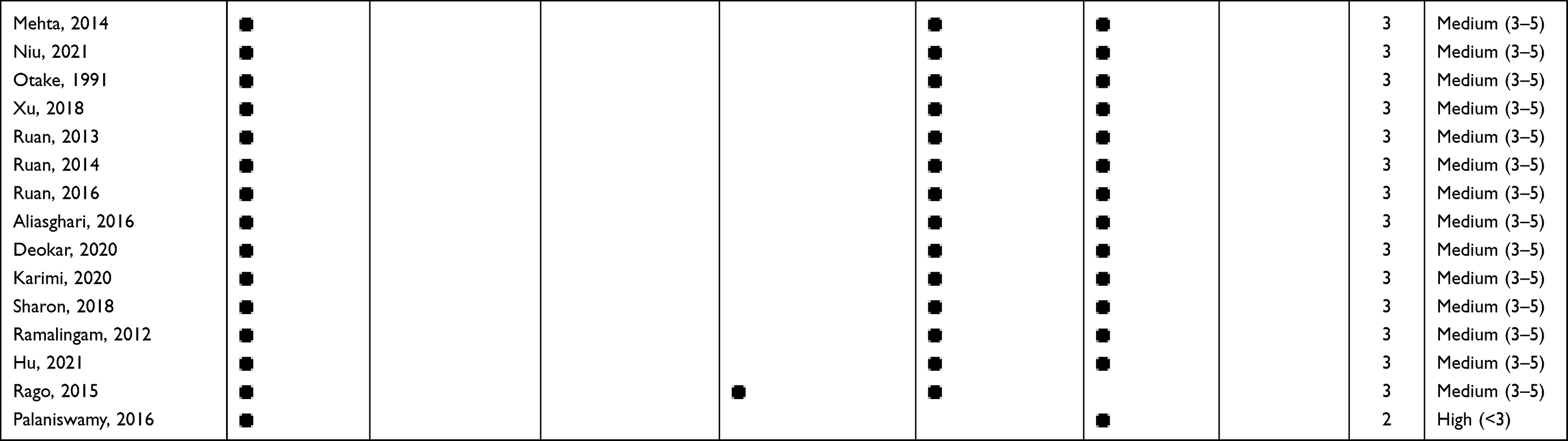 |
Table 1 Risk of Bias of the Included Publications |
 |
Figure 3 Number of publications of the 4 non-metallic cariostatic nanomaterials. |
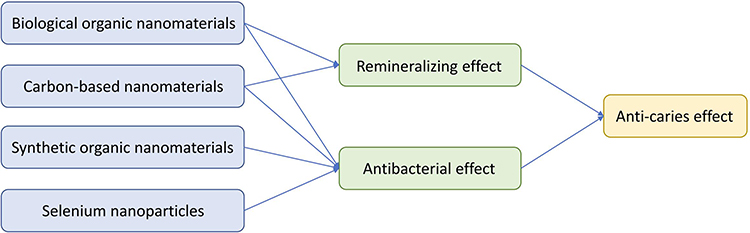 |
Figure 4 Mechanism of the 4 non-metallic cariostatic nanomaterials. |
Discussion
Biological Organic Nanomaterials
Biological organic nanomaterials are nano-scale materials derived from organisms. These materials have attracted widespread attention due to their great biocompatibility, biodegradability and bioavailability.
Amino Acid-Based Nanomaterials
Amino acid-based nanomaterials are amino acids and their derivatives, including peptides and proteins. Amino acid-based nanomaterials are novel materials used for caries management. Table 2 summarizes 28 publications on the amino acid-based nanomaterials for caries management. Free amino acids, such as D-cysteine, process antibacterial properties. D-cysteine can inhibit the metabolic activity and lactic acid production of Streptococcus mutans (S. mutans) and Streptococcus sanguinis (S. sanguinis) biofilms.13
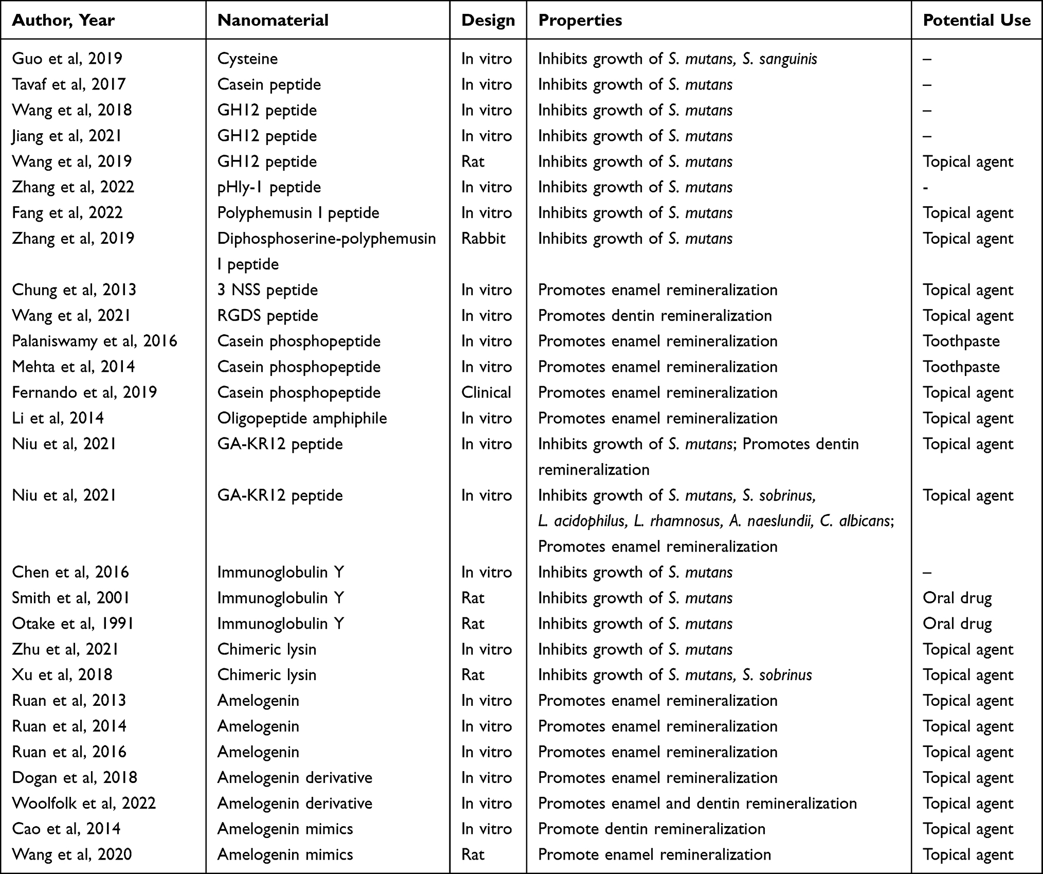 |
Table 2 Studies on Amino Acid-Based Nanomaterials for Caries Management |
Peptides are short chains of amino acids that the amide type of covalent chemical bonds link. The constitutions of peptides vary based on types and sequences of amino acids, which generate unique properties of each peptide. Antimicrobial peptides have potent, broad-spectrum antimicrobial properties against oral pathogens. Researchers obtain antimicrobial peptides from extracting immune responses to microbial infections or artificial synthesis. A casein peptide that the partial proteolysis of milk caseins produces inhibits biofilm formation independently. It can be combined and used with silver nanoparticles to improve significantly the antibacterial effect against S. mutans.14 Some antimicrobial peptides, including GH12 peptide, pHly-1 peptide, polyphemusin I peptide and diphosphoserine-polyphemusin I peptide, have shown an antibacterial effect on the biomass and cariogenic activity of S. mutans in vitro.15–20 Notably, the GH-12 peptide exhibits good biocompatibility and a strong antibacterial effect against S. mutans on the molars of rats in vivo. GH-12 peptide is a potential topical agent to inhibit dental caries.16 Moreover, topical use of diphosphoserine-polyphemusin I peptide reduced the plaque on incisors of rabbits in vivo.20 Besides a strong antimicrobial effect, some peptides have remineralising properties that can be used for caries management. Two studies demonstrated that adding 3 NSS peptide and RGDS peptide into bioactive glass can enhance bio-remineralization of enamel and dentin.21,22 Casein phosphopeptide remineralizes the enamel when combined with stannous fluoride and amorphous calcium phosphate nanoparticles in toothpaste.23–25 Oligopeptide amphiphile is a biomimetic remineralising material. It can self-assemble into nano-fibers in the presence of calcium ions and work with the calcium ions to foster the biomimetic mineralization on enamel surface.26 Furthermore, GA-KR12 peptide is a dual-function peptide with an antibacterial and remineralising effect. It can inhibit the growth of multiple cariogenic microorganisms and enhance the remineralization on enamel and dentin.27,28
Some proteins can inhibit bacterial growth. Researchers used immunoglobulin Y as an oral drug and found that it can inhibit the growth of S. mutans through passive immunity in rats.29,30 Loading immunoglobulin Y on hydroxyapatite also improved the antibacterial property of hydroxyapatite.31 Chimeric lysin can cause high lytic activity in cariogenic microorganisms. The S. mutans and Streptococcus sobrinus (S. sobrinus) biofilm viability of teeth treated with chimeric lysin significantly decreased on the premolars of rats.32 The chimeric lysin can be combined with amorphous calcium phosphate in carboxymethyl chitosan nanogel to inhibit biofilm formation and promote remineralization.33
Apart from their antimicrobial properties, some proteins can act as biomimetic materials to regulate precipitation of calcium phosphate and protect teeth from demineralization. They can induce remineralization and form an enamel-like apatite under a physiological condition to prevent caries. Therefore, proteins are bioavailable alternatives to repair defective enamel. Amelogenin is a critical protein for controlling the organized growth of apatite crystals, which play a direct role in nucleation, crystal growth and the stretch spacing of hydroxyapatite crystallites.34 Amelogenin-containing chitosan hydrogels can induce the apatite crystals to form on the damaged human enamel.35–37 Amelogenin derivative is a small domain derived from native amelogenin. It can construct a mineral layer on the demineralized enamel.38 Amelogenin derivative also generates biomimetical remineralization on silver diamine fluoride-treated dentin.39 Besides, amelogenin mimics can bind to collagen fibrils and mimic the bio-mineralization process on enamel and dentin.40,41
Natural Extractive-Based Nanomaterials
Although nanoparticles can be obtained using various physicochemical methods, green synthesis of nanoparticles using natural extracts minimizes the usage of toxic chemicals.42 Table 3 summarizes the 17 publications on the natural extractive-based nanomaterials for managing dental caries.
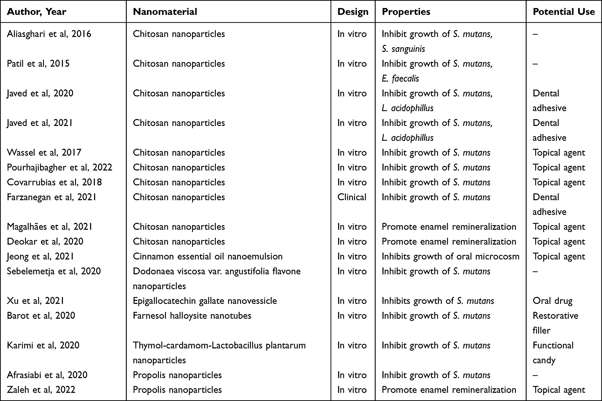 |
Table 3 Studies on Natural Extractive-Based Nanomaterials for Caries Management |
Chitosan is a natural polymer obtained by the alkaline hydrolysis of chitin. Chitosan is widely used in biomedicine materials because it has promising remineralising, antibacterial and biocompatible properties without toxicity.43,44 Chitosan nanoparticles have shown a remineralization ability on demineralized enamel.45,46 Due to their nanometric dimensions, chitosan nanomaterials exhibit a greater ability to penetrate the oral biofilm than ordinary chitosan does. Chitosan nanomaterials can be used as antimicrobial agents for caries management because they inhibit the formation of S. mutans and S. sanguinis biofilm.47 Moreover, chitosan nanoparticles can be applied as the matrix loaded with propolis, miswak, rutin and emodin to develop the anti-caries agent and dental varnish.48–50 The chitosan nanoparticles have also been used as the capping agent combined with copper, copper oxide, zinc oxide and titanium dioxide nanoparticles to enhance the antibacterial property of dentin adhesive.51–54 A clinical study demonstrated that orthodontic adhesive containing chitosan-titanium dioxide nanoparticles could prevent white spot lesions. The orthodontic adhesive containing chitosan-titanium dioxide nanoparticles significantly inhibits the S. mutans compared to the commercial orthodontic adhesive.54
Moreover, plant extractives attract researchers’ interest due to their antibacterial properties for caries management. Cinnamon essential oil nanoemulsion exhibited a strongly inhibited the growth of an oral microcosm.55 Dodonaea viscosa var. angustifolia flavone nanoparticles significantly reduced the formation and acid production of S. mutans biofilm.56 Epigallocatechin gallate is an extracted polymer from green tea that shows remarkable anti-cariogenic bioactivity with poor stability.57 To increase the stability and efficacy of epigallocatechin gallate, researchers entrapped it in the oral nanovesicle to inhibit the production of glucan and formation of S. mutans biofilm.57 Farnesol is present in essential oils from many plants. Adding Farnesol halloysite nanotubes into composite resin creates antibacterial activity against S. mutans without compromising mechanical properties.58 Thymol-cardamom-Lactobacillus plantarum nanoparticles are prepared as functional candies to inhibit the growth of S. mutans for managing dental caries.59 In addition, propolis nanoparticles reduce the virulence of S. mutans and promote remineralization on enamel.60,61
Synthetic Organic Nanomaterials
Apart from natural extractive organic compounds, many synthetic organic nanomaterials have been investigated as anti-caries agents. They are often used as active components with carriers to construct hybrid nanomaterials. Table 4 summarizes 15 publications on the synthetic organic nanomaterials for managing dental caries. All were in vitro studies, and the systematic search yielded no clinical or animal studies on synthetic organic nanomaterials for caries management.
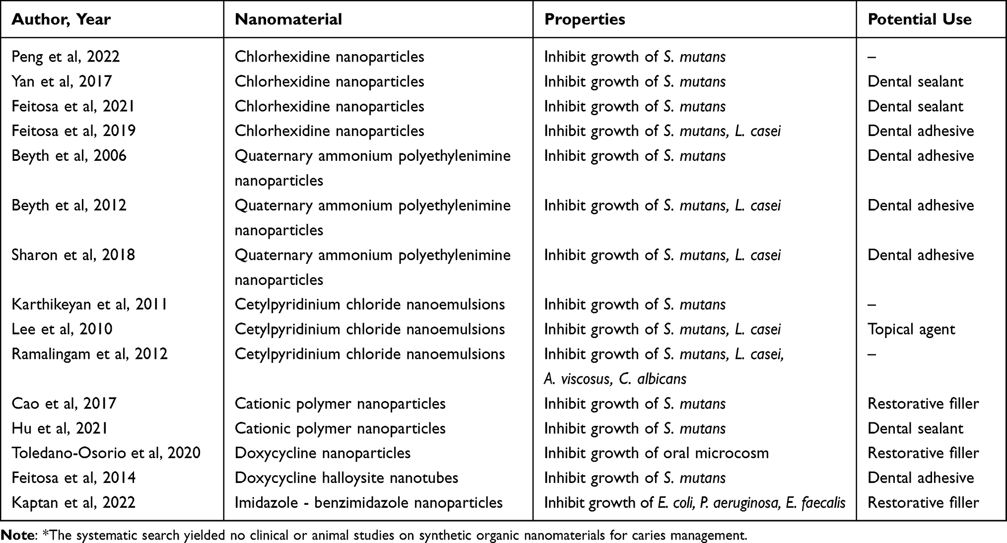 |
Table 4 In vitro Studies* on Synthetic Organic Nanomaterials for Caries Management |
Chlorhexidine is an antibacterial agent commonly used in dental practice as an antiseptic and disinfectant. Chlorhexidine nanoparticles inhibit S. mutans and Lactobacillus casei (L. casei) from growing and from forming their biofilms. They can be used as the filler in dentin adhesive and dental sealant for caries management.62,63 Nano-carriers loaded with chlorhexidine can build a drug delivery system for advanced antibacterial properties.64 Researchers have used various types of nano-carriers to synthesize a desirable anti-caries agent, including nanotubes, mesoporous nanoparticles and polymers. Chlorhexidine-loaded halloysite nanotubes effectively kill S. mutans, which can be used in a pit-and-fissure sealant. Additionally, some researchers have utilized poly (N,N-dimethylaminoethyl methacrylate-co-2-hydroxyethyl methacrylate), a bio reagent for cell culture, as the pH-sensitive nano-carrier and chlorhexidine as the active ingredients. The system could control the release rate of chlorhexidine according to the pH value and it could exhibit lower cytotoxicity against human oral keratinocytes than free chlorhexidine does. The chlorhexidine in these delivery systems showed the same antibacterial effects on S. mutans biofilms as free chlorhexidine.65 A research group synthesized the chlorhexidine encapsulated with mesoporous silica and found it could improve the anti-microbial performance of glass ionomer cement without affecting its mechanical properties.66
Quaternary ammonium compound nanoparticles are cationic surfactants used to prevent caries. They are broad-spectrum antimicrobials with low toxicity.67 Quaternary ammonium polyethylenimine nanoparticles inhibit oral pathogens, such as S. mutans and L. casei. They can be incorporated into glass ionomer cements and resin composite.68,69 Adding quaternary ammonium polyethylenimine nanoparticles did not compromize the composite materials’ flexural modulus or the flexural strength.70 Cetylpyridinium chloride is another quaternary ammonium compound used in mouthwashes and toothpastes. Cetylpyridinium chloride nanoemulsions can inhibit S. mutans, L. casei, Actinomyces viscosus (A. viscosus) and Candida albicans (C. albicans) when applied to tooth surfaces.71–73 Furthermore, quaternary ammonium compound nanoparticles can cooperate with silver to synthesis a core-shell silver bromide–cationic polymer nanocomposite. This composite showed long-term antimicrobial properties against S. mutans. The cationic-polymer shell provides a sustained antibacterial effect in dental resins and sealants.74,75
Researchers also developed synthetic nanomaterials against cariogenic microorganisms. Doxycycline-nanoparticles-doped dental composite resins can reduce viability of oral biofilm.76 Another study encapsulated doxycycline in the halloysite nanotube to enhance the antibacterial effect of dental adhesive against S. mutans.77 Many fungicides contain imidazole and benzimidazole. Researchers synthesized imidazole and benzimidazole nanoparticles and incorporated them to inhibit bacteria. Studies have shown these antibacterial composite resins inhibit Escherichia coli (E. coli), Pseudomonas aeruginosa (P. aeruginosa) and Enterococcus faecalis (E. faecalis).78
Carbon-Based Nanomaterials
Researchers also developed carbon-based nanomaterials for caries management. Carbon-based nanomaterials exhibit high mechanical strength.79,80 They have antibacterial and remineralising properties. Table 5 summarizes 13 publications on carbon-based nanomaterials for managing dental caries. All were in vitro studies, and the systematic search yielded no clinical or animal studies on carbon-based nanomaterials for caries management. The carbon-based nanomaterials in these publications can be categorized as carbon nanoparticles, graphene nanoplatelets, graphene oxide nanoplatelets and reduced graphene oxide nanoplatelets.
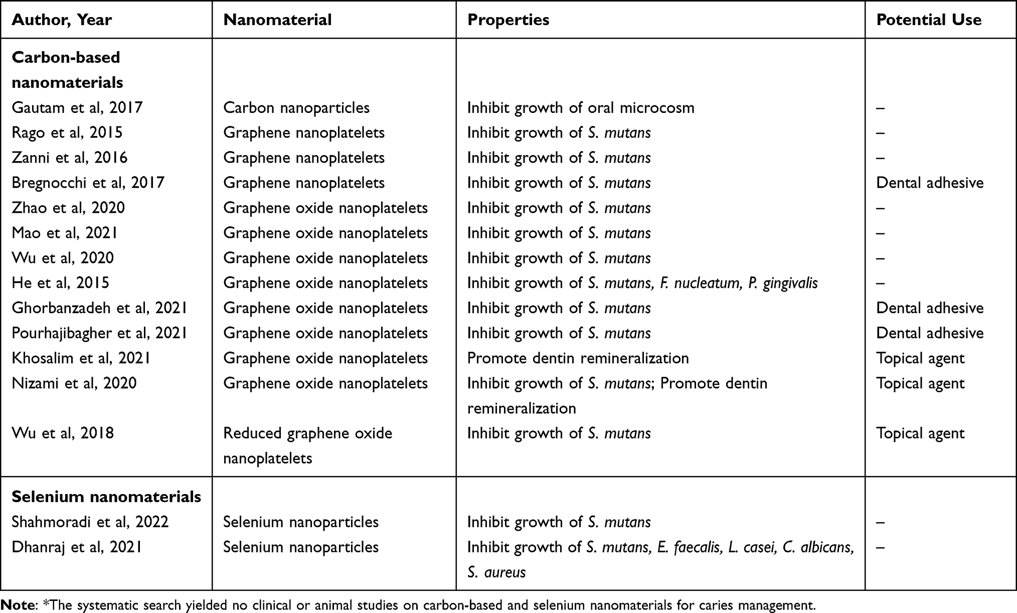 |
Table 5 In vitro Studies* on Carbon-Based and Selenium Nanomaterials for Caries Management |
Carbon nanoparticles consist of pure carbon in various structures. They inhibit the growth of oral microcosms without toxicity to human cells.81 Graphene nanoplatelets are single layers of carbon allotrope arranged in a 2-dimensional honeycomb lattice nanoplatelet. Graphene nanoplatelets provide antibacterial protection against S. mutans.82 They can be used as the filler in dental adhesive to prevent caries without affecting the mechanical properties of adhesive.83,84 Graphene nanoplatelets can be platform-doped with other nanoparticles, such as zinc oxide nanoparticles, to obtain the enhanced inhibitory effect of biofilm formation.85 Graphene oxide nanoplatelets are oxidized graphene nanoplatelets.86,87 They can be added into an orthodontic adhesive to prevent white spot lesions after orthodontic treatment because of their antibacterial effects against S. mutans.88,89 Graphene oxide nanoplatelets can destroy the cell wall and membrane of S. mutans, Fusobacterium nucleatum (F. nucleatum) and Porphyromonas gingivalis (P. gingivalis) to inhibit biofilm formation.90–92 In addition, graphene oxide nanoplatelets showed a remineralising ability to occlude the exposed dentinal tubules and to promote the growth of hydroxyapatite crystal on demineralized dentin.93 Due to abundant oxygen groups on the surface of graphene oxide nanoplatelets, graphene oxide nanoplatelets can be functionalized with diverse antiseptics to give them enhanced antibacterial properties. Graphene oxide nanoplatelets combined with silver and copper nanoparticles have antimicrobial effects against S. mutans, with a superior aesthetic effect on dentin compared to silver diamine fluoride.94,95 A report also showed that loading graphene oxide nanoplatelets with polyethylenimine complexes and with antisense vicR RNA can reduce virulent-associated gene expressions and reduce the S. mutans biofilm from forming.96 Reduced graphene oxide nanoplatelets are another graphene derivative reduced from graphene oxide nanoplatelets. Reduced graphene oxide nanoplatelets have greater surface area and better mechanical strength than graphene oxide nanoplatelets do. Researchers found that the reduced graphene oxide nanoplatelets could link with silver nanoparticles to inhibit the growth of S. mutans biofilm.97
Selenium Nanomaterials
Selenium is an indispensable trace element for the human body’s cellular functions. Selenium in antioxidant enzymes and functional protein molecules plays a crucial role in reducing oxidative stress.98 Table 5 shows the 2 publications on selenium nanomaterials for managing dental caries. All were in vitro studies, and the systematic search yielded no clinical or animal studies on selenium nanomaterials for caries management. Selenium nanoparticles can inhibit growth of S. mutans, E. faecalis, L. casei, C. albicans and Staphylococcus aureus (S. aureus) and their biofilms.98,99 However, these studies were not performed on tooth tissue. Further studies are needed to explore the application of selenium nanomaterials for management of dental caries.
Conclusion
The most common type of non-metallic nanomaterials for caries management is organic nanomaterials. The literature showed non-metallic nanomaterials have antibacterial and/or remineralising properties. Non-metallic nanomaterials can be incorporated into dental sealants, toothpaste, dental adhesives, topical agents and even candies and drugs. However, the majority of the publications are in vitro studies, and only 2 publications are clinical studies.
Acknowledgments
This study is supported by the National Natural Science Foundation of China (NSFC) – General Program No. 81870812.
Disclosure
The authors report no conflicts of interest in this work.
References
1. Collaborators GDaIIaP. Global, regional, and national incidence, prevalence, and years lived with disability for 354 diseases and injuries for 195 countries and territories, 1990–2017: a systematic analysis for the global burden of disease study 2017. Lancet. 2018;392(10159):1789–1858. doi:10.1016/S0140-6736(18)32279-7
2. Selwitz RH, Ismail AI, Pitts NB. Dental caries. Lancet. 2007;369(9555):51–59. doi:10.1016/S0140-6736(07)60031-2
3. Niu JY, Yin IX, Wu WKK, Li QL, Mei ML, Chu CH. Antimicrobial peptides for the prevention and treatment of dental caries: a concise review. Arch Oral Biol. 2021;122:105022. doi:10.1016/j.archoralbio.2020.105022
4. Petersen PE. World Health Organization global policy for improvement of oral health--World Health Assembly 2007. Int Dent J. 2008;58(3):115–121. doi:10.1111/j.1875-595X.2008.tb00185.x
5. Hurlbutt M, Young DA. A best practices approach to caries management. J Evid Based Dent Pract. 2014;14:77–86. doi:10.1016/j.jebdp.2014.03.006
6. Pitts N, Zero D. White paper on dental caries prevention and management. FDI World Dent Fed. 2016;1:3–9.
7. Zhu T, Huang Z, Shu X, Zhang C, Dong Z, Peng Q. Functional nanomaterials and their potentials in antibacterial treatment of dental caries. Colloids Surf B Biointerfaces. 2022;218:112761. doi:10.1016/j.colsurfb.2022.112761
8. Makkar H, Patri G. Fabrication and appraisal of poly (lactic-co-glycolic acid) - moxifloxacin nanoparticles using vitamin E-TPGS: a potential intracanal drug delivery agent. J Clin Diagn Res. 2017;11(6):Zc05–Zc08. doi:10.7860/JCDR/2017/27633.9957
9. Yin IX, Zhang J, Zhao IS, Mei ML, Li Q, Chu CH. The antibacterial mechanism of silver nanoparticles and its application in dentistry. Int J Nanomedicine. 2020;15:2555–2562. doi:10.2147/IJN.S246764
10. Mammari N, Lamouroux E, Boudier A, Duval RE. Current knowledge on the oxidative-stress-mediated antimicrobial properties of metal-based nanoparticles. Microorganisms. 2022;10:2. doi:10.3390/microorganisms10020437
11. Clarkson BH, Exterkate RA. Noninvasive dentistry: a dream or reality? Caries Res. 2015;49(Suppl 1):11–17. doi:10.1159/000380887
12. AlShwaimi E, Bogari D, Ajaj R, Al-Shahrani S, Almas K, Majeed A. In vitro antimicrobial effectiveness of root canal sealers against Enterococcus faecalis: a systematic review. J Endod. 2016;42(11):1588–1597. doi:10.1016/j.joen.2016.08.001
13. Guo X, Liu S, Zhou X, et al. Effect of D-cysteine on dual-species biofilms of Streptococcus mutans and Streptococcus sanguinis. Sci Rep. 2019;9(1):1–8.
14. Tavaf Z, Tabatabaei M, Khalafi-Nezhad A, Panahi F. Evaluation of antibacterial, antibofilm and antioxidant activities of synthesized silver nanoparticles (AgNPs) and casein peptide fragments against Streptococcus mutans. Eur J Integr Med. 2017;12:163–171. doi:10.1016/j.eujim.2017.05.011
15. Wang Y, Wang X, Jiang W, et al. Antimicrobial peptide GH12 suppresses cariogenic virulence factors of Streptococcus mutans. J Oral Microbiol. 2018;10(1):1442089. doi:10.1080/20002297.2018.1442089
16. Wang Y, Zeng Y, Wang Y, et al. Antimicrobial peptide GH12 targets Streptococcus mutans to arrest caries development in rats. J Oral Microbiol. 2019;11(1):1549921. doi:10.1080/20002297.2018.1549921
17. Jiang W, Luo J, Wang Y, et al. The pH-responsive property of antimicrobial peptide GH12 enhances its anticaries effects at acidic pH. Caries Res. 2021;55(1):21–31. doi:10.1159/000508458
18. Zhang P, Wu S, Li J, et al. Dual-sensitive antibacterial peptide nanoparticles prevent dental caries. Theranostics. 2022;12(10):4818–4833. doi:10.7150/thno.73181
19. Fang Z, Zhang Y, Cao CY, Li QL, Wong HM. Constructing an anti-S. mutans and mineralizing membrane by combination self-assembled lysozyme with antimicrobial peptide. Mater Des. 2022;220:110891.
20. Zhang LY, Fang ZH, Li QL, Cao CY. A tooth-binding antimicrobial peptide to prevent the formation of dental biofilm. J Mater Sci Mater Med. 2019;30(4):45. doi:10.1007/s10856-019-6246-6
21. Chung HY, Li CC. Microstructure and nanomechanical properties of enamel remineralized with asparagine-serine-serine peptide. Mater Sci Eng C Mater Biol Appl. 2013;33(2):969–973. doi:10.1016/j.msec.2012.11.031
22. Wang SN, Sheng XY, Huang GB, Li QJ, Dong YM. Dentin remineralization induced by nanobioactive glass in association with RGDS peptide. Mater Today Commin. 2021;28:102515.
23. Palaniswamy UK, Prashar N, Kaushik M, Lakkam SR, Arya S, Pebbeti S. A comparative evaluation of remineralizing ability of bioactive glass and amorphous calcium phosphate casein phosphopeptide on early enamel lesion. Dent Res J. 2016;13(4):297–302. doi:10.4103/1735-3327.187872
24. Mehta AB, Kumari V, Jose R, Izadikhah V. Remineralization potential of bioactive glass and casein phosphopeptide-amorphous calcium phosphate on initial carious lesion: an in-vitro pH-cycling study. J Conserv Dent. 2014;17(1):3–7. doi:10.4103/0972-0707.124085
25. Fernando JR, Shen P, Sim CPC, et al. Self-assembly of dental surface nanofilaments and remineralisation by SnF(2) and CPP-ACP nanocomplexes. Sci Rep. 2019;9(1):1285. doi:10.1038/s41598-018-37580-w
26. Li QL, Ning TY, Cao Y, Zhang WB, Mei ML, Chu CH. A novel self-assembled oligopeptide amphiphile for biomimetic mineralization of enamel. BMC Biotechnol. 2014;14:32. doi:10.1186/1472-6750-14-32
27. Niu JY, Yin IX, Wu WKK, Li QL, Mei ML, Chu CH. A novel dual-action antimicrobial peptide for caries management. J Dent. 2021;111:103729. doi:10.1016/j.jdent.2021.103729
28. Niu JY, Yin IX, Wu WKK, Li QL, Mei ML, Chu CH. Remineralising dentine caries using an artificial antimicrobial peptide: an in vitro study. J Dent. 2021;111:103736.
29. Smith DJ, King WF, Godiska R, Tuomanen EL. Passive transfer of immunoglobulin Y antibody to Streptococcus mutans glucan binding protein B can confer protection against experimental dental caries. Infect Immun. 2001;69(5):3135–3142. doi:10.1128/IAI.69.5.3135-3142.2001
30. Otake S, Nishihara Y, Makimura M, et al. Protection of rats against dental caries by passive immunization with hen-egg-yolk antibody (IgY). J Dent Res. 1991;70(3):162–166. doi:10.1177/00220345910700030101
31. Chen X, Yang B, Qi C, et al. DNA-templated microwave-hydrothermal synthesis of nanostructured hydroxyapatite for storing and sustained release of an antibacterial protein. Dalton Trans. 2016;45(4):1648–1656. doi:10.1039/C5DT03357H
32. Xu J, Yang H, Bi Y, Li W, Wei H, Li Y. Activity of the chimeric lysin ClyR against common gram-positive oral microbes and its anticaries efficacy in rat models. Viruses. 2018;10(7):380. doi:10.3390/v10070380
33. Zhu Y, Yan J, Mujtaba BM, Li Y, Wei H, Huang S. The dual anti-caries effect of carboxymethyl chitosan nanogel loaded with chimeric lysin ClyR and amorphous calcium phosphate. Eur J Oral Sci. 2021;129(3):e12784. doi:10.1111/eos.12784
34. Moradian-Oldak J. Protein-mediated enamel mineralization. Front Biosci. 2012;17(6):1996–2023. doi:10.2741/4034
35. Ruan QC, Zhang YZ, Yang XD, Nutt S, Moradian-Oldak J. An amelogenin-chitosan matrix promotes assembly of an enamel-like layer with a dense interface. Acta Biomater. 2013;9(7):7289–7297. doi:10.1016/j.actbio.2013.04.004
36. Ruan Q, Moradian-Oldak J. Development of amelogenin-chitosan hydrogel for in vitro enamel regrowth with a dense interface. J Vis Exp. 2014;89. doi:10.3791/51606
37. Ruan Q, Liberman D, Bapat R, Chandrababu KB, Phark JH, Moradian-Oldak J. Efficacy of amelogenin-chitosan hydrogel in biomimetic repair of human enamel in pH-cycling systems. J Biomed Eng Inform. 2016;2(1):119–128. doi:10.5430/jbei.v2n1p119
38. Dogan S, Fong H, Yucesoy DT, et al. Biomimetic tooth repair: amelogenin-derived peptide enables in vitro remineralization of human enamel. ACS Biomater Sci Eng. 2018;4(5):1788–1796. doi:10.1021/acsbiomaterials.7b00959
39. Woolfolk SK, Cloyd AK, Ye Q, et al. Peptide-enabled nanocomposites offer biomimetic reconstruction of silver diamine fluoride-treated dental tissues. Polymers. 2022;14(7):1368. doi:10.3390/polym14071368
40. Cao Y, Liu W, Ning T, et al. A novel oligopeptide simulating dentine matrix protein 1 for biomimetic mineralization of dentine. Clin Oral Investig. 2014;18(3):873–881. doi:10.1007/s00784-013-1035-y
41. Wang D, Deng J, Deng X, Fang C, Zhang X, Yang P. Controlling enamel remineralization by amyloid-like amelogenin mimics. Adv Mater. 2020;32(31):e2002080. doi:10.1002/adma.202002080
42. Nguyen NTT, Nguyen LM, Nguyen TTT, Liew RK, Nguyen DTC, Tran TV. Recent advances on botanical biosynthesis of nanoparticles for catalytic, water treatment and agricultural applications: a review. Sci Total Environ. 2022;827:154160. doi:10.1016/j.scitotenv.2022.154160
43. Kou SG, Peters LM, Mucalo MR. Chitosan: a review of sources and preparation methods. Int J Biol Macromol. 2021;169:85–94. doi:10.1016/j.ijbiomac.2020.12.005
44. Li Y, Chi YQ, Yu CH, et al. Drug-free and non-crosslinked chitosan scaffolds with efficient antibacterial activity against both gram-negative and gram-positive bacteria. Carbohydr Polym. 2020;241:116386. doi:10.1016/j.carbpol.2020.116386
45. Deokar KK, Shashikiran ND, Maurya A, et al. Comparative evaluation of chitosan nanoparticles, silver diamine fluoride and acidulated phosphate fluoride gel on microhardness of artificial carious lesions created on extracted teeth. J Clin Diagn Res. 2020;14(11):ZC20–ZC23.
46. Magalhães TC, Teixeira NM, França RS, et al. Synthesis of a chitosan nanoparticle suspension and its protective effects against enamel demineralization after an in vitro cariogenic challenge. J Appl Oral Sci. 2021;29:e20210120. doi:10.1590/1678-7757-2021-0120
47. Aliasghari A, Rabbani Khorasgani M, Vaezifar S, Rahimi F, Younesi H, Khoroushi M. Evaluation of antibacterial efficiency of chitosan and chitosan nanoparticles on cariogenic streptococci: an in vitro study. Iran J Microbiol. 2016;8(2):93–100.
48. Wassel MO, Khattab MA. Antibacterial activity against Streptococcus mutans and inhibition of bacterial induced enamel demineralization of propolis, miswak, and chitosan nanoparticles based dental varnishes. J Adv Res. 2017;8(4):387–392. doi:10.1016/j.jare.2017.05.006
49. Patil AG, Jobanputra AH. Rutin-chitosan nanoparticles: fabrication, characterization and application in dental disorders. Polym Plast Technol Eng. 2015;54(2):202–208. doi:10.1080/03602559.2014.935425
50. Pourhajibagher M, Keshavarz Valian N, Bahador A. Theranostic nanoplatforms of emodin-chitosan with blue laser light on enhancing the anti-biofilm activity of photodynamic therapy against Streptococcus mutans biofilms on the enamel surface. BMC Microbiol. 2022;22(1):68. doi:10.1186/s12866-022-02481-6
51. Covarrubias C, Trepiana D, Corral C. Synthesis of hybrid copper-chitosan nanoparticles with antibacterial activity against cariogenic Streptococcus mutans. Dent Mater J. 2018;37(3):379–384. doi:10.4012/dmj.2017-195
52. Javed R, Rais F, Fatima H, et al. Chitosan encapsulated ZnO nanocomposites: fabrication, characterization, and functionalization of bio-dental approaches. Mater Sci Eng C Mater Biol Appl. 2020;116:111184. doi:10.1016/j.msec.2020.111184
53. Javed R, Rais F, Kaleem M, et al. Chitosan capping of CuO nanoparticles: facile chemical preparation, biological analysis, and applications in dentistry. Int J Biol Macromol. 2021;167:1452–1467. doi:10.1016/j.ijbiomac.2020.11.099
54. Farzanegan F, Shahabi M, Niazi AE, Soleimanpour S, Shafaee H, Rangrazi A. Effect of the addition of Chitosan and TiO 2 nanoparticles on antibacterial properties of an orthodontic composite in fixed orthodontic treatment: a randomized clinical trial study. Biomed Phys Eng Express. 2021;7(4):045017. doi:10.1088/2057-1976/ac0609
55. Jeong YJ, Kim HE, Han SJ, Choi JS. Antibacterial and antibiofilm activities of cinnamon essential oil nanoemulsion against multi-species oral biofilms. Sci Rep. 2021;11(1):5911. doi:10.1038/s41598-021-85375-3
56. Sebelemetja M, Moeno S, Patel M. Anti-acidogenic, anti-biofilm and slow release properties of Dodonaea viscosa var. angustifolia flavone stabilized polymeric nanoparticles. Arch Oral Biol. 2020;109:104586. doi:10.1016/j.archoralbio.2019.104586
57. Xu X, Dai Z, Zhang Z, et al. Fabrication of oral nanovesicle in-situ gel based on epigallocatechin gallate phospholipid complex: application in dental anti-caries. Eur J Pharmacol. 2021;897:173951. doi:10.1016/j.ejphar.2021.173951
58. Barot T, Rawtani D, Kulkarni P, Hussain CM, Akkireddy S. Physicochemical and biological assessment of flowable resin composites incorporated with farnesol loaded halloysite nanotubes for dental applications. J Mech Behav Biomed Mater. 2020;104:103675. doi:10.1016/j.jmbbm.2020.103675
59. Karimi N, Jabbari V, Nazemi A, et al. Thymol, cardamom and Lactobacillus plantarum nanoparticles as a functional candy with high protection against Streptococcus mutans and tooth decay. Microb Pathog. 2020;148:104481. doi:10.1016/j.micpath.2020.104481
60. Afrasiabi S, Pourhajibagher M, Chiniforush N, Bahador A. Propolis nanoparticle enhances the potency of antimicrobial photodynamic therapy against Streptococcus mutans in a synergistic manner. Sci Rep. 2020;10(1):15560. doi:10.1038/s41598-020-72119-y
61. Zaleh AA, Salehi-Vaziri A, Pourhajibagher M, Bahador A. The synergistic effect of nano-propolis and curcumin-based photodynamic therapy on remineralization of white spot lesions: an ex vivo study. Photodiagnosis Photodyn Ther. 2022;38:102789. doi:10.1016/j.pdpdt.2022.102789
62. Feitosa S, Carreiro AFP, Martins VM, Platt JA, Duarte S. Effect of a chlorhexidine-encapsulated nanotube modified pit-and-fissure sealant on oral biofilm. Dent Mater J. 2021;40(3):758–765. doi:10.4012/dmj.2020-241
63. Feitosa SA, Palasuk J, Geraldeli S, Windsor LJ, Bottino MC. Physicochemical and biological properties of novel chlorhexidine-loaded nanotube-modified dentin adhesive. J Biomed Mater Res B Appl Biomater. 2019;107(3):868–875. doi:10.1002/jbm.b.34183
64. Lim KS, Kam PC. Chlorhexidine--pharmacology and clinical applications. Anaesth Intensive Care. 2008;36(4):502–512. doi:10.1177/0310057X0803600404
65. Peng X, Han Q, Zhou X, et al. Effect of pH-sensitive nanoparticles on inhibiting oral biofilms. Drug Deliv. 2022;29(1):561–573. doi:10.1080/10717544.2022.2037788
66. Yan H, Yang H, Li K, Yu J, Huang C. Effects of chlorhexidine-encapsulated mesoporous silica nanoparticles on the anti-biofilm and mechanical properties of glass ionomer cement. Molecules. 2017;22(7):1225. doi:10.3390/molecules22071225
67. Zhu GY, Lu BY, Zhang TX, et al. Antibiofilm effect of drug-free and cationic poly (D,L-lactide-co-glycolide) nanoparticles via nano-bacteria interactions. Nanomedicine. 2018;13(10):1093–1106. doi:10.2217/nnm-2017-0391
68. Beyth N, Yudovin-Farber I, Bahir R, Domb AJ, Weiss EI. Antibacterial activity of dental composites containing quaternary ammonium polyethylenimine nanoparticles against Streptococcus mutans. Biomaterials. 2006;27(21):3995–4002. doi:10.1016/j.biomaterials.2006.03.003
69. Beyth N, Pilo R, Weiss EI. Antibacterial activity of dental cements containing quaternary ammonium polyethylenimine nanoparticles. J Nanomater. 2012;2012:1–6. doi:10.1155/2012/814763
70. Sharon E, Sharabi R, Eden A, et al. Antibacterial activity of orthodontic cement containing quaternary ammonium polyethylenimine nanoparticles adjacent to orthodontic brackets. Int J Environ Res Public Health. 2018;15(4):606. doi:10.3390/ijerph15040606
71. Karthikeyan R, Amaechi BT, Rawls HR, Lee VA. Antimicrobial activity of nanoemulsion on cariogenic Streptococcus mutans. Arch Oral Biol. 2011;56(5):437–445. doi:10.1016/j.archoralbio.2010.10.022
72. Lee VA, Karthikeyan R, Rawls HR, Amaechi BT. Anti-cariogenic effect of a cetylpyridinium chloride-containing nanoemulsion. J Dent. 2010;38(9):742–749. doi:10.1016/j.jdent.2010.06.001
73. Ramalingam K, Amaechi BT, Ralph RH, Lee VA. Antimicrobial activity of nanoemulsion on cariogenic planktonic and biofilm organisms. Arch Oral Biol. 2012;57(1):15–22. doi:10.1016/j.archoralbio.2011.07.001
74. Cao W, Zhang Y, Wang X, et al. Development of a novel resin-based dental material with dual biocidal modes and sustained release of Ag(+) ions based on photocurable core-shell AgBr/cationic polymer nanocomposites. J Mater Sci Mater Med. 2017;28(7):103. doi:10.1007/s10856-017-5918-3
75. Hu YT, Yu F, Tang XY, et al. The antibacterial effect and physical performance of pit and fissure sealants based on an antibacterial core-shell nanocomposite. J Mech Behav Biomed Mater. 2021;117:104414. doi:10.1016/j.jmbbm.2021.104414
76. Toledano-Osorio M, Osorio R, Aguilera FS, et al. Polymeric nanoparticles protect the resin-dentin bonded interface from cariogenic biofilm degradation. Acta Biomater. 2020;111:316–326. doi:10.1016/j.actbio.2020.05.002
77. Feitosa SA, Palasuk J, Kamocki K, et al. Doxycycline-encapsulated nanotube-modified dentin adhesives. J Dent Res. 2014;93(12):1270–1276. doi:10.1177/0022034514549997
78. Kaptan Usul S, Aslan A, Lüleci HB, et al. Investigation of antimicrobial and mechanical effects of functional nanoparticles in novel dental resin composites. J Dent. 2022;123:104180. doi:10.1016/j.jdent.2022.104180
79. Lee SM, Yoo KH, Yoon SY, et al. Enamel anti-demineralization effect of orthodontic adhesive containing bioactive glass and graphene oxide: an in-vitro study. Materials. 2018;11(9):1728. doi:10.3390/ma11091728
80. Sun L, Yan Z, Duan Y, Zhang J, Liu B. Improvement of the mechanical, tribological and antibacterial properties of glass ionomer cements by fluorinated graphene. Dent Mater. 2018;34(6):e115–e127. doi:10.1016/j.dental.2018.02.006
81. Gautam G, Jha D, Gaurav SS, Sharma AK, Kumar P, Gautam HK. Synthesis of carbon nanoparticles from mustard oil and evaluation of their antibacterial activity against dental caries. Micro Nano Lett. 2017;12(10):799–802. doi:10.1049/mnl.2017.0293
82. Xia MY, Xie Y, Yu CH, et al. Graphene-based nanomaterials: the promising active agents for antibiotics-independent antibacterial applications. J Control Release. 2019;307:16–31. doi:10.1016/j.jconrel.2019.06.011
83. Rago I, Bregnocchi A, Zanni E, et al. Antimicrobial activity of graphene nanoplatelets against streptococcus mutans.
84. Bregnocchi A, Zanni E, Uccelletti D, et al. Graphene-based dental adhesive with anti-biofilm activity. J Nanobiotechnology. 2017;15(1):89. doi:10.1186/s12951-017-0322-1
85. Zanni E, Chandraiahgari CR, De Bellis G, et al. Zinc oxide nanorods-decorated graphene nanoplatelets: a promising antimicrobial agent against the cariogenic bacterium Streptococcus mutans. Nanomaterials. 2016;6(10):179. doi:10.3390/nano6100179
86. Lu B-Y, Zhu G-Y, Yu C-H, et al. Functionalized graphene oxide nanosheets with unique three-in-one properties for efficient and tunable antibacterial applications. Nano Res. 2021;14(1):185–190. doi:10.1007/s12274-020-3064-6
87. Alam K, Jo YY, Park CK, Cho H. Synthesis of graphene oxide using atmospheric plasma for prospective biological applications. Int J Nanomedicine. 2020;15:5813–5824. doi:10.2147/IJN.S254860
88. Ghorbanzadeh R, Hosseinpour Nader A, Salehi-Vaziri A. The effects of bimodal action of photodynamic and photothermal therapy on antimicrobial and shear bond strength properties of orthodontic composite containing nano-graphene oxide. Photodiagnosis Photodyn Ther. 2021;36:102589. doi:10.1016/j.pdpdt.2021.102589
89. Pourhajibagher M, Bahador A. Orthodontic adhesive doped with nano-graphene oxide: physico-mechanical and antimicrobial properties. Folia Med. 2021;63(3):413–421. doi:10.3897/folmed.63.e53716
90. He J, Zhu X, Qi Z, et al. Killing dental pathogens using antibacterial graphene oxide. ACS Appl Mater Interfaces. 2015;7(9):5605–5611. doi:10.1021/acsami.5b01069
91. Zhao M, Shan T, Wu Q, Gu L. The antibacterial effect of graphene oxide on Streptococcus mutans. J Nanosci Nanotechnol. 2020;20(4):2095–2103. doi:10.1166/jnn.2020.17319
92. Yu CH, Chen GY, Xia MY, et al. Understanding the sheet size-antibacterial activity relationship of graphene oxide and the nano-bio interaction-based physical mechanisms. Colloids Surf B Biointerfaces. 2020;191:111009. doi:10.1016/j.colsurfb.2020.111009
93. Khosalim IP, Zhang YY, Yiu CKY, Wong HM. Electrophoresis-aided biomimetic mineralization system using graphene oxide for regeneration of hydroxyapatite on dentin. Materials. 2021;15(1):199. doi:10.3390/ma15010199
94. Nizami MZI, Nishina Y, Yamamoto T, Shinoda-Ito Y, Takashiba S. Functionalized graphene oxide shields tooth dentin from decalcification. J Dent Res. 2020;99(2):182–188. doi:10.1177/0022034519894583
95. Mao M, Zhang W, Huang Z, et al. Graphene oxide-copper nanocomposites suppress cariogenic Streptococcus mutans biofilm formation. Int J Nanomedicine. 2021;16:7727–7739. doi:10.2147/IJN.S303521
96. Wu S, Liu Y, Zhang H, Lei L. Nano-graphene oxide with antisense vicR RNA reduced exopolysaccharide synthesis and biofilm aggregation for Streptococcus mutans. Dent Mater J. 2020;39(2):278–286. doi:10.4012/dmj.2019-039
97. Wu R, Zhao Q, Lu S, Fu Y, Yu D, Zhao W. Inhibitory effect of reduced graphene oxide-silver nanocomposite on progression of artificial enamel caries. J Appl Oral Sci. 2018;27:e20180042. doi:10.1590/1678-7757-2018-0042
98. Dhanraj G, Rajeshkumar S, Omri A. Anticariogenic effect of selenium nanoparticles synthesized using brassica oleracea. J Nanomater. 2021;2021:1–9. doi:10.1155/2021/8115585
99. Shahmoradi S, Shariati A, Amini SM, Zargar N, Yadegari Z, Darban-Sarokhalil D. The application of selenium nanoparticles for enhancing the efficacy of photodynamic inactivation of planktonic communities and the biofilm of Streptococcus mutans. BMC Res Notes. 2022;15(1). doi:10.1186/s13104-022-05973-w
 © 2022 The Author(s). This work is published and licensed by Dove Medical Press Limited. The
full terms of this license are available at https://www.dovepress.com/terms
and incorporate the Creative Commons Attribution
- Non Commercial (unported, 3.0) License.
By accessing the work you hereby accept the Terms. Non-commercial uses of the work are permitted
without any further permission from Dove Medical Press Limited, provided the work is properly
attributed. For permission for commercial use of this work, please see paragraphs 4.2 and 5 of our Terms.
© 2022 The Author(s). This work is published and licensed by Dove Medical Press Limited. The
full terms of this license are available at https://www.dovepress.com/terms
and incorporate the Creative Commons Attribution
- Non Commercial (unported, 3.0) License.
By accessing the work you hereby accept the Terms. Non-commercial uses of the work are permitted
without any further permission from Dove Medical Press Limited, provided the work is properly
attributed. For permission for commercial use of this work, please see paragraphs 4.2 and 5 of our Terms.
Recommended articles
Nanomaterials: Promising Tools for the Diagnosis and Treatment of Myocardial Infarction
Ge Y, Wu L, Mei S, Wu J
International Journal of Nanomedicine 2025, 20:1747-1768
Published Date: 11 February 2025
The Emerging Roles of Nano Drug Delivery Systems in Treatment of Osteoporosis-Current Knowledge, Challenges and Future Perspectives
Yin P, Dong S, Yu J, Zhao Z, Hu Y
International Journal of Nanomedicine 2025, 20:11061-11079
Published Date: 10 September 2025
Advances in the Application of Multimodal Nano-Antimicrobial Strategies in Prosthetic Joint Infections: A Systematic Review
Zhang Z, Sun J, Luo Y, Huang C, Xiao W, Wang W
International Journal of Nanomedicine 2025, 20:13989-14013
Published Date: 20 November 2025
Melittin-Loaded Fe3O4-Aushell Nanocomposite Hydrogel for Multifunctional Treatment of Atopic Dermatitis
Xu WC, Duan XQ, Zhong L, Li YC, Ran L, Xu HH, Wu Q, Huang K, Miao NN, Jiang T, Chen QH, Zhang Y, Zhang HZ, Wang RP, Gong M
International Journal of Nanomedicine 2026, 21:606172
Published Date: 9 June 2026