
Back to Journals » International Journal of Nanomedicine » Volume 21
Cardiotoxicity Challenges in Nanomaterial: Risk Assessment, Mechanisms, and Mitigation Strategies
Authors Li Z  , Liu L , Zhang P , Wen Z, Jia Y, Liao X
Received 11 January 2026
Accepted for publication 14 March 2026
Published 20 March 2026 Volume 2026:21 595349
DOI https://doi.org/10.2147/IJN.S595349
Checked for plagiarism Yes
Review by Single anonymous peer review
Peer reviewer comments 3
Editor who approved publication: Professor Eng San Thian
Zhichao Li,1,2,* Lidi Liu,1,2,* Peng Zhang,3 Zhongjie Wen,4 Yu Jia,3 Xiaoyang Liao3
1Day Surgery Center, General Practice Medical Center, West China Hospital of Sichuan University, Chengdu, People’s Republic of China; 2Day Surgery Center, Shang Jin Hospital of West China Hospital, Sichuan University, Chengdu, People’s Republic of China; 3General Practice Ward/International Medical Center Ward, Teaching and Research Section of General Practice, General Practice Research Institute, and General Practice Medical Center, West China Hospital of Sichuan University, Chengdu, People’s Republic of China; 4West China School of Medicine, West China Hospital, Sichuan University, Chengdu, People’s Republic of China
*These authors contributed equally to this work
Correspondence: Xiaoyang Liao, Email [email protected] Yu Jia, Email [email protected]
Abstract: The growing use of nanomaterials (NMs) in consumer, medical and industrial products raises significant concerns about human exposure and the risk of cardiovascular toxicity. This narrative review synthesizes three critical and interconnected aspects of nanomaterial-induced cardiotoxicity—risk assessment models, mechanisms, and mitigation strategies—with the overarching goal of advancing fundamental knowledge and supporting the development of safer NMs. A variety of assessment models are explored, ranging from traditional in vitro and in vivo systems to emerging organ-on-a-chip platforms. A tiered, decision-driven strategy for model selection is based on risk-stage, objective-orientation, evidence complementarity, and ethical optimization, with emphasis on the critical need to assess toxicity under pathological conditions. Key mechanisms include oxidative stress, mitochondrial dysfunction, inflammatory responses, disruption of ion homeostasis, and induction of cell death. The specific pathway is often dictated by the physicochemical properties of nanoparticle. Potential mitigation strategies include surface engineering, elemental substitution/doping, morphological design, the use of chelating agents/antioxidants, and adopting Safe-by-Design principles. Interdisciplinary collaboration is crucial during the developmental phase to balance the immense application potential of NMs with the imperative to address their associated toxicity challenges.
Keywords: nanomaterial, nanoparticle, cardiotoxicity, mechanism, risk assessment, mitigation strategies
Introduction
Nanomaterials (NMs) are materials possessing at least one dimension between 1 and 100 nm, which confers unique properties and functions. Those NMs with all three dimensions within the nanoscale are referred to as nanoparticles (NPs).1 Based on their chemical composition, NPs can be broadly categorized into five classes:2 (1) Metallic or alloy NPs, such as silver (Ag) NPs and gold NPs. (2) Oxide and composite metal oxide NPs, including titanium dioxide (TiO2), zinc oxide (ZnO), and iron oxide NPs. (3) Carbide NPs, including carbon NPs and graphene. (4) Inorganic salt NPs, such as silica NPs (SiO2 NPs). (5) Organic NPs, which comprise novel nanomaterials based on lipids, proteins, polysaccharides, and organic polymers. Among these, the first four categories have been reported to induce cardiotoxicity.3 In contrast, organic NPs generally exhibit higher biocompatibility and lower cytotoxicity.4 Some organic nanomaterials, such as those based on liposomes5 or polysaccharides,6 may even help alleviate cardiac injury. Research on the cardiotoxic of organic NPs remains limited. However, existing evidence indicates that certain organic types, such as polystyrene nanoparticles (PS-NPs)7,8 and polyethylene glycol (PEG)-coated NPs,9 may present potential cardiotoxicity risks.
The expanding incorporation of nanomaterials into consumer products, medical applications, and industrial processes has increased the likelihood of human exposure, thereby raising significant concerns about their potential toxicological implications—particularly for the cardiovascular system—which ultimately hinders the clinical translation of nanomaterials. For instance, the accumulation of NMs has been detected in human atherosclerotic plaques.10 Studies indicate that various NMs—including TiO2 NPs,11,12 ZnO NPs,13,14 silica NPs,15,16 polymeric NPs,17 and carbon nanotubes18—can contribute to dyslipidemia, promote foam cell formation, and exacerbate the development of atherosclerotic lesions in vivo. Certain metal-based NPs and carbon nanotubes have been implicated in inducing oxidative stress, mitochondrial dysfunction, and inflammatory cascades, which may exacerbate cardiac injury.3,19 Furthermore, the passive accumulation of nanomaterials in mononuclear phagocyte systems and their potential to disrupt vascular permeability underscore the need for rigorous toxicological evaluation.19,20 However, nanotoxicology remains a nascent field,21 and research specifically aimed at mitigating nanotoxic effects in the cardiovascular system is still limited.22 Mitigation strategies, such as surface functionalization, biocompatible coatings,23 and stimuli-responsive nanosystems,24,25 are being actively explored to enhance the safety profile of nanomaterials. This narrative review aims to integrate three critical and interconnected aspects of nanomaterial-induced cardiotoxicity—risk assessment, underlying mechanisms, and subsequent mitigation strategies—thereby establishing a comprehensive framework that advances our understanding, supports the development of safer nanomaterials, and contributes to regulatory decision-making.
Methods
A narrative review was conducted following the Scale for the Assessment of Narrative Review Articles(SANRA).26 While this review follows a narrative synthesis approach, we employed a systematic and transparent literature search and screening process to enhance methodological rigor, minimize selection bias, and ensure reproducibility. This hybrid approach, consistent with practices for improving the quality of narrative reviews,26 as detailed below. A literature search was performed in the PubMed database on July 2, 2025, covering articles published from January 1, 1900, to July 1, 2025. The search strategy employed a combination of MeSH terms including “Nanostructures”, “Nanoparticles”, and “Cardiotoxicity”, along with free-text terms such as “Nanomaterial”, “Nanostructured Materials”, “Nanotechnology”, “Nanocrystalline Materials”, “Nanocrystals”, “Heart Toxicit*”, and “Myocard* Toxicit*” identified in Title/Abstract sections. To exclude studies investigating the use of nanomaterials in mitigating chemotherapy-induced cardiotoxicity, we applied exclusion terms comprising the following chemotherapeutic agents (with wildcards): “doxorubicin”, “cisplatin”, “paclitaxel”, “adriamycine”, and “daunorubicin”. Further details are available in Supplementary Table S1. The eligibility criteria included original research or review focusing on cardiovascular toxicity induced by nanomaterials. Studies were excluded if they utilized nanomaterials to mitigate cardiotoxicity, or exhibited low relevance to the topic despite mentioning related terminology, or were not available in full text. All records were imported into EndNote 20 for initial management. The titles and abstracts of each article were screened to assess eligibility. In instances where relevance could not be determined from the abstract alone, a full-text review was performed. All processes involving literature retrieval, screening, and data extraction were carried out independently by two researchers. Any discrepancies during the process were resolved through discussion with the corresponding author until a consensus was achieved. Owing to the considerable methodological diversity across the included studies, standardized risk of bias assessment tools were not suitable Accordingly, we opted for a narrative synthesis of the available evidence over a formal quality assessment, supplementing our findings with a critical appraisal of the literature. Potential limitations of the methodology were also addressed. The methods were consistent with those reported in earlier studies.27
A total of 160 records were identified through the search. Following the exclusion of 52 articles based on predefined criteria, 108 articles were retained for final inclusion (Supplementary Figure S1). The subsequent sections review the risk assessment models, mechanisms, key influencing factors, and potential mitigation strategies associated with nanomaterial-induced cardiotoxicity.
Toxicity Assessment Models of Nanomaterials
Toxicity assessment models for NMs encompass in vitro, in vivo, and emerging approaches (Figure 1), tailored to evaluate acute and chronic effects, target organ toxicity, and mechanistic insights. In vitro models employ cell cultures28 or organ-on-a-chip systems for mechanistic studies under controlled conditions, while in vivo models utilize whole organisms to simulate systemic responses.29 Emerging technologies integrate computational and high-throughput methods to enhance predictive accuracy.3 Cardiac toxicity, a critical focus, is evaluated through functional, histological, and molecular biomarkers, such as changes in heart rate, oxidative stress markers, and apoptosis. Each model must account for NM characteristics (size, shape, composition) and exposure routes to ensure reliability.
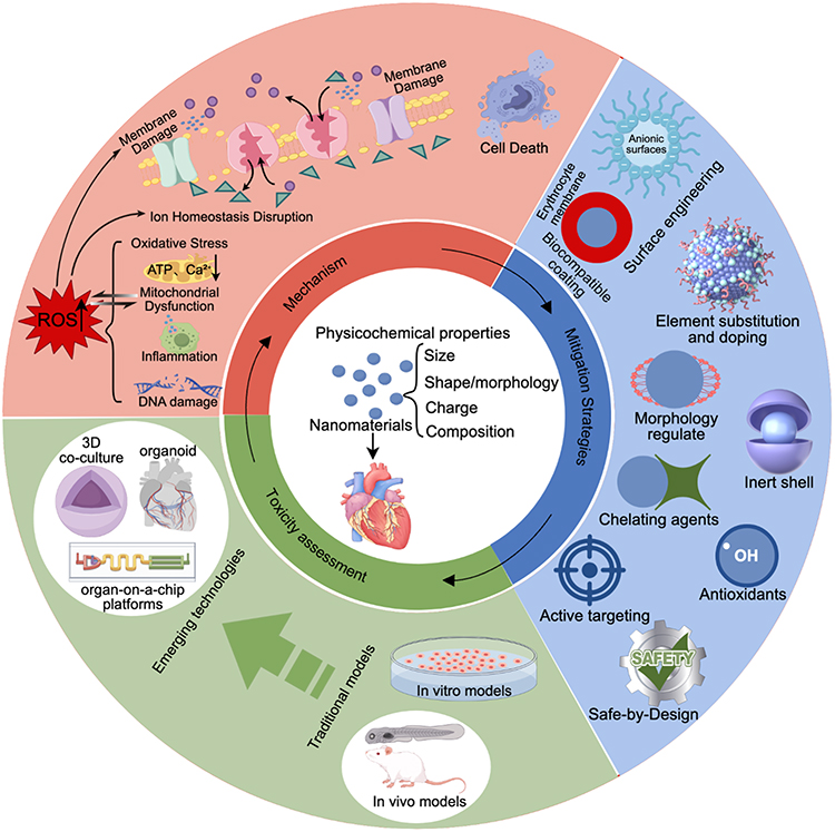 |
Figure 1 Assessment, mechanisms, and mitigation strategies of nanomaterial-induced cardiotoxicity. Upward arrows indicating an increase in levels, while downward arrows indicating a decrease in levels. |
In vitro models provide rapid screening for nanoparticle toxicity, focusing on direct cellular interactions and molecular pathways. These models primarily use cultured cardiomyocytes to assess key mechanisms like oxidative stress, mitochondrial dysfunction, inflammation, and apoptosis. For instance, studies employing isolated adult rat cardiomyocytes revealed that silica nanoparticles impair mitochondrial function and disrupt calcium handling, leading to cardiotoxic effects.28 Another notable example involves the development of “in vitro” blood flow models to study mesoporous silica nanoparticles (MSNs).30 These physiologically relevant systems incorporate shear stress to mimic vascular conditions, allowing for the assessment of particle shape-dependent toxicity. Spherical MSNs showed greater oxidative damage than rodlike MSNs.30 While “in vitro” approaches provide foundational mechanistic insights, caution must be exercised when extrapolating these findings to the “in vivo” settings. Therefore, their integration with “in vivo” and emerging technologies, such as multi-organ-on-a-chip systems, is essential to bridge the gap between cellular mechanisms and systemic cardiotoxicity.
In vivo models are indispensable for assessing systemic toxicity and organ-specific effects, particularly cardiovascular outcomes. Rodent models, such as mice and rats, provide insights into chronic exposures and disease contexts. For instance, Wistar rats exposed to ZnO NPs (39nm) via oral administration demonstrate dose-dependent cardiotoxicity, as evidenced by significant changes in electrocardiographic parameters, increased biochemical indices of cardiac function, and dysregulated oxidative stress.31 Another example involves ApoE mice treated with intratracheal instillation of silica nanoparticles (Si-60, 60 nm), which showed significant declines in cardiac function metrics, such as cardiac output, stroke volume and left ventricular internal diameter in a sub-chronic exposure scenario. This finding indicates nanoparticle-induced, size-dependent cardiotoxicity, with Si-60 demonstrating greater toxicity than Si-300 (600 nm).32 Zebrafish (Danio rerio) is a widely adopted model due to its transparency, high fecundity, low cost, and gene manipulation accessibility, enabling rapid screening of acute and chronic effects. For example, a study testing the acute toxicity of 31 different NPs—covering four types excluding organic NPs—on zebrafish during both adult and early life stages revealed that metallic and metal-oxide particles, such as copper (Cu), Ag, copper oxide (CuO), copper zinc iron oxide, magnesium oxide, calcium oxide, and nickel oxide, induced cumulative mortality. Among these, Cu NPs and Ag NPs exhibited toxicity, with median lethal concentration values of approximately 3 mg/L in zebrafish.33 A recent research indicates that acute and chronic exposure to nano-cerium oxide induces cardiovascular toxicity in zebrafish, which impairs hematopoietic function and cardiac development.34 Notably, the resemblance of the zebrafish heart to the human embryonic heart, along with the direct microscopical observability of cardiac morphology and rhythm (eg., heartbeats, vasculature, and cell activity) in transparent embryos, has greatly enhanced NPs toxicity evaluation. Therefore, zebrafish embryos are highly suitable models for assessing nanoparticle-induced cardiac developmental toxicity.29
Recent advances have established emerging non-animal platforms for evaluating environmental toxicant impacts on human health, including 3D co-cultures, organoids, and organ-on-a-chip (OoC) platforms.3,35 These systems recapitulate human organ structure and key functions, surpassing traditional 2D cultures by modeling cell-cell interactions, secretory dynamics, and tissue-specific microenvironments.36 Furthermore, disease models based on cardiac organoids37,38 serve as powerful tools to elucidate the susceptibility of high-risk populations under pathological conditions.35 Crucially, they circumvent species disparities and ethical constraints inherent in animal studies. Additionally, OoC platforms offer distinct advantages such as high throughput, reduced reagent consumption, and enhanced physiological relevance.3,35 Among these models, OoC indicates exceptional promise due to its ability to emulate organ-level functionality. For instance, employing three-dimensional fibrous scaffolds, Ahn et al engineered a heart-on-a-chip platform to evaluate the effects of Ag NPs and TiO2 NPs on cardiac contractile function.39 Utilizing the cardiac organoid-on-a-chip platform, Zhang et al dynamically observed cardiotoxicity from short- and long-term exposure to PS-NPs.35 Their research indicate that oxidative stress, inflammation, calcium homeostasis disruption, and mitochondrial dysfunction are potential mechanisms and early critical events, whereas cardiac fibrosis is a prominent late-stage feature.35 Despite its potential, OoC faces several limitations. (1) Individual organ platforms are confined to modeling specific organ functions and represent only partial aspects of real tissues.40 (2) Maintaining long-term tissue viability and functionality remains challenging. (3) The restricted sample volumes complicate certain analytical procedures. (4) Adoption in standardized cardiotoxicity assessment is still limited.
Integrated Approaches to Testing and Assessment (IATAs) are being developed to guide the safety assessment of NMs. For example, an IATA for nanomaterials with potential for direct blood contact has been developed to assess haemocompatibility. This approach follows an iterative process, gathering relevant information for safety assessment, including existing experimental data, in silico model predictions, and new experimental procedures to fill data gaps.41 Machine learning-integrated omics can also play a role in the risk and safety assessment of nanomaterials. By combining integrative omics with machine learning, it is possible to profile nanomaterial safety and risk assessment more accurately. This approach can provide insights into the perturbations of delicate biological functions after integration with nanomaterials, which is valuable for regulatory authorities in making decisions regarding the safety of nanomaterials.42
The selection of cardiotoxicity assessment models for nanomaterials should be guided by a hierarchical decision-making process based on the evaluation objectives, physicochemical properties of the nanomaterials, exposure scenarios, and regulatory requirements. In vitro models are well-suited for initial high-throughput screening, elucidation of mechanistic pathways, and establishment of dose–response relationships, particularly during preliminary stages with limited resources. When the assessment requires evaluation of systemic toxicity, inter-organ interactions, or chronic pathological progression, In vivo models remain indispensable. Emerging technologies, such as OoC, serve as physiologically relevant platforms that bridge in vitro and in vivo systems. OoC platforms are particularly valuable as key validation tools when nanomaterials exhibit complex physicochemical characteristics (eg., facile protein corona formation), when disease states need to be recapitulated, or when predictions from conventional in vitro models deviate from in vivo outcomes.43 The IATA framework emphasizes the weight-of-evidence integration from multiple sources, making it suitable for regulatory decision-making and comprehensive evaluation of high-risk nanomaterials. When a single model is insufficient to fully capture the complex interactions between nanomaterials and the cardiovascular system—particularly for high-risk materials—a tiered strategy integrating “computational prediction—in vitro screening—mechanistic exploration—in vivo validation—integrated assessment” is recommended. In summary, model selection should adhere to the principles of “risk-stage, objective-orientation, evidence complementarity, and ethical optimization”.
Mechanisms of Nanomaterial-Induced Cardiotoxicity
The cardiovascular system, due to its limited regenerative capacity and role as the primary contact target following systemic exposure, is highly vulnerable to the toxicity of nanoparticles. Studies indicates that NMs inducing cardiotoxicity mainly include TiO2,44 silicon dioxide,45 iron oxide,46 silver,47 and carbon-based nanomaterials like carbon nanotubes48 and graphene oxide.49 Key mechanisms include oxidative stress, inflammatory responses, membrane damage, ion homeostasis disruption, and cell death (Figure 1). Table 1 illustrates the potential cardiotoxic mechanisms of common nanoparticles.
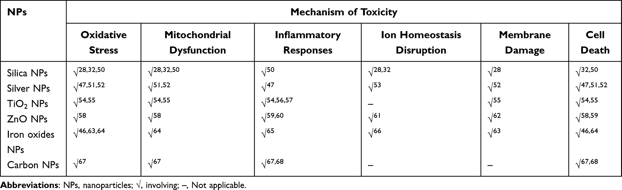 |
Table 1 Potential Mechanisms of Cardiotoxicity Induced by Common Nanoparticles |
Oxidative Stress and Mitochondrial Dysfunction
A fundamental mechanism driving nanomaterial cardiotoxicity is the induction of oxidative stress. Various NPs, including PS-NPs,7,8 Ag NPs, TiO2 NPs,54 SiO2 NPs,32,50 lead halide perovskites,69 nano-sized iron oxide,66 and graphene oxide, have been reported to significantly elevate intracellular levels of reactive oxygen species (ROS) within cardiomyocytes. This excessive ROS generation overwhelms endogenous antioxidant defenses, causing oxidative damage to vital cellular components such as lipids, proteins, and DNA.22 Exposure to TiO2 NPs was associated with increased cardiac muscle levels of ROS, including superoxide radicals and hydrogen peroxide, along with decreased malondialdehyde content and increased DNA peroxidation. Furthermore, TiO2 exposure reduced the activity of antioxidant enzymes,54 particularly superoxide dismutase and glutathione S-transferases.70 SiO2 NPs rank among the top five most utilized nanomaterials in commercial products, finding applications in drug delivery, gene therapy, and diagnostics.71 Additionally, due to their higher biocompatibility compared to other imaging NPs, SiO2 NPs have been identified as ideal NPs for medical imaging.71 Studies show that SiO2 NPs induce oxidative stress, impair mitochondrial function, disrupt energy production and interfer with calcium ion (Ca2⁺) handling in mice cardiomyocytes.32 This mitochondrial dysfunction leads to reduced cardiac contractility, manifesting as decreased stroke volume and cardiac output, and is implicated in heart failure, arrhythmias, and sudden death.28 Additionally, SiO2 NPs are associated with cardiac hypertrophy.50 Key influencing factors include nanoparticle size, exposure duration (acute or chronic exposures), and dose (higher concentrations correlate with increased oxidative damage).3 For example, a size-dependent cardiotoxicity has been observed, wherein SiO2 NPs measuring 60 nm in diameter exhibit greater myocardial toxicity compared to their 300 nm counterparts.32 In vitro studies indicate that all ultrasmall NPs of Fe3O4, SiO2, and gold (<5 nm) significantly induce ROS generation, an effect not observed with their larger counterparts.63 This size-dependent phenomenon is also supported by in vivo findings. An acute exposure study revealed that ultrasmall iron oxide NPs (2.3 and 4.2 nm Fe3O4, administered intravenously to mice) induced severe acute oxidative stress in multiple organs, exhibiting high toxicity characterized particularly by substantial hydroxyl radical production in the heart. In contrast, no obvious toxicity was observed for larger (9.3 nm) iron oxide NPs.63 Furthermore, a chronic exposure study indicates that the repeated intraperitoneal administration of iron oxide NPs (Fe2O3 NPs, <50 nm) at the dose of 25 and 50 mg/kg body weight for 30 days at seven days interval led to dose-dependent cardiotoxicity in mice, manifested as myocardial oxidative stress, mitochondrial damage, and apoptosis.64 Consequently, in addition to minimizing exposure, modification of nanomaterials’ physicochemical properties to suppress ROS generation represents an imperative strategy for mitigating nanomaterial-induced toxicity.
NPs often target mitochondria, leading to structural and functional damage characterized by mitochondrial membrane hyperpolarization, impaired respiration, and reduced ATP production.3,72 Mitochondrial dysfunction further amplifies ROS production, creating a detrimental cycle that culminates in cardiomyocyte injury and death. Mitochondrial damage is associated with severe cardiac diseases, including myocardial hypertrophy, heart failure, arrhythmias, sudden cardiac death, and other serious heart conditions.3
Inflammatory Responses and Cell Death
Inflammatory responses, essential for combating harmful stimuli, involve the secretion of inflammatory mediators and the infiltration of inflammatory cells. NPs can trigger such responses through various mechanisms, as exemplified by TiO2 NPs, ZnO NPs, cadmium NPs (Cd NPs), carbon NPs (CNPs), and SiO2 NPs. For instance, C-reactive protein (CRP), a key inflammatory marker, shows elevated serum levels with increasing nano-TiO2 exposure.73 Another rat study revealed increases in serum tumor necrosis factor α (TNF-α) and interleukin 6 (IL-6) after daily oral administration of TiO2 NPs for 90 days, further supporting the induction of systemic inflammation.56 Mechanistically, research in mice has shown that cardiac lesions induced by TiO2 NPs (administered intragastrically at 2.5, 5, or 10 mg/kg body weight for 90 days) are mediated through nuclear factor-κB (NF-κB) activation via the protein kinase C epsilon or ERK1/2 signaling cascades. This pathway leads to the upregulation of pro-inflammatory cytokines, including TNF-α, IL-6, IL-1β, and and interferon-α.57 This may cause cardiac damage, such as disordered myocardial fibre arrangement, swelling, cardiomyocyte hypertrophy, tissue necrosis, myocardial haemorrhage, and cardiac fragmentation.57 Oral administration of ZnO NPs induces cardiac inflammation, DNA damage, and apoptosis in rat models, culminating in myocardial injury.59 Rats exposed to ZnO NPs exhibit significantly elevated levels of inflammatory mediators, including TNF-α, IL-6, and CRP. These biomarkers may play pivotal roles in ZnO NPs-induced cardiotoxicity. In vitro studies using cultured cardiomyocytes indicate that TNF-α amplifies ROS generation, thereby triggering DNA damage.74 Subsequent to DNA damage, apoptosis emerges as a critical pathological endpoint. Exposure to Cd NPs has been shown to induce pathological changes and functional impairments in newborn chicks hearts, triggering upregulation of inflammatory mediators including IL-6, IL-8, TNF-α, and NF-κB.75 Histopathological examination reveals myofibrillar lysis and inflammatory cell infiltration. After 60 days of CNPs exposure, adult zebrafish showed cardiac endothelial uptake and heart deposition of CNPs.68 CNPs also caused significant myocardial ultrastructural changes and dose-dependently induced inflammatory cytokine expression, leading to sub - endocardial inflammation and apoptosis.68 It is worth noting that pathway activation may be size-dependent. For example, ultrasmall gold NPs (<10 nm; eg., 4.5 nm particles) preferentially and markedly activate the NLRP3 inflammasome and induce IL-1β secretion by mature Caspase-1 in a dose-dependent manner.76 One proposed mechanism is their direct cytoplasmic penetration, which promotes robust ROS generation to activate the NLRP3 inflammasome and targets the degradation of the microtubule-associated protein 1-light chain 3 (LC3), thereby relieving LC3-mediated inhibition of NLRP3.76 In contrast, larger gold NPs (>10 nm) primarily trigger the NF-κB signalling pathway.76 Moreover, a study investigated the combined toxicity of SiO2 NPs and methylmercury (MeHg) on the cardiovascular system of zebrafish embryos.77 Results showed that co-exposure led to a significant decrease in cardiac output, a pronounced increase in vascular endothelial damage, and substantial neutrophil activation.77 This indicates that the cardiac toxicity of combined SiNP and MeHg exposure is more severe than that of individual exposures.77
Persistent inflammation is a well-established contributor to atherosclerosis, myocardial remodeling, heart failure, and arrhythmias. Collectively, evidence suggests nanoparticle-induced cardiac inflammation and injury. However, unlike primary exposure sites (such as skin, gastrointestinal tract, and lungs) with well-established nanoparticle-inflammatory response relationships, the mechanistic details of cardiac inflammation remain poorly characterized. Current research delineates only inflammatory processes, whereas comprehensive assessment of cardiac-inflammatory responses is warranted. Crucially, the influence of nanoparticle physicochemical properties (size, shape, surface chemistry) on cardiac inflammation is inadequately investigated. Future studies should elucidate the entire inflammatory cascade.22
Membrane Damage, Ion Homeostasis Disruption, and Cell Death
Several NMs, notably SiO2 NPs, Ag NPs, ZnO NPs, iron oxide NPs, and CNPs, have been linked to cardiotoxicity through pathways involving membrane integrity compromise and ion dysregulation. These effects are often mediated by oxidative stress, mitochondrial dysfunction, and inflammatory responses, leading to structural and functional cardiac impairments. For example, SiO2 NPs induce cardiotoxicity through membrane damage and disruption of ion homeostasis, particularly calcium (Ca2⁺) handling. Exposure to SiO2 NPs results in direct association and internalization into cardiomyocyte membranes, triggering ROS generation. This oxidative stress impairs mitochondrial function, reducing sarcoplasmic reticulum Ca2⁺-ATPase activity and shortening Ca2⁺ release, which compromises cellular shortening and energy status.28 This SiO2-induced cardiotoxicity mechanism potentially contributes to the pathogenesis of heart failure, arrhythmias, and sudden cardiac death.28 Ag NPs, especially PEG-coated variants, cause cardiotoxicity via membrane damage and ion imbalance triggered by oxidative stress. Upon pulmonary exposure, PEG-Ag NPs translocate to the heart, inducing ROS overproduction and inflammation. This disrupts ion channels, leading to altered cardiac electrophysiology and Ca2⁺ homeostasis. The toxicity is amplified in hypertension models due to heightened oxidative stress and inflammatory cytokine release, further compromising membrane integrity and ion regulation.78 Thereby, PEG coating, while enhancing biocompatibility, does not eliminate toxicity in sensitive populations.78 ZnO NPs can disrupt Ca2⁺ homeostasis, leading to cytosolic Ca2⁺ accumulation, which is associated with cardiomyocyte injury.74 Similarly, the myocardial delivery of magnetic iron oxide NPs might produce iron overload-induced myocardial injury.66 CNPs damage membrane structures from within cellular organelles.22 Membrane damage caused by NPs is influenced by their chemical composition, dimension, surface charge, and morphology.22 As particle size decreases, the augmented surface area exposes more surface atoms and structural defects, thereby promoting the formation of additional reactive sites. Positively charged NPs exhibit enhanced toxicity profiles, indicating more easy internalization into the cytosol of neonatal rat cardiomyocytes and consequent severe cellular damage.22 This phenomenon is likely attributable to electrostatic attraction between positively charged NPs and negatively charged plasma membranes, facilitating membrane adherence and subsequent lipid extraction.22
In total, NPs induce cardiotoxicity through multiple pathways, varying by particle type and physicochemical properties. This heterogeneity underscores the complexity of NP-mediated cardiovascular toxicity. A predominant mechanism involves oxidative stress, while mitochondrial dysfunction represents another critical pathway that directly compromises cardiac energy metabolism and contractile function. Concurrently, disturbances in ion channel activity and calcium handling alter cardiac electrophysiology and contractility. Notably, distinct mechanistic profiles exist across different nanoparticle classes. SiO2 NPs primarily target mitochondrial and calcium pathways,28,32 whereas metallic and metal-oxide NPs (eg., Ag, iron oxides, and ZnO NPs) involve multiple mechanisms. For instance, Ag NPs, widely used in medical materials, antibiotics, and wound dressings, can induce cardiac oxidative stress,51 inflammation,47 and fibrosis.74 Despite their usefulness as MRI contrast agents and in targeted delivery and hyperthermia therapy,79 iron oxide NPs can also cause myocardial injury through mechanisms including oxidative stress, mitochondrial dysfunction,64 inflammatory responses,65 iron overload66 and membrane damage.63 Oxidative stress is considered to be the main mechanism.66 Collectively, these insights highlight that NPs induce cardiotoxicity through interconnected pathways involving oxidative stress, mitochondrial impairment, inflammation, apoptosis, and ionic disturbance, each of which is uniquely associated with the NP’s physicochemical characteristics. This underscores the necessity of implementing safe-by-design strategies to mitigate toxicity, based on a thorough understanding of nanoparticle physicochemical properties and their corresponding toxicological mechanisms.
Strategies to Mitigate Cardiovascular Toxicity of Nanomaterials
Based on mechanistic insights into nanomaterial-biological interactions, several key mitigation strategies have emerged,23 such as surface engineering strategies, elemental substitution and doping, morphological and structural design, chelation and antioxidant strategies, and exposure control and safe-by-design integration (Figure 1, Table 2).
 |
Table 2 Potential Strategies for Mitigating the Cardiovascular Toxicity of Nanomaterials |
Surface Engineering Strategies
Surface engineering serves as a foundational strategy for mitigating cardiotoxicity, as surface properties critically determine NPs interactions with biological membranes92 and subsequent toxicity pathways.23 Surface modification techniques are designed to alter or confer novel surface properties to NPs, enabling functionalization, targeting capability, biocompatibility, and other relevant attributes.22 Coating with biocompatible materials can mask reactive surface sites, modulate surface charge, prevent particle aggregation, and diminish cellular uptake—collectively contributing to a reduction in NP toxicity.22 For instance, designing NPs with anionic surfaces has been shown to lessen electrostatic interactions with negatively charged cell membranes, thus reducing membrane disruption and subsequent cardiotoxicity.23 Similarly, chitosan-coated ZnO NPs exhibited minimal cardiotoxicity even at elevated concentrations.6 Protein corona formation represents an alternative strategy for NP surface coating. Researchers frequently incubate NPs with specific sera or proteins in vitro to promote the adsorption of a protein corona.80,81 The proteins occupy reactive sites on the NP surface, thereby reducing their bioreactivity upon administration.81 Another extensively investigated coating strategy utilizes natural cell membranes—derived from red blood cells, stem cells, immune cells, among others—which has emerged as an effective method to improve biocompatibility and evade immune recognition.82,83 Consequently, a primary objective of surface engineering strategies is to minimize direct interactions between NPs and cellular membranes, effectively preventing cellular internalization and ultimately mitigating nanotoxicity.
Elemental Substitution and Doping
Replacing toxic elements within NPs with less hazardous alternatives or incorporating stabilizing dopants can effectively reduce ion-mediated toxicity. For instance, enriching lithium nickel manganese cobalt oxide with manganese at the expense of cobalt and nickel has been found to reduce the release of toxic metal ions and subsequently decreased toxicity to model bacteria Shewanella oneidensis MR-1.23 Incorporating dopants can alter the electronic structure or dissolution kinetics of NPs. Doping iron into CuO NPs formed a CuFe2O4 phase on the surface, which stabilized the structure and reduced the release of cytotoxic Cu2⁺ ions, leading to lower toxicity despite potentially increased cellular uptake.84 Similarly, iron doping in ZnO NPs has been shown to suppress the dissolution of Zn2⁺ ions, thereby mitigating associated oxidative stress and inflammatory responses.85
Morphological and Structural Design
The physical form of NPs influences dissolution rates and membrane interactions. Nanoparticle shape and curvature impact dissolution kinetics and membrane interactions.93 Nanospheres generally exhibit higher specific surface area and dissolution rates compared to nanorods or nanocubes. Silver nanoplatelets indicate higher dissolution and toxicity than nanocubes, correlating with their increased surface area.86 Designing NPs with lower surface area-to-volume ratios can reduce dissolution of toxic ions. Furthermore, morphology significantly influences membrane interactions. Theoretical studies have indicated that nanospheres exhibit greater membrane interactions compared to nanoplates; however, nanoplates indicate substantially higher redox activity, thereby representing a more toxic morphological form despite their reduced membrane engagement.23 This finding underscores the importance of identifying the primary toxicity mechanisms for the targeted redesign of nanomaterials to mitigate their toxicity. Coating a toxic core material with an inert shell (eg., ZnS shell on CdSe quantum dots,87 silica shells on metal NPs88) creates a physical barrier that impedes dissolution and direct contact with biological components, thereby reducing ion release.87,88 Silica-coated nickel (Ni) NPs showed significantly reduced Ni2⁺ dissolution and minimal toxicity in zebrafish embryo models compared to uncoated Ni NPs or those attached to silica surfaces.88 Polymer shells (eg., PEG) can also stabilize NPs and reduce dissolution.94
Chelating Agents or Antioxidant Addition
Counteracting the toxic effects of released ions or generated ROS is a complementary approach. Attaching chelating agents specific to toxic metal ions (eg., deferoxamine for Fe3⁺,22 tetrathiomolybdate for Cu2⁺89) directly to the NP surface can complex released ions immediately upon dissolution, preventing their interaction with cellular components and mitigating toxicity.95 Chelating agents for cationic metal ions exhibit negative charge characteristics, further reducing nonspecific cellular binding.23 Tethering antioxidant molecules (eg., Trolox - a vitamin E analog,90 vitamin C,23 or other ROS scavengers) to NPs provides localized protection against NP-induced oxidative stress. These antioxidants can neutralize ROS generated either by the NP itself or as a secondary consequence of NP exposure.66,90,96,97 For example, gold NPs functionalized with Trolox demonstrate enhanced ability to scavenge ROS.90 Co-loading the antioxidant N-acetylcysteine with magnetic iron oxide NPs was shown to alleviate oxidative stress and reduce apoptosis in hypoxia/reoxygenation cardiomyocytes.66 Additionally, certain NPs, such as those composed of ceria or yttria, possess intrinsic antioxidant properties.23
Exposure Control and Safe-by-Design Integration
Minimizing unintended cardiac exposure and designing safety into NMs from the outset are crucial. For non-cardiac medications, utilizing localized administration routes instead of systemic delivery can effectively reduce the overall burden of NMs reaching the cardiovascular system.22 Furthermore, actively targeting NMs to specific diseased sites enhances efficacy at lower doses and minimizes off-target accumulation in sensitive organs like the heart.91
Integrating “Safe(r)-by-Design” strategies early in the NM development process is advocated by regulatory bodies like the Organization for Economic Co‐operation and Development.22 This involves selecting inherently less toxic materials, optimizing physicochemical properties (size, charge, stability), and conducting thorough in vitro and in vivo cardiotoxicity assessments using relevant models (eg., cardiomyocytes, zebrafish embryos, rodent models) throughout the design phase. Assessing NM behavior under pathological conditions (eg., hypertension, ischemia) is also critical, as vascular permeability and cardiac susceptibility may increase.35,98,99 For example, low-dose PS-NPs exacerbated myocardial infarction symptoms under pathological models without exhibiting significant cardiotoxicity in healthy ones.35 A notable gap in the current literature is the limited investigation of nanomaterial-induced cardiotoxicity in susceptible populations and comorbidities. Future research should prioritize the use of disease-relevant comorbidity models to elucidate how underlying pathologies modulate nanomaterial toxicity, as this is crucial for comprehensive risk assessment and the protection of vulnerable groups. Establishing standardized protocols for assessing the cardiotoxicity potential of NPs, including specific endpoints like arrhythmogenicity,22 is essential for regulatory approval and safe clinical translation. Comprehensive characterization (size, charge, dissolution, ROS potential) and understanding of transformation products in biological fluids are vital for accurate risk assessment.
Limitations
This narrative review acknowledges certain methodological limitations that warrant careful consideration. First, the literature search was confined to the PubMed database, which may not encompass relevant studies from other databases or sources, thereby introducing potential selection bias. Second, owing to the absence of a formal risk of bias assessment, a rigorous evaluation of evidence quality may be somewhat constrained; nevertheless, we sought to mitigate potential subjective interpretation biases through critical discussions during evidence synthesis. For future research, we recommend expanding the search scope to include multiple databases—such as PubMed, Web of Science, EMBASE, Cochrane Library, and clinical trial registries—to improve the representativeness and coverage of the evidence. Whenever possible, priority should be given to systematic reviews and meta-analyses, complemented by rigorous and standardized risk of bias assessments, to better quantify effect sizes and evaluate between-study heterogeneity. Such methodological refinements would enhance the reliability of subsequent reviews and provide more robust support for clinical and policy-related decision-making.
Conclusion
This review has established a comprehensive framework for understanding and mitigating nanomaterial-induced cardiotoxicity by integrating three interconnected pillars: risk assessment, underlying mechanisms, and mitigation strategies. The selection of appropriate assessment models is guided by a tiered, decision-driven strategy based on risk stage, objective orientation, evidence complementarity, and ethical optimization, with emphasis on the critical need to evaluate toxicity under pathological conditions relevant to susceptible populations and comorbidities—an aspect essential for translating experimental findings to human health risk assessment. Mitigating the cardiovascular toxicity of nanomaterials requires a multifaceted approach grounded in understanding the underlying mechanisms (membrane damage, ion release, oxidative stress, inflammation, cell death). Strategies include engineering surface properties (charge, coatings), modifying core composition (element substitution, doping), optimizing morphology, employing chelators or antioxidants, and controlling exposure through targeted delivery and Safe-by-Design principles. The integration of these strategies, validated through robust in vitro and in vivo cardiotoxicity testing using standardized methods, is paramount for developing effective and safe nanomedicines and other nano-enabled products. However, the implementation of these strategies can be constrained by their conflict with core nanotherapeutic design goals, which requires extensive product-specific analysis and customization. The early collaboration of a multidisciplinary team is therefore critical. By linking risk assessment, mechanisms, and mitigation strategies within a decision-making framework, this review provides a holistic perspective that we believe will guide future research toward safer nanomaterial design. The continued development of sophisticated models and innovative techniques promises substantial progress in the mitigation of nanocardiotoxicity, ultimately enabling the safe and effective use of nanomaterials.
Abbreviations
NMs, nanomaterials; NPs, nanoparticles; Ag, silver; TiO2, titanium dioxide; ZnO, zinc oxide; SiO2 NPs, silica NPs; PS-NPs, polystyrene nanoparticles; PEG, polyethylene glycol; SANRA, Scale for the Assessment of Narrative Review Articles; MSNs, mesoporous silica nanoparticles; OoC, organ-on-a-chip; IATAs, Integrated Approaches to Testing and Assessment; ROS, reactive oxygen species; CRP, C-reactive protein; TNF-α, tumor necrosis factor α; IL-6, interleukin 6; MeHg, methylmercury.
Author Contributions
Zhichao Li and Lidi Liu are co-first authors. All authors made a significant contribution to the work reported, whether that is in the conception, study design, execution, acquisition of data, analysis and interpretation, or in all these areas; took part in drafting, revising or critically reviewing the article; gave final approval of the version to be published; have agreed on the journal to which the article has been submitted; and agree to be accountable for all aspects of the work. All authors have confirmed and agreed to be responsible for this manuscript. All authors have read and agreed to the published version of the manuscript.
Funding
This work was supported by grants from Noncommunicable Chronic Diseases-National Science and Technology Major Project (No. 2024ZD0524702/2024ZD0524700), Sichuan Science and Technology Program (No. 2024NSFSC1534).
Disclosure
The authors declare that they have no competing interests.
References
1. Bleeker EAJ, de Jong WH, Geertsma RE, et al. Considerations on the EU definition of a nanomaterial: science to support policy making. Regul Toxicol Pharmacol. 2013;65(1):119–15. doi:10.1016/j.yrtph.2012.11.007
2. Li B, Tang M. Research progress of nanoparticle toxicity signaling pathway. Life Sci. 2020;263:118542. doi:10.1016/j.lfs.2020.118542
3. Cheng Y, Chen Z, Yang S, et al. Nanomaterials-induced toxicity on cardiac myocytes and tissues, and emerging toxicity assessment techniques. Sci Total Environ. 2021;800:149584. doi:10.1016/j.scitotenv.2021.149584
4. Chereddy KK, Payen VL, Preat V. PLGA: from a classic drug carrier to a novel therapeutic activity contributor. J Control Release. 2018;289:10–13. doi:10.1016/j.jconrel.2018.09.017
5. Su X, Zhang X, Liu W, et al. Advances in the application of nanotechnology in reducing cardiotoxicity induced by cancer chemotherapy. Semin Cancer Biol. 2022;86(Pt 2):929–942. doi:10.1016/j.semcancer.2021.08.003
6. Younes N, Pintus G, Al-Asmakh M, et al. “Safe” chitosan/zinc oxide nanocomposite has minimal organ-specific toxicity in early stages of zebrafish development. ACS Biomater Sci Eng. 2020;6(1):38–47. doi:10.1021/acsbiomaterials.8b01144
7. Wu H, Guo J, Yao Y, Xu S. Polystyrene nanoplastics induced cardiomyocyte apoptosis and myocardial inflammation in carp by promoting ROS production. Fish Shellfish Immunol. 2022;125:1–8. doi:10.1016/j.fsi.2022.04.048
8. Xiong Z, Kong Q, Hua J, Chen Q, Wang D. Cardiotoxicity of polystyrene nanoplastics and associated mechanism of myocardial cell injury in mice. Ecotoxicol Environ Saf. 2025;290:117712. doi:10.1016/j.ecoenv.2025.117712
9. Yang C, Tian A, Li Z. Reversible cardiac hypertrophy induced by PEG-coated gold nanoparticles in mice. Sci Rep. 2016;6(1):20203. doi:10.1038/srep20203
10. Kooi ME, Cappendijk VC, Cleutjens KB, et al. Accumulation of ultrasmall superparamagnetic particles of iron oxide in human atherosclerotic plaques can be detected by in vivo magnetic resonance imaging. Circulation. 2003;107(19):2453–2458. doi:10.1161/01.CIR.0000068315.98705.CC
11. Yu X, Zhao X, Ze Y, et al. Changes of serum parameters of TiO2 nanoparticle-induced atherosclerosis in mice. J Hazard Mater. 2014;280:364–371. doi:10.1016/j.jhazmat.2014.08.015
12. Mikkelsen L, Sheykhzade M, Jensen KA, et al. Modest effect on plaque progression and vasodilatory function in atherosclerosis-prone mice exposed to nanosized TiO2. Part Fibre Toxicol. 2011;8(1):32. doi:10.1186/1743-8977-8-32
13. Yan Z, Wang W, Wu Y, et al. Zinc oxide nanoparticle-induced atherosclerotic alterations in vitro and in vivo. Int J Nanomed. 2017;12:4433–4442. doi:10.2147/IJN.S134897
14. Ceballos-Gutierrez A, Rodriguez-Hernandez A, Alvarez-Valadez MDR, et al. ZnO nanoparticles induce dyslipidemia and atherosclerotic lesions leading to changes in vascular contractility and cannabinoid receptors expression as well as increased blood pressure. Nanomaterials. 2021;11(9):2319. doi:10.3390/nano11092319
15. Ma R, Qi Y, Zhao X, et al. Amorphous silica nanoparticles accelerated atherosclerotic lesion progression in ApoE−/− mice through endoplasmic reticulum stress-mediated CD36 up-regulation in macrophage. Part Fibre Toxicol. 2020;17(1):50. doi:10.1186/s12989-020-00380-0
16. Stachyra K, Wisniewska A, Kiepura A, et al. Inhaled silica nanoparticles exacerbate atherosclerosis through skewing macrophage polarization towards M1 phenotype. Ecotoxicol Environ Saf. 2022;230:113112. doi:10.1016/j.ecoenv.2021.113112
17. Yin T, Li Y, Ren Y, et al. Phagocytosis of polymeric nanoparticles aided activation of macrophages to increase atherosclerotic plaques in ApoE−/− mice. J Nanobiotechnology. 2021;19(1):121. doi:10.1186/s12951-021-00863-y
18. Suzuki Y, Tada-Oikawa S, Hayashi Y, et al. Single- and double-walled carbon nanotubes enhance atherosclerogenesis by promoting monocyte adhesion to endothelial cells and endothelial progenitor cell dysfunction. Part Fibre Toxicol. 2015;13(1):54. doi:10.1186/s12989-016-0166-0
19. Chen S, Su Y, Zhang M, et al. Insights into the toxicological effects of nanomaterials on atherosclerosis: mechanisms involved and influence factors. J Nanobiotechnol. 2023;21(1):140. doi:10.1186/s12951-023-01899-y
20. Yang P, Ren J, Yang L. Nanoparticles in the new era of cardiovascular therapeutics: challenges and opportunities. Int J Mol Sci. 2023;24(6):5205. doi:10.3390/ijms24065205
21. Najahi-Missaoui W, Arnold RD, Cummings BS. Safe nanoparticles: are we there yet? Int J Mol Sci. 2020;22(1):385. doi:10.3390/ijms22010385
22. Zheng Z, Zhu S, Wang X, et al. Understanding the mechanism of cardiotoxicity induced by nanomaterials: a comprehensive review. Small Sci. 2025;5(5):2400498. doi:10.1002/smsc.202400498
23. Buchman JT, Hudson-Smith NV, Landy KM, Haynes CL. Understanding nanoparticle toxicity mechanisms to inform redesign strategies to reduce environmental impact. Acc Chem Res. 2019;52(6):1632–1642. doi:10.1021/acs.accounts.9b00053
24. Grzelczak M, Liz-Marzan LM, Klajn R. Stimuli-responsive self-assembly of nanoparticles. Chem Soc Rev. 2019;48(5):1342–1361. doi:10.1039/C8CS00787J
25. Jiang M, Wang Y, Zhang J, et al. Temperature and ultrasound-responsive nanoassemblies for enhanced organ targeting and reduced cardiac toxicity. Int J Nanomed. 2024;19:11397–11413. doi:10.2147/IJN.S470465
26. Baethge C, Goldbeck-Wood S, Mertens S. SANRA—a scale for the quality assessment of narrative review articles. Res Integrity Peer Rev. 2019;4(1):5. doi:10.1186/s41073-019-0064-8
27. Liu L, Zhang P, Jia Y, et al. Artificial intelligence in ambulatory surgery: current applications, challenges, and future directions. J Med Syst. 2025;49(1):146. doi:10.1007/s10916-025-02286-w
28. Guerrero-Beltran CE, Bernal-Ramirez J, Lozano O, et al. Silica nanoparticles induce cardiotoxicity interfering with energetic status and Ca 2+ handling in adult rat cardiomyocytes. Am J Physiol Heart Circ Physiol. 2017;312(4):H645–H61. doi:10.1152/ajpheart.00564.2016
29. Bai C, Tang M. Toxicological study of metal and metal oxide nanoparticles in zebrafish. J Appl Toxicol. 2020;40(1):37–63. doi:10.1002/jat.3910
30. Cheng Y, Tao J, Zhang Y, et al. Shape and shear stress impact on the toxicity of mesoporous silica nanoparticles: in vitro and in vivo evidence. Mol Pharm. 2023;20(6):3187–3201. doi:10.1021/acs.molpharmaceut.3c00180
31. Hussein AA, Moatamed ER, El-Desoky MM, El Khayat Z. Electrophysiological and biochemical effect of zinc oxide nanoparticles on heart functions of male Wistar rats. Sci Rep. 2024;14(1):15416. doi:10.1038/s41598-024-65189-9
32. Qi Y, Xu H, Li X, et al. Silica nanoparticles induce cardiac injury and dysfunction via ROS/Ca2+/CaMKII signaling. Sci Total Environ. 2022;837:155733. doi:10.1016/j.scitotenv.2022.155733
33. Kovriznych JA, Sotnikova R, Zeljenkova D, Rollerova E, Szabova E, Wimmerova S. Acute toxicity of 31 different nanoparticles to zebrafish (Danio rerio) tested in adulthood and in early life stages – comparative study. Interdiscip Toxicol. 2013;6(2):67–73. doi:10.2478/intox-2013-0012
34. Liu Y, Liao Y-L, Wang J-X, et al. The effect and mechanisms of nano cerium dioxide (Nano-CeO2) on cardiovascular development in zebrafish (Danio rerio). Environ Pollut. 2025;374:126268. doi:10.1016/j.envpol.2025.126268
35. Zhang T, Yang S, Ge Y, et al. Unveiling the heart’s hidden enemy: dynamic insights into polystyrene nanoplastic-induced cardiotoxicity based on cardiac organoid-on-a-chip. ACS Nano. 2024;18(45):31569–31585. doi:10.1021/acsnano.4c13262
36. Lu RXZ, Radisic M. Organ-on-a-chip platforms for evaluation of environmental nanoparticle toxicity. Bioact Mater. 2021;6(9):2801–2819. doi:10.1016/j.bioactmat.2021.01.021
37. Song M, Choi DB, Im JS, et al. Modeling acute myocardial infarction and cardiac fibrosis using human induced pluripotent stem cell-derived multi-cellular heart organoids. Cell Death Dis. 2024;15(5):308. doi:10.1038/s41419-024-06703-9
38. Lewis-Israeli YR, Wasserman AH, Gabalski MA, et al. Self-assembling human heart organoids for the modeling of cardiac development and congenital heart disease. Nat Commun. 2021;12(1):5142. doi:10.1038/s41467-021-25329-5
39. Ahn S, Ardona HAM, Lind JU, et al. Mussel-inspired 3D fiber scaffolds for heart-on-a-chip toxicity studies of engineered nanomaterials. Anal Bioanal Chem. 2018;410(24):6141–6154. doi:10.1007/s00216-018-1106-7
40. Chen X, Zhang YS, Zhang X, Liu C. Organ-on-a-chip platforms for accelerating the evaluation of nanomedicine. Bioact Mater. 2021;6(4):1012–1027.
41. Powell LG, Gillies S, Fernandes TF, et al. Developing integrated approaches for testing and assessment (IATAs) in order to support nanomaterial safety. Nanotoxicology. 2022;16(4):484–499. doi:10.1080/17435390.2022.2103470
42. Ahmad F, Mahmood A, Muhmood T. Machine learning-integrated omics for the risk and safety assessment of nanomaterials. Biomater Sci. 2021;9(5):1598–1608. doi:10.1039/D0BM01672A
43. Park J, Wu Z, Steiner PR, Zhu B, Zhang JXJ. Heart-on-chip for combined cellular dynamics measurements and computational modeling towards clinical applications. Ann Biomed Eng. 2022;50(2):111–137. doi:10.1007/s10439-022-02902-7
44. Herrera-Rodriguez MA, Del Pilar Ramos-Godinez M, Cano-Martinez A, et al. Food-grade titanium dioxide and zinc oxide nanoparticles induce toxicity and cardiac damage after oral exposure in rats. Part Fibre Toxicol. 2023;20(1):43. doi:10.1186/s12989-023-00553-7
45. Feng L, Ning R, Liu J, et al. Silica nanoparticles induce JNK-mediated inflammation and myocardial contractile dysfunction. J Hazard Mater. 2020;391:122206. doi:10.1016/j.jhazmat.2020.122206
46. Nemmar A, Beegam S, Yuvaraju P, et al. Ultrasmall superparamagnetic iron oxide nanoparticles acutely promote thrombosis and cardiac oxidative stress and DNA damage in mice. Part Fibre Toxicol. 2015;13(1):22. doi:10.1186/s12989-016-0132-x
47. Ferdous Z, Al-Salam S, Greish YE, Ali BH, Nemmar A. Pulmonary exposure to silver nanoparticles impairs cardiovascular homeostasis: effects of coating, dose and time. Toxicol Appl Pharmacol. 2019;367:36–50. doi:10.1016/j.taap.2019.01.006
48. Thompson LC, Sheehan NL, Walters DM, Lust RM, Brown JM, Wingard CJ. Airway exposure to modified multi-walled carbon nanotubes perturbs cardiovascular adenosinergic signaling in mice. Cardiovasc Toxicol. 2019;19(2):168–177. doi:10.1007/s12012-018-9487-6
49. Bangeppagari M, Park SH, Kundapur RR, Lee SJ. Graphene oxide induces cardiovascular defects in developing zebrafish (Danio rerio) embryo model: in-vivo toxicity assessment. Sci Total Environ. 2019;673:810–820. doi:10.1016/j.scitotenv.2019.04.082
50. Wang F, Liang Q, Ma Y, et al. Silica nanoparticles induce pyroptosis and cardiac hypertrophy via ROS/NLRP3/Caspase-1 pathway. Free Radic Biol Med. 2022;182:171–181. doi:10.1016/j.freeradbiomed.2022.02.027
51. Khan AA, Alanazi AM, Alsaif N, Al-Anazi M, Sayed AYA, Bhat MA. Potential cytotoxicity of silver nanoparticles: stimulation of autophagy and mitochondrial dysfunction in cardiac cells. Saudi J Biol Sci. 2021;28(5):2762–2771. doi:10.1016/j.sjbs.2021.03.021
52. He J, Ma Y, Niu X, et al. Silver nanoparticles induce endothelial cytotoxicity through ROS-mediated mitochondria-lysosome damage and autophagy perturbation: the protective role of N-acetylcysteine. Toxicology. 2024;502:153734. doi:10.1016/j.tox.2024.153734
53. Lin C-X, Yang S-Y, Gu J-L, Meng J, Xu H-Y, Cao J-M. The acute toxic effects of silver nanoparticles on myocardial transmembrane potential, INa and IK1 channels and heart rhythm in mice. Nanotoxicology. 2017;11(6):827–837. doi:10.1080/17435390.2017.1367047
54. Zhang Q, Liu Z, Du J, et al. Dermal exposure to nano-TiO2 induced cardiovascular toxicity through oxidative stress, inflammation and apoptosis. J Toxicol Sci. 2019;44(1):35–45. doi:10.2131/jts.44.35
55. Huerta-Garcia E, Zepeda-Quiroz I, Sanchez-Barrera H, et al. Internalization of titanium dioxide nanoparticles is cytotoxic for H9c2 rat cardiomyoblasts. Molecules. 2018;23(8):1955. doi:10.3390/molecules23081955
56. Chen Z, Wang Y, Zhuo L, et al. Effect of titanium dioxide nanoparticles on the cardiovascular system after oral administration. Toxicol Lett. 2015;239(2):123–130. doi:10.1016/j.toxlet.2015.09.013
57. Yu X, Hong F, Zhang Y-Q. Cardiac inflammation involving in PKCε or ERK1/2-activated NF-κB signalling pathway in mice following exposure to titanium dioxide nanoparticles. J Hazard Mater. 2016;313:68–77. doi:10.1016/j.jhazmat.2016.03.088
58. Nagarajan M, Maadurshni GB, Tharani GK, et al. Exposure to zinc oxide nanoparticles (ZnO-NPs) induces cardiovascular toxicity and exacerbates pathogenesis – role of oxidative stress and MAPK signaling. Chem Biol Interact. 2022;351:109719. doi:10.1016/j.cbi.2021.109719
59. Baky NA, Faddah LM, Al-Rasheed NM, Al-Rasheed NM, Fatani AJ. Induction of Inflammation, dna damage and apoptosis in rat heart after oral exposure to zinc oxide nanoparticles and the cardioprotective role of α-lipoic acid and vitamin E. Drug Res. 2013;63(5):228–236. doi:10.1055/s-0033-1334923
60. Chuang H-C, Juan H-T, Chang C-N, et al. Cardiopulmonary toxicity of pulmonary exposure to occupationally relevant zinc oxide nanoparticles. Nanotoxicology. 2014;8(6):593–604. doi:10.3109/17435390.2013.809809
61. Liu X, Deng H, Zhao Z, et al. ZnO nanoparticles induce acute arrhythmia and heart failure in mice by disturbing cardiac ion channels. Front Cardiovasc Med. 2025;12:1569265. doi:10.3389/fcvm.2025.1569265
62. Ziglari T, Anderson DS, Holian A. Determination of the relative contribution of the non-dissolved fraction of ZnO NP on membrane permeability and cytotoxicity. Inhal Toxicol. 2020;32(2):86–95. doi:10.1080/08958378.2020.1743394
63. Wu L, Wen W, Wang X, et al. Ultrasmall iron oxide nanoparticles cause significant toxicity by specifically inducing acute oxidative stress to multiple organs. Part Fibre Toxicol. 2022;19(1):24. doi:10.1186/s12989-022-00465-y
64. Manickam V, Periyasamy M, Dhakshinamoorthy V, Panneerselvam L, Perumal E. Recurrent exposure to ferric oxide nanoparticles alters myocardial oxidative stress, apoptosis and necrotic markers in male mice. Chem Biol Interact. 2017;278:54–64. doi:10.1016/j.cbi.2017.10.003
65. Zhu M-T, Wang B, Wang Y, et al. Endothelial dysfunction and inflammation induced by iron oxide nanoparticle exposure: risk factors for early atherosclerosis. Toxicol Lett. 2011;203(2):162–171. doi:10.1016/j.toxlet.2011.03.021
66. Shen Y, Gong S, Li J, et al. Co-loading antioxidant N-acetylcysteine attenuates cytotoxicity of iron oxide nanoparticles in hypoxia/reoxygenation cardiomyocytes. Int J Nanomed. 2019;14:6103–6115. doi:10.2147/IJN.S209820
67. Xu Z, Li J, Su B, et al. A role of ROS-dependent defects in mitochondrial dynamic and autophagy in carbon black nanoparticle-mediated myocardial cell damage. Free Radic Biol Med. 2024;220:249–261. doi:10.1016/j.freeradbiomed.2024.04.241
68. Zhou W, Tian D, He J, et al. Prolonged exposure to carbon nanoparticles induced methylome remodeling and gene expression in zebrafish heart. J Appl Toxicol. 2019;39(2):322–332. doi:10.1002/jat.3721
69. He R, Ding X, Zhang T, et al. Study on myocardial toxicity induced by lead halide perovskites nanoparticles. Nanotoxicology. 2023;17(5):449–470. doi:10.1080/17435390.2023.2255269
70. Rolo D, Assuncao R, Ventura C, et al. Adverse outcome pathways associated with the ingestion of titanium dioxide nanoparticles—a systematic review. Nanomaterials. 2022;12(19):3275. doi:10.3390/nano12193275
71. Duan J, Yu Y, Li Y, Yu Y, Sun Z. Cardiovascular toxicity evaluation of silica nanoparticles in endothelial cells and zebrafish model. Biomaterials. 2013;34(23):5853–5862. doi:10.1016/j.biomaterials.2013.04.032
72. Li Y, Li F, Zhang L, et al. Zinc oxide nanoparticles induce mitochondrial biogenesis impairment and cardiac dysfunction in human iPSC-derived cardiomyocytes. Int J Nanomed. 2020;15:2669–2683. doi:10.2147/IJN.S249912
73. Chang X, Xie Y, Wu J, Tang M, Wang B. Toxicological characteristics of titanium dioxide nanoparticle in rats. J Nanosci Nanotechnol. 2015;15(2):1135–1142. doi:10.1166/jnn.2015.8998
74. Bostan HB, Rezaee R, Valokala MG, et al. Cardiotoxicity of nano-particles. Life Sci. 2016;165:91–99. doi:10.1016/j.lfs.2016.09.017
75. Guo K, Ge J, Zhang C, et al. `Cadmium induced cardiac inflammation in chicken (Gallus gallus) via modulating cytochrome P450 systems and Nrf2 mediated antioxidant defense. Chemosphere. 2020;249:125858. doi:10.1016/j.chemosphere.2020.125858
76. Zhu M, Du L, Zhao R, et al. Cell-penetrating nanoparticles activate the inflammasome to enhance antibody production by targeting microtubule-associated protein 1-light chain 3 for degradation. ACS Nano. 2020;14(3):3703–3717. doi:10.1021/acsnano.0c00962
77. Duan J, Hu H, Li Q, et al. Combined toxicity of silica nanoparticles and methylmercury on cardiovascular system in zebrafish (Danio rerio) embryos. Environ Toxicol Pharmacol. 2016;44:120–127. doi:10.1016/j.etap.2016.05.004
78. Nemmar A, Al-Salam S, Greish YE, Beegam S, Zaaba NE, Ali BH. Impact of intratracheal administration of polyethylene glycol-coated silver nanoparticles on the heart of normotensive and hypertensive mice. Int J Mol Sci. 2023;24(10):8890. doi:10.3390/ijms24108890
79. Valdiglesias V, Fernandez-Bertolez N, Kilic G, et al. Are iron oxide nanoparticles safe? Current knowledge and future perspectives. J Trace Elem Med Biol. 2016;38:53–63. doi:10.1016/j.jtemb.2016.03.017
80. Ge C, Du J, Zhao L, et al. Binding of blood proteins to carbon nanotubes reduces cytotoxicity. Proc Natl Acad Sci USA. 2011;108(41):16968–16973. doi:10.1073/pnas.1105270108
81. Duan G, Kang S-G, Tian X, et al. Protein Corona mitigates the cytotoxicity of graphene oxide by reducing its physical interaction with cell membrane. Nanoscale. 2015;7(37):15214–15224. doi:10.1039/C5NR01839K
82. Zou D, Wu Z, Yi X, et al. Nanoparticle elasticity regulates the formation of cell membrane-coated nanoparticles and their nano-bio interactions. Proc Natl Acad Sci USA. 2023;120(1):e2214757120. doi:10.1073/pnas.2214757120
83. Jimenez-Jimenez C, Manzano M, Vallet-Regi M. Nanoparticles coated with cell membranes for biomedical applications. Biology. 2020;9(11):406. doi:10.3390/biology9110406
84. Naatz H, Lin S, Li R, et al. Safe-by-design CuO nanoparticles via Fe-Doping, Cu–O bond length variation, and biological assessment in cells and zebrafish embryos. ACS Nano. 2017;11(1):501–515. doi:10.1021/acsnano.6b06495
85. Xia T, Zhao Y, Sager T, et al. Decreased dissolution of ZnO by iron doping yields nanoparticles with reduced toxicity in the rodent lung and zebrafish embryos. ACS Nano. 2011;5(2):1223–1235. doi:10.1021/nn1028482
86. Helmlinger J, Sengstock C, Gross-Heitfeld C, et al. Silver nanoparticles with different size and shape: equal cytotoxicity, but different antibacterial effects. RSC Adv. 2016;6(22):18490–18501. doi:10.1039/C5RA27836H
87. Pramanik S, Hill SKE, Zhi B, et al. Comparative toxicity assessment of novel Si quantum dots and their traditional Cd-based counterparts using bacteria models Shewanella oneidensis and Bacillus subtilis. Environ Sci Nano. 2018;5(8):1890–1901.
88. Mahoney S, Najera M, Bai Q, Burton EA, Veser G, Xu B. The developmental toxicity of complex silica-embedded nickel nanoparticles is determined by their physicochemical properties. PLoS One. 2016;11(3):e0152010. doi:10.1371/journal.pone.0152010
89. He H, Zou Z, Wang B, et al. Copper Oxide nanoparticles induce oxidative DNA damage and cell death via copper ion-mediated P38 MAPK activation in vascular endothelial cells. Int J Nanomed. 2020;15:3291–3302. doi:10.2147/IJN.S241157
90. Nie Z, Liu KJ, Zhong C-J, et al. Enhanced radical scavenging activity by antioxidant-functionalized gold nanoparticles: a novel inspiration for development of new artificial antioxidants. Free Radic Biol Med. 2007;43(9):1243–1254. doi:10.1016/j.freeradbiomed.2007.06.011
91. Di Martino P. Active nanotargeting in medicine. Curr Pharm Des. 2019;25(17):1913–1914. doi:10.2174/138161282517190822161247
92. Melby ES, Lohse SE, Park JE, et al. Cascading effects of nanoparticle coatings: surface functionalization dictates the assemblage of complexed proteins and subsequent interaction with model cell membranes. ACS Nano. 2017;11(6):5489–5499. doi:10.1021/acsnano.7b00231
93. Misra SK, Dybowska A, Berhanu D, Luoma SN, Valsami-Jones E. The complexity of nanoparticle dissolution and its importance in nanotoxicological studies. Sci Total Environ. 2012;438:225–232. doi:10.1016/j.scitotenv.2012.08.066
94. Li Y, Zhang W, Li K, Yao Y, Niu J, Chen Y. Oxidative dissolution of polymer-coated CdSe/ZnS quantum dots under UV irradiation: mechanisms and kinetics. Environ Pollut. 2012;164:259–266. doi:10.1016/j.envpol.2012.01.047
95. Mahendra S, Zhu H, Colvin VL, Alvarez PJ. Quantum dot weathering results in microbial toxicity. Environ Sci Technol. 2008;42(24):9424–9430. doi:10.1021/es8023385
96. Sistanipour E, Meshkini A, Oveisi H. Catechin-conjugated mesoporous hydroxyapatite nanoparticle: a novel nano-antioxidant with enhanced osteogenic property. Colloids Surf B Biointerfaces. 2018;169:329–339. doi:10.1016/j.colsurfb.2018.05.046
97. Viglianisi C, Di Pilla V, Menichetti S, et al. Linking an α-tocopherol derivative to cobalt(0) nanomagnets: magnetically responsive antioxidants with superior radical trapping activity and reduced cytotoxicity. Chemistry. 2014;20(23):6857–6860. doi:10.1002/chem.201402289
98. Sukriti S, Tauseef M, Yazbeck P, Mehta D. Mechanisms regulating endothelial permeability. Pulm Circ. 2014;4(4):535–551. doi:10.1086/677356
99. Laubertova L, Dvorakova M, Balis P, Puzserova A, Zitnanova I, Bernatova I. Preliminary findings on the effect of ultrasmall superparamagnetic iron oxide nanoparticles and acute stress on selected markers of oxidative stress in normotensive and hypertensive rats. Antioxidants. 2022;11(4):751. doi:10.3390/antiox11040751
 © 2026 The Author(s). This work is published and licensed by Dove Medical Press Limited. The
full terms of this license are available at https://www.dovepress.com/terms
and incorporate the Creative Commons Attribution
- Non Commercial (unported, 4.0) License.
By accessing the work you hereby accept the Terms. Non-commercial uses of the work are permitted
without any further permission from Dove Medical Press Limited, provided the work is properly
attributed. For permission for commercial use of this work, please see paragraphs 4.2 and 5 of our Terms.
© 2026 The Author(s). This work is published and licensed by Dove Medical Press Limited. The
full terms of this license are available at https://www.dovepress.com/terms
and incorporate the Creative Commons Attribution
- Non Commercial (unported, 4.0) License.
By accessing the work you hereby accept the Terms. Non-commercial uses of the work are permitted
without any further permission from Dove Medical Press Limited, provided the work is properly
attributed. For permission for commercial use of this work, please see paragraphs 4.2 and 5 of our Terms.
Recommended articles
Revolutionizing Retinal Therapy: The Role of Nanoparticle Drug Carriers in Managing Vascular Retinal Disorders
Lu H, He N, Zhang L, You Y, Lv Z
Clinical Ophthalmology 2025, 19:1565-1582
Published Date: 15 May 2025
Mechanistic Insights into Flavonoid Subclasses as Cardioprotective Agents Against Doxorubicin-Induced Cardiotoxicity: A Comprehensive Review
Shang W, Li XH, Zeng LH, Li Z, Hu Y, Wen HM, Cao FJ, Wan GX
Drug Design, Development and Therapy 2025, 19:5553-5596
Published Date: 1 July 2025