
Back to Journals » Drug Design, Development and Therapy » Volume 20

Biomimetic Scaffolds and Extracellular Matrix-Based Strategies for Myofiber Regeneration in Volumetric Muscle Loss
Authors Pandey GA, Kashikar PM, Nathani K, Mangrulkar S  , Sawarkar SP, Omri A
Received 25 December 2025
Accepted for publication 16 March 2026
Published 27 March 2026 Volume 2026:20 544862
DOI https://doi.org/10.2147/DDDT.S544862
Checked for plagiarism Yes
Review by Single anonymous peer review
Peer reviewer comments 4
Editor who approved publication: Dr Muzammal Hussain
Gaurav Anilkumar Pandey,1 Purva Mayur Kashikar,1 Khushali Nathani,1 Shubhada Mangrulkar,2 Sujata Pralhad Sawarkar,1 Abdelwahab Omri3
1Department of Pharmaceutics, SVKM’s Dr. Bhanuben Nanavati College of Pharmacy, Mumbai, MH, India; 2Department of Pharmacology, Smt. Kishoritai Bhoyar College of Pharmacy, Nagpur, MH, India; 3The Novel Drug & Vaccine Delivery System Systems Facility, Department of Chemistry and Biochemistry, Laurentian University, Sudbury, ON, Canada
Correspondence: Abdelwahab Omri, The Novel Drug & Vaccine Delivery System Systems Facility, Department of Chemistry and Biochemistry, Laurentian University, Sudbury, ON, P3E 2C6, Canada, Email [email protected] Sujata Pralhad Sawarkar, Department of Pharmaceutics, SVKM’s Dr. Bhanuben Nanavati College of Pharmacy, Mumbai, MH, India, Email [email protected]
Abstract: Volumetric Muscle Loss presents a critical challenge involving the traumatic or surgical loss of over 20% of skeletal muscle mass by overwhelming the body’s natural regenerative capacity. It causes functional decline of skeletal muscles leading to reduced quality of life. Current surgical interventions, such as autograft and allograft muscle transfers, often fall short of restoring full mobility frequently causing donor site morbidity and graft failure. The objective of this manuscript is to discuss the role of emerging regenerative strategies focusing on restoring muscle structure and regenerative microenvironment. Recent advances emphasize on extracellular matrix-based therapies that promote myogenesis and vascularization because of their ability to replicate the native structural as well as biochemical attributes leading to muscle fiber regeneration and innervation. Further, incorporation of growth factors like vascular endothelial growth factor (VEGF), insulin-like growth factor-1 (IGF-1), or stem cells in the scaffolds help to recapitulate the complex structure and signaling of extracellular matrix promoting accelerated healing and recovery as observed in pre-clinical trials. However, despite of positive outcomes, there are challenges like immunogenicity, issues with batch to batch reproducibility, which hinder scalability and translation. Interdisciplinary collaboration between biomaterials science, tissue engineering, and clinical research can serve as solution to resolve this critical issue and will be helpful to advance these technologies potentially shifting the approach of VML therapeutic management from palliative to curative.
Plain Language Summary: Volumetric Muscle Loss leads to more than 20% loss of skeletal muscle fibers caused due to trauma or surgical removal. This leads to persistent weakness, impaired movement, and reduced quality of life. Traditional treatments such as muscle autografts or allografts often do not fully restore strength or coordinated function causing complications, including donor site damage, limited graft integration, or immune-related issues. Emerging regenerative approaches aim to rebuild both muscle tissue and its supportive environment. Biomaterial scaffolds designed to mimic the extracellular matrix can promote muscle fiber formation, blood vessel growth, and nerve integration. Incorporating growth factors like VEGF and IGF-1 or therapeutic cells into these scaffolds has shown encouraging results in preclinical studies. However, concerns regarding immune compatibility and manufacturing consistency must be addressed to enable reliable clinical translation.
Keywords: skeletal muscle, hyaluronic acid, fibrin, laminin, natural scaffold, synthetic scaffold
Introduction
Volumetric Muscle Loss (VML) is a severe musculoskeletal condition marked by the irreversible destruction of over 20% of muscle mass, surpassing the body’s natural regenerative capacity.1,2 Typically caused by traumatic injuries (eg, blast wounds, motor vehicle accidents), surgical resections (eg, tumor removal), or congenital defects, VML leads to chronic disability, impaired mobility, and long-term functional deficits.3–6 Among trauma patients, it contributes to nearly 65% of long-term disabilities, particularly in cases of severe open fractures.7,8 In 2022, WHO reported that the global incidence of musculoskeletal injuries affects 1.71 billion individuals and more than three-quarters of these involve open wound injuries.9 In the US, VML-associated injuries arise to approximately 150,000 open fractures, 30,000 gunshot wounds, and 13,000 soft tissue sarcoma excisions annually.10,11 Similarly, in countries like India, trauma-related musculoskeletal injuries primarily from road accidents account for 61.9% of fatalities in adults aged 18–45 years, underscoring the global impact on productivity and healthcare systems.12 For high-income countries, healthcare systems are well-funded to provide timely access to advanced diagnostic and treatment options, as well as specialist as well as multidisciplinary care, to ensure measurable outcomes.13 Advanced palliative care and trauma care centers improve patient outcomes by reducing the country’s mortality rate.14 In contrast, healthcare infrastructure in low- and middle-income countries is limited, burdened with partially developed trauma centers and a shortage of trained specialists, limiting access to quality care.15
Existing therapies for VML, such as free functional muscle transfer, offer only partial functional recovery and are plagued by complications like donor site morbidity, graft failure, and inconsistent reinnervation.16–18 Rehabilitation and physical therapy, though essential, often fail to restore lost muscle mass or strength, leaving many patients with permanent impairments. Compounding these challenges, VML injuries frequently involve concurrent fractures, vascular damage, and systemic inflammation, which further hinder recovery. Secondary injury mechanisms—ischemia-reperfusion injury, oxidative stress, and calcium overload—worsen muscle degeneration and impede regeneration.19–21 Given these shortcomings, regenerative medicine has emerged as a transformative approach for VML. Strategies leveraging stem cells, growth factors, and bioengineered scaffolds aim to restore functional muscle tissue rather than merely managing symptoms.22,23 Among these, extracellular matrix (ECM)-based therapies stand out due to the ECM’s critical role in muscle repair. The ECM not only provides structural support but also regulates satellite cell activation, angiogenesis, and fibrosis prevention—key processes involved in skeletal muscle regeneration. Recent advances in biomimetic scaffolds—engineered to replicate native ECM architecture—have shown remarkable preclinical success. These constructs, often combined with growth factors eg, VEGF, IGF-1, or stem cells, enhance muscle regrowth, vascularization, and histological muscle repair.24–26
This review examines the potential of ECM-based therapies for VML, focusing on their biological foundations and clinical applications. We first outline the mechanisms of skeletal muscle regeneration and the ECM’s role in repair. Next, we discuss how VML disrupts these processes and evaluate current biomimetic scaffold designs. Finally, we analyze preclinical and clinical outcomes, address translational challenges, and highlight future directions for ECM-based solutions in VML treatment.
ECM Biology in VML
Structure and Function of the ECM in Skeletal Muscle
Skeletal muscle regeneration requires the activation and proliferation of muscle stem cells known as satellite cells essential to repair and rejuvenate the muscle function following an acute injury. Satellite cells were first identified in 1961 by Alexander Mauro as residing between the sarcolemma (plasma membrane) and the basal lamina, a form of external lamina that surrounds all muscle cells.27 The injury influences the release of inflammatory markers particularly cytokines which contribute to muscle regeneration and their rapid expansion yields new myotubes and fuses with injured myofibers to regenerate muscle fibers.28 The ECM forms a complex network of structural proteins (collagen, elastin), adhesive glycoproteins (fibronectin, laminin), and proteoglycans (hyaluronic acid) that provide both mechanical support and biochemical signaling in skeletal muscle. The ECM serves as a reservoir for growth factors and mediates critical functions including cell adhesion, migration, proliferation, and differentiation—all essential for muscle homeostasis and repair.29
Table 1 summarizes the primary ECM components involved in skeletal muscle regeneration and highlighting the respective structural and biological roles in tissue repair. Together, these ECM elements underscore the importance from maintaining structural integrity to regulating cellular signaling, inflammation, and growth factor availability.
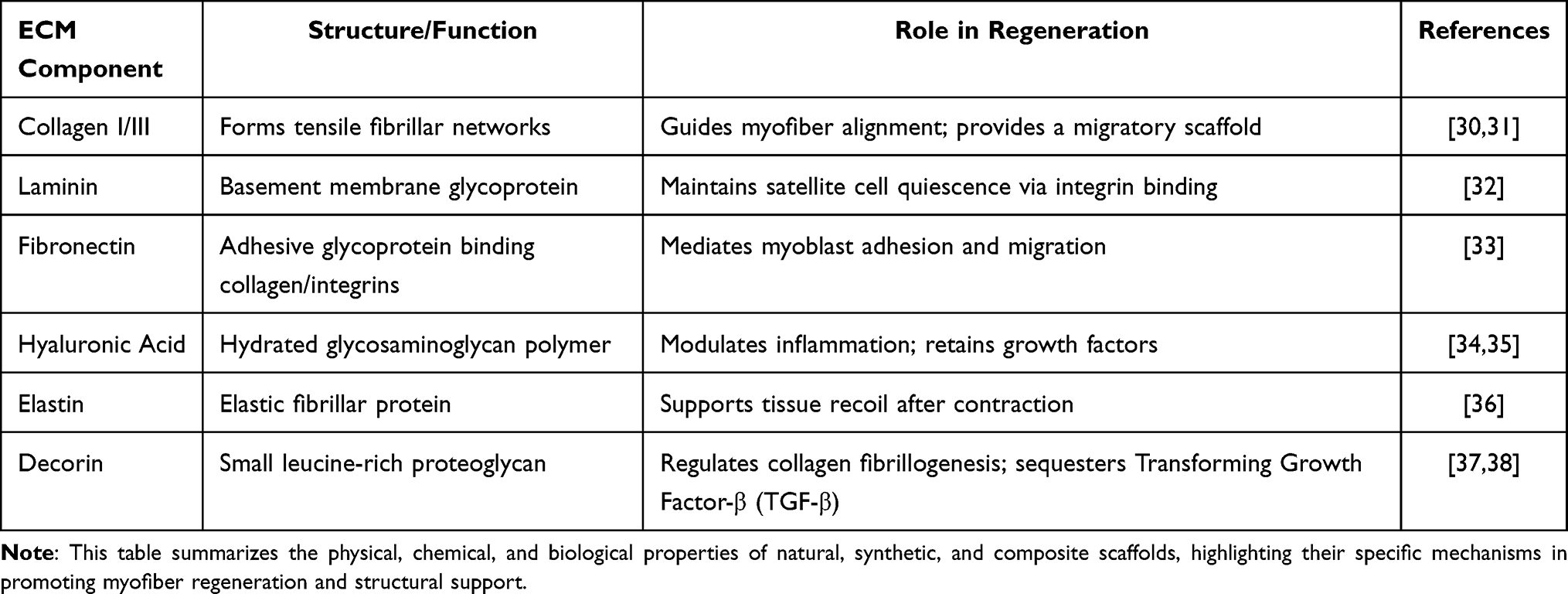 |
Table 1 Classification and Functional Attributes of Biomaterials for VML Repair |
Mechanism of Muscle Regeneration
The ECM, besides being a structural building block for tissues also have been recognized as one of the important factors in cell-to-cell communication during muscle regeneration acting as both a structural scaffold and a signaling hub.39,40 Its receptors, such as integrin,41 are abundant and it interacts with muscle cells to carry out the signaling pathways that control fundamental cellular processes like proliferation, differentiation, and migration.42 These interactions are essential to orchestrate the integrated set of responses that contribute collectively towards effective tissue repair.
Figure 143 shows the ability of skeletal muscle fibers to repair and regulate their activity through a multistep process involving degeneration and subsequent repair due to necrosis. The underlying mechanism by which skeletal muscle-based scaffolds regulate the immune system involves macrophage polarization that is strongly influenced by the immunomodulatory effects of mesenchymal cells.44,45 These cells contribute significantly to suppressing immune activity by secretion of several mediators and cell–scaffold interactions.46 The acute inflammatory response initiated after injury is characterized by infiltration of neutrophils and macrophages.46 M1 macrophages promote debris clearance by releasing pro-inflammatory cytokines such as Tumor Necrosis Factor—α (TNF-α), Interleukins (IL) like IL-1β and IL-6 and M2 macrophages enhance muscle tissue repair by transforming under the influence of mesenchymal cells, releasing anti-inflammatory cytokines such as IL-10 and IL-13 which facilitates regeneration.29,47,48 The innate capacity for muscle repair and satellite cell migration along with proliferation controls inflammation while releasing growth factors like TGF-β1, Fibroblast Growth Factor (FGF), VEGF, and promoting structural organization and regeneration.43 These factors guide satellite cell activation, proliferation, and differentiation into myoblasts that repair damaged matrix by enabling matrix breakdown and new matrix synthesis thus to make myofibers.49,50
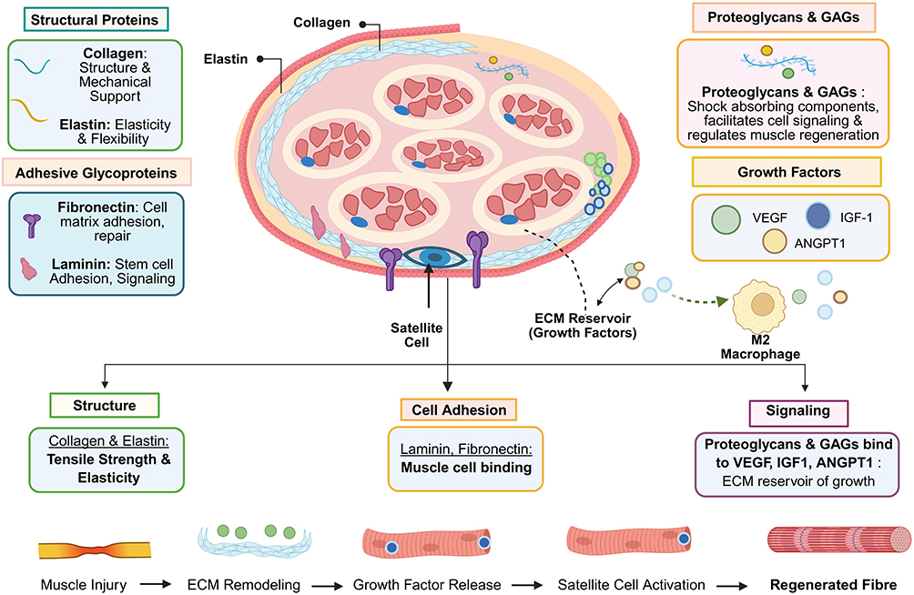 |
Figure 1 ECM composition: schematic representation of skeletal muscle ECM components and their functional roles (created with BioRender.com). The extracellular matrix (ECM) of skeletal muscle is a complex, hierarchical network comprising structural proteins (collagen, elastin), adhesive glycoproteins (laminin, fibronectin), and specialized molecules (proteoglycans and GAGs). This illustration highlights the dynamic interplay between the ECM and resident cells, including myoblasts, fibroblasts, and satellite cells. Key signaling pathways—such as VEGF for vascularization, IGF-1 for myogenesis, and ANGPT1 for immune modulation—are sequestered and presented by the ECM to orchestrate tissue repair and homeostasis. |
ECM Dysregulation in VML
In VML, ECM functions are severely compromised. Traumatic injuries disrupt the ECM scaffold, causing an imbalance between degradation and synthesis that often leads to fibrosis.51 Excessive collagen deposition creates a stiff matrix that impairs fiber alignment, cell migration, and neuromuscular junction reformation.52 This aberrant remodeling limits satellite cell function and regenerative capacity, presenting a major recovery barrier. ECM-based or biomimetic scaffolds offer promising solutions to restore the muscle microenvironment.53 The muscle injury triggers dynamic reorganization of the stem cell niche. This specialized microenvironment contains multiple interacting components: resident muscle stem cells, recruited immune cells, and supportive stromal cells, all embedded within a carefully balanced ECM.54 Together, these elements coordinate the complex process of tissue regeneration through precise cell-ECM signaling.
Table 2 outlines the sequential phases of skeletal muscle regeneration following an injury. The detailed coordinated events highlight the regulated immune responses that initiate debris clearance, activate myogenic progenitors guiding extracellular matrix deposition. The regenerative and remodeling phases enable myofiber formation, vascularization restoring impaired regeneration.
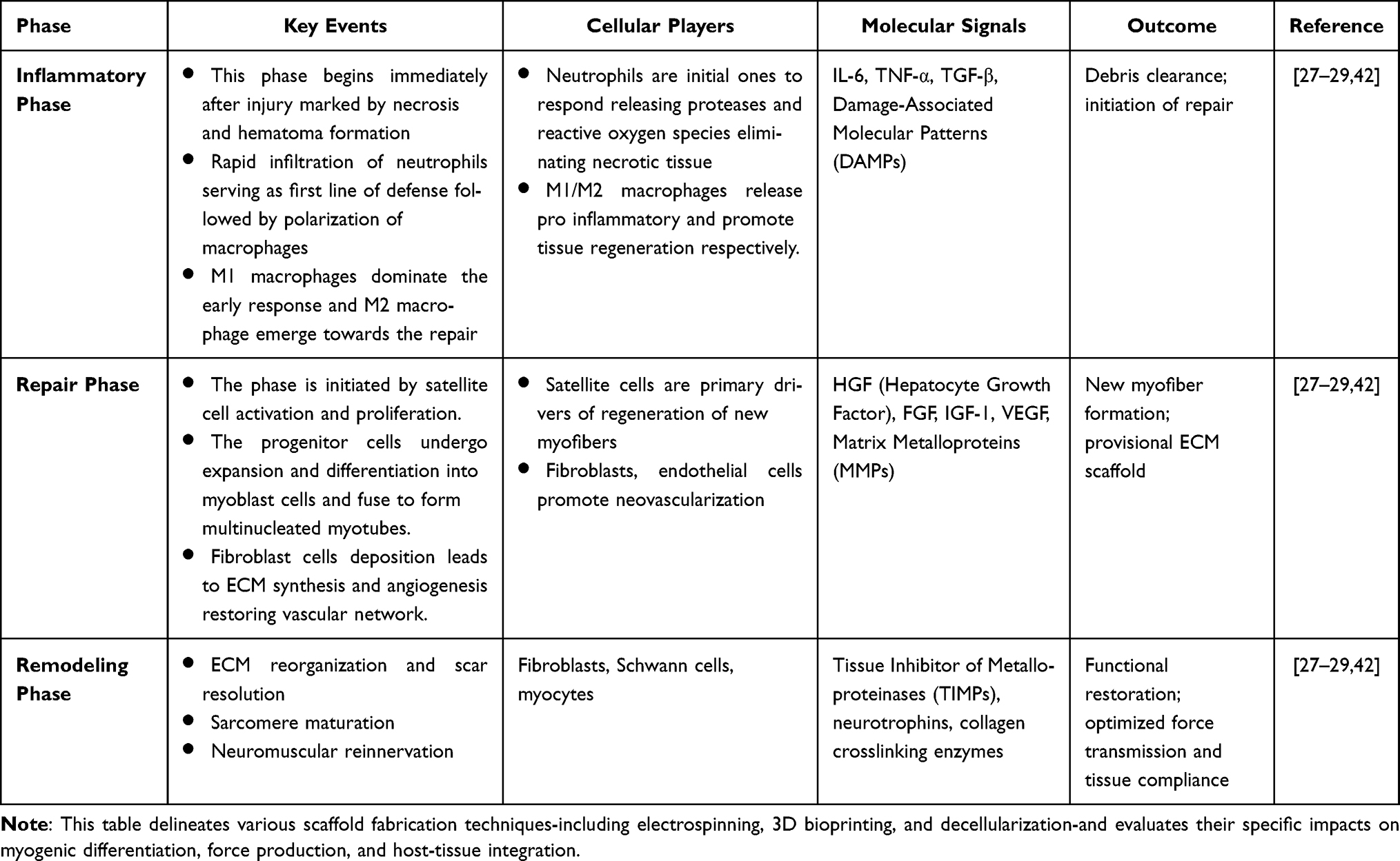 |
Table 2 Engineering Strategies and Biological Outcomes of Biomimetic Scaffolds for VML |
Any disruption in the equilibrium between ECM degradation and synthesis can result in fibrosis, a condition marked by an overabundance of collagen and other ECM elements, leading to stiff and non-functional tissue.55 Fibrosis poses a significant obstacle to muscle function and presents a major challenge in scenarios where regeneration is hindered. The importance of the ECM in muscle regeneration is clear across various scenarios. In the context of aging and specific disorders like muscular dystrophy, the ECM tends to become stiffer and more fibrotic, which hinders the muscle’s ability to regenerate effectively.56 This decline in ECM quality is a contributing factor to the progressive muscle weakness seen in these conditions. Gaining insights into the ECM’s role in these mechanisms have paved the way for innovative therapeutic strategies.40
Figure 257 describes that following injury, the muscle stem cell niche undergoes significant reorganization, involving multiple cell types like fibroblasts, macrophages, neutrophils, fibro-adipogenic progenitors (FAPs), and glial cells interacting within a remodeled ECM. Key ECM components including fibronectin, collagens (I, III, IV), laminins, elastins, tenascin-C, versican, proteoglycans, fibrillins, and thrombospondins (TSP-1, −2, −4) collectively mediate cell adhesion, migration, and signaling during repair. This schematic illustrates the complex interplay between cellular and extracellular components in the regenerating muscle microenvironment.57
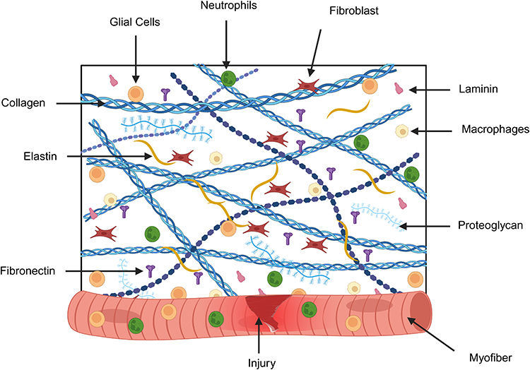 |
Figure 2 The skeletal muscle stem cell niche and ECM microenvironment (created with BioRender.com). This schematic illustrates the specialized microenvironment, or niche, that regulates muscle satellite cell behavior following injury. Structural and Adhesive Proteins: The niche is composed of a dense network of Collagen fibers and Fibrillins providing mechanical integrity, while Laminin and Fibronectin serve as critical anchors for MuSC adhesion and polarity. Specialized ECM Molecules: Proteoglycans and Elastin facilitate the sequestration of growth factors and provide the tissue with necessary recoil and elasticity. Cellular Constituents: The microenvironment is populated by a diverse array of resident and infiltrating cells. Fibroblasts actively remodel the matrix, while Macrophages, Neutrophils, and Glial cells provide biochemical cues that dictate the transition from the inflammatory phase to the regenerative phase. Regenerative Focus: The interaction between the cells and the damaged Myofiber at the site of Injury is highlighted, demonstrating how the biochemical and mechanical properties of the ECM scaffold are essential for directing stem cell activation, proliferation, and successful tissue integration. |
Material Classes and Functionalization
Design Principles of Biomimetic Scaffolds
Biomimetic scaffolds for VML are designed to replicate the structural, mechanical, and biochemical properties of native skeletal muscle ECM, promoting functional tissue repair.58,59 These scaffolds are to mimic the complexity of the ECM by adding necessary factors like collagen,60 fibrin,61 and glucosamine glycans62 essential for muscle tissue generation and remodeling. These biomimetic scaffolds also feature mechanism-aligned topographies that direct the alignment of myoblasts to further boost muscle regenerative property during myotube formation.63 An optimal scaffold must be biocompatible to avoid immune rejection, biodegradable to degrade in sync with tissue remodeling, and mechanically robust to withstand physiological stresses.59 Porosity and microarchitecture should enable nutrient exchange, cellular infiltration, and aligned myofiber growth. Crucially, scaffolds must facilitate vascularization and innervation while supporting cell proliferation and differentiation.64–66 These criteria ensure the scaffold not only fills the defect but also actively aids regeneration.
Figure 3 describes several scaffold fabrication approaches for VML repair and how they balance structural control, biological activity, and clinical practicality. Three-dimensional bioprinting allows precise, layer-by-layer placement of biomaterials and cells, enabling controlled pore size and spatial organization, although it can be limited by material properties and scalability.67 Electrospinning produces fibrous meshes that resemble the alignment of native ECM, offering high surface area but often restricting deep cell infiltration due to small pore sizes. Freeze-drying creates highly porous, interconnected foam-like scaffolds that are relatively simple and scalable to produce, yet they may lack sufficient mechanical strength.68 Decellular scaffolds, in contrast, retain the natural three-dimensional architecture and biochemical signals of donor tissue, enhancing bioactivity, but it is constrained by donor availability and potential immunogenicity. Together, these techniques provide complementary strategies, and their selection depends on the required balance between structural precision, biological function, and translational feasibility.
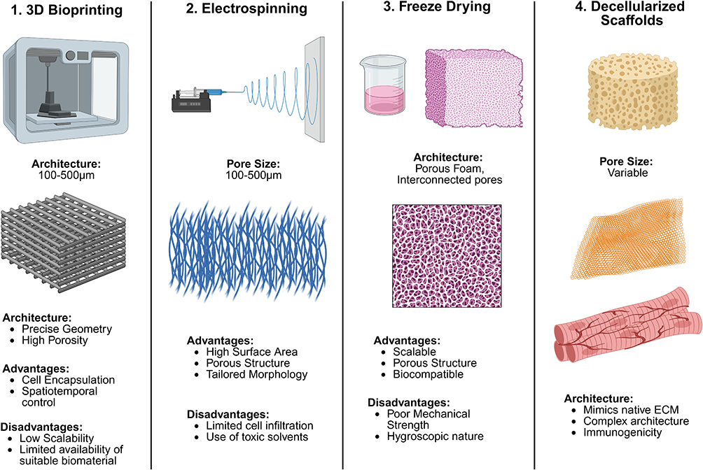 |
Figure 3 Scaffold fabrication techniques: comparative overview of scaffold fabrication techniques for VML repair (created with BioRender.com). A side-by-side comparison of four prominent fabrication methods: (1) 3D Bioprinting, providing high precision and cell encapsulation; (2) Electrospinning, producing fibrous meshes that mimic natural ECM alignment; (3) Freeze-drying, creating highly porous foam architectures; and (4) Decellularization, preserving the native complex architecture and bioactivity of donor tissue. The workflow, resulting architecture, and inherent advantages/limitations (e.g, scalability vs bioactivity) are detailed for each technique to guide scaffold selection. |
Effective scaffold selection for VML treatment requires the use of robust materials that address specific defect characteristics and consider patient biology to maximize regenerative potential. The first step in defect analysis is to evaluate its size.69 Depending on the area of muscles lost, scaffolds with mechanical integrity and stability can support the load required to support tissue growth.70 Anatomical location is critical to understanding the type of scaffold required, its mechanical demands along with the specific biochemical environment.71 To design a scaffold for the quadriceps muscle, one with high tensile strength and resilience is required to prevent the scaffold from failing and causing permanent functional impairment.
The study by Anderson S.E et al determined the regenerative, fibrotic, vascular, and neuromuscular responses by creating a complete muscle defect in quadriceps of female C57BL/6J mice with different defect sizes of 2 mm, 3 mm, and 4 mm in diameter. The relative quadriceps loss was 4.44 ± 1.85%, 15.49 ± 2.04%, and 32.16 ± 5.14% for a defect size of 2 mm, 3 mm, and 4 mm, respectively. Quantitative evaluation of wet muscle weight demonstrated the reduction in defect size for all groups on day 7, but the 4 mm defect size remained damaged and deteriorated on day 28. Among the three defect sizes, the 2 mm lesion largely dissolved fibrosis and inflammation on day 28, while the 3 mm lesion exhibited incomplete myofiber formation and deposition of collagen with unresolved CD68+ macrophage infiltration that was analyzed by histological examinations. Vascular analysis using micro CT angiography increased vascular volume confirming angiogenesis. The neuromuscular reinnervation at end of 28 days post injury was 44.3% with 2 mm defect and 55.7% in 3 mm defect due to the presence of fibrotic tissue. The overall study concludes 3 mm defect as the critical threshold for nonhealing VML in mouse quadriceps and 4mm injury as regenerative failure due to disorganized vascularization and hindered regeneration.72
The study conducted by Panayi AC et al highlighted the improved functional performance of the quadriceps muscle by treatment with collagen-glucosamine glycan (CGAG)-based scaffold in a murine model of VML. Histological results are presented as the presence of fibrotic scar deposition with disorganized muscle architecture in the untreated group, whereas the scaffold-based intervention improved muscle hypertrophy after injury. Two weeks after injury, the scaffold group displayed lower fiber density (114 ± 7 fibers/HPF) compared to the uninjured control group (268 ± 13 fibers/HPF) and untreated VML (196 ± 18 fibers/HPF). Finally, by functional treadmill testing at 6 weeks, muscle repair was improved by 20–25% in the scaffold group compared to untreated VML. At the molecular level, upregulation of tissue inhibitor of metalloproteinase 1 (TIMP1) and colony stimulating factor 2 (CSG2) leads to ECM turnover by stimulating the differentiation of progenitor cells into macrophages and granulocytes, thereby increasing immune cell recruitment. Angiogenic factors such as VEGF, ANGPT1, HGF, and myogenic factors such as IGF1 and STAT3 ensured vascularization and gradual progression from injury to regeneration in scaffold treated animals, supported by histological evidence. In a broader context, the researchers also reinforce the fact that scaffolds not only support structural integrity but also regulate the host immune response, which decellularized ECM-based scaffolds lack, providing inconsistent and variable cell-scaffold integration and coherent regenerative gene expression.73
At the same time, patient attributes including age, coexisting medical conditions such as hypertension, diabetes, other vascular ailments, muscle regeneration capacity, and immune system response are also taken into account before selecting the scaffold. A person’s aging significantly affects the body’s response to regenerate muscle fibers efficiently and existing comorbidities can also hinder the results associated with the healing process. This eventually leads to a drastic change in the healing trajectory characterized by increased fibrosis and scar tissue deposition.74 All of these factors are useful in governing the architecture of the scaffold, its stiffness, and resilience further shaping the biocompatibility profile essential to ensure fibrosis-free integration.
Researchers Kim J.T. et al, carried out a study to evaluate the modulating effect of age on VML muscle repair with a 3- and 18-month-old rat model with tibialis anterior defects. Decellularized skeletal muscle scaffold with minced muscle revealed a superior outcome in young 3-month-old rats by restoring torque to 79.9 ± 12.2% compared to the untreated 3-month-old rat that had a torque of 62.3 ± 10.1%. On the other hand, the 18-month-old rat model did not show significant muscle recovery compared to the untreated group, with an achieved torque of only 57.1 ± 8.7% and 58.6 ± 14%, respectively. The molecular mechanism implicated for superior regenerative outcomes in the 3-month-old rat model was the upregulation of myogenic genes such as MyoD, MyoG, and inflammatory biomarkers such as IL-β1 and TNF-α. Collagen deposition in young rats maintained normal collagen levels compared to older rats with markedly elevated fibrosis. The overall conclusion drawn from the study is the pronounced age-related response to the same treatment intervention.75
Types of Biomimetic Scaffolds
Natural Scaffolds
Elastin
In tissue engineering, elastin is a crucial ECM protein that provides elasticity and recoil to tissues-derived materials are increasingly used for their flexibility and biocompatibility. A key advancement involves methacryloyl-functionalized elastin (MeTro) combined with gelatin methacryloyl (GelMA) to create elastic hydrogels for 3D bioprinting. These scaffolds exhibit controlled degradation (12.7% by day 1, 17.9% by day 14) and maintain >90% cell viability, demonstrating strong cytocompatibility and structural support for muscle regeneration.76–78
Additionally, elastin-like recombinases (ELRs)—synthetic analogs mimicking elastin’s properties—have been shown to modulate macrophage behavior in rat models, shifting from pro-inflammatory M1 to regenerative M2 phenotypes, reducing fibrosis, and enhancing myofiber formation.79,80 Recent work with human elastin-like polypeptides integrated into collagen scaffolds has improved mechanical stability and vascularization, supporting aligned ECM remodeling for muscle regeneration.81
In recent years, elastin-like recombiners (ELRs) have been investigated as a biomaterial solution for enabling muscle regrowth in the context of volumetric muscle loss VML, which arises from chronic inflammation and subsequent fibrotic scar formation.77 The study proposed that the biodegradable amphiphilic ELR-based hydrogels are able to provide a switching niche for converting of M1 macrophages into their pro-regenerative M2 counterpart. Rat tibialis anterior muscle model was utilized to introduce chemically and physically crosslinked ELR hydrogels. These results show that the muscle treated with ELR hydrogels a higher percentage of M2 macrophages at 14 days and cell increases, which are responsible for reducing inflammation seconds to collagen production.78,79
More recently, a biomimetic human elastin-like polypeptide was proposed as an alternative to muscular volume loss assistance and enriched vascular tissue engineering. The objective of the current investigation was to develop human elastin-like polypeptide with improved mechanical properties, biodegradability, and cytocompatibility using several recombinant strategies. The results demonstrate that human elastin-like polypeptide addition to collagen scaffolds dramatically improves their tensile strength and stress resistance, approaching native tissue protein composition. These scaffolds can be useful in applications such as regenerative medicine, which require a certain mechanical integrity of the scaffold to ensure proper tissue regeneration. Human elastin-like polypeptide possesses thermosensitive characteristics which are beneficial for purification and scaffold integration.82
The enhancement of these biomaterials by combining them with the elements described here could further increase their regenerative properties towards vascular tissue damage and volumetric muscle atrophy, without which effective treatments are scarce. Through the developments using human elastin-like polypeptide, a versatile platform to design successful strategies for tissue engineering and regenerative medicine that can reconcile problems specific to native elastin in biomedical applications.82,83
An important leap in tissue engineering is achieved through the development of biomimetic elastin-like proteins that exhibit superior mechanical properties and biocompatibility for vascular as well as muscle regeneration applications. These modified elastin materials find wider scope to enhance clinical outcomes in regenerative therapies with the ongoing research work, particularly for reinstating function of lost or injured tissues.
Hyaluronic Acid
Hyaluronic acid, a naturally occurring biopolymer, is valued for its biocompatibility, hydration capacity, and low immunogenicity.84
In a study, a bioinspired semisynthetic acrylated hyaluronic acid (AcHyA) hydrogel was introduced as prospective therapy for VML of craniofacial muscles by researchers. In a novel rat injury model AnHyA was applied to a 5 mm × 5 mm lesion in superficial masseter. The results were shown to be an improvement relative to untreated controls following 16 weeks. From the images, muscle fibrosis was visibly reduced in hydrogel-treated muscles and there also appeared to be less damage (smaller defects) as well as higher myofiber cross-sectional area. AcHyA hydrogel promotes muscle sparing and reduces scarring in craniofacial VML injury, indicating a possible regenerative benefit to the damaged musculature. The findings highlight hydrogels as a therapeutic approach and provide hope for enhanced patient recovery from this disorder.85
Researchers in another study looked into the combination of 3D bioprinting and electrospinning with semisynthetic acrylated hyaluronic acid (AcHyA) hydrogels for VML repair. In rat masseter muscle injury models, AcHyA reduced fibrosis by 50% and defect size by 20%, while increasing fiber cross-sectional area (30%). These strategies allow creation of scaffolds that can more closely replicate the ECM, and as a result improve adhesion and alignment for muscle regeneration. There are also, investigations into whether using biological scaffolds with growth factors and cell therapies will increase muscle recovery even more. Yet, there are challenges to meet such that long-term clinical efficacy and immune responses toward the implanted materials can be more optimized on behalf of any effective VML treatment.86
Case studies have reported hyaluronic acid-based hydrogels are effective in treating volumetric muscle loss and enhancement of muscle lipogenesis by reducing fibrosis-associated features which can reduce the integration of better tissue.34,87 In the future, advanced HA formulations and their combination with 3D bioprinting or even cell therapy could be promising to enhance tissue regeneration.
Collagen
Collagen, a vital component of the extracellular matrix, is crucial for tissue regeneration, especially in treating VML. Its types, particularly type I and III, is a fundamental ECM protein that provides structural support and facilitates cell adhesion.30 The assembled cell-decorated collagen (AC-DC) bioprinting technique uses collagen microfibers to create aligned, high-strength implants, which have improved muscle function in injury models.88 However, collagen alone may lack sufficient mechanical mimicry of muscle, as seen in studies where collagen I hydrogels with minced muscle grafts yielded limited muscle repair.89 A notable improvement is photo cross-linkable collagen methacrylate (CMA) hydrogels, which offer tunable stiffness and enhance vascularization. CMA scaffolds significantly boost angiogenesis and myogenesis compared to traditional collagen, making them promising for VML repair.90
Minced muscle grafts and collagen I hydrogels composite has recently been suggested for treatment of volumetric muscle loss (VML) by researchers. A more recent study showed that despite the gains in muscle recovery in grafts comprising 50% minced muscle, only about half as many new muscle fibers were created compared with whole grafts. These activity constraints were observed due to the limited ability of the collagen hydrogel in supporting muscle growth (though it had a strong angiogenic capacity). The defect of our hydrogels for mimicking the mechanical properties of native muscle led to limited migration and regeneration ability.91
In biomimetic VML treatments, collagen serves as a whorl to be remodeled in the same way that is seen with native tissue and integrates well into new vasculature development along with promulgating cell growth. This improves its effectiveness in muscle regeneration as it can be tailored for different forms such as hydrogels with adjustable properties.31,92 The secondary studies should focus on improving collagen-derived materials involving better mechanical properties and integration with native tissue.92 Breakthroughs in collagen tissue engineering may lead to improved VML therapies and other regenerative procedures. Given the vast unmet medical needs and tremendous potential benefits of fully developed organ bioengineering, such incremental innovation in this area can bring huge leaps forward to regenerative medicine or tissue engineering.
Laminin
Laminin is an essential building block of the extracellular matrix and participates in many cellular events like adhesion, differentiation, or migration. Its importance is most substantiated with respect to muscle repair and regeneration, especially in the setting of VML that results from severe forms of trauma or surgical excision. However, treatments for VML are unfortunately few and far between. More recent studies have begun to investigate the ability of laminin–111 in determining muscle regeneration and functional outcomes for these challenging cases.32,93
In a study, it was evaluated that a lower dose of minced muscle grafts with hyaluronic acid hydrogel augmented with laminin-111 (HA+LMN) in comparison to autologous muscle transfer, and interestingly found the gel was effective at increasing skeletal myogenesis/volume re-growth. The most striking result is the 42% relative improvement in peak tetanic torque with respect to untreated limbs. It was discovered that the combination treatment dramatically and synergistically improved functional outcome, though did not surpass minced muscle graft control. Histological analysis showed the HA+LMN compound was associated with reduced macrophage activity and, in particular, an increase satellite cell density primarily outside of rather than within the defected area. The results indicated a remarkable 42% improvement in peak tetanic torque compared to untreated limbs. Interestingly, while the combination treatment showed significant benefits, it did not outperform the control group that received only minced muscle grafts. Histological analyses revealed that the HA+LMN treatment led to decreased macrophage activity and increased satellite cell density, predominantly in the surrounding muscle tissue rather than the defect area itself. The results collectively demonstrate that HA+LMN could be a promising therapeutic option for VML, although further research is needed to fully understand its mechanisms and optimize its effectiveness.94
In another study, where the tibialis anterior muscle of rats was treated for VML using fibrin hydrogels enriched with laminin-111 at a concentration of 450 μg/mL. At 28 days post-injury, the treatment group exhibited augmentation in myogenic protein expression and an increase in contractile area. Supplementation of hydrogels with laminin-111 was most effective, enhancing muscle strength (measured as torque production) by up to 60%. Additional studies of laminin-111 enriched hydrogels for VML should be pursued given the results that are suggestive in nature but also promising (eg, functional gain, muscle regeneration).34,83,87 These data have significant implications and suggest that laminin-111 may offer this population novel therapeutic approaches to address the consequences of VML.93
When introducing laminin-111 into hydrogels it creates a microenvironment that supports muscle regeneration. These findings imply that laminin-111 supplemented hydrogels might provide a new treatment strategy for the restoration of VML-related functions by enhancing myogenic activity, contractile tissue formation, and muscular strength. The factors, such as concentration of laminin 111 to use for optimal effects, composition of the hydrogel in which they are loaded and long-term efficacy required detailed investigation.95
Synthetic Scaffolds
Synthetic polymers serve as controlled, reproducible platforms for muscle tissue engineering due to their tunable mechanical properties, degradation kinetics, and structural architecture. Their compatibility with fabrication techniques like 3D printing and electrospinning makes them valuable for VML repair.60 However, their limited bioactivity requires surface modifications or incorporation of biological molecules to improve cellular interactions.
Poly ε Caprolactone (PCL)
Poly ε caprolactone (PCL), an aliphatic polyester, is valued for its mechanical strength, slow degradation, and compatibility with electrospinning and 3D printing. Its ability to form aligned nanofibers replicates the anisotropic structure of native skeletal muscle, promoting myoblast alignment and fusion into myotubes.96,97 For example, electrospun PCL/gelatin scaffolds with aligned fibers significantly enhanced myotube formation by guiding cell orientation. Mechanical stimulation further increased myotube width (12.92 ± 3.29 μm) and nuclear fusion index (95.73 ± 1.05%), underscoring the importance of mechanical cues in regeneration. Despite these advantages, PCL’s hydrophobic surface and lack of cell-adhesive motifs limit bioactivity. Strategies to address this include surface modification with ECM proteins (eg, collagen, laminin) or blending with natural polymers. PCL/gelatin composites, for instance, improved wettability, myotube contractility, and actin density compared to pure PCL, demonstrating the benefits of hybrid materials.98
The study carried out by Kim I et al, demonstrated the study carried out using myotubes induced myogenic progenitor cell embedded in PCL scaffold using electrospinning technique for skeletal muscle regeneration by incorporation of Matrigel. The scaffold rapidly induced myofiber formation enabling its alignment upto 12.8° ± 5.3° possibly due to cell signaling highlighting the role of biomimetic agents along with mechanical strength and biodegradation offered by PCL.99
Polylactic Acid (PLA)
Polylactic Acid (PLA), an FDA-approved biodegradable polymer, degrades faster than PCL, making it suitable for temporary mechanical support in muscle regeneration. Electrospun PLA scaffolds facilitate myoblast adhesion and alignment, while porous structures enhance vascular infiltration.100 However, PLA’s brittleness and acidic degradation byproducts can cause localized inflammation. Blending PLA with hydrophilic polymers like polyethylene glycol (PEG) improves elasticity and reduces acidic microenvironments.101 For example, PLA/PEG/RosA/GO membranes exhibited a threefold increase in tensile strength (2.6 MPa) and improved hydrophilicity, reducing inflammation while enhancing tissue compatibility. Incorporating ceramics like β-TCP or hydroxyapatite further strengthens PLA scaffolds for musculoskeletal applications. However, replicating muscle-like elasticity remains challenging, prompting research into crimped fiber geometries or elastomeric additives.100
Polyethylene Glycol
Polyethylene glycol (PEG) PEG, a hydrophilic and non-toxic polymer, is widely used in hydrogel scaffolds due to its tunable crosslinking and high water content, which supports nutrient exchange—critical for VML repair. However, PEG’s bioinert nature necessitates functionalization with adhesive peptides (eg, RGD) to promote cell adhesion. PEG-modified PLA scaffolds, for instance, enhanced endothelial cell attachment and collagen deposition, improving vascularization in vivo. Beyond structural support, PEG-based scaffolds serve as drug delivery platforms.102 The PLA/PEG/RosA/GO system delivered rosmarinic acid and graphene oxide, achieving 99% antibacterial efficacy and accelerating wound healing in murine models. Despite its advantages, PEG’s low mechanical strength requires reinforcement with stiffer polymers like PLA or PCL for load-bearing applications in large muscle defects.100
In the study carried out by Alarcon Y et al, the potential solution for skeletal muscle regeneration is PEG diacrylate. The authors fabricated copolymer of PEG diacrylate and acrylic acid with collagen methacrylate further promoting cell adhesion and cell proliferation. The results positively showed C2C12 myoblast adhesion and differentiation with sustained metabolic activity over 14 day in vitro study.103
Hybrid Scaffolds
Hybrid scaffolds integrate the strengths of both natural and synthetic materials, creating composite platforms that balance mechanical performance with bioactivity. By combining the structural integrity and tunability of synthetic polymers with the cell-instructive properties of natural extracellular matrix (ECM) components, these scaffolds address the limitations of single-material systems.104 This synergistic approach is particularly valuable for volumetric muscle loss (VML) repair, as it enhances both mechanical support and biological integration.
Collagen–PCL Hybrid Scaffolds
Collagen–PCL hybrid scaffolds illustrate this strategy effectively. Electrospun PCL fibers provide mechanical strength and structural alignment for myotube formation, while collagen enhances hydrophilicity and offers integrin-binding sites to improve cell adhesion and signaling. Studies show that collagen-coated or blended PCL scaffolds significantly improve myoblast proliferation, alignment, and differentiation compared to pure PCL.104 In VML animal models, these scaffolds increase muscle fiber density, reduce fibrosis, suggesting strong clinical potential.92 Advanced fabrication techniques, such as co-electrospinning or coaxial electrospinning, allow for composite fibers with gradient or layered architectures that better mimic native muscle tissue. For example, research comparing PCL/collagen and PCL/gelatin scaffolds found that collagen incorporation reduced fiber size, increased hydrophilicity, and softened mechanical properties, all of which promote cell growth and musculoskeletal tissue development.105,106
Fibrin-Based Hybrid Scaffolds
Fibrin-based hybrids, often combined with synthetic polymers like PLA, PLGA, or PEG, are widely explored due to fibrin’s natural role in wound healing and its bioactive properties. These scaffolds enhance vascular infiltration, ECM remodeling, and satellite cell recruitment in VML defects.107 Fibrin–PLGA constructs, for instance, act as transient matrices that degrade gradually while supporting early regeneration phases, eventually allowing native tissue deposition. Additionally, fibrin’s ability to encapsulate and release growth factors (eg, VEGF, IGF-1) makes it useful for modulating the regenerative microenvironment. This controlled delivery can enhance muscle repair by promoting angiogenesis and myogenesis.108
Hyaluronic Acid Hybrids
HA-based hybrid scaffolds are gaining traction due to HA’s anti-inflammatory, pro-angiogenic, and cell-adhesive properties. When incorporated into synthetic networks like PEG hydrogels or PLGA frameworks, HA improves scaffold integration and host–tissue interaction. These systems enhance myogenic differentiation, vascularization, and immune modulation in vivo. For example, HA-modified PEG hydrogels functionalized with RGD peptides and loaded with muscle progenitor cells have demonstrated superior tissue integration and myofiber organization in murine VML models. Similarly, HA-chondroitin sulfate hydrogels accelerate skeletal muscle healing by promoting cell migration and angiogenesis.109,110
Hybrid scaffolds offer significant advantages, including customizable architectures (gradient or multi-layered designs) and controlled release capabilities, better mimicking native muscle complexity. However, their fabrication can be technically challenging, and ensuring consistent material integration without compromising functionality remains a key hurdle. Future research should focus on optimizing fabrication techniques and improving scalability for clinical translation.111,112
A comparative assessment of scaffold classes for VML repair is described in Figure 4 underscoring clear material-dependent trade-offs across mechanical competence, biological performance, and translational readiness. Natural scaffolds demonstrate strong bioactivity due to inherent cell-binding ability and tissue-specific attributes, supporting adhesion, proliferation, and differentiation. However, they generally exhibit limited mechanical strength and present challenges related to large-scale manufacturing and immunological variability. In contrast, synthetic scaffolds provide tunable and reproducible mechanical properties with high structural precision and scalability, yet they lack intrinsic bio-instructive signals and often require functionalization to enhance cellular interactions. Hybrid scaffolds integrate components from both systems to balance structural robustness with biological functionality, thereby offering improved overall performance and enhanced clinical applicability. This comparative framework highlights the importance of aligning material selection with the specific mechanical and regenerative demands of VML repair.
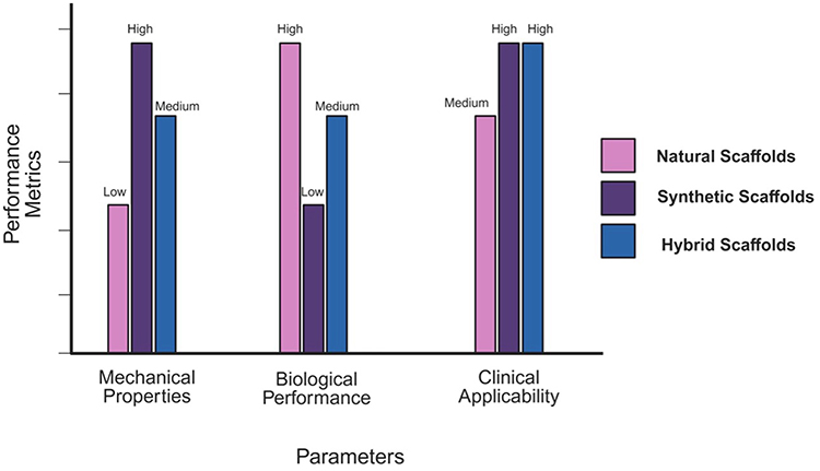 |
Figure 4 Performance metrics of natural, synthetic, and hybrid scaffolds for VML repair (created with BioRender.com). |
Functionalization Strategies
Functionalization is a critical strategy in enhancing the therapeutic potential of biomimetic scaffolds, transforming them from passive structural supports into bioactive platforms capable of directing tissue regeneration. One common approach involves the incorporation of growth factors, such as VEGF, insulin-like growth factor-1, and FGF. These signaling molecules orchestrate key regenerative processes: VEGF stimulates angiogenesis, promoting the formation of blood vessels necessary for oxygen and nutrient delivery; IGF-1 activates satellite cells and enhances myogenic differentiation; and FGF supports the proliferation and migration of muscle progenitor cells. Controlled and localized delivery of these factors from scaffolds ensures sustained biological activity at the injury site.113,114
Moreover, electrical and mechanical stimulation has emerged as a powerful tool to mimic the native physiological environment of skeletal muscle. Electrical stimulation of scaffolds enhances myotube formation, contractile function, and alignment, mimicking neuromuscular signaling.104 Similarly, mechanical loading promotes fiber alignment, cellular orientation, and ECM deposition, all of which are essential for functional muscle tissue. Incorporating conductive materials or designing dynamic bioreactor systems enables the delivery of these physical cues during in vitro maturation or post-implantation.115
The list of key biomimetic scaffolds used in VML are described in Table 3. It provides an overview of several scaffold types highlighting the material composition. Further the mechanism by which the scaffold supports modulation of myogenesis, vascularization, and innervation are discussed.
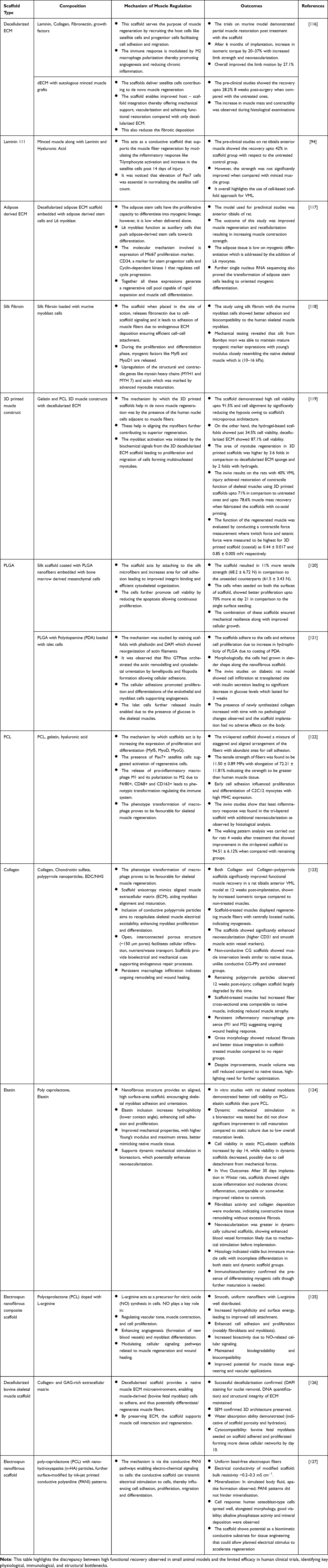 |
Table 3 Critical Analysis of Preclinical Success vs Clinical Translation Barriers in VML |
The scaffolds act as engineered biomimetic ECM-based guides for muscle regeneration by promoting muscle repair and functional restoration. Regeneration capacity depends on the type of material, its composition, presence of any growth factors, cellular incorporation, and distribution.128 Since the architecture of the scaffold is highly influenced by the type of biomaterial used, it directly regulates cell signaling, adhesion, migration, proliferation, and differentiation.129 The decision matrix for deciding the type of scaffold to be used is described in Table 4. The decision framework for selecting biomimetic scaffolds for VML was based on defect size, injury complexity, and patient-specific factors. It integrates material choice, scaffold architecture, and bioactive incorporation to align mechanical support with regenerative demands. This stratified approach emphasizes the necessity of personalized scaffold strategies to achieve functional muscle restoration.
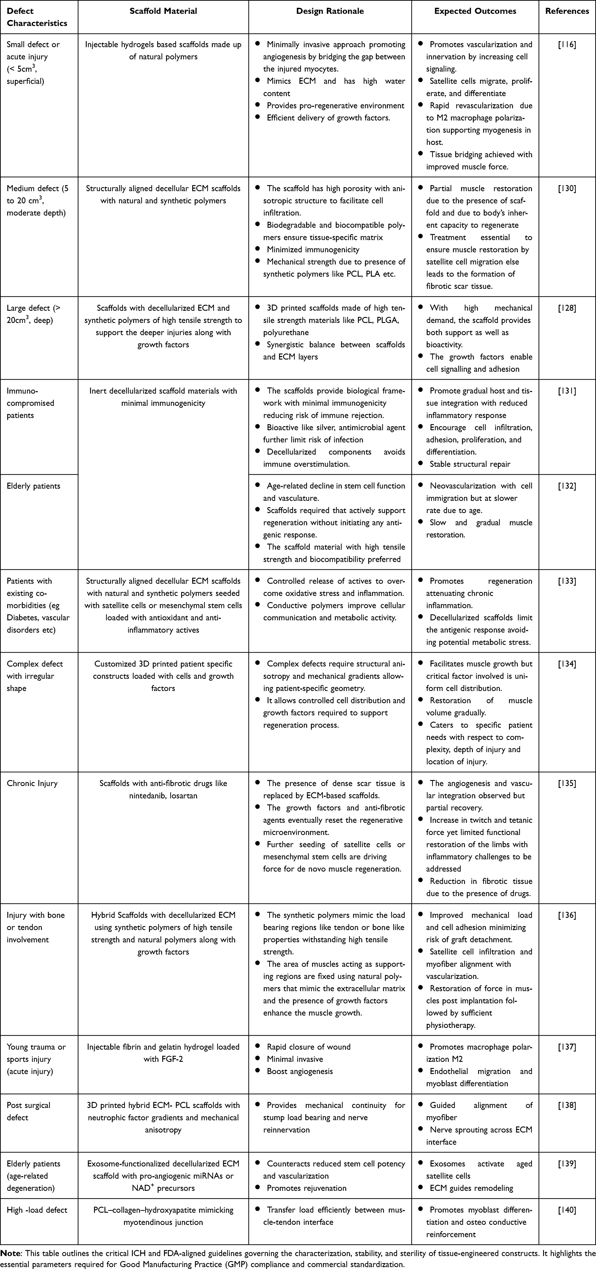 |
Table 4 Regulatory Framework and Quality Control Standards for Scaffold-Based Muscle Therapies |
While advances in scaffold design have been significantly observed and shown promise in preclinical studies, clinical translation for VML treatment faces persistent knowledge gaps in understanding the host’s immune response to implanted material, inflammation, and fibrosis due to chronic immune activation, thereby ensuring biocompatibility to avoid immunogenic reactions.141 While biomimetic scaffolds mimic the ECM, cell adhesion, and differentiation with adequate vasculature and long-term functional integration still remains a challenge. Approaches such as customizable 3D printed scaffolds show promising properties but have not yet been clinically established.142,143 A major unresolved challenge in VML repair is the restoration of functional neuromuscular units rather than mere tissue filling. While scaffold-based systems often promote myofiber formation, they frequently fail to achieve synchronized vascularization and reinnervation, which are essential for sustained contractile function. Additionally, fibrotic signaling pathways remain insufficiently controlled in large defect models, particularly in chronic injury settings where immune dysregulation persists.43 To overcome these limitations, future scaffold designs should incorporate dynamic, responsive materials capable of modulating the inflammatory milieu and adapting to the evolving regenerative microenvironment. The inclusion of mechanoresponsive elements may further enhance alignment and maturation of regenerating myofibers.144 However, translating such multifunctional constructs to clinical settings demands careful balancing between biological complexity and manufacturability. There is also a need for standardized protocols for comprehensive biomolecular characterization to better understand host scaffold interactions in clinical trials. Multidisciplinary research focusing on precise biomimicry at the molecular and structural levels is needed to address these knowledge gaps, improved manufacturing technologies for clinical translation of biomimetic scaffolds to evaluate the long-term performance and safety of these scaffolds in muscle regeneration.145 Strategies like integrating angiogenic factors or pre-vascularizing scaffolds are essential to ensure sufficient blood supply.
Preclinical to Clinical Translation
Critical Evaluation of Scaffold Performance
Biomimetic scaffolds for treating VML are extensively tested in preclinical animal models to assess how well they integrate with tissue, promote muscle regrowth, and support blood vessels and nerve formation.146 Rodents, such as mice and rats with surgically created defects in muscles like the quadriceps or tibialis anterior, are popular choices due to their cost-effectiveness and genetic uniformity.147 For example, studies using scaffolds made from small intestinal submucosa extracellular matrix (SIS-ECM) in these models have shown new muscle tissue forming with functional blood vessels and nerves. However, rodents differ significantly from humans in immune responses and tissue structure, limiting how well findings translate. Larger animals like pigs, dogs, and sheep better mimic human biology, particularly in processes like scar tissue formation and blood vessel remodeling.148 A study in mini-pigs, for instance, revealed that muscle injuries healed with extensive scarring and minimal regeneration, mirroring what happens in humans. Sheep models are especially useful for testing large scaffolds, as they highlight challenges like ensuring nutrients reach the core of implanted materials to prevent cell death.149–151 Table 5 summarizes key animal models used in VML research. This table highlights how each model contributes unique insights into muscle degeneration, fibrosis, muscle repair, and biomaterial performance. By comparing reproducibility, anatomical relevance, and regenerative outcomes, it supports the discussion on preclinical validation of biomimetic scaffolds. These models collectively bridge mechanistic understanding and translational assessment of VML therapies.
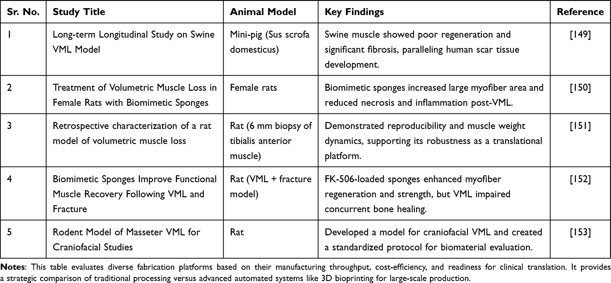 |
Table 5 Industrial Scalability and Clinical Adoption Matrix for VML Scaffolds |
Despite encouraging preclinical outcomes, several biomimetic scaffolds have underperformed due to inadequate immunomodulation, insufficient vascularization, and poor neuromuscular integration. Many constructs successfully replicate the structural aspects of native extracellular matrix but fail to actively direct macrophage polarization toward a pro-regenerative phenotype, resulting in prolonged inflammation and fibrotic encapsulation. Animal studies remain critical for evaluating the safety and efficacy of biomimetic scaffolds but raise ethical concerns, prompting adherence to the 3Rs principle—Replacement (using non-animal alternatives where possible), Reduction (minimizing animal use), and Refinement (enhancing welfare) to address welfare issues.154 However, interspecies differences, such as faster healing in rodents compared to humans, complicate translation, leading researchers to prioritize larger models like pigs and dogs that better replicate human fibrosis and regeneration.155 Additionally, human variables like age, comorbidities, and individual healing capacity further challenge the replication of clinical conditions in animals. Emerging alternatives, such as patient-derived organoids and humanized mice, offer promising avenues to bridge this gap, though.156 Solutions include adaptive clinical trial designs, collaborative efforts between engineers and regulators, and post-market surveillance to ensure therapies meet safety and efficacy standards.
The clinical application of biomimetic scaffolds for VML remains experimental, with ongoing and completed trials yielding mixed outcomes.157
Regulatory and Manufacturing Considerations
Regenerative approaches for VML that include cell-based therapies, biomaterial scaffolds, and combination products occupy a complex regulatory space. This leads to inconsistency in product quality and results due to lack of standardized protocols related to safety, efficacy, and sustainability.158 The International Council for Harmonization of Technical Requirements for Pharmaceuticals for Human Use (ICH) is recognized for accelerating clinical translation for global access by ensuring the safety of biomaterials for VML-based therapies.159 Current ICH initiatives related to quality and manufacturing of biologics and cell-based products include ICH Q5A (R2) which addresses genetically engineered viral vectors and other biotechnology products associated with cell-based therapies, ICH Q5D related to the derivation and characterization of cell substrate with detailed documentation, ICH Q5E when changes are made to the scaffold material and ICH Q6B to establish important quality characteristics of organic products.160
It highlights the importance of understanding regulatory pathways, manufacturing standardization, and implementation timelines. Regulatory authorities such as the FDA and EMA require a clear understanding of the frameworks typically under investigation of new drugs (IND). In the United States, the FDA oversees these constructs as combination products through the Office of Combination Products, classifying them based on their mode of action.161 Developers must comply with current Good Manufacturing Practices (cGMP; 21 CFR Part 4) and submit appropriate regulatory submissions, either an Investigational Device Exemption (IDE) or an IND application, depending on whether the scaffold functions primarily as a device or as a biologic.162
In the European Union, the European Medicines Agency (EMA) governs these scaffolds under the Advanced Therapy Medicinal Products (ATMP) framework, which requires simultaneous compliance with GMP for biologics and ISO 13485 quality standards for device components.163 Both regulatory bodies emphasize manufacturing standardization, including validated sourcing of biomaterials, process reproducibility, and rigorous sterility and performance testing, to ensure safety and effectiveness.164 Early collaboration with regulators helps clarify classification, expedite documentation, and avoid delays during clinical progression. At the preclinical stage, researchers face the difficult balance of adhering to ethical animal research principles while generating meaningful data designed to protect animals often forces studies in unrealistically healthy specimens that poorly predict human outcomes.149 Combination products that pair scaffolds with cells or biological factors fall into regulatory grey areas between device and drug classifications, requiring reviews from multiple FDA divisions. Cellular therapies bring additional layers of complexity—from ensuring stem cells will not form tumors to verifying genetic modifications will not cause unintended effects.165 Manufacturing these complex products under strict quality standards drives costs into prohibitive ranges, with each component from biological materials to specialized equipment requiring rigorous validation.166 Despite these substantial barriers, the field continues advancing through innovations in smart materials and precision manufacturing. However, realizing the full potential of these technologies will require more than scientific breakthroughs—it demands new frameworks for regulatory evaluation, standardized testing methods, and international cooperation to streamline the path from lab to patient while maintaining rigorous safety and ethical standards.167 The coming years will test whether our current systems can adapt quickly enough to deliver these promising therapies to those who need them.
Realistically, the full clinical translation journey spans 8 to 12 years, encompassing approximately 2 years of preclinical optimization, 3 to 5 years of Phase I to III clinical evaluation, and 1 to 2 years of regulatory review and marketing authorization, followed by long-term post-market surveillance to ensure continued safety and performance.168,169 Accelerating this process requires proactive regulatory engagement, automated and scalable manufacturing systems, and coordinated multidisciplinary collaboration between engineers, biologists, and clinicians to refine design, improve functionality, and validate efficacy through adaptive clinical trial frameworks.170
Challenges and Translational Potential
The clinical application of biomimetic scaffolds for VML faces several persistent biological and technical challenges. A primary concern is immune rejection, as scaffolds derived from animal or donor tissues frequently trigger inflammatory responses that lead to fibrosis and impaired integration with host tissue.40,171,172 This hostile immune environment not only compromises scaffold stability but also disrupts the delicate process of muscle regeneration, highlighting the urgent need for advanced biocompatible materials with immunomodulatory properties. Equally problematic is the difficulty in establishing functional vascular and neural networks within implanted constructs.29,173 The absence of rapid blood vessel formation results in oxygen deprivation and cell death, while inadequate nerve connections prevent proper muscle reinnervation, ultimately leading to tissue atrophy and loss of contractile function. Current strategies have yet to reliably replicate the body’s natural ability to restore these critical networks in large-scale defects.174 Another major hurdle lies in recreating the native muscle ECM, which provides essential mechanical and biochemical guidance for regenerating tissue. Scaffolds must precisely mimic the ECM’s unique combination of structural anisotropy, elasticity, and dynamic signaling cues.175,176 Even minor deviations in stiffness or architecture can misdirect cell alignment and impair muscle fiber maturation, significantly reducing regenerative outcomes. These challenges are further complicated by the stark differences between preclinical models and human patients.168,177 Although, preclinical studies in VML consistently demonstrate substantial histological improvements following scaffold-based interventions, including increased myofiber cross-sectional area, enhanced vascularization, and improved innervation density. However, these structural and cellular gains frequently fail to translate into proportional recovery of functional outputs such as twitch and tetanic force. This divergence highlights a persistent translational gap between tissue-level regeneration and meaningful restoration of contractile performance as shown in Figure 5. This gap calls for better human-relevant models and precision medicine approaches to turn research breakthroughs into successful clinical outcomes.
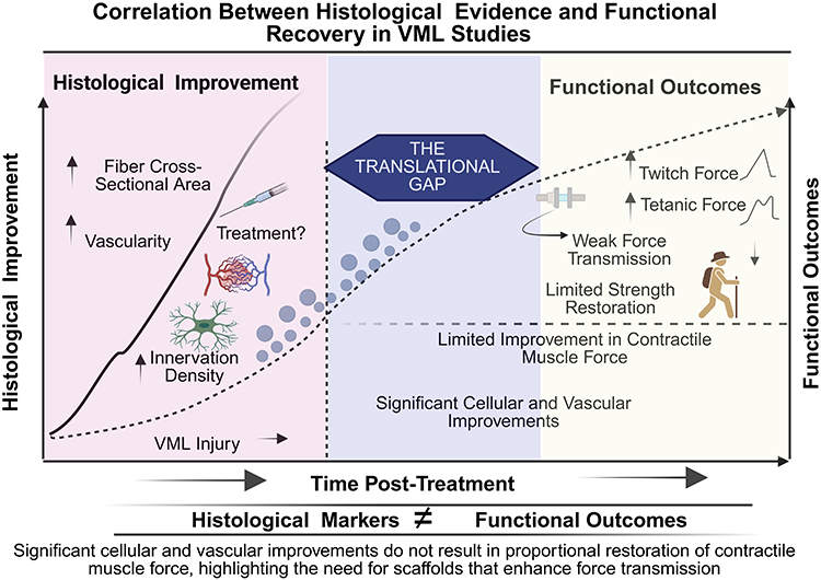 |
Figure 5 Functional recovery vs histological evidence (created with BioRender.com). Correlation between histological evidence and functional recovery in VML Studies. A dual-axis representation mapping histological markers (e.g, fiber cross-sectional area, innervation density) against functional outcomes (twitch/tetanic force). The overlay illustrates the “translational gap” often observed in preclinical studies, where significant cellular and vascular improvements do not always result in a proportional restoration of contractile muscle force, emphasizing the need for scaffolds that improve force transmission. |
Addressing these multifaceted biological barriers will require innovations in material science, vascularization techniques, neural integration strategies, and ECM fabrication, along with improved translational models that better reflect human pathophysiology. Only through such comprehensive advances can biomimetic scaffolds fulfill their potential for treating VML in clinical practice.59,63,178
Scalability remains a critical bottleneck while advanced fabrication techniques like electrospinning and 3D bioprinting enable precise control over microarchitecture, they struggle to produce clinically relevant volumes without compromising structural integrity, particularly when attempting to replicate the anisotropic organization of native muscle tissue.179 Material variability further complicates production, as batch-to-batch inconsistencies in natural polymers lead to unpredictable mechanical properties and cellular responses, undermining therapeutic reliability.180 Controlled delivery of bioactive factors adds another layer of complexity, as current encapsulation strategies for growth factors (VEGF, FGF, PDGF) lack the spatiotemporal precision required for optimal tissue guidance.181 The technical challenges are related to manufacturing and commercialization barriers, including exorbitant GMP production costs (>$50,000 per batch for decellularized ECM scaffolds and supply chain limitations for clinical-grade reagents.182 While emerging innovations like stimuli-responsive smart scaffolds, spatially patterned bioinks, and hybrid natural-synthetic systems offer promising solutions, significant development is still required to address the interrelated challenges of scalable production, material consistency, degradation control, bioactive delivery, and perfusion— all of which must be overcome to advance these technologies from promising laboratory prototypes to clinically viable therapies.67,183
Future Directions
The treatment of VML remains one of the most complex and unmet challenges in regenerative medicine. Despite significant progress in developing biomimetic scaffolds that recapitulate the native ECM and support muscle regeneration, substantial hurdles remain before these therapies can be widely implemented clinically.184 Future research will depend on the integration of emerging technologies, advanced regenerative therapies, and personalized medicine approaches—underpinned by strong interdisciplinary collaboration and continued investment in translational research. To speed up the process, 1–3 years are necessary for refining scaffold composition, mechanical characteristics, and thorough pre-clinical testing. Followed by which another 3–5 years focus to be shifted towards advanced fabrication technologies which includes AI guided design and 3D bioprinting for production of patient-specific scaffolds with controlled cellular and biological cues. In long term (5–10 years), the scaffold should be clinically translated with adaptive biomaterials that is able to restore full muscle function.185,186
Technological advancements are revolutionizing how scaffolds for VML can be designed to match the geometry of the injury site and incorporate spatial gradients of cells, growth factors, and ECM proteins. This level of customization enhances tissue integration and may improve functional outcomes.187 Another key innovation is the development of smart scaffolds engineered to possess responsive or adaptive properties. These include pH-sensitive,188,189 thermosensitive,190,191 and enzyme-responsive192 materials that can alter their structure, stiffness, or release profiles in response to the local microenvironment. New emerging technologies like artificial intelligence and machine learning are helpful for transforming the scaffold design.193 These advanced tools help in predicting the optimal scaffold architecture along with the materials to be used for catering various patient needs by integrating therapeutic elements like myoblast cells, growth factors, stem cells.
Recent advances in regenerative medicine demonstrate that combining biomimetic scaffolds with other innovative therapeutic approaches can significantly improve outcomes for VML treatment. A particularly effective strategy involves incorporating mesenchymal stem cells into scaffold designs. These cells show remarkable myogenic potential and secrete bioactive factors that enhance tissue repair. Research indicates that mesenchymal stem cells -seeded scaffolds promote muscle regeneration through multiple mechanisms: stimulating blood vessel formation, regulating immune responses, and facilitating the recruitment and differentiation of host cells.46,194 Multifunctional scaffold integrating several cells, gene-editing technologies that provide a microenvironment to restore complex muscle architecture has shown potential in breakthrough applications. Gene therapy approaches, including CRISPR-Cas9 gene editing systems, represent another promising avenue for enhancing scaffold-based treatments.195 These technologies enable precise modification of genetic pathways controlling muscle regeneration, such as activating pro-regenerative genes or correcting genetic defects that hinder repair processes. When delivered via biomimetic scaffolds, genetically modified cells can provide targeted therapeutic effects at the injury site.196
Emerging research highlights the potential of extracellular vesicle (EVs) and exosomes as cell-free therapeutic alternatives. These naturally occurring nanoparticles, derived from stem cells or immune cells, contain bioactive molecules (proteins, lipids, and RNAs) that can reduce inflammation, promote blood vessel growth, and stimulate muscle formation.197,198 Incorporating EVs into scaffold systems offers advantages including localized, controlled release of therapeutic factors while potentially avoiding some of the immunological and regulatory challenges associated with cell-based therapies.198
The effective treatment of VML requires a multidisciplinary approach, integrating expertise from biomedical engineering, regenerative biology, materials science, rehabilitation medicine, and clinical practice to develop solutions that restore not only tissue structure but also functional strength and endurance.11,148,199 Biomimetic scaffolds, when combined with tailored rehabilitation protocols, have demonstrated potential in enhancing neuromuscular integration and recovery, highlighting the need for close collaboration between tissue engineers and rehabilitation specialists to optimize both biological regeneration and mechanical performance.200 Additionally, translational efforts must prioritize scalability, regulatory compliance, and cost-effectiveness by standardizing fabrication processes, establishing reproducible preclinical models, and conducting rigorous long-term safety and efficacy assessments. Ensuring scaffold systems meet Good Manufacturing Practice (GMP) standards and align with clinical workflows will be critical for successful real-world implementation.158
Despite significant progress in the field, critical challenges remain in developing optimal ECM-based scaffolds, including determining ideal composition, mechanical properties, and degradation kinetics, as well as refining the dosage, timing, and delivery of adjunctive therapies like stem cells and growth factors to ensure long-term functional integration with native muscle and vasculature. Addressing these challenges requires increased investment in interdisciplinary research, with collaboration among funding agencies, academic institutions, and industry partners to accelerate next-generation regenerative therapies for VML.111 Advancing clinical trials, streamlining regulatory pathways, and fostering public–private partnerships will be essential to translate these innovations into clinical practice. Biomimetic strategies that recreate native-like ECM environments, combined with stem cell therapy, gene editing, and personalized scaffold design within a collaborative translational framework, offer a transformative approach to restoring muscle function and improving the quality of life for patients with VML.201
Emerging Technologies
To further align biomimetic scaffold strategies for VML with emerging frontiers in regenerative engineering, it is essential to incorporate recent technological breakthroughs that reshape skeletal muscle regeneration. 3D bioprinting has emerged as a reliable approach for fabricating scaffolds with precise regulation of fiber alignment, pore geometry, and cell distribution. These features are crucial for recreating the anisotropic structure and contractile function of native muscle tissue. The three principal bioprinting modalities used for skeletal muscle engineering differ substantially in their printing mechanics, bioink requirements, and performance outcomes. Extrusion-based systems dispense continuous filaments of highly viscous bioinks through pneumatic or mechanical pressure, enabling fabrication of large, cell-dense constructs but exposing encapsulated cells to elevated shear stress and relatively coarse resolution.202 Inkjet-based platforms generate discrete droplets using thermal or piezoelectric actuation, offering rapid and cost-effective patterning with reduced shear forces, although they are restricted to low-viscosity formulations and moderate structural resolution.203 In contrast, laser-assisted bioprinting employs pulsed laser energy to transfer bioink droplets in a nozzle-free manner, minimizing mechanical stress on cells and achieving superior spatial precision, albeit with increased technical complexity and cost.204 These distinctions highlight the need to align bioprinting modality with the specific architectural, mechanical, and cellular requirements of skeletal muscle regeneration which are described in Figure 6.
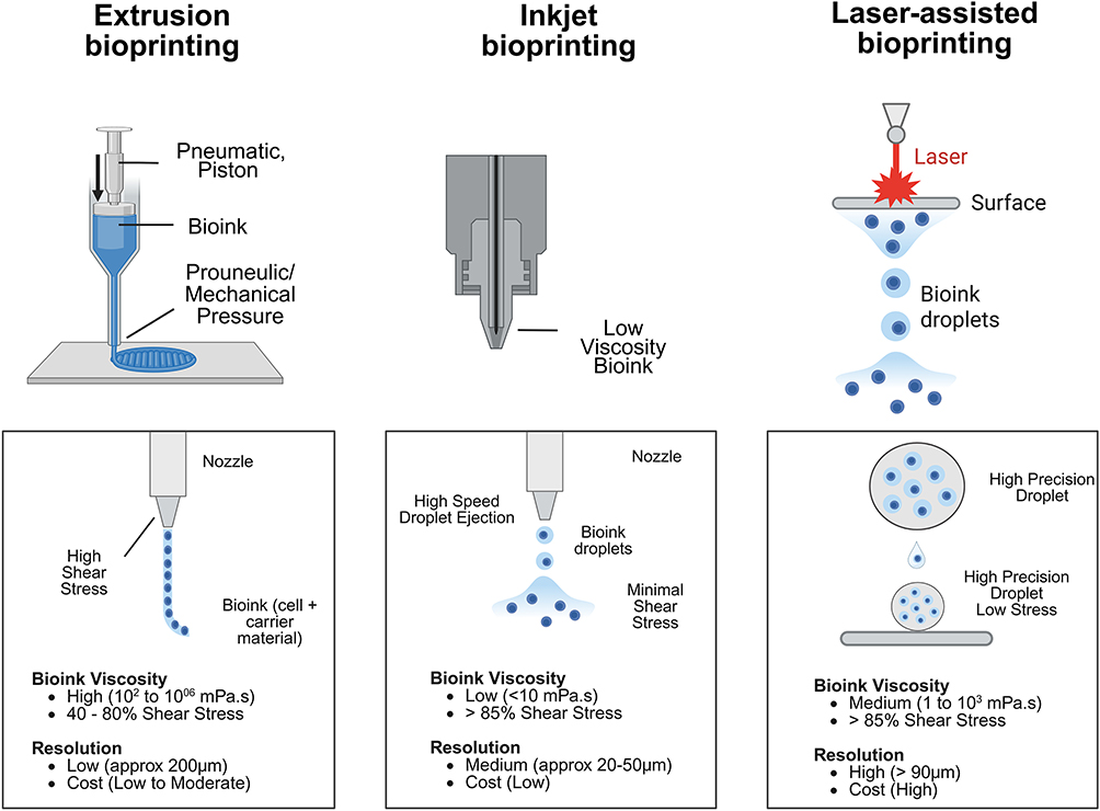 |
Figure 6 Technical comparison of the three primary 3D Bioprinting modalities for skeletal muscle engineering (created with BioRender.com). This illustration summarizes the mechanisms and comparative technical parameters of the three dominant bioprinting modalities used in the fabrication of skeletal muscle scaffolds: Extrusion-Based Bioprinting: Utilizes a piston or pneumatic-driven syringe to deposit continuous filaments of high-viscosity bioinks. It is highly effective for creating large-scale structural scaffolds but subject cells to high shear stress at the nozzle tip, which can lower viability (40–80%). It supports very high cell densities but offers lower resolution (~ 200um). Inkjet-Based Bioprinting: Employs thermal or piezoelectric actuators to generate discrete droplets of low-viscosity bioinks. While it provides faster printing speeds and lower costs, the low viscosity of the bioink often results in poor mechanical integrity of the final construct and limits resolution to a medium range (~ 20–50um). Laser-Assisted Bioprinting: Uses a laser pulse to create a high-pressure vapor bubble on a donor “ribbon”, propelling precise droplets onto a substrate. This nozzle-free approach eliminates shear stress, maintaining high cell viability (> 95%) and achieving the highest resolution (< 10um), though it is characterized by higher operational costs and technical complexity. The summary metrics below the schematics provide a quick reference for Bioink Viscosity, Resolution, Cell Viability, and Manufacturing Cost to assist in selecting the optimal modality based on specific muscle tissue engineering requirements. |
Apart from this, one of the most transformative advancements is 4D bioprinting, an evolution of conventional 3D bioprinting in which printed constructs are engineered to undergo controlled, time-dependent structural or functional transformations in response to external stimuli. In the context of VML, 4D bioprinted muscle constructs can be designed using shape-memory polymers or stimuli-responsive hydrogels that are engineered to respond to biochemical (pH, enzymes, reactive oxygen species), mechanical, or electrical signals present within injured muscle tissue.205–207 This dynamic adaptability more closely mimics the native muscle microenvironment, where mechanical loading and electrical activity continuously influence fiber maturation and alignment. By enabling implanted scaffold to reorganize, 4D bioprinting addresses the limitation of conventional scaffolds, i.e. their inability to adapt to the evolving regenerative environment.208
For example, redox-responsive polymers can modulate growth factor release in oxidative inflammatory environments characteristic of acute VML, while mechanoresponsive matrices can alter stiffness in response to contractile forces, thereby promoting myogenic differentiation. Electrically conductive biomaterials incorporating graphene, polypyrrole, or gold nanostructures further enable synchronized myotube formation and neuromuscular integration by facilitating electrical signal propagation.209
Another transformative development is the application of organ-on-a-chip technologies to model VML pathophysiology and therapeutic screening. Microfluidic muscle-on-chip systems recreate the architectural alignment, vascular perfusion, and mechanical loading conditions of skeletal muscle in vitro, allowing real-time assessment of contractility, calcium flux, and metabolic activity.210 These platforms enable high-resolution evaluation of scaffold–cell interactions under dynamic conditions that better approximate in vivo physiology than traditional static culture models.211 Moreover, integrating immune or endothelial compartments into multi-organ chips can elucidate the complex inflammatory and angiogenic cascades following volumetric muscle injury. Such systems not only accelerate preclinical optimization of biomimetic scaffolds but also reduce reliance on large-animal models by providing predictive, human-relevant data on functional restoration.212
Artificial intelligence (AI) and machine learning–driven scaffold design further represent a paradigm shift in regenerative biomaterials.168 AI algorithms can analyze multidimensional datasets encompassing material composition, pore architecture, mechanical properties, and biological outcomes to identify optimal design parameters for muscle regeneration.213 Generative modeling approaches enable rapid virtual prototyping of scaffold architectures tailored to patient-specific defect geometries derived from imaging data. Additionally, predictive modeling of growth factor kinetics, degradation profiles, and cell matrix interactions can streamline translational development by minimizing empirical trial-and-error experimentation. By integrating computational modeling with advanced manufacturing platforms such as bioprinting, AI-guided design fosters precision-engineered scaffolds capable of delivering reproducible and scalable therapeutic outcomes.214
Collectively, these emerging technologies signal a transition from static, structure-focused constructs to adaptive, data-driven, and physiologically integrated regenerative systems. Incorporating 4D bioprinting, smart scaffolds and AI-enabled design into VML research frameworks not only enhances mechanistic understanding but also establishes a forward-looking roadmap for clinically translatable muscle regeneration strategies which is described in Figure 7.
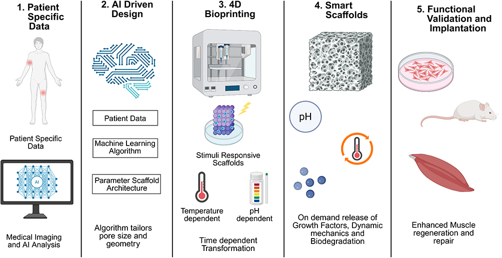 |
Figure 7 Emerging technologies revolutionizing scaffold design for VML treatment (created with BioRender.com). Integration of advanced technologies into the scaffold design workflow. (1) Patient Specific data is generated using medical imaging techniques (2) AI-Driven Design utilizes machine learning to optimize pore geometry based on patient-specific imaging. (3) 4D Bioprinting enables time-dependent structural changes in response to external stimuli. (4) Smart Scaffolds incorporate stimuli-responsive materials (pH, temperature, enzymes) for on-demand growth factor release, (5) validating it further for personalized and functional tissue engineering solutions. |
Conclusion and Outlook
VML remains a significant clinical challenge, characterized by irreversible tissue damage and functional impairment that profoundly impacts patients’ lives. Current surgical treatments, including autologous muscle transfers, often provide incomplete recovery and introduce additional complications.
This review underscores the critical role of the ECM in muscle regeneration, serving as both a structural framework and a bioactive signaling platform that guides satellite cell behavior, angiogenesis, and neuromuscular reinnervation. Advances in biomimetic scaffold design, incorporating key ECM components such as laminin, elastin, fibrin, have demonstrated improved muscle fiber alignment, vascular network formation, and functional outcomes in preclinical studies.
This review emphasizes ECM-based biomimetic scaffolds as a transformative strategy in volumetric muscle loss (VML) management, shifting the paradigm from passive tissue replacement to active, instructive regeneration. The combination of the structural architecture, mechanical properties, and the signaling pathway of native ECM, substantiate the scaffolds to promote myogenesis, immune regulation, and muscle remodeling. The major outcome is the successful VML repair depends not only on replacing lost tissue volume but also by coordinating cellular interactions that restore muscle integrity.
However, replicating the native ECM’s intricate architecture, controlling immune responses, ensuring consistent graft integration, scalability and compliance of regulatory requirements still remain key challenges. Overcoming these barriers demands a multidisciplinary approach, uniting experts in bioengineering, materials science, immunology, and clinical rehabilitation to optimize scaffold design, manufacturing processes, and therapeutic protocols. Future progress will depend on integrating innovative techniques such as 3D bioprinting, dynamic biomaterials, and cell-based therapies to create patient-specific solutions. By aligning technological advancements with clinical needs, researchers can develop more effective and scalable treatments. In summary, ECM-inspired regenerative strategies represent a transformative opportunity in VML treatment. These approaches not only address structural repair but also aim to restore full muscle function, offering patients a meaningful improvement in quality of life. Equally important is developing the scalable and regulatory compliant materials of clinical relevance. The clinical trial should ensure robust outcomes with respect to strength and muscle recovery along with functional restoration thereby transforming the standard of care for patients suffering from VML.
Author Contributions
All authors made a significant contribution to the work reported, whether that is in the conception, study design, execution, acquisition of data, analysis and interpretation, or in all these areas; took part in drafting, revising, or critically reviewing the article; gave final approval of the version to be published; have agreed on the journal to which the article has been submitted; and agree to be accountable for all aspects of the work.
Disclosure
The authors declare that they have no conflicts of interest in this work.
References
1. Testa S, Fornetti E, Fuoco C, et al. The war after war: volumetric muscle loss incidence, implication, current therapies and emerging reconstructive strategies, a comprehensive review. Biomedicines. 2021;9(5):564. doi:10.3390/biomedicines9050564
2. Sicherer ST, Venkatarama RS, Grasman JM. Recent trends in injury models to study skeletal muscle regeneration and repair. Bioengineering. 2020;7(3):1–35. doi:10.3390/bioengineering7030076
3. Grogan BFMD, Hsu JR. Volumetric muscle loss. Am Acad Orthop Surg. 2011;19:35–37. doi:10.5435/00124635-201102001-00007
4. Corona BT, Rivera JC, Owens JG, Wenke JC, Rathbone CR. Volumetric muscle loss leads to permanent disability following extremity trauma. J Rehabil Res Dev. 2015;52(7):785–792. doi:10.1682/JRRD.2014.07.0165
5. Corona BT, Wenke JC, Ward CL. Pathophysiology of volumetric muscle loss injury. Cells Tissues Organs. 2016;202(3–4):180–188. doi:10.1159/000443925
6. Btc BEP. Volumetric muscle loss. Methods Mol Biol. 2016;1460:19–31. doi:10.1007/978-1-4939-3810-0_2
7. Garg K, Ward CL, Hurtgen BJ, et al. Volumetric muscle loss: persistent functional deficits beyond frank loss of tissue. J Orthop Res. 2015;33(1):40–46. doi:10.1002/jor.22730
8. Corona BT, Ward CL, Baker HB, Gjc TJW, Christ GJ. Implantation of in vitro tissue engineered muscle repair constructs and bladder acellular matrices partially restore in vivo skeletal muscle function in a rat model of volumetric muscle loss injury. Tissue Eng Part A. 2014;20(3):705–715. doi:10.1089/ten.TEA.2012.0761
9. Cieza A, Causey K, Kamenov K, Hanson SW, Chatterji S, Vos T. Global estimates of the need for rehabilitation based on the global burden of disease study 2019: a systematic analysis for the global burden of disease study 2019. Lancet. 2020;396(10267):2006–2017. doi:10.1016/S0140-6736(20)32340-0
10. Sorensen JR, Mcfaline-Figueroa J, Call JA. Pathophysiology of volumetric muscle loss and targets for regenerative rehabilitation. In: Regenerative rehabilitation: From basic science to the clinic. 2022:177–225. doi:10.1007/978-3-030-95884-8_6
11. Greising SM, Warren GL, Southern WM, Nichenko AS, Qualls AE, Jac BTC. Early rehabilitation for volumetric muscle loss injury augments endogenous regenerative aspects of muscle strength and oxidative capacity. BMC Musculoskelet Disord. 2018;19(1):173. doi:10.1186/s12891-018-2095-6
12. Dash DK. Road crashes account for over 43% unintentional injury deaths in India: health ministry. Times of India. 2024. Available from: https://timesofindia.indiatimes.com/india/road-crashes-account-for-over-43-of-unintentional-injury-deaths-in-india-health-ministry/articleshow/113003065.cms.
13. Dongqing G, Gl SO. Global burden of road injuries and their attributable risk factors from 1990 to 2021: a systematic analysis for the global burden of disease study 2021. Prev Med Reports. 2025;53. doi:10.1016/j.pmedr.2025.103051
14. Collaborators G 2021 D and I. Global incidence, prevalence, years lived with disability (YLDs), disability-adjusted life-years (DALYs), and healthy life expectancy (HALE) for 371 diseases and injuries in 204 countries and territories and 811 subnational locations, 1990-2021: a systema. Lancet. 2024;403(10440):2133–2161. doi:10.1016/S0140-6736(24)00757-8
15. Shanthakumar D, Payne A, Patricia Leitch MAW, Alfa-Wali M. Trauma care in low- and middle-income countries. Surg J. 2021;7(4):e281–e285. doi:10.1055/s-0041-1732351
16. Mase VJ Jr, Hsu JR, Wolf SE, et al. Clinical application of an acellular biologic scaffold for surgical repair of a large, traumatic quadriceps femoris muscle defect. Orthopedics. 2010;33(7):511. doi:10.3928/01477447-20100526-24
17. Das S, Browne KD, Laimo FA, et al. Pre-innervated tissue-engineered muscle promotes a pro-regenerative microenvironment following volumetric muscle loss. Commun Biol. 2020;3:330. doi:10.1038/s42003-020-1056-4
18. Aurora A, Garg K, Tjw BTC, Walters TJ. Physical rehabilitation improves muscle function following volumetric muscle loss injury. BMC Sport Sci Med Rehabil. 2014;6(1):41. doi:10.1186/2052-1847-6-41
19. Downing K, Prisby R, Varanasi V, Zhou J, Pan Z, Brotto M. Old and new biomarkers for volumetric muscle loss. Curr Opin Pharmacol. 2021;59:61–69. doi:10.1016/j.coph.2021.05.001
20. Greising SM, Corona BT. Regenerative and rehabilitative medicine: a necessary synergy for functional recovery from volumetric muscle loss injury. Cells Tissues Organs. 2016;202(3–4):237–249. doi:10.1159/000444673
21. Somers SM, Gilbert-Honick J, Choi IY, et al. Engineering skeletal muscle grafts with PAX7::GFP-sorted human pluripotent stem cell-derived myogenic progenitors on fibrin microfiber bundles for tissue regeneration. Bioengineering. 2022;9(11):693. doi:10.3390/bioengineering9110693
22. Golebiowska A, Intravaia JT, Sathe VM, Nukavarapu SP. Decellularized extracellular matrix biomaterials for regenerative therapies: advances, challenges and clinical prospects. Bioact Mater. 2024;32:98–123. doi:10.1016/j.bioactmat.2023.09.017
23. Guitart M, Lloreta J, Mañas-Garcia L, Barreiro E. Muscle regeneration potential and satellite cell activation profile during recovery following hindlimb immobilization in mice. J Cell Physiol. 2018;233(5):4360–4372. doi:10.1002/jcp.26282
24. Quarta M, Brett JO, DiMarco R, et al. An artificial niche preserves the quiescence of muscle stem cells and enhances their therapeutic efficacy. Nat Biotechnol. 2016;34(7):752–759. doi:10.1038/nbt.3576
25. Chevallay B, Herbage D. Collagen-based biomaterials as 3D scaffold for cell cultures: applications for tissue engineering and gene therapy. Med Biol Eng Comput. 2000;38:211–218. doi:10.1007/BF02344779
26. Kim JH, Seol Y-J, In Kap K, et al. 3D bioprinted human skeletal muscle constructs for muscle function restoration. Sci Rep. 2018;8(12307). doi:10.1038/s41598-018-29968-5
27. Schüler SC, Liu Y, Dumontier S, Grandbois M, Moal EL, Ddw Cornelison CFB. Extracellular matrix: brick and mortar in the skeletal muscle stem cell niche. Front Cell Dev Biol. 2022;10. doi:10.3389/fcell.2022.1056523
28. Ahmad K, Shaikh S, Ahmad SS, Choi I. Cross-talk between extracellular matrix and skeletal muscle: implications for myopathies. Front Pharmacol. 2020;11. doi:10.3389/fphar.2020.00142
29. Forcina L, Cosentino M, Musarò A. Mechanisms regulating muscle regeneration: insights into the interrelated and time-dependent phases of tissue healing. Cells. 2020;9(5):1297. doi:10.3390/cells9051297
30. Amirrah IN, Lokanathan Y, Zulkiflee I, Wee MFMR, Motta A, Fauzi MB. A comprehensive review on collagen type i development of biomaterials for tissue engineering: from biosynthesis to bioscaffold. Biomedicines. 2022;10(9):2307. doi:10.3390/biomedicines10092307
31. Yuliya Nashchekina AN, Wei Q. Collagen fibril orientation in vitro: from formation to advanced biomaterial development. Biomimetics. 2025;10(10):664. doi:10.3390/biomimetics10100644
32. Ishii K, Sakurai H, Suzuki N. Recapitulation of extracellular LAMININ environment maintains stemness of satellite cells in vitro. Stem Cell Reports. 2018;10(2):568–582. doi:10.1016/j.stemcr.2017.12.013
33. Longstreth JH, Wang K. The role of fibronectin in mediating cell migration. Am J Physiol. 2024;326(4):C1212–C1225. doi:10.1152/ajpcell.00633.2023
34. Snetkov P, Zakharova K, Morozkina S, Olekhnovich R. Hyaluronic acid: the influence of molecular weight and degradable properties of biopolymer. Polymers. 2020;12:1800.
35. Alhakamy NA, Hosny KM, Rizg WY, et al. Development and optimization of hyaluronic acid-poloxamer in-situ gel loaded with voriconazole cubosomes for enhancement of activity against ocular fungal infection. Gels. 2022;8(4):241. doi:10.3390/gels8040241
36. Krymchenko R, Coşar Kutluoğlu G, van Hout N, Manikowski D, Doberenz C, van Kuppevelt TH, Daamen WF. Elastogenesis in focus: navigating elastic fibers synthesis for advanced dermal biomaterial formulation. Adv Healthc Mater. 2024;13(27). doi:10.1002/adhm.202400484
37. Xie C, Mondal DK, Ulas M, Neill T, Iozzo RV. Oncosuppressive roles of decorin through regulation of multiple receptors and diverse signaling pathways. Am J Physiol. 2022;322(3):C554–C566. doi:10.1152/ajpcell.00016.2022
38. Tjahjono NS, Subramanian D, Shihabeddin TZ. Effect of decorin and aligned collagen fibril topography on TGF-β1 activation of corneal keratocytes. Bioengineering. 2025;12(3):259. doi:10.3390/bioengineering12030259
39. Ghajarieh A, Habibi S, Talebian A. Biomedical applications of nanofibers. Russ J Appl Chem. 2021;94(7):847–872. doi:10.1134/S1070427221070016
40. Mukund K, Subramaniam S. Skeletal muscle: a review of molecular structure and function, in health and disease. Wiley Interdiscip Rev Syst Biol Med. 2020;12(1). doi:10.1002/wsbm.1462
41. Pereira RF, Barrias CC, Bártolo PJ, Granja PL. Cell-instructive pectin hydrogels crosslinked via thiol-norbornene photo-click chemistry for skin tissue engineering. Acta Biomater. 2018;66:282–293. doi:10.1016/j.actbio.2017.11.016
42. Csapo R, Matthias Gumpenberger BW, Wessner B. Skeletal muscle extracellular matrix – what do we know about its composition, regulation, and physiological roles? A narrative review. Front Physiol. 2020;11. doi:10.3389/fphys.2020.00253
43. Laumonier T, Menetrey J. Muscle injuries and strategies for improving their repair. J Exp Orthop. 2016;3(1). doi:10.1186/s40634-016-0051-7
44. Kheradmandi M, Vasheghani-Farahani E, Ghiaseddin A, Ganji F. Skeletal muscle regeneration via engineered tissue culture over electrospun nanofibrous chitosan/PVA scaffold. J Biomed Mater Res. 2016;104(7):1720–1727. doi:10.1002/jbm.a.35702
45. Cai A, Schneider P, Zheng ZM, et al. Myogenic differentiation of human myoblasts and mesenchymal stromal stem cells under GDF11 on poly-caprolactone-collagen I-polyethylene-nanobers.
46. Qiu X, Liu S, Zhang H, et al. Mesenchymal stem cells and extracellular matrix scaffold promote muscle regeneration by synergistically regulating macrophage polarization toward the M2 phenotype. Stem Cell Res Ther. 2018;3(1):88. doi:10.1186/s13287-018-0821-5
47. Philippou A, Maridaki M, Theos A, Koutsilieris M. Cytokines in Muscle Damage. Vol. 58.
48. Atri C, Guerfali FZ, Laouini D. Role of human macrophage polarization in inflammation during infectious diseases. Int J Mol Sci. 2018;19(6):1801. doi:10.3390/ijms19061801
49. Fu X, Wang H, Hu P. Stem cell activation in skeletal muscle regeneration. Cell Mol Life Sci. 2015;72(9):1663–1677. doi:10.1007/s00018-014-1819-5
50. Qazi TH, Duda GN, Ort MJ, Perka C, Geissler S, Winkler T. Cell therapy to improve regeneration of skeletal muscle injuries. J Cachexia Sarcopenia Muscle. 2019;10(3):501–516. doi:10.1002/jcsm.12416
51. El Ayadi A, Jay JW, Prasai A. Current approaches targeting the wound healing phases to attenuate fibrosis and scarring. Int J Mol Sci. 2020;21(3):1105. doi:10.3390/ijms21031105
52. Corona BT, Garg K, Ward CL, McDaniel JS, Walters TJ, Rathbone CR. Autologous minced muscle grafts: a tissue engineering therapy for the volumetric loss of skeletal muscle. Am J Physiol. 2013;305(7):761–775. doi:10.1152/ajpcell.00189.2013
53. Li N, Chen Y, Wang Q, et al. Microenvironment-driven satellite cell regeneration and repair in aging-related sarcopenia: mechanisms and therapeutic frontiers. Stem Cell Res Ther. 2025;16(545). doi:10.1186/s13287-025-04481-5
54. Wenjing L, Minyou C, Zhang L. Muscle stem cell microenvironment and functions in muscle regeneration. Biomolecules. 2025;15(6):765. doi:10.3390/biom15060765
55. Mayorca-Guiliani AE, Leeming DJ, Henriksen K, et al. ECM formation and degradation during fibrosis, repair, and regeneration. Metab Heal Dis. 2025;3(25). doi:10.1038/s44324-025-00063-4
56. Wynn TA, Vannella KM. Macrophages in tissue repair, regeneration, and fibrosis. Immunity. 2016;44(3):450–462. doi:10.1016/j.immuni.2016.02.015
57. Mafalda Loreti AS. The jam session between muscle stem cells and the extracellular matrix in the tissue microenvironment. Regen Med. 2022;7. doi:10.1038/s41536-022-00204-z
58. Shin YC, Lee JH, Jin L, et al. Stimulated myoblast differentiation on graphene oxide-impregnated PLGA-collagen hybrid fibre matrices matrices. J Nanobiotechnology. 2015;13(1):1–11. doi:10.1186/s12951-015-0081-9
59. Hwtm GDM. Biomimetic scaffolds for skeletal muscle regeneration. Discov J. 2019;7(1):e90. doi:10.15190/d.2019.3
60. Sell SA, Wolfe PS, Garg K, McCool JM, Rodriguez IA, Bowlin GL. The use of natural polymers in tissue engineering: a focus on electrospun extracellular matrix analogues. Polymers. 2010;2(4):522–553. doi:10.3390/polym2040522
61. Manning CN, Schwartz AG, Liu W, et al. Controlled delivery of mesenchymal stem cells and growth factors using a nanofiber scaffold for tendon repair. Acta Biomater. 2013;9(6):6905–6914. doi:10.1016/j.actbio.2013.02.008
62. Narayanan N, Jia Z, Kim KH, et al. Biomimetic glycosaminoglycan-based scaffolds improve skeletal muscle regeneration in a Murine volumetric muscle loss model. Bioact Mater. 2021;6(4):1201–1213. doi:10.1016/j.bioactmat.2020.10.012
63. Grasman JM, Zayas MJ, Page RL, Pins GD. Biomimetic scaffolds for regeneration of volumetric muscle loss in skeletal muscle injuries. Acta Biomater. 2015;25:2–15. doi:10.1016/j.actbio.2015.07.038
64. Jain R, Shetty S, Yadav KS. Unfolding the electrospinning potential of biopolymers for preparation of nanofibers. J Drug Deliv Sci Technol. 2020;57(January):101604. doi:10.1016/j.jddst.2020.101604
65. Sm GCV. Skeletal muscle tissue engineering: strategies for volumetric constructs. Definitions. 2020. doi:10.32388/d7r0n5
66. Deng Z, Guo Y, Zhao X, et al. Stretchable degradable and electroactive shape memory copolymers with tunable recovery temperature enhance myogenic differentiation. Acta Biomater. 2016;46:234–244. doi:10.1016/j.actbio.2016.09.019
67. El-Husseiny HM, Mady EA, Hamabe L, et al. Smart/stimuli-responsive hydrogels: cutting-edge platforms for tissue engineering and other biomedical applications. Mater Today Bio. 2022;13(December 2021):100186. doi:10.1016/j.mtbio.2021.100186
68. Nemati S, Kim S, Shin YM, Shin H. Current progress in application of polymeric nanofibers to tissue engineering. Nano Converg. 2019;6(1). doi:10.1186/s40580-019-0209-y
69. Niknezhad SV, Mehrali M, Khorasgani FR, et al. Enhancing volumetric muscle loss (VML) recovery in a rat model using super durable hydrogels derived from bacteria. Bioact Mater. 2024;38:540–558. doi:10.1016/j.bioactmat.2024.04.006
70. Chen N, Menglu W, Williams R, Yan J, Jiayi Zhou DZ, Ding Y. Strong living scaffolds for load-bearing musculoskeletal tissue regeneration. Mater Today Bio. 2025;35. doi:10.1016/j.mtbio.2025.102571
71. Eltom A, Zhong G, Muhammad A. Scaffold techniques and designs in tissue engineering functions and purposes: a review. Adv Mater Sci Eng. 2019;2019:1–13. doi:10.1155/2019/3429527
72. Anderson SE, Han WM, Srinivasa V, et al. Determination of a critical size threshold for volumetric muscle loss in the mouse quadriceps. Tissue Eng - Part C Methods. 2019;25(2):59–70. doi:10.1089/ten.TEC.2018.0324
73. Panayi AC, Smit L, Hays N, et al. A porous collagen-GAG scaffold promotes muscle regeneration following volumetric muscle loss injury. Wound Repair Regen. 2020;28(1):61–74. doi:10.1111/wrr.12768
74. Tidball JG, Flores I, Welc SS, Eo MW-H. Aging of the immune system and impaired muscle regeneration: a failure of immunomodulation of adult myogenesis. Exp Gerontol. 2021;145. doi:10.1016/j.exger.2020.111200
75. Kim JT, Kasukonis B, Dunlap G, Perry R, Tyrone W, Wolchok JC. Regenerative repair of volumetric muscle loss injury is sensitive to age. Tissue Eng Part A. 2020;26(1–2):3–14. doi:10.1089/ten.TEA.2019.0034
76. Lee S, Sani ES, Spencer AR, Guan Y, Annabi N. Human-recombinant-elastin-based bioinks for 3D bioprinting of vascularized soft tissues. Adv Mater. 2020;32(45). doi:10.1002/adma.202003915
77. Ibáñez-Fonseca A, Santiago Maniega S, Gorbenko del Blanco D, et al. Elastin-like recombinamer hydrogels for improved skeletal muscle healing through modulation of macrophage polarization. Front Bioeng Biotechnol. 2020;8:413. doi:10.3389/fbioe.2020.00413
78. Marsico G, Jin C, Abbah SA, et al. Elastin-like hydrogel stimulates angiogenesis in a severe model of critical limb ischemia (CLI): an insight into the glyco-host response. Biomaterials. 2021;269. doi:10.1016/j.biomaterials.2020.120641
79. Rodríguez-Cabello JC, González I, Cipriani F, Poocza L. Elastin-like materials for tissue regeneration and repair. In: Peptides and Proteins as Biomaterials for Tissue Regeneration and Repair; 2018:309–327. doi:10.1016/B978-0-08-100803-4.00012-7
80. Mbundi L, Gonzalez-Perez M, Gonzalez-Perez F, Juanes-Gusano D, Rodriguez-Cabello JC. Trends in the development of tailored elastin-like recombinamer–based porous biomaterials for soft and hard tissue applications. Front Mater. 2021. doi:10.3389/fmats.2020.601795
81. Dai M, Belaïdi J-P, Fleury G, Garanger E, Rielland M, Xavier Schultze SL. Elastin-like polypeptide-based bioink: a promising alternative for 3D bioprinting. Biomacromolecules. 2021;22(12):4956–4966. doi:10.1021/acs.biomac.1c00861
82. Boccafoschi F, Ramella M, Sibillano T, et al. Human elastin polypeptides improve the biomechanical properties of three-dimensional matrices through the regulation of elastogenesis. J Biomater Res Part A. 2015;103(3):1218–1230. doi:10.1002/jbm.a.35257
83. Lima LF, Sousa MG, Rodrigues GR, et al. Elastin-like polypeptides in development of nanomaterials for application in the medical field. Front Nanotechnol. 2022;4. doi:10.3389/fnano.2022.874790
84. Khunmanee S, Jeong Y, Park H. Crosslinking method of hyaluronic-based hydrogel for biomedical applications. J Tissue Eng. 2017;8. doi:10.1177/2041731417726464
85. Chantre CO, Gonzalez GM, Ahn S, Cera L, Campbell PH, KKP SPH. Porous biomimetic hyaluronic acid and extracellular matrix protein nanofiber scaffolds for accelerated cutaneous tissue repair. ACS Appl Mater Interfaces. 2019;11(49):45498–45510. doi:10.1021/acsami.9b17322
86. Rohrer L, Kato S, Browne SA, Striedinger-Melo K, Kevin Healy JHP, Pomerantz JH. Acrylated hyaluronic-acid based hydrogel for the treatment of craniofacial volumetric muscle loss. Tissue Eng Part A. 2024;30(21–22):704–711. doi:10.1089/ten.TEA.2023.0241
87. Varghese SA, Rangappa SM, Siengchin S, Parameswaranpillai J. Natural polymers and the hydrogels prepared from them. In: Hydrogels Based on Natural Polymers. Elsevier Inc.: 2019. doi10.1016/B978-0-12-816421-1.00002-1
88. Christensen KW, Turner J, Coughenour K, et al. Assembled Cell-Decorated Collagen (AC-DC) fiber bioprinted implants with musculoskeletal tissue properties promote functional recovery in volumetric muscle loss. Adv Healthc Mater. 2022;11(3). doi:10.1002/adhm.202101357
89. Chen C-L, Wei S-Y, Chen W-L, YCC T-LH. Reconstructing vascular networks promotes the repair of skeletal muscle following volumetric muscle loss by pre-vascularized tissue constructs. J Tissue Eng. 2023;14. doi:10.1177/20417314231201231
90. Kajave NS, Schmitt T, Nguyen TU, Kishore V. Dual crosslinking strategy to generate mechanically viable cell-laden printable constructs using methacrylated collagen bioinks. Mater Sci Eng C. 2020;107:110290. doi:10.1016/j.msec.2019.110290
91. Ward CL, BTC LJ, Corona BT. An autologous muscle tissue expansion approach for the treatment of volumetric muscle loss. Biores Open Access. 2015;4(1):198–208. doi:10.1089/biores.2015.0009
92. Sionkowska A. Collagen blended with natural polymers: recent advances and trends. Prog Polym Sci. 2021;122:101452. doi:10.1016/j.progpolymsci.2021.101452
93. Marcinczyk M, Dunn A, Haas G, et al. The effect of laminin-111 hydrogels on muscle regeneration in a murine model of injury. Tissue Eng Part A. 2019;25(13–14):1001–1012. doi:10.1089/ten.tea.2018.0200
94. Goldman SM, Henderson BEP, BTC TJW, Corona BT. Co-delivery of a laminin-111 supplemented hyaluronic acid based hydrogel with minced muscle graft in the treatment of volumetric muscle loss injury. PLoS One. 2018;13(1):e0191245. doi:10.1371/journal.pone.0191245
95. Marcinczyk M, Elmashhady H, Talovic M, Dunn A, Bugis F, Garg K. Laminin-111 enriched fibrin hydrogels for skeletal muscle regeneration. Biomaterials. 2017;141:233–242. doi:10.1016/j.biomaterials.2017.07.003
96. Augustine R, Malik HN, Singhal DK, et al. Electrospun polycaprolactone/ZnO nanocomposite membranes as biomaterials with antibacterial and cell adhesion properties. J Polym Res. 2014;21(3). doi:10.1007/s10965-013-0347-6
97. Meng C. Electrospinning PLLA/PCL blend fibre-based materials and their biomedical application: a mini review. Polymers. 2025;17(20):2802. doi:10.3390/polym17202802
98. José Calero-Castro F, Manuel Perez-Puyana V, Laga I, Ruiz JP, de la P de J ARF, de la Portilla de Juan F. Mechanical stimulation and aligned poly(ε-caprolactone)-gelatin electrospun scaffolds promote skeletal muscle regeneration. ACS Appl Bio Mater. 2024;7(10):6430. doi:10.1021/acsabm.4c00559
99. Kim I, Lee SS, Ghosh A, Ferguson SJ, Bar‐Nur O. A self-renewing biomimetic skeletal muscle construct engineered using induced myogenic progenitor cells. Adv Funct Mater. 2023;34(1). doi:10.1002/adfm.202300571
100. Liu C, Du G, Guo Q, Li R, Li C, He H. Fabrication and characterization of polylactic acid electrospun wound dressing modified with polyethylene glycol, rosmarinic acid and graphite oxide. Nanomaterials. 2023;13(13):2000. doi:10.3390/nano13132000
101. Darabian B, Hamed Bagheri SM, Mohammadi S. Improvement in mechanical properties and biodegradability of PLA using poly(ethylene glycol) and triacetin for antibacterial wound dressing applications. Prog Biomater. 2020;9(1–2):45–64. doi:10.1007/s40204-020-00131-6
102. Lee S, Xinming Tong FY, Yang F. Effects of the poly(ethylene glycol) hydrogel crosslinking mechanism on protein release. Biomater Sci. 2016;4(3):405–411. doi:10.1039/c5bm00256g
103. Miranda Alarcón YS, Jazwinska D, Lymon T. The use of collagen methacrylate in actuating polyethylene glycol diacrylate-acrylic acid scaffolds for muscle regeneration. Ann Biomed Eng. 2023;51(6):1165–1180. doi:10.1007/s10439-023-03139-8
104. Perez-Puyana V, Wieringa P, Yuste Y, et al. Fabrication of hybrid scaffolds obtained from combinations of PCL with gelatin or collagen via electrospinning for skeletal muscle tissue engineering. J Biomed Mater Res. 2021;109(9):1600–1612. doi:10.1002/jbm.a.37156
105. Lee MC, Jodat YA, Endo Y, et al. Engineering large-scale hiPSC-derived vessel-integrated muscle-like lattices for enhanced volumetric muscle regeneration. Trends Biotechnol. 2024;42(12):1715–1744. doi:10.1016/j.tibtech.2024.08.001
106. Cady C, Nair K, Rodriguez HC, Rust B, Ghandour S, Anish Potty AG. Optimization of polycaprolactone and type I collagen scaffold for tendon tissue regeneration. Cureus. 2024;16(3):e56930. doi:10.7759/cureus.56930
107. Bayer IS. Advances in fibrin-based materials in wound repair: a review. Molecules. 2025;27(14):4504. doi:10.3390/molecules27144504
108. Songjie L, Dan X, Chen H, et al. Developing fibrin-based biomaterials/scaffolds in tissue engineering. Bioact Mater. 2024;40:597–623. doi:10.1016/j.bioactmat.2024.08.006
109. Liu J, Feng Y, Peng Q, et al. Application of biomaterials in vascularization of cardiac organoids. iScience. 2025;28(10):113531. doi:10.1016/j.isci.2025.113531
110. Movahedi M, Asefnejad A, Rafienia M, Khorasani MT. Potential of novel electrospun core-shell structured polyurethane/starch (hyaluronic acid) nanofibers for skin tissue engineering: in vitro and in vivo evaluation. Int J Biol Macromol. 2020;146:627–637. doi:10.1016/j.ijbiomac.2019.11.233
111. Xie X, Wang Y, Ziyan L, et al. Recent advances in gradient biomimetic scaffolds for tendon-bone interface regeneration. Front Bioeng Biotechnol. 2025;13. doi:10.3389/fbioe.2025.1629816
112. Fang L, Lin X, Ruian X, Liu L, Zhang Y, JX FTJJL&. Advances in the development of gradient scaffolds made of nano-micromaterials for musculoskeletal tissue regeneration. Nano-Micro Lett. 2025;17(75). doi:10.1007/s40820-024-01581-4
113. Kiryk J, Michalak M, Majchrzak Z. Functionalization strategies of chitosan-based scaffolds with growth factors for bone regeneration: a systematic review. Mar Drugs. 2025;23(10):396. doi:10.3390/md23100396
114. Liu Y, Wan Y, Li C, et al. Gradient scaffolds in bone-soft tissue interface engineering: structural characteristics, fabrication techniques, and emerging trends. J Orthop Transl. 2025;50:333–353. doi:10.1016/j.jot.2024.10.015
115. MVAPMCW Ś, Paradiso A, Costantini M, Świȩszkowski W. Hydrogel-based fiber biofabrication techniques for skeletal muscle tissue engineering. ACS Biomater Sci Eng. 2022;8(2):379–405. doi:10.1021/acsbiomaterials.1c01145
116. Zhu C, Sklyar K, Karvar M, Endo Y, Sinha I. Scaffold tissue engineering strategies for volumetric muscle loss. Plast Aesthetic Res. 2023;10(58). doi:10.20517/2347-9264.2022.89
117. Liang W, Han M, Li G, et al. Perfusable adipose decellularized extracellular matrix biological scaffold co-recellularized with adipose-derived stem cells and L6 promotes functional skeletal muscle regeneration following volumetric muscle loss. Biomaterials. 2024;307. doi:10.1016/j.biomaterials.2024.122529
118. Chaturvedi V, Naskar D, Kinnear BF. Silk fibroin scaffolds with muscle-like elasticity support in vitro differentiation of human skeletal muscle cells. J Tissue Eng Regen Med. 2017;11(11):3178–3192. doi:10.1002/term.2227
119. Choi Y-J, Jun Y-J, Kim DY. A 3D cell printed muscle construct with tissue-derived bioink for the treatment of volumetric muscle loss. Biomaterials. 2019;206:160–169. doi:10.1016/j.biomaterials.2019.03.036
120. Sahoo S, JCHG SLT, Hong Goh JC. PLGA nanofiber-coated silk microfibrous scaffold for connective tissue engineering. J Biomed Mater Res Part B Appl Biomater. 2010;95(1):19–28. doi:10.1002/jbm.b.31678
121. Zhang M, Du H, Guan Y, et al. Study on the effect of PDA-PLGA scaffold loaded with islet cells for skeletal muscle transplantation in the treatment of diabetes. Front Bioeng Biotechnol. 2022;10. doi:10.3389/fbioe.2022.927348
122. Jin S, Cai Y, Li Y. A sandwich-like nanofibrous scaffold with macrophage phenotype transformation and myogenic differentiation for skeletal muscle regeneration. Bioact Mater. 2025;51:211–230. doi:10.1016/j.bioactmat.2025.05.008
123. Basurto IM, Bandara GC, Boudreau RD. Freeze-dried porous collagen scaffolds for the repair of volumetric muscle loss injuries. ACS Biomater Sci Eng. 2025;11(3):1598–1611. doi:10.1021/acsbiomaterials.4c01601
124. Perez-Puyana V, Villanueva P, Jiménez-Rosado M, de la Portilla F, Romero A. Incorporation of elastin to improve polycaprolactone-based scaffolds for skeletal muscle via electrospinning. Polymers. 2021;13(9):1501. doi:10.3390/polym13091501
125. Goreninskii SI, Bolbasov EN, Sudarev EA, et al. Fabrication and properties of L-arginine-doped PCL electrospun composite scaffolds. Mater Lett. 2017;214:64–67. doi:10.1016/j.matlet.2017.11.115
126. de Melo LF, Almeida GHDR, Azarias FR. Decellularized bovine skeletal muscle scaffolds: structural characterization and preliminary cytocompatibility evaluation. Cells. 2024;13(8):688. doi:10.3390/cells13080688
127. Rajzer I, Rom M, Menaszek E, Fabia J, Kwiatkowski R. Conductive polyaniline patterns on electrospun polycaprolactone/hydroxyapatite scaffolds for bone tissue engineering. Materials. 2021;14(17):4837. doi:10.3390/ma14174837
128. Kozan NG, Joshi M, Sicherer ST, Grasman JM. Porous biomaterial scaffolds for skeletal muscle tissue engineering. Front Bioeng Biotechnol. 2023;11. doi:10.3389/fbioe.2023.1245897
129. Patel KH, Dunn AJ, Talovic M, et al. Aligned nanofibers of decellularized muscle ECM support myogenic activity in primary satellite cells in vitro. Biomed Mater. 2019;14(3):035010. doi:10.1088/1748-605X/ab0b06
130. Gahlawat S, Oruc D, Paul N. Tissue engineered 3D constructs for volumetric muscle loss. Ann Biomed Eng. 2024;52(9):2325–2347. doi:10.1007/s10439-024-03541-w
131. Ankur Singh NAP, Peppas NA. Hydrogels and scaffolds for immunomodulation. Adv Mater. 2014;26(38):6530–6541. doi:10.1002/adma.201402105
132. Papa EV, Dong X, Hassan M. Skeletal muscle function deficits in the elderly: current perspectives on resistance training. J Nat Sci. 2017;3(1):e272.
133. Xu Y, Chen X, Qian Y, et al. Melatonin-based and biomimetic scaffold as muscle–ECM implant for guiding myogenic differentiation of volumetric muscle loss. Adv Funct Mater. 2020;30(27). doi:10.1002/adfm.202002378
134. Reed C, Huynh T, Schluns J, Phelps P, Jamie Hestekin JCW, Wolchok JC. Cell-derived extracellular matrix fiber scaffolds improve recovery from volumetric muscle loss. Tissue Eng Part A. 2024;30(5–6):181–191. doi:10.1089/ten.tea.2022.0227
135. Nicholson PR, Raymond-Pope CJ, Lillquist TJ, Bruzina AS, Call JA, Greising SM. In sequence antifibrotic treatment and rehabilitation after volumetric muscle loss injury. Eur PMC. 2024;14(2):101–113. doi:10.1089/wound.2024.0109
136. Pien N, Krzyslak H, Kallaje SS, et al. Tissue engineering of skeletal muscle, tendons and nerves: a review of manufacturing strategies to meet structural and functional requirements. Appl Mater Today. 2023;31. doi:10.1016/j.apmt.2023.101737
137. Li Z, Qu T, Ding C, Ma C, Sun H, Li S, Liu X. Injectable gelatin derivative hydrogels with sustained vascular endothelial growth factor release for induced angiogenesis. Acta Biomater. 2016;13:88–100. doi:10.1016/j.actbio.2014.11.002
138. Sun Y, Wu Q, Zhang Y, Dai K, Wei Y. 3D-bioprinted gradient-structured scaffold generates anisotropic cartilage with vascularization by pore-size-dependent activation of HIF1α/FAK signaling axis. Nanomedicine Nanotechnol Biol Med. 2021;37. doi:10.1016/j.nano.2021.102426
139. Shen Z, Shan Y, Lu Y. Decellularized extracellular matrix-loaded exosome hydrogel for cell-free tracheal scaffold in tracheal defect reconstruction and repair. J Nanobiotechnology. 2025;23(23):289. doi:10.1186/s12951-025-03328-8
140. Iwasaki N, Roldo M, Karali A, Sensini A, Blunn G. Development of muscle tendon junction in vitro using aligned electrospun PCL fibres. Eng Regen. 2024;5(3):409–420. doi:10.1016/j.engreg.2024.01.004
141. Altamirano DE, Davis DE, WLG H-QM, Grayson WL. Engineering the immune and fibrotic response in VML. J Physiol. 2025;603(23):7409–7420. doi:10.1113/JP286608
142. Diaz-Gomez L, Gonzalez-Prada I, Millan R, et al. 3D printed carboxymethyl cellulose scaffolds for autologous growth factors delivery in wound healing. Carbohydr Polym. 2022;278:118924. doi:10.1016/j.carbpol.2021.118924
143. Sabetkish S, Peter Currie LM, Meagher L. Recent trends in 3D bioprinting technology for skeletal muscle regeneration. Acta Biomater. 2024;181:46–66. doi:10.1016/j.actbio.2024.04.038
144. Soliman E, Bianchi F, Sleigh JN, et al. Engineered method for directional growth of muscle sheets on electrospun fibers. J Biomed Mater Res. 2018;106(5):1165–1176. doi:10.1002/jbm.a.36312
145. Hossen SMM, Khaleque MA, Lim M-S, Kang J-K, Kim D-K, YYK H-HL. Biomimetic strategies for bone regeneration: smart scaffolds and multiscale cues. Biomimetics. 2025;11(1):12. doi:10.3390/biomimetics11010012
146. Liang W, Rigele A, Mengli X, et al. Bifunctional adECM bioscaffold with STIM1-ASCs and IGF-2 promotes functional masseter VML repair via myogenesis and fibrosis suppression. Bioact Mater. 2025;54:466–491. doi:10.1016/j.bioactmat.2025.08.019
147. Narayanan N, Jiang C, Wang C, et al. Harnessing fiber diameter-dependent effects of myoblasts toward biomimetic scaffold-based skeletal muscle regeneration. Front Bioeng Biotechnol. 2020;8(March):1–12. doi:10.3389/fbioe.2020.00203
148. Sicari BM, Agrawal V, Siu BF, Medberry CJ, Dearth CL, SFB NJT. A murine model of volumetric muscle loss and a regenerative medicine approach for tissue replacement. Tissue Eng Part A. 2012;18(19–20):1941–1948. doi:10.1089/ten.TEA.2012.0475
149. De Paolis F, Testa S, Guarnaccia G, et al. Long-term longitudinal study on swine VML model. Biol Direct. 2023;18(42). doi:10.1186/s13062-023-00399-1
150. Johnson D, Dunn A, Haas G, et al. Treatment of volumetric muscle loss in female rats with biomimetic sponges. Eur Cells Mater. 2023;46:24–39. doi:10.22203/eCM.v046a02
151. Dolan CP, SMG CLDBTC&, Salame N, Rafei M, Shammaa R. Retrospective characterization of a rat model of volumetric muscle loss. BMC Musculoskelet Disord. 2022;23(1):23. doi:10.1186/s12891-022-05760-5
152. Dunn A, Haas G, Madsen J. Biomimetic sponges improve functional muscle recovery following composite trauma. J Orthop Res. 2022;40(5):1039–1052. doi:10.1002/jor.25143
153. Rohrer L, Striedinger K, Pomerantz J. Rodent model of masseter volumetric muscle loss for studying bioengineering materials. J Vis Exp. 2024;31(207):e66450. doi:10.3791/66450
154. EC RCH. The 3Rs and humane experimental technique: implementing change. Anim. 2019;9(10):754. doi:10.3390/ani9100754
155. Farahnaz Rayatdoost OG, Grottke O. The use of large animal models in trauma and bleeding studies. Hamostaseologie. 2023;43(5):360–373. doi:10.1055/a-2118-1431
156. Chunhui cai XH. Organoids in biomedicine: bridging innovation, disease modeling, and regulatory transformation. Cell Transplant. 2025;34. doi:10.1177/09636897251376507
157. Capella H, Crum RJ, Hussey G, Badylak S. Advances, challenges, and future directions in the clinical translation of ECM biomaterials for regenerative medicine applications. Adv Drug Deliv Rev. 2024;211(7). doi:10.1016/j.addr.2024.115347
158. Kakroodi FA, Khodadoust E, Alizadeh M, Tehrani RSH, Sarabi PA, Mohammad Rahmanian MV. Current challenges and future directions of ATMPs in regenerative medicine. Regen Med. 2025;9(30):358–370. doi:10.1016/j.reth.2025.06.017
159. European Medical Agency. International Council on Harmonisation of Technical Requirements for Registration of Pharmaceuticals for Human Use (ICH). 2023. Available from: https://www.ema.europa.eu/en/partners-networks/international-activities/multilateral-coalitions-initiatives/international-council-harmonisation-technical-requirements-registration-pharmaceuticals-human-use-ich.
160. ICH. International council for harmonization of technical requirements for pharmaceuticals for human use. Available from: https://www.ich.org/page/quality-guidelines.
161. Costa E, Ajith V, Khaldi AFA, et al. Addressing global regulatory challenges in rare disease drug development. Drug Discov Today. 2025;30(10):104462. doi:10.1016/j.drudis.2025.104462
162. Food and Drug Administration. Current Good Manufacturing Practice (CGMP) regulations. 2025. Available from: https://www.fda.gov/drugs/pharmaceutical-quality-resources/current-good-manufacturing-practice-cgmp-regulations.
163. European Medicial Agency. Advanced therapy medicinal products: overview. 2020. Available from: https://www.ema.europa.eu/en/human-regulatory-overview/advanced-therapy-medicinal-products-overview.
164. Leka papazisi FT. Safeguarding bioproduction modalities: comprehensive testing strategies require reliable analytical processes and reference materials. BioProcess International. Available from: https://www.bioprocessintl.com/process-development/safeguarding-bioproduction-modalities-comprehensive-testing-strategies-require-reliable-analytical-processes-and-reference-materials.
165. Food and Drug Administration. Combination products guidance documents. 2025. Available from: https://www.fda.gov/combination-products/guidance-regulatory-information/combination-products-guidance-documents.
166. Rodríguez-Gómez FD, Monferrer D, Oriol Penon PRG. Regulatory pathways and guidelines for nanotechnology-enabled health products: a comparative review of EU and US frameworks. Front Med. 2025;12. doi:10.3389/fmed.2025.1544393
167. Rozhok A, Abate R, Manoli E, Nele L. A review of recent advanced applications in smart manufacturing systems. J Manuf Mater Process. 2026;10(1):1. doi:10.3390/jmmp10010001
168. Vijayan RSK, Kihlberg J, Cross JB, Poongavanam V. Enhancing preclinical drug discovery with artificial intelligence. Drug Discov Today. 2022;27(4):967–984. doi:10.1016/j.drudis.2021.11.023
169. Deore AB, Dhumane JR, Wagh R, Sonawane R. The stages of drug discovery and development process. Asian J Pharm Res Dev. 2019;7(6):62–67. doi:10.22270/ajprd.v7i6.616
170. Mire-Sluis A, Dobbins J, Moore CMV, et al. Patient-centric quality standards. J Pharm Sci. 2024;113(4):837–855. doi:10.1016/j.xphs.2024.01.006
171. Xing H, Lee H, Luo L, Kyriakides TR. Extracellular matrix-derived biomaterials in engineering cell function and skeletal muscle development. Biotechnol Adv. 2020;42:107421. doi:10.1016/j.biotechadv.2019.107421
172. Kiran S, Dwivedi P, Kumar V, Price RL, Singh UP. Immunomodulation and biomaterials: key players to repair volumetric muscle loss. Cells. 2021;10(8):2016.doi:10.3390/cells10082016
173. Mottel LG, BJK BRS, Kwee BJ. Immunomodulatory biomaterials for vascularized and innervated skeletal muscle repair. Front Immunol. 2025;16. doi:10.3389/fimmu.2025.1657015
174. Lei Q, Zhang F, Wang K, et al. Advancements in skeletal muscle tissue engineering: strategies for repair and regeneration of skeletal muscle beyond self-repair. Regen Biomater. 2025;12. doi:10.1093/rb/rbaf050
175. Mishra A, Modi U, Sharma R, Bhatia D, Solanki R. Biochemical and biophysical cues of the extracellular matrix modulates stem cell fate: progress and prospect in extracellular matrix mimicking biomaterials. Biomed Eng Adv. 2025;9. doi:10.1016/j.bea.2024.100143
176. Yousefi F, Foster LA, CZ OAS. Integrating Physical and Biochemical Cues for Muscle Engineering: scaffolds and Graft Durability. Bioeng. 2024;11(12). doi:10.3390/bioengineering11121245
177. Yamakawa H, Kusumoto D, Hashimoto H, Yuasa S. Stem cell aging in skeletal muscle regeneration and disease. Int J Mol Sci. 2020;21(5):1830. doi:10.3390/ijms21051830
178. Yeo M, Kim GH. Anisotropically aligned cell-laden nanofibrous bundle fabricated via cell electrospinning to regenerate skeletal muscle tissue. Small. 2018;14(48):1–13. doi:10.1002/smll.201803491
179. dos SAEA, Cotta T, Santos JPF, et al. Bioactive cellulose acetate nanofiber loaded with annatto support skeletal muscle cell attachment and proliferation. Front Bioeng Biotechnol. 2023;11(February):1–13. doi:10.3389/fbioe.2023.1116917
180. Wang Z, Wang X, Xu W. Translational challenges and prospective solutions in the implementation of biomimetic delivery systems. Pharmaceutics. 2023;15(11):2623. doi:10.3390/pharmaceutics15112623
181. Marjanovic J, Jurczuk V, Tose LV, et al. Scaffolds with spatiotemporally controlled growth factor delivery and cyclodextrin-enabled antagonism of growth factor receptor sequestration promote cutaneous wound healing. Regen Med. 2025;10. doi:10.1038/s41536-025-00431-0
182. van Hengel EVA, van der Laan LJW, de Jonge J, Verstegen MMA. Towards safety and regulation criteria for clinical applications of decellularized organ-derived matrices. Bioengineering. 2025;12(2):136. doi:10.3390/bioengineering12020136
183. AlAnsari R, Hasan B, Deen GR, Torsten U. Hydrogel- and nanocomposite-based drug-delivery strategies in the treatment of vaginal infections. Polymers. 2024;16(6):775. doi:10.3390/polym16060775
184. Zhou Z, Liu J, Xiong T, Liu Y, ZAL RST. Engineering innervated musculoskeletal tissues for regenerative orthopedics and disease modeling. Wiley. 2024;20(23). doi:10.1002/smll.202310614
185. Ahmed Y, Ankah NK, Ogunlakin N, Toor IU, Farooq W. Exploring the future of metallic implants: a review of biodegradable and non-biodegradable solutions. Corros Rev. 2025;43(5):495–521. doi:10.1515/corrrev-2024-0125
186. Cartmell SH. Regenerative technologies: future grand challenges and emerging strategies. Front Med Technol. 2020;2. doi:10.3389/fmedt.2020.603580
187. Ye CKS. Exploring the 3D bioprinting landscape in the delivery of active pharmaceutical compounds for therapeutic and regenerative medicine applications. J Biomed Mater Res Part B Appl Biomater. 2025;113(10). doi:10.1002/jbm.b.35654
188. Al-arjan WS, Umar M, Khan A, et al. pH-responsive PVA / BC- f -GO dressing materials for burn and chronic wound healing with curcumin release kinetics. Polymers. 2022;14(10):1949. doi:10.3390/polym14101949
189. Kocak G, Tuncer C, Bütün V. PH-responsive polymers. Polym Chem. 2017;8(1):144–176. doi:10.1039/c6py01872f
190. Chatterjee S, Hui PCL, Kan C-W. Thermoresponsive hydrogels and their biomedical applications: special insight into their applications in textile based transdermal therapy. Polymers. 2018;10(5):480. doi:10.3390/polym10050480
191. Russo E, Villa C. Poloxamer hydrogels for biomedical applications. Pharmaceutics. 2019;11(12):671. doi:10.3390/pharmaceutics11120671
192. Wang ZG, Wan LS, Liu ZM, Huang XJ, Xu ZK. Enzyme immobilization on electrospun polymer nanofibers: an overview. J Mol Catal B Enzym. 2009;56(4):189–195. doi:10.1016/j.molcatb.2008.05.005
193. Du Y, Ge J, Li Y, Ma PX, Lei B. Biomimetic elastomeric, conductive and biodegradable polycitrate-based nanocomposites for guiding myogenic differentiation and skeletal muscle regeneration. Biomaterials. 2018;157:40–50. doi:10.1016/j.biomaterials.2017.12.005
194. Shuqing L, Wang J, Chen J, Yunyuan Y, Huang X, YX GZXZ&. Mesenchymal stem cell-derived extracellular matrix for musculoskeletal tissue regeneration. Commun Biol. 2026;9. doi:10.1038/s42003-026-09638-3
195. Mohammadi A, ZJ MAH, Jamalpoor Z. Genetic engineering frontiers in cell manipulation-based tissue engineering: a comprehensive review. Bioimpacts. 2025;15:30973. doi:10.34172/bi.30973
196. Nayak M, Banerjee D, Venugopal V, Barui AK, Mukherjee S. Cell-engineered technologies for wound healing and tissue regeneration. Biomed Innov. 2025;2(1):38. doi:10.1038/s44385-025-00042-w
197. Shaffi SC, Hairuddin ON, Mansor SF, BHY TMFS, Yahaya BH. Unlocking the potential of extracellular vesicles as the next generation therapy: challenges and opportunities. Tissue Eng Regen Med. 2024;21(4):513–527. doi:10.1007/s13770-024-00634-4
198. Ebrahimi F, Kumari A, Ghadami S, KD SAA. The potential for extracellular vesicles in nanomedicine: a review of recent advancements and challenges ahead. Adv Biol. 2024;9(8). doi:10.1002/adbi.202400623
199. Nakayama KH, Shayan M, Huang NF. Engineering biomimetic materials for skeletal muscle repair and regeneration. Adv Healthc Mater. 2019;8(5):1–14. doi:10.1002/adhm.201801168
200. Park S, Rahaman KA, HJ Y-CK, Han HS, Han H-S. Fostering tissue engineering and regenerative medicine to treat musculoskeletal disorders in bone and muscle. Bioact Mater. 2024;40:345–365. doi:10.1016/j.bioactmat.2024.06.022
201. Chung WK, Huh KY, Park J, Oh J, Yu K-S. Establishment of Advanced Regulatory Innovation for Clinical Trials Transformation (ARICTT): a multi-stakeholder public-private partnership-based organization to accelerate the transformation of clinical trials. Transl Clin Pharmacol. 2024;32(1):30–40. doi:10.12793/tcp.2024.32.e1
202. Wang P, Sun Y, Li D, et al. Extrusion-based 3D co-printing: printing material design and novel workflow for fabricating patterned heterogeneous tissue structures. Mater Des. 2023;227:111737. doi:10.1016/j.matdes.2023.111737
203. Zub K, Stephanie Hoeppener USS, Schubert US. Inkjet printing and 3D printing strategies for biosensing, analytical, and diagnostic applications. Adv Mater. 2022;34(31). doi:10.1002/adma.202105015
204. Zhuang Q, Zhang Y, Liu X. Laser-assisted direct three-dimensional printing of free-standing thermoset devices. Nat Electron. 2025;8(11):1059–1071. doi:10.1038/s41928-025-01491-2
205. Zhang S, Zhang Y, Wang Z, et al. Temperature-sensitive gel-loaded composite nanomedicines for the treatment of cervical cancer by vaginal delivery. Int J Pharm. 2020;586(March):119616. doi:10.1016/j.ijpharm.2020.119616
206. Bustamante-Torres M, Romero-Fierro D, Arcentales-Vera B, Palomino K, Magaña H, Bucio E. Hydrogels classification according to the physical or chemical interactions and as stimuli-sensitive materials. Gels. 2021;7(4):1–25. doi:10.3390/gels7040182
207. Aftab M, Ikram S, Ullah M, Khan SU, Wahab A, Naeem M. Advancement of 3D bioprinting towards 4d bioprinting for sustained drug delivery and tissue engineering from biopolymers. J Manuf Mater Process. 2025;9(8):285. doi:10.3390/jmmp9080285
208. Ashammakhi N, Ahadian S, Zengjie F, et al. Advances and future perspectives in 4D bioprinting. Biotechnol J. 2018;13(12). doi:10.1002/biot.201800148
209. Swami Vetha BS, Adam AG, Aileru A. Redox responsive copolyoxalate smart polymers for inflammation and other aging-associated diseases. Int J Mol Sci. 2021;22(11):5607. doi:10.3390/ijms22115607
210. Ingber DE. Human organs-on-chips for disease modelling, drug development and personalized medicine. Nat Rev Genet. 2022;23(8):467–491. doi:10.1038/s41576-022-00466-9
211. Gilmore KJ, Kita M, Han Y, et al. Skeletal muscle cell proliferation and differentiation on polypyrrole substrates doped with extracellular matrix components. Biomaterials. 2009;30(29):5292–5304. doi:10.1016/j.biomaterials.2009.06.059
212. Agrawal G, Aereas Aung SV, Varghese S. Skeletal muscle-on-a-chip: an in vitro model to evaluate tissue formation and injury. Lab Chip. 2017;17(20):3447–3461. doi:10.1039/c7lc00512a
213. Parvin N, Joo SW, Jung JH, Mandal TK. Multimodal AI in biomedicine: pioneering the future of biomaterials, diagnostics, and personalized healthcare. Nanomaterials. 2025;15(12):895. doi:10.3390/nano15120895
214. Vyas J, Raytthatha N, Vyas P. Biomaterial-based additive manufactured composite/scaffolds for tissue engineering and regenerative medicine: a comprehensive review. Polymers. 2025;17(8):1090. doi:10.3390/polym17081090
 © 2026 The Author(s). This work is published and licensed by Dove Medical Press Limited. The
full terms of this license are available at https://www.dovepress.com/terms
and incorporate the Creative Commons Attribution
- Non Commercial (unported, 4.0) License.
By accessing the work you hereby accept the Terms. Non-commercial uses of the work are permitted
without any further permission from Dove Medical Press Limited, provided the work is properly
attributed. For permission for commercial use of this work, please see paragraphs 4.2 and 5 of our Terms.
© 2026 The Author(s). This work is published and licensed by Dove Medical Press Limited. The
full terms of this license are available at https://www.dovepress.com/terms
and incorporate the Creative Commons Attribution
- Non Commercial (unported, 4.0) License.
By accessing the work you hereby accept the Terms. Non-commercial uses of the work are permitted
without any further permission from Dove Medical Press Limited, provided the work is properly
attributed. For permission for commercial use of this work, please see paragraphs 4.2 and 5 of our Terms.