
Back to Journals » International Journal of Nanomedicine » Volume 21

Biological Safety Analysis of Nanoparticles: Exploring Toxicity, Mechanisms, and Safety Factors for Pharmaceuticals
Authors Huang A, Jiang Z, Liang Z  , Tang N, Liu J, Yu XA , Wang B, Wang X
Received 10 February 2026
Accepted for publication 17 April 2026
Published 25 May 2026 Volume 2026:21 602693
DOI https://doi.org/10.2147/IJN.S602693
Checked for plagiarism Yes
Review by Single anonymous peer review
Peer reviewer comments 2
Editor who approved publication: Prof. Dr. Anderson Oliveira Lobo
Anxian Huang,1– 3,* Ziying Jiang,1,2,4,* Zhiyuan Liang,2 Naping Tang,1,3 Jingjing Liu,2 Xie-an Yu,2 Bing Wang,2 Xiaowei Wang1,2,4
1China State Institute of Pharmaceutical Industry, Shanghai, People’s Republic of China; 2NMPA Center for Innovation and Research in Regulatory Science, Shenzhen Institute for Drug Control, Shenzhen, People’s Republic of China; 3Shanghai Innostar Bio-Tech Co. Ltd, Shanghai, People’s Republic of China; 4Fudan University, Shanghai, People’s Republic of China
*These authors contributed equally to this work
Correspondence: Bing Wang, NMPA Center for Innovation and Research in Regulatory Science, Shenzhen Institute for Drug Control, Shenzhen, People’s Republic of China, Email [email protected] Xiaowei Wang, NMPA Center for Innovation and Research in Regulatory Science, Shenzhen Institute for Drug Control, Shenzhen, People’s Republic of China, Email [email protected]
Background: With the rapid advancement of nanotechnology, nanoparticles (NPs) have been widely applied in fields such as drug delivery, medical imaging, and disease therapy, owing to their unique physicochemical and biological properties. However, as NPs become increasingly prevalent in industrial production and daily life, the frequency and routes of human exposure have significantly increased, making the safety issues arising from their interactions with biological systems a major focus of public health concern.
Methods: This review systematically retrieved relevant literature from databases including PubMed and Web of Science over the past 15 years, focusing on toxicological studies of NPs at the organ and system levels.
Results: It summarizes the key mechanisms of nanotoxicity and the critical factors influencing safety evaluation. The findings indicate that NPs toxicity primarily targets the liver, kidneys, nervous system, and immune system. Oxidative stress, inflammatory responses, and DNA damage represent the major common mechanisms of nanotoxicity, while physicochemical properties such as particle size, surface charge, and protein corona formation are the core factors affecting safety.
Conclusion: Based on these findings, this review analyzes the limitations of existing research in the context of the current research landscape, aiming to provide theoretical support for the safe application of NPs and to offer a reference for the establishment of a systematic nanotoxicity safety evaluation framework.
Keywords: nanoparticles, biological safety, toxicity mechanism, safety factors
Introduction
Nanoparticles (NPs) refer to a class of tiny particles where at least one dimension in their three-dimensional spatial scale ranges from 1 to 100 nanometers (nm), with sizes falling between individual atoms/molecules and macroscopic materials. This unique scale endows NPs with distinctive physical, chemical, and biological properties: the small-size effect enables them to easily penetrate biological barriers, the high specific surface area enhances their interactions with biomolecules, and the surface activity allows flexible regulation of their biological targeting ability.1 Leveraging these advantages, NPs have been widely applied in fields such as drug delivery, medical imaging, and food additives, and have become one of the core materials driving technological innovations across multiple disciplines.2–4
However, with the extensive penetration of NPs into manufacturing fields and daily life scenarios, they have transformed from a type of specialized technical material into non-negligible environmental and biological exposure factors. Throughout their entire life cycle (production, use, and disposal), NPs may enter organisms through multiple pathways, including inhalation, dermal contact, oral ingestion, and intravenous injection. Early studies, limited by detection technologies and experimental models, once assumed that some NPs possessed good biocompatibility and were even used as “safe carriers” in the food and pharmaceutical fields. In recent years, however, with the in-depth development of nanotoxicology research, a growing body of evidence has demonstrated that due to their nanoscale size and unique surface properties, NPs are difficult to be efficiently degraded or cleared by the metabolic systems of organisms, leading to gradual accumulation in organs or tissues such as the liver, kidneys, and spleen. This accumulation effect further amplifies the interactions between NPs and cells, tissues, and organs, disrupts the physiological balance of the organism, and triggers potential toxic risks.
Existing studies have shown that NPs exposure can induce adverse effects such as hepatotoxicity, nephrotoxicity, neuroinflammation, and immunotoxicity. However, several critical knowledge gaps remain. First, the causal links between specific physicochemical properties and toxicological outcomes are poorly understood due to the lack of systematic structure–activity relationship studies. Second, the long-term health effects of chronic low-dose NPs exposure—a scenario more relevant to real-world conditions—remain largely unexplored. Third, most nanotoxicity assessments focus on single NPs types under idealized conditions, while real-world exposure involves complex NPs mixtures, whose potential synergistic or antagonistic effects are virtually unknown. Meanwhile, regulatory frameworks face major challenges, including the absence of standardized testing protocols, validated in vitro–in vivo extrapolation models, and appropriate dose metrics. These issues collectively impede the development of systematic nanotoxicity safety evaluation frameworks and the safe translation of nanotechnology.
Therefore, greater emphasis must be placed on the toxicological assessment of NPs to ensure their safety in applications across various fields. This issue has been identified by institutions such as the United States Environmental Protection Agency (EPA), the World Health Organization (WHO), and the Organization for Economic Co-operation and Development (OECD), and has attracted widespread attention in the scientific community. According to surveys, the number of scientific articles published on “nanotoxicity” or “nanotoxicology” in the past decade has gradually increased (based on the Web of Science database, there are approximately 6148 such articles to date, while the number was almost negligible before 2005).
Applications
Owing to their unique physicochemical properties, NPs have been widely applied in multiple critical fields of production and daily life, covering the pharmaceutical, food, and industrial sectors. Their specific application scenarios are shown in Figure 1 and Table 1. Among these fields, the pharmaceutical sector is where the application of NPs is most concentrated and the research is most in-depth, mainly manifested in the construction of drug delivery systems, disease treatment and adjuvant therapy, diagnostic imaging, and other aspects. The details of these applications are illustrated in Figure 2, and the representative marketed nanomedicines are also presented in Table 2.
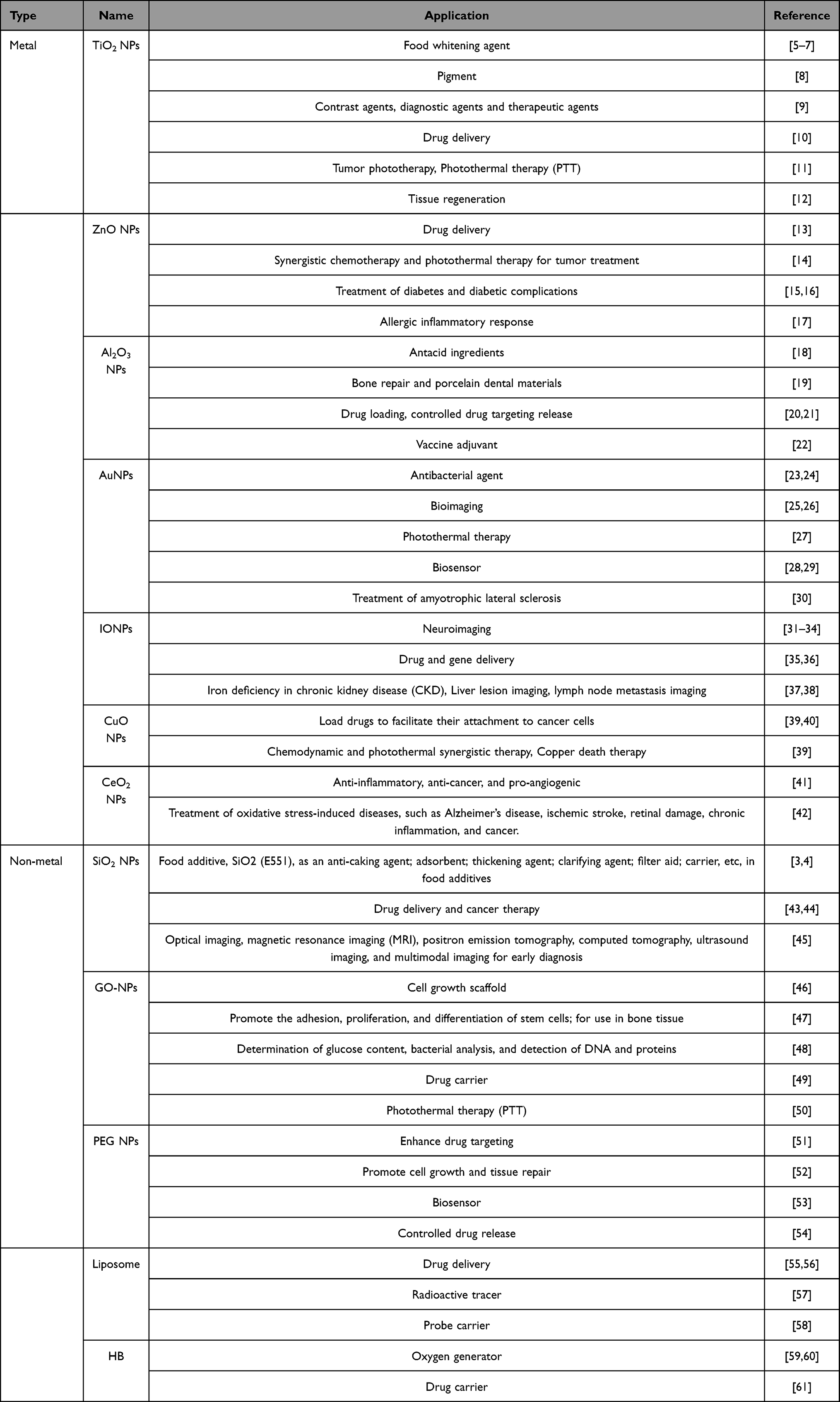 |
Table 1 Application of Representative Nanoparticles |
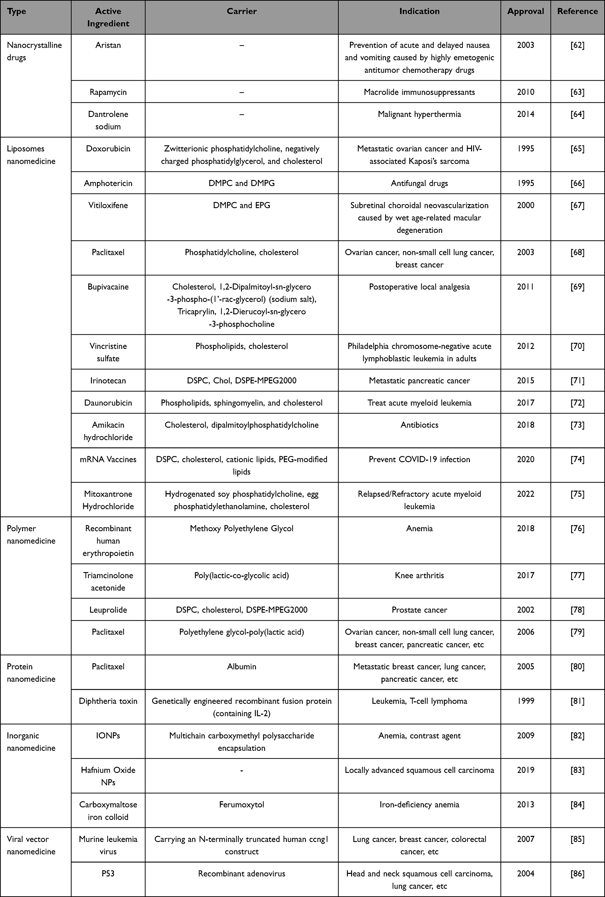 |
Table 2 Representative Marketed Nanomedicine |
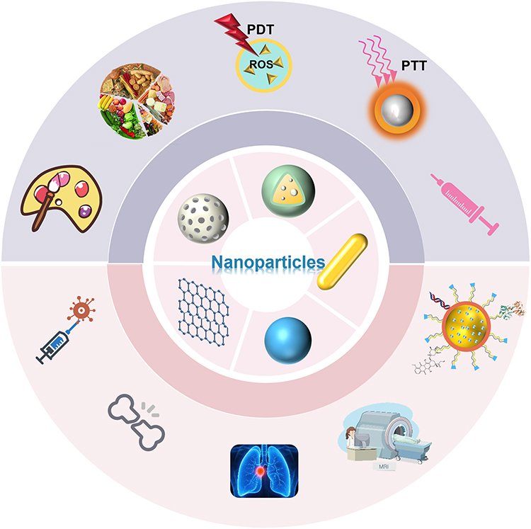 |
Figure 1 The Applications of Nanoparticles. Nanoparticles come in various shapes such as spherical, rod-like, and mesh-like etc, they are widely used, not only used as pigments and food additives, but also extensively applied in fields such as photothermal therapy, therapeutic agents, drug delivery systems, molecular imaging, biosensors, tissue engineering, and vaccine adjuvants. |
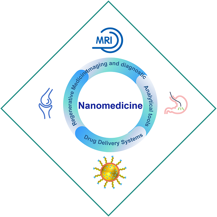 |
Figure 2 Application of Nanotechnology in Human Medicine. Nanotechnology is now widely used in medicine in regenerative medicine, imaging and diagnostics, analytical tools and drug delivery systems. |
In terms of disease treatment, NPs can exert therapeutic effects on a variety of diseases, including diabetes and its complications, allergic inflammatory responses, amyotrophic lateral sclerosis (ALS), and diseases induced by oxidative stress (eg, Alzheimer’s disease, ischemic stroke, retinal damage, chronic inflammation, and cancer).15,17,30,42 Meanwhile, NPs also exhibit significant advantages in adjuvant therapy: they can assist in tumor phototherapy, synergize with chemotherapy and photothermal therapy, and enhance the efficacy of chemodynamic therapy and photodynamic therapy.11,14,27,39 As drug and gene delivery carriers, certain NPs can function as oxygen generators to alleviate hypoxic tumor microenvironments, while also loading drugs to facilitate their attachment to cancer cells.40,59 Alternatively, NPs can be used in the field of bone tissue engineering to promote cell growth and tissue regeneration.19,47 Furthermore, within tissues, they can act as nanozyme, exerting anti-inflammatory, anticancer, and angiogenesis-promoting effects.41 In the fields of diagnostic imaging and therapy, the application of metal NPs is widespread. They can serve as contrast agents and diagnostic tools for optical imaging, Magnetic Resonance Imaging (MRI), Positron Emission Tomography (PET), Computed Tomography (CT), ultrasound imaging, and multimodal imaging for early diagnosis.9,25,26,37,45
In addition to their role in medicine, NPs can be applied to food as anti-caking agents, adsorbents, thickening agents, and other additives in food products.3,4 In industrial applications, metal NPs, as pigments, can fully leverage their unique optical properties, excellent stability, and advantages of the small size effect.8
Systemic Toxicity
NPs can enter the body by inhalation, skin, oral and intravenous injection. On the one hand, it will have toxic side effects on target organs or metabolic organs such as lungs, liver and kidneys. On the other hand, it can also reach other tissues through the blood circulation and cause tissue damage and systemic damage. Certain NPs with special properties can also cross the Blood-Brain Barrier (BBB) and cause nerve damage. In this section, the damage of NPs to various tissues or systems is summarized by classifying toxicity in general tissues from a macroscopic perspective. The organ and systemic toxicity of NPs is detailed in Figure 3 and Table 3.
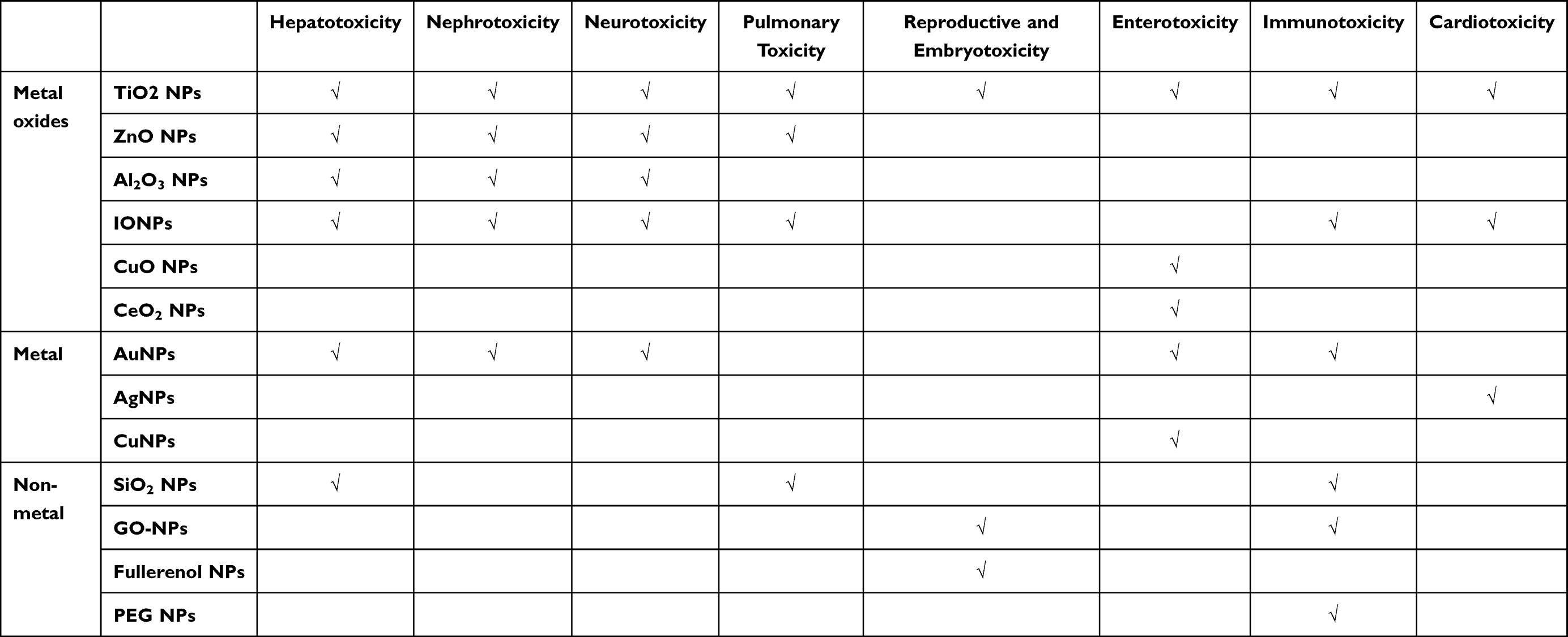 |
Table 3 OrganSystem Toxicity of Nanoparticles |
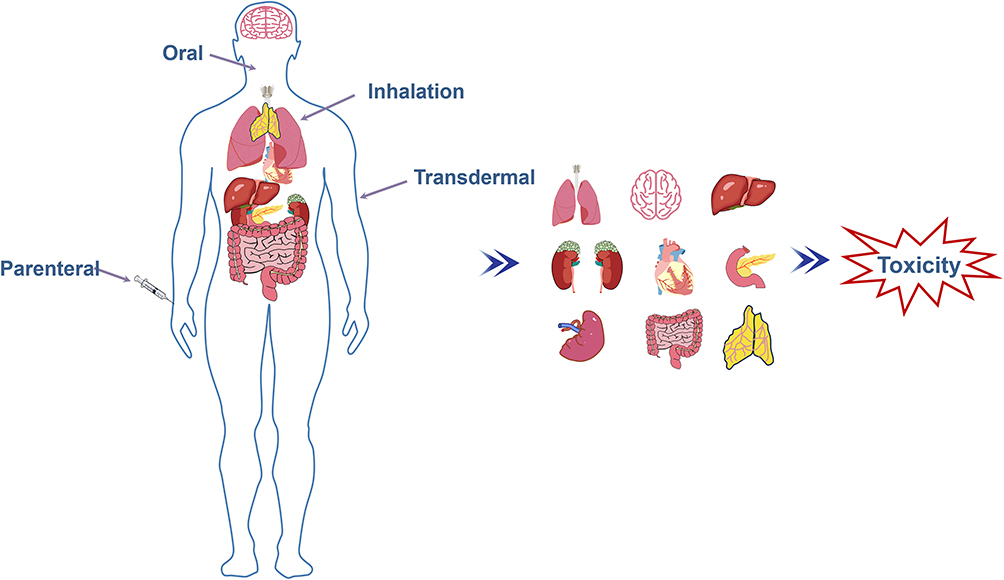 |
Figure 3 Systemic Toxicity of Nanoparticles. Nanoparticles can enter the human body through various means such as oral ingestion, inhalation, injection, and transdermal absorption, thereby causing toxic effects on numerous tissues and organs including the lungs, brain and nervous system, liver, kidneys, heart, intestines, reproductive system, as well as embryos and the immune system. |
Hepatotoxicity
Upon ingestion and subsequent entry into the systemic circulation as drug carriers, NPs may accumulate in potential target organs. Notably, NPs tend to accumulate in the liver to a greater extent than in other organs, where they may interact with hepatocytes. This interaction plays a critical role in determining the in vivo fate of NPs and the manifestation of hepatotoxicity. Presently, research on the hepatotoxicity of NPs primarily focuses on organ- and cellular-level investigations.
Titanium dioxide (TiO2) NPs were initially considered harmless substances; however, with the advancement of research, their low toxicity has been called into question. Studies have found that TiO2 NPs are a major component of environmental pollutants, with as much as 760 tons of these particles entering soils each year through wastewater and sludge.87,88 TiO2 NPs have been classified by the International Agency for Research on Cancer (IARC) as a Group 2B carcinogen, meaning they are possibly carcinogenic to humans.89 The European Union (EU) announced a ban on its use in food starting in August 2022. Oral administration of TiO2 NPs induces hepatotoxicity, manifested as disruption of biomarkers and imbalance in the oxidative and antioxidant systems,90 while intraperitoneal injection of TiO2 NPs has been observed to result in mitochondrial damage in the liver and hepatocyte apoptosis.91 A single administration of 5 g/kg of TiO2 NPs according to the OECD procedure revealed eosinophilic degeneration and patchy necrosis of hepatocytes around the central vein of the liver in mice.92 Furthermore, studies have detected residual TiO2 in the liver of humans who have received titanium implants.93
The hepatotoxicity of ZnO NPs has been reported across a range of toxicological studies, from cellular models to animal studies.94 The inclusion of ZnO NPs in the diet has been linked to liver damage, while both intraperitoneal (i.p.) and intravenous (i.v.) administration of ZnO NPs have been shown to induce hepatotoxicity.95,96 Inflammatory cell infiltration, dilated hepatic sinusoids, Kupffer cell proliferation, necrosis and other changes in liver tissues were observed in rats after 21 days of exposure to ZnO NPs.97 Additionally, studies have found that ZnO NPs can induce various symptoms of liver damage, such as decreased GSH levels and increased Lactate Dehydrogenase (LDH) and Malondialdehyde (MDA) levels.98 In addition to single-agent administration, co-administration of ZnO NPs (80 μg/kg) and CeO2 NPs (50 μg/kg) at extremely low doses has also been shown to induce oxidative stress-mediated inflammation and result in liver necrosis.99 However, modification of ZnO NPs can reduce their hepatotoxicity. For instance, MgO/ZnO core/shell NPs (MgO/ZnO CS NPs), synthesized from MgO and ZnO, exhibited no severe degeneration or necrosis at a dose of 10 mg/mL, with toxicity significantly lower than that of ZnO NPs administered alone.100
The liver has been identified as the primary organ where Al2O3 NPs accumulate during biodistribution within the body.101 Some studies have demonstrated its ability to increase p53 and Nrf2 levels, to reduce Hsp70 levels, and to promote apoptosis in liver tissue.102 Al2O3 NPs also induced a significant decrease in all antioxidant parameters (eg, Superoxide Dismutase (SOD), and Glutathione (GSH), and Catalase Activity (CA) in rat liver, and an even lower decrease in antioxidant parameters was observed after combining ZnO NPs.103
Hepatotoxicity of Iron Oxide Nanoparticles (IONPs) may be correlated with COX-2 mediated ER-mitochondrial Ca2+.104 Focal areas of perivascular round cell aggregates as well as necrosis in the liver of mice exposed orally to IONPs for 28 days.105 Moreover, IONPs can disrupt lipid metabolism through the gut-liver axis, indirectly contributing to liver injury.106
In addition to metal oxide NPs, the hepatotoxicity of AuNPs has also been reported. Following injection, AuNPs can persist in the liver and spleen for up to 7 days, inducing acute inflammatory responses and hepatocyte apoptosis.107 AuNPs exacerbate other disease-induced liver injury, such as aggravating the hepatotoxic effects of LPS by amplifying Reactive Oxygen Species (ROS)-dependent crosstalk in hepatic macrophages and hepatocytes.108 Alternatively, it worsens adriamycin-induced liver injury by inhibiting metabolic enzyme activities and the Nrf2/antioxidant axis in the liver.109–111 SiO2 NPs are also capable of promoting hepatic steatosis and liver injury, which exacerbate the progression of metabolic fatty liver disease.112
Nowadays, a multitude of new techniques have been used to evaluate the hepatotoxicity of NPs, including inductively coupled plasma-atomic emission spectroscopy,96 liver organoids,113 signaling pathways analyzed by Gene Expression Omnibus Dataset,114 Liquid Chromatography-Tandem Mass Spectrometry (LC-MS/MS) Metabolomics-based analysis,115 various hepatocyte models,116 etc. It is believed that with the development of the technology, the mechanism of liver injury by NPs will become better understood, which will be of greater help for future research.
Nephrotoxicity
The kidney, as an important eliminatory organ of nanomedicines, depends on size in terms of both the clearance pathway and intra-organ transport.117–119 In general, NPs smaller than 6 nm are more readily filtered and cleared by the kidneys, while larger NPs are unable to pass through the glomerular filtration barrier (GFB), leading to their accumulation in glomerular and tubular cells, which disrupts normal kidney function by causing oxidative stress, DNA damage, inflammation, and autophagy.119 Exploring the damage caused by NPs to the kidneys is essential for a more comprehensive understanding of the clearance process of NPs in vivo.
TiO2 NPs exhibit only slight toxicity at lower concentrations when co-cultured with IP15 cells.120 However, prolonged administration or high doses may lead to oxidative stress and cause damage to the kidneys. Small-sized TiO2 NPs (5 nm, 6.9 nm) accumulate in the kidneys and induce significant histopathological changes.121,122 The renal toxicity of TiO2 NPs is manifested in several ways, including the upregulation of IL-2, IL-4, IL-6, and NF-κB, as well as the attenuation of antioxidant stress responses in renal tissue cells.123,124 It causes injury to proximal renal tubular cells, as well as tubular dilation and cellular desquamation;125 and activation of the TGF-β/SMAD/p38MAPK and Wnt signaling pathways, which contribute to renal fibrosis in rats.126–128 Additionally, studies have reported on the safety of TiO2 NPs in relation to kidney function.129 Therefore, the nephrotoxicity of TiO2 NPs should not be generalized. Their potential effects and mechanisms of action on the kidneys must be thoroughly investigated, using the human maximum exposure criterion, to gain a deeper understanding of their safety and potential risks.
ZnO NPs are among the most nephrotoxic metal NPs when compared to other types of NPs.120,130–132 Lipidomic analysis revealed that 3 nm ZnO NPs exhibited the highest toxicity in renal cells, primarily by disrupting sphingolipid metabolism and autophagy processes through the elevation of ceramide levels.133 It was demonstrated that the toxicity of ZnO NPs is associated with TRPML1 channel activation, which mediates the release of Zn2+ and induces autophagy and cell death in human kidney cells.134,135 HIF is one of the targets through which ZnO NPs induce kidney injury. Treatment with ZnO NPs in HEK-293 cells and BALB/c mice resulted in a significant increase in the expression of HIF-1α, along with changes in renal pathology, serum creatinine, and blood urea nitrogen levels.136,137 CuO/ZnO core/shell NPs (CuO/ZnO CS NPs), synthesized from CuO and ZnO, induced significant kidney injury in mice when administered via gavage at a dose of 20 mg/L. However, at a dose of 40 mg/L, the extent of kidney injury was alleviated, exhibiting a unique pattern of “more significant injury at lower doses.” This phenomenon may be attributed to the adaptive response of mice to nanoparticle exposure at higher doses, specifically through “reduced absorption or accelerated excretion”.138
Polyethylene Glycol-AuNPs (PEG-AuNPs) (45 nm) significantly induced fuzzy swelling, edema, and vacuolar degeneration in multiple regions of the epithelial lining of the mouse renal tubules.139 LA-ICP-MS imaging clearly delineated the pathways of PEG-AuNPs in the kidney. One hour after injection, PEG-AuNPs rapidly entered the kidney via the bloodstream, accumulating primarily in the renal cones and renal pelvis. Four hours later, the signal in the renal pelvis weakened, while the signal in the renal cone region increased. After 24 hours, the signal was concentrated in the renal medulla and renal cortex.140 It was found that AuNPs also exacerbate renal damage caused by certain nephrotoxic substances (CDPP, PQ, 5-ASA) by increasing levels of renal blood urea nitrogen (BUN), creatinine (Cr), and IL-6.141 Therefore, treatments or diagnostic imaging involving preparations containing AuNPs should be avoided in patients with renal injury induced by related substances.
IONPs, commonly used as MRI contrast agents, can also exacerbate kidney damage to some extent. Szalay et al found that IONPs induced a viable, concentration-dependent decrease in Vero cells (African green monkey kidney cell line) after 24 hours of exposure.142 Kumari et al showed that 28 days of repeated oral administration of IONPs led to focal tubular injury and red medullary congestion in the kidneys.105 Another study found that a ten-day gavage of IONPs increased MDA levels, a marker of oxidative stress, altered blood chemistry biomarkers (such as total bilirubin [TBil], blood urea nitrogen [BUN], and creatinine [CREA]), and induced damage and desquamation of renal tubular epithelial cells in mice. Additionally, the study suggested that pomegranate extracts could help alleviate the kidney damage caused by IONPs.143
Aluminum is added to various daily products and is now reported to enter the human body through contaminated food and water. Its effects on cellular structures and macromolecules have been observed in both in vitro and in vivo studies, contributing to nephrotoxicity, oxidative stress, apoptosis, and other potential hazards.144–148 Environmental Al2O3 NPs lead to elevated levels of catalase, superoxide dismutase, and thiobarbituric acid-reactive substances, as well as reduced glutathione concentrations in the kidneys of aquatic animals.149 In addition, Al2O3 NPs caused significant increases in mammalian serum urea, creatinine, chloride, calcium, MDA, DNA damage, LDH, Tumor Necrosis Factor (TNF), and Proliferating Cell Nuclear Antigen (PCNA) expression, along with significant decreases in serum potassium, renal SOD, and GSH.101,150,151 Co-exposure to Al2O3 NPs and ZnO NPs results in more pronounced hepatorenal toxicity and systemic inflammation.103 Similar to many NPs, the relatively high surface area and reactivity of Al2O3 NPs contribute to their toxic effects. Therefore, co-exposures of this kind should be minimized to protect biological health and safety.
Neurotoxicity
Neurotoxicity is a potential adverse effect that may be either reversible or irreversible, and it can impact the structure, function, or biochemistry of neurons within the nervous system.152 Numerous studies have demonstrated that the neurotoxicity of NPs is primarily driven by oxidative stress induced by free radicals.89 TiO2 NPs not only reduce PC12 cell viability, induce apoptosis, inhibit synaptic growth, and disrupt the ubiquitin-proteasome system,153 but they also increase HT22 cell apoptosis through calcium imbalance-mediated endoplasmic reticulum stress.154 Additionally, TiO2 NPs enhance BBB permeability and induce mitochondrial damage, autophagy, neuroinflammation, and apoptosis in primary rat cortical astrocyte cells.155–157 Compared to other metals, TiO2 NPs are more likely to induce neuronal apoptosis, cognitive impairment, and synaptic plasticity dysfunction in the offspring of rodents.158,159 Exposure to TiO2 NPs during gestation results in neurobehavioral deficits in both the dams and their offspring, which are linked to damage to the gut-brain axis.160,161 TiO2 NPs cause anxiety-like behavior, cognitive impairments, and oxidative damage in the hippocampus, particularly during the adolescent stages of neurodevelopment.162 Neurotoxicity, including mitochondrial dysfunction, apoptosis, and alterations in neuronal structure and function, is observed with adult exposure.163–165
As early as 1980, Kozik et al observed morphological changes in the hippocampal cortex and basal ganglia of rats following the ingestion of high doses of ZnO.166 In 2009, the toxicity of ZnO NPs to neural stem cells was revealed to be related to Zn2+.167 The neurotoxic effects of ZnO NPs are associated with multiple signaling pathways, primarily including JNK, cAMP/CREB, PINK1/parkin-mediated mitochondrial autophagy, Ca2+-dependent NF-κB, ERK, p38, AK2-STAT3, and CAMK2A/CAMK2B signaling pathways.168–173 The neurotoxicity of ZnO NPs has also been confirmed in 3D brain organoids.174
Research indicates that, in contrast to other crystal forms, η-Al2O3 NPs manifest more pronounced cytotoxic effects on N2A neuroblastoma cells.175 γ-Al2O3 NPs affect neural stem cell viability and structure at high concentrations.176 Al2O3 NPs impair neurobehavioral function in ICR mice and are more toxic to the brain compared to nanocarbon and micron-sized Al2O3 of the same size.177 Its neurotoxicity is also manifested by altering neurotransmitter levels in rodents, affecting the expression of antioxidant mRNA, causing morphological changes in neuronal cell nuclei and acetylcholinesterase activity, and increasing the expression of β-amyloid protein.178–180
AuNPs have relatively low neurological damage than other metals, and can treat neurodegenerative diseases at low doses, yet some neurotoxicity has been reported.181,182 Dose-dependent toxicity of AuNPs on glial cells and neural progenitor cells.183 Glucose-coated AuNPs enter the rat brain through the BBB within 10 min of carotid artery injection, resulting in damage to the rat brain antioxidant enzyme glutathione peroxidase, and may further lead to oxidative stress and DNA damage.184,185
It has been confirmed that IONPs can cross the BBB and reach the brain directly via the olfactory nerves after nebulized administration. They can also cause pathological changes in the olfactory bulb, hippocampus, and striatum. The mechanism behind their ability to traverse the BBB may be related to the damage they cause to endothelial cells.186,187 Several studies have reported that exposure to IONPs in rats induces neuroinflammation, activates antioxidant responses and increases α-synuclein expression, decreases dopamine levels, causes degeneration in the hippocampus and striatum.105,188–191
Pulmonary Toxicity
As the main organs of the respiratory system, the lungs undertake the key function of gas exchange and are also an important gateway for various substances in the environment to enter the body. Compared with other particle sizes, NPs are more likely to enter and be deposited in the lungs and cause higher toxicity due to their smaller size and higher specific surface area.192,193
The pulmonary toxicity of many NPs has been demonstrated. Inhalation of TiO2 NPs resulted in the formation of two distinct histopathological features of pneumoconiosis in rats, namely Fibrotic Pulmonary Dust Foci and Dust Macules.194 Inhalation of various doses of TiO2 NPs resulted in distinct toxic effects. Low doses (0.5 mg/kg) primarily induced lymphocyte and macrophage aggregation, leading to emphysema and rupture of the alveolar septa. In contrast, high doses (4 mg/kg) caused thickening of the alveolar walls and interstitial tissue, along with collapse of the terminal bronchioles.195 Inhalation of IONPs and ZnO NPs induces considerable lung tissue damage in rats.196 It was observed that intratracheal instillation of ZnO NPs caused inflammatory cell infiltration in the alveoli of rats.197 Research has shown that IONPs (22 nm) cause greater destruction of lung epithelial cells compared to submicron (280 nm) IONPs.198 However, inhalation of micrometer-sized ZnO particles triggered a more pronounced systemic inflammatory response compared to ZnO NPs.199 This difference in toxicity may be related to the intrinsic properties of the metal particles themselves. The short-term pulmonary toxicity of CuO NPs was primarily characterized by acute lung inflammation and injury, whereas long-term exposure led to chronic inflammation and fibrosis. This toxic effect was associated with increased overexpression and secretion of MMP-3 in the lungs of mice.200
In addition to metal oxide NPs, the lungs are also highly sensitive to the toxicity of inhaled SiO2 NPs. This toxicity primarily occurs through the activation of the VEGFC/D-VEGFR3 signaling pathway in lung and lymphatic tissues, leading to pulmonary inflammation, lymphatic endothelial cell damage, and the formation and remodeling of pulmonary lymphatic vessels.201 Cationic liposomes can also induce pulmonary toxicity, with polyvalent cationic liposomes being more likely to cause dose-dependent toxicity and lung inflammation compared to monovalent cationic liposomes.202
Reproductive and Embryotoxicity
The reproductive system consists of various organs, each with varying sensitivities to potential harmful factors and substances. Currently, research on the reproductive toxicity of nanomaterials primarily focuses on their effects on reproductive organs, germ cells, and fetal development.203–206
Reproductive toxicity of TiO2 NPs has been observed in both male and female animals. In female mice, exposure to TiO2 NPs for 60 days results in decreased ovarian weight and disrupts the development of ovarian follicles via the TGF-β signaling pathway.207 In addition, TiO2 NPs caused a significant increase in MDA and estrogen levels in female mice, and significantly reduced fertilization rate, pregnancy rate, and number of deliveries.208 TiO2 NPs primarily impacted testicular morphology, sperm characteristics, and reproductive hormones in male mice. This was demonstrated by significant pathological changes and histomorphometric alterations in the testes, a reduction in sperm count, abnormalities in sperm quality, and a decrease in luteinizing hormone levels.209–212 Furthermore, studies have suggested that the reproductive toxicity of TiO2 NPs may be linked to mechanisms involving oxidative stress, apoptosis, inflammation, and interference with steroidogenesis.213–215
Carbon-based nanomaterials, such as graphene oxide and fullerenes, have also been a recent focus of research regarding their potential toxicity to the reproductive system. They have been shown to affect reproductive cells in mice, the pregnancy process, and fetal development to some extent.216,217 Exposure to Graphene Quantum Dots results in a significant decrease in the rate of first polar body extrusion in mouse oocytes and notably affects the mean fetal length. However, this effect does not carry over to the second generation of offspring.218 On the other hand, exposure to graphene oxide NPs (GO-NPs) caused dose-dependent pregnancy complications in pregnant mice, impacting placental barrier function. This was evidenced by a 30–80% reduction in the expression of tight junction proteins and vascular endothelial growth factor in placental tissue.219 In male mice, GO-NPs can cause significant histological damage to the testes, along with a notable loss of mitochondrial membrane potential in cells. This is accompanied by a decrease in sperm count and an increase in sperm deformity rates. However, the effects on sperm can be reversed after exposure is discontinued.220,221 Fullerenol NPs have been found to interfere with the strict process of oocyte meiotic resumption, possibly through the modulation of the EGFR-ERK1/2 signaling pathway and the expression and distribution of CX43.222
Enterotoxicity
The intestine, as a core component of the digestive system, plays a vital role in food digestion, nutrient absorption, and waste excretion. The potential intestinal toxicity of NPs has become one of the current research hotspots. The mechanisms through which they impact intestinal health are diverse, including direct damage to intestinal tissue, disruption of the gut barrier function, induction of intestinal inflammation, and alterations to the composition of the gut microbiota.223,224
In the food industry and drug delivery field, metal NPs, which are widely used, can cause intestinal toxicity when they reach a certain concentration.225–228 Among them, TiO2 NPs exhibit the most noticeable toxicity. Their intestinal toxic effects include increased mucosal permeability, impaired intestinal barrier function, induced gut microbiota imbalance, and elevated levels of lipopolysaccharides. These effects further lead to intestinal oxidative stress, inflammation, and intestinal-related metabolic disturbances. Further studies have found that the toxic mechanism of TiO2 NPs may be associated with the activation of the intestinal PKC/TLR4/NF-κB signaling pathway.227,229,230 TiO2 NPs have a significant impact on the gut microbiota of juvenile grouper, particularly on the genera Lactobacillus and Nautella. Changes in these bacterial populations can further trigger immune responses and alterations in metabolic products.231 CuO NPs can induce intestinal toxicity in zebrafish by altering the microbial abundance of Short-Chain Fatty Acids and Lipopolysaccharide (LPS) metabolism, which inhibits the key immune-regulatory pathway TLR4/MyD88/NF-κB.232 CeO2 NPs cause shedding of the intestinal epithelial tissue in rats, damage or disappearance of the glandular structure in the lamina propria, and induce changes in the structure and abundance of the intestinal microbiota.233
In addition to common metal oxide nanomaterials, the intestinal toxicity of metal NPs has been demonstrated. Cu NPs for smart drug delivery systems were found to cause dose-dependent damage to piglet intestinal epithelial cells accompanied by an increase in the oxidative stress markers MDA content and Metallothionein values.234,235 The toxic effects of Au-NPs on intestinal cells were less severe, with a decrease in colony-forming ability observed only after prolonged exposure to high concentrations.236
Immunotoxicity
The immunotoxicity of NPs is a complex and multifaceted phenomenon. Different nanoparticle drug carriers have varying effects on immune cells; some can activate immune responses, while others may suppress immune activity.237–239
The immunotoxicity of TiO2 NPs manifests as the inhibition of lymphocyte proliferation and macrophage Nitric Oxide (NO) production in vitro, with this suppressive effect being dose-dependent. In mice exposed to TiO2 NPs, significant impairments in the development and proliferation of B cells and T cells are observed, along with a reduction in macrophage activity and a decrease in Natural Killer cell numbers. This disruption leads to a diminished immune response against tumor cells, ultimately resulting in enhanced tumor growth in the mice.240 The immune toxicity induced by TiO2 NPs is reduced when they are embedded in mesoporous silica NPs (MSN) to form new TiO2@MSN particles, showing lower toxicity compared to TiO2 NPs alone.241
In studies on the immunotoxicity of SiO2 NPs, SNP50, SNP100 (non-porous SiO2 NPs), and Meso100, HMSNP100 (mesoporous SiO2 NPs) exhibited no signs of immunotoxicity.242 However, MSN with an average diameter of 160 nm can induce severe skin inflammatory responses, leading to a significant increase in immunoglobulins, serum histamine, and Th1/Th2/Th17 cytokines. This toxic response is associated with the protein corona on the MSN surface.243
Gold nanorods (Au NRs) can cause significant changes in nearly all immune cell subpopulations in the peripheral blood of mice, further disrupting both innate and adaptive immune responses.244 A study by Massich et al found that compared to lipid complexes carrying the same DNA sequence, densely functionalized, oligonucleotide-modified AuNPs triggered a significant 25% reduction in innate immune responses.245 There have also been reports on the immunotoxicity of non-metallic NPs, such as GO, carbon black NPs, and carbon nanotubes.246–248
Exosomes induced by IONPs can trigger immune activation and inflammatory responses associated with nanoparticle exposure.249 Park et al found that IONPs trigger the autophagic process before apoptosis by causing mitochondrial dysfunction and endoplasmic reticulum stress in RAW264.7 cells, leading to an increase in leukocyte and neutrophil levels, IL-8 secretion, and lactate dehydrogenase release.250 Follow-up studies have confirmed the immune-stimulating effects of IONPs on the spleen and lungs. These effects are mainly characterized by mitogen-induced proliferation of splenic lymphocytes, increased IL-1β secretion, the induction of a Th1-polarized inflammatory response in the lungs, enhanced chemokine secretion, and the upregulation of antigen-presenting proteins, leading to elevated levels of neutrophils, lymphocytes, and eosinophils.251–254
Most NPs carriers are considered non-toxic and biocompatible; however, there have still been reports of immune toxicity associated with certain NPs carriers. For example, Ambisome (amphotericin B liposome) has been reported to cause allergic reactions not previously associated with amphotericin B. This is likely due to the lipid components of the drug, which may trigger a direct and potentially fatal reaction.255 Lipid-based NPs (LNPs), which are commonly used for their low immunogenicity, are more prone to triggering the body’s immune response following surface modification.239,256 Cationic liposomes can trigger macrophage cytotoxicity by reducing the synthesis of NO and TNF-α in macrophages activated by LPS/IFN-γ.257 The immunotoxicity of PEG-modified NPs is also one of the current research focuses. Currently, allergic or hypersensitivity reactions have been reported for PEG 6000, PEG 3350, and PEG-containing barium contrast agents. The primary allergic symptoms include urticaria, dizziness, hypotension, angioedema, and transient episodes of tachycardia.258–260 The formation of anti-PEG antibodies after human exposure to PEG additives is one of the causes of allergic reactions associated with relevant drugs. Reports have shown that anti-PEG antibodies are present in the plasma of 25% of healthy blood donors, and these pre-existing anti-PEG antibodies may exacerbate the immune response of subjects to PEG-containing drugs.261–265
The mechanism behind PEG drug allergic reactions and hypersensitivity caused by anti-PEG antibodies is closely related to complement activation. Kozma et al revealed its role in complement activation-related pseudo-allergy (CARPA) induced by PEGylated liposomes and PEG-G-CSF (polyethylene glycol-conjugated granulocyte colony-stimulating factor).266
Cardiotoxicity
At present larger number of studies have reported the cardiotoxicity of TiO2 NPs, IONPs and AgNPs.267,268 Acute exposure to TiO2 NPs can induce dose-dependent cardiac toxicity, manifested as damage and alterations to myocardial fibers and cardiomyocytes.269 Long-term exposure to TiO2 NPs leads to the accumulation of titanium in the heart, triggering inflammatory responses, myocardial cell necrosis, and biochemical dysfunction of the heart.270 Nichols et al observed that the diastolic dysfunction induced by TiO2 NPs in the heart could be alleviated through the overexpression of a novel mitochondrial-targeted antioxidant enzyme, phospholipid hydroperoxide glutathione peroxidase (mPHGPx).271 IONPs can induce a reduction in heart rate, cardiac blood accumulation, and pericardial edema in zebrafish embryos. Intraperitoneal injection of IONPs in mice leads to dose-dependent oxidative damage to myocardial cells.157,272 The research conducted by Mohamed et al found that after administering IONPs to rats, cardiac toxicity was observed, characterized by a significant increase in Creatine Kinase Isoenzyme MB and LDH levels, along with an upregulation of TNF-α expression and a downregulation of HSP70 in heart tissue. Coating the NPs with rutin (Ru) significantly mitigated these toxic effects.273 The primary cardiac toxicity of Ag NPs is the induction of significant DNA base oxidation in the heart, primarily manifested by an increase in 8-Hydroxy-2’-deoxyguanosine levels. This toxic effect is more pronounced when Ag NPs are used in combination with IONPs.274
Factors Influencing the Safety of NPs
The interaction between NPs and the body is closely related to the physico-chemical properties of NPs. Different physicochemical properties lead to different mechanisms and outcomes of toxicity induced by NPs in the organism. This section summarizes the key factors affecting the safety of NPs, in order to provide references for ensuring the safety and effectiveness of NPs and establishing a strict and comprehensive quality control system. Factors influencing the safety of NPs is illustrated in Figure 4.
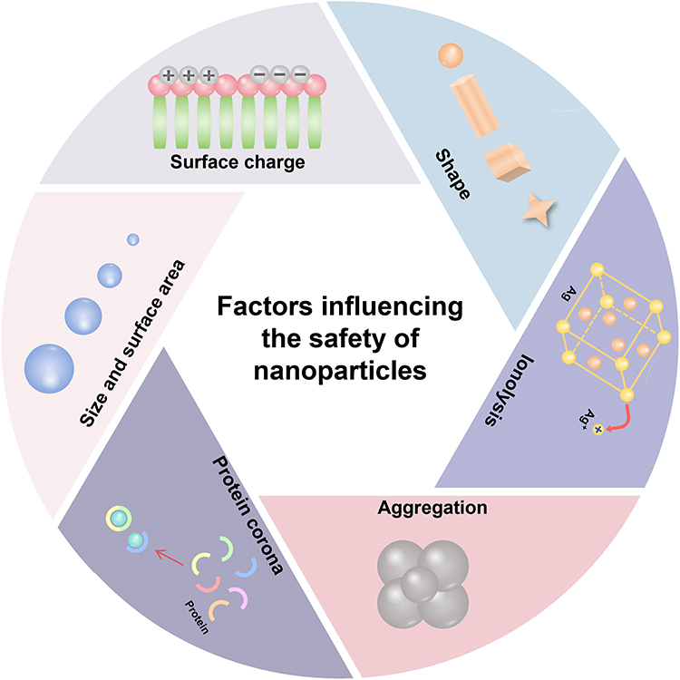 |
Figure 4 Factors Influencing the Safety of Nanoparticles. The different physicochemical properties of nanoparticles, such as size and surface area, surface charge, shapes, Ion dissolution, aggregation, and protein Corona, determine the differences in their distribution and behavior patterns in various tissues and organs of the body, thereby leading to the differences in toxicity of different nanoparticles in the body. |
Size and Surface Area
The size and surface characteristics of NPs influence their interaction with biological systems. Their size impacts both distribution and toxicity. NPs with varying sizes accumulate differently in organs such as the lungs, liver, and kidneys, which is partly due to the filtration structures in these organs.275 The transmembrane transport of NPs varies with particle size. Smaller particles primarily pass through the membrane via passive diffusion, while larger particles enter the cell through endocytosis.276 Typically, the toxicity of NPs is positively correlated with their size. For instance, the larger the particle size of TiO2 NPs, the greater the toxicity to the liver.92,277,278 Larger-sized IONPs are more easily absorbed by the spleen.279,280 The larger the size of Al2O3 NPs, the more pronounced their effect in inducing inflammation in the kidneys.281
However, the impact of size on the toxicity of NPs is also a subject of debate. For human dermal fibroblasts, 45 nm AuNPs are more toxic than 13 nm AuNPs.282 Nevertheless, smaller-sized AuNPs exhibit greater toxicity to macrophages, fibroblasts, melanoma cells, and epithelial cells. This may be due to the fact that smaller AuNPs are more likely to enter the cell nucleus and disrupt DNA structure.278,283 Smaller-sized AuNPs are also more likely to accumulate in the kidneys and liver. One of the reasons for this phenomenon is the different bridging reactions mediated by the Wnt/β-catenin signaling pathway.284,285 In addition, the size of AuNPs also affects the body’s response in terms of activating innate immune signaling pathways. 4.5 nm AuNPs preferentially activate the NLRP3 inflammasome to promote Caspase-1 maturation and IL-1β production, while AuNPs larger than 10 nm are more likely to trigger the NF-κB signaling pathway.286 The effect of SiO2 NPs particle size on their toxicity follows a similar pattern to that of AuNPs. Hepatotoxicity and nephrotoxicity of SiO2 NPs are more severe for small particle size, and the smaller the particle size of SiO2 NPs in amorphous structure, the more toxic it is.287,288 But some studies have also shown that large-sized SiO2 NPs exhibit greater toxicity due to increased surface contact area, easier cellular uptake, and susceptibility to membrane disruption.289–291
In summary, the impact of size on the toxicity of NPs is not absolute; it is also influenced by multiple factors such as cell and tissue type and the properties of the NPs. Therefore, a comprehensive, multifaceted approach is required to gain a deeper understanding of the mechanisms behind the various toxic effects of NPs and further promote the development of nanomedicine.
Surface Charge
The interaction of NPs with biological systems depends strongly on their surface charge, which in turn is influenced by the molecular structure of the coating material. The surface charge influences colloidal behavior, selective absorption, plasma protein binding, transmembrane permeability, and the integrity of the BBB.292 Based on charge properties, surface charge can be classified into three categories: positive, negative, and neutral charge.293,294 Positively charged NPs are more likely to interact with negatively charged glycoproteins on cell membranes, making them more readily absorbed by cells. As a result, they exhibit stronger cytotoxicity compared to negatively charged or neutral NPs, and are also more likely to participate in the regulation and replenishment processes in blood and biological fluids.295–297 Cationic functional groups in the structure of AuNPs are more likely to cause cell membrane rupture. The membrane-disrupting activity is influenced by the nature of the positive charge and the characteristics of the underlying chains. The higher the charge density, the more severe the damage to the cell membrane.298–300 For example, carboxyl-PEG-modified AuNPs display greater cytotoxicity than citrate-coated AuNPs, while cationic BPEI (branched polyethyleneimine)-modified AuNPs show the highest cellular uptake and toxicity response.301,302 Except for AuNPs, positively charged AgNPs, IONPs and CeO2 NPs are more toxic than their negatively charged counterparts.303–305
It has been demonstrated that the toxicity of NPs’ surface charge is influenced by their chemical composition, with cationic AuNPs being toxic and anionic ones non-toxic when the surface groups contain only the external chemical composition of carbon, hydrogen and oxygen.306 Whereas, when the surface groups consisted of carbon, hydrogen, oxygen and sulfur, both positively and negatively charged AuNPs were cytotoxic, and the negative charge was more toxic.307 In addition to this, the surface properties of the NPs interact with the protein crowns adsorbed on their surfaces, leading to altered cellular uptake mechanisms, loss of enzyme activity, and ultimately disruption of biological processes.308–310
Shapes
NPs have characteristic shapes such as spherical, cylindrical, ellipsoidal, cubic, rod-shaped and flaky, and different shapes have different effects on their toxicity magnitude.311 It has been shown that the nephrotoxicity produced by mesoporous SiO2 NPs in ICR mice depends strongly on their shape.312 In general, spherical NPs are more amenable to cytophagy, but are less toxic than other shapes, especially NPs with higher aspect ratios (including tubes, ribbons, rods, and wires and polyhedra, etc).313 Spherical TiO2 NPs are much less toxic to macrophages than dendritic and spindle-shaped.314 Different shapes of NPs displayed different toxicity in different types of cells. Rod-shaped CeO2 NPs exhibited more significant toxicity to macrophages than cubic and octahedral CeO2 NPs particles, whereas wire bundles and cubic CeO2 NPs with sharp edges were more toxic to human bone marrow cell lines.315,316 Flower-shaped AuNPs were more toxic to human endothelial cells, whereas spherical and rod-shaped AuNPs were much more toxic than stellate prismatic and mirror-shaped AuNPs in HEK293T cells.317,318
Therefore, in the safety evaluation of nanopharmaceuticals, the influence brought by their shapes needs to be paid close attention to, and advanced mathematical models can be developed with the help of various imaging techniques to predict their behaviors in organisms, which can provide the basis for the establishment of an accurate and efficient method for the characterization of the shapes and homogeneity of NPs.
Ion Dissolution
The dissolution and ionization properties of nanomaterials are determined by their chemical composition, which can be influenced by environmental factors such as pH and ionic strength. These properties are crucial in assessing the potential toxicity of NPs. Cho et al investigated the toxicity of 15 different metal/metal oxide NPs, exploring the relationship between various physicochemical parameters, including zeta potential and solubility, and their potential to induce pulmonary inflammation The results revealed that under acidic conditions (pH 5.5), a higher positive zeta potential increased the likelihood of lysosomal dissolution and the release of toxic metal ions, which, in turn, triggered a more pronounced inflammatory response.319 The dissociated Ag+ ions have been identified as the principal cause behind the mitochondrial damage, reactive oxygen species production, and apoptosis induced by AgNPs.320
Similarly, the dissolved Zn2+ ions are the predominant cause of neurotoxicity in mouse neural stem cells triggered by ZnO NPs. Surface coating with iron effectively alleviates this detrimental effect.167,321 However, for Cu NPs, their predominant form of toxicity is not the dissociated ions, but rather the particles themselves.322 In addition to ions, the release of surface modifications on NPs can also contribute to significant cytotoxicity. For instance, spherical CTAB (cetyltrimethylammonium bromide)-functionalized AuNPs demonstrate enhanced toxicity compared to their rod-shaped equivalents. This can be attributed to the more facile release of CTAB from the surface of the spherical particles.323 When polystyrene sulfonate and polyallylamine hydrochloride coatings are applied to the surface of these AuNPs, their toxicity is markedly diminished.324 In addition to coating modification, the toxicity of NPs can also be mitigated by designing a shell structure to encapsulate the NPs. For instance, designing a MgO shell around AgO NPs can slow down the release rate of Ag⁺, ensuring that Ag⁺ is released in a “slow and sustained” manner and thereby avoiding the toxicity caused by excessively high local Ag⁺ concentrations.325
Toxicity Related to the Biotransformation of Nanomaterials
Aggregation
The influence of aggregation on the toxicity of NPs remains a contentious issue. The interactions of aggregates with receptors and membrane proteins may differ from the internalization pathways observed with individual NPs.326 AuNPs, transported through the endolysosomal pathway, are susceptible to disintegration in the acidic microenvironment (pH 4.5–6). This degradation induces the formation of aggregates that remain sequestered within the cell, owing to the disruption of intracellular structures.327,328 The aggregates that form may lead to vascular thrombosis, subsequently impairing the uptake and targeted delivery of the NPs.329 In general, aggregates can attenuate adverse effects by decreasing the specific surface area and restricting interactions with cellular structures.330 For instance, larger aggregates of AgNPs can markedly reduce hemolytic toxicity compared to their smaller counterparts.331,332 However, in macrophages, larger aggregates can enhance the delivery of the smaller initial NPs, leading to a more pronounced toxic effect.333
Protein Corona
NPs with elevated surface energy, upon introduction into intricate physiological fluids, are capable of progressively and selectively adsorbing biomolecules from the surrounding milieu, thereby forming a biomolecular corona composed of proteins, lipids, carbohydrates, nucleic acids, and other components. The protein fraction of this corona is specifically designated as the protein corona.334,335 The protein corona can influence the toxicity of NPs through mechanisms such as modulating cellular recognition, triggering immune responses, altering the biocompatibility of the particles, and affecting hepatic metabolism.109,336–339 The protein corona formed by the binding of ZnO NPs with proteins in brain homogenates can also impact physiological functions by altering protein conformation and either inhibiting or enhancing enzymatic activity.340
The influence of the protein corona on NPs toxicity is dual-faceted. On one hand, it can mitigate toxicity by preventing the dissolution and release of ions from the NPs, thereby reducing the likelihood of their uptake by cells.109 On the other hand, the protein corona has been demonstrated to activate the endoplasmic reticulum stress pathway and the TGF-β/Smad 2 pathway in rats, subsequently inducing pulmonary fibrosis.341 For mesoporous SiO2 NPs, the protein corona formed on their surface can significantly elevate the expression of cytokines specific to atopic dermatitis in mice, as well as increase the infiltration of immune cells.243 Studies have further indicated that the protein corona can modulate hemolysis, platelet activation, and tumor metastasis, thereby intensifying the toxicity to cells or the organism.342–344
Distinct forms of protein coronas can provoke varied toxicological outcomes. In contrast to the Human Serum Albumin corona, the Haptoglobin corona is correlated with increased cellular internalization and a diminished hepatocyte survival rate.109 The CdS NPs coated with PC proteins primarily induce apoptosis by enhancing the expression of the FcγRIIB receptor on macrophage surfaces, thereby activating the AKT/Caspase-3 signaling pathway.345
Mechanisms of Nanoparticle Toxicity
NPs can cause damage to different tissues or systems through a variety of mechanisms. At present, research on these mechanisms mainly focuses on oxidative stress, DNA damage, and inflammatory responses. This section summarizes the toxicity mechanisms of NPs, providing certain references for researchers to better understand the interactions between NPs and the body. The toxicity mechanism of NPs is illustrated in Figure 5.
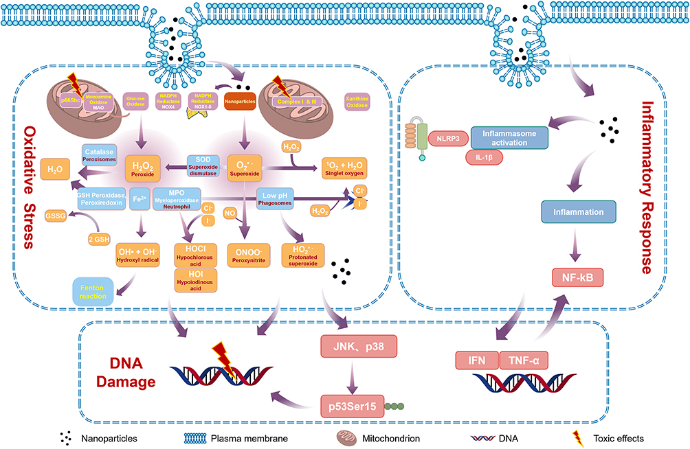 |
Figure 5 Toxicity Mechanism of Nanoparticles. Nanoparticles mainly exert toxic effects through oxidative stress, DNA damage, and inflammatory responses, leading to the destruction of cell structure and function, and subsequently inducing tissue and organ toxicity. |
Oxidative Stress
ROS in the body are a group of highly reactive oxygen-containing molecules that mainly function in signal regulation. Under normal physiological conditions, the production and clearance of ROS maintain a dynamic balance, thereby preserving homeostasis. However, when ROS production exceeds the body’s capacity for clearance, it can lead to an imbalance in homeostasis, triggering oxidative stress and subsequently damaging cellular structures.346 Studies have demonstrated that NPs can induce intracellular oxidative stress by activating oxidative stress signaling pathways or suppressing the activity of antioxidant systems. This results in the excessive accumulation of intracellular ROS or an imbalance in the NO/NOS system, ultimately leading to tissue or organ toxicity.347 For example, compared to conventional ZnO, ZnO NPs exhibit more severe oxidative stress at the cellular level, which in turn induces more pronounced liver damage in rats.348 The transcriptional level of the SOD1 gene in mice induced by Ag NPs was significantly higher than that in the control group, accompanied by more severe lung damage.349 Exposure to TiO2 NPs induces oxidative stress, which causes hippocampal cell apoptosis in rats and mice, ultimately leading to impairments in motor function and spatial recognition memory.349 TiO2 NPs can downregulate the levels and activities of antioxidant enzymes such as Catalase, Glutathione Peroxidase 1, and SOD in human thyroid follicular epithelial cells, while upregulating the levels of ROS and MDA induced by oxidative stress. This, in turn, leads to damage to the cell’s structure and function.350 Moreover, certain metal NPs can release metal ions, participate in Fenton or Fenton-like reactions, and promote the accumulation of ROS within cells, thereby further inducing oxidative stress.351,352
DNA Damage
Humans are typically exposed to NPs through inhalation (respiratory tract), ingestion (gastrointestinal tract), dermal contact, and injection (circulatory system).353–356 When NPs enter the human body, they may traverse various cellular barriers and reach sensitive organs such as the lungs, liver, and kidneys. This can result in mitochondrial damage, DNA mutations, and ultimately lead to cell apoptosis or death.357–359
NPs-induced DNA damage can be categorized into direct and indirect damage. Direct damage refers to covalent interactions between NPs and DNA molecules, resulting in alterations to the structure and properties of the DNA. The hepatotoxicity mechanism of TiO2 NPs in mice involves direct DNA damage. Specifically, TiO2 NPs insert base pairs into DNA or bind to DNA nucleotides, accumulating in liver DNA through Ti-O(N) and Ti-P bonds. This leads to DNA strand breaks in the liver and alters the conformation of the DNA.360 ZnO NPs have been found to significantly increase the mutation frequency of the HGPRT gene and, to some extent, interfere with DNA damage and repair processes.361,362 Positively charged AuNPs have the ability to strongly interact with the negatively charged DNA in cells, resulting in DNA damage and disrupting the normal progression of the cell cycle, which causes an extension of certain cell cycle phases. This process could also play a role in promoting carcinogenesis.295,363
Indirect damage refers to DNA damage that occurs as a consequence of other toxic effects caused by NPs. The principal mechanism through which NPs induce toxicity is the damage to DNA caused by oxidative stress and inflammation triggered by ROS.364–366 For instance, ZnO NPs can trigger oxidative stress, activate JNK and p38 pathways, and facilitate the phosphorylation of p53Ser15, ultimately resulting in DNA damage and apoptosis in human liver cells.367 Al2O3 NPs induce DNA damage in mouse neuronal cells via ROS-mediated indirect injury, while Co and Cr NPs trigger the release of IL-6, which indirectly causes DNA damage in neurons and astrocytes derived from neural progenitor cells.368
Inflammatory Response
Inflammation is a defense mechanism against the invasion of harmful substances into the body. It helps eliminate harmful factors and damaged tissues, restoring cells and tissues to their normal function. The vast majority of nanomedicines are exogenous substances that have the potential to trigger a series of excessive inflammatory responses upon entering the body.369 It has been found that NPs predominantly trigger inflammatory responses by promoting the release of inflammatory factors and inducing the activation of inflammatory vesicles.200,370,371 For example, TiO2 NPs upregulate the expression of inflammatory factors such as TNF-α, IL-1β, NF-κB, IFN-α, and IFN-β. Moreover, the entry of MnO2 NPs into the organism results in a significant increase in IL-1β and TNF-α levels.372,373 SiO2 NPs can regulate the transcription and release of pro-inflammatory cytokines by participating in the activation of inflammasomes, as well as activate key pathways related to inflammation and cell death.371 CuO NPs induce pulmonary inflammation via triggering cellular cuproptosis.374
Apart from the previously discussed mechanisms, NPs can also impair cellular structure and function through multiple routes, including gene mutations, mitochondrial damage, and fibrosis, which in turn can cause toxicity across various organs. As nanotechnology continues to evolve and its applications broaden, there is an increasing necessity to thoroughly elucidate the toxicity mechanisms of NPs, investigate their potential toxic pathways, and establish accurate methods to quantify the degree of influence from each mechanism, thereby gaining a more profound understanding of the inherent toxicity of NPs.
Conclusion and Discussion
The toxicity and safety of NPs represent a critical issue that limits their widespread application. This review systematically synthesizes the toxicological characteristics, regulatory factors, and underlying mechanisms of NPs. The main findings are as follows: in terms of toxic manifestations, NPs exhibit significant organ- and system-specific toxicity, with the liver, kidneys, and nervous system being the primary target organs. Regarding regulatory factors, physicochemical properties such as particle size, surface charge, shape, and ion release are key variables determining toxic outcomes. In terms of mechanisms, oxidative stress, DNA damage, and inflammatory responses constitute the core pathways of NPs-induced toxicity.
These findings provide important insights for further research. First, the specificity of target organs suggests that safety evaluation of NPs should prioritize the toxic risks to specific organs based on the application scenario. Second, the correlation between physicochemical properties and toxic effects indicates that toxicity reduction can be achieved through optimization of parameters such as particle size and surface modification while preserving functionality. Third, elucidation of the core toxicity mechanisms provides a theoretical basis for the development of in vitro alternative testing methods and the identification of early biomarkers.
To address the toxicity challenges associated with NPs, future efforts can focus on the following strategies. The first is design optimization: modulating the biodistribution and metabolic behavior of NPs through surface modification or smart responsive designs to reduce accumulation in non-target organs. The second is the establishment of a full life-cycle assessment framework: integrating emerging technologies such as organ-on-a-chip and multi-omics analysis to construct a toxicity evaluation system covering the entire process from synthesis to application, metabolism, and excretion. Finally, the development of “smart detoxification” strategies leveraging emerging technologies offers a promising approach for toxicity intervention.
In summary, based on a systematic summary of research progress in NPs toxicity, this review identifies key scientific issues and existing limitations, and proposes feasible pathways for toxicity reduction and evaluation optimization, thereby providing theoretical support for improving the safety assessment of nanoparticles.
Funding
This work was supported by the National Natural Science Foundation of China (82104357), Shenzhen Science and Technology Program (KJZD20240903102714019), and Science and Technology Innovation Project of the Guangdong Provincial Drug Administration (2022TDB65).
Disclosure
The authors declare that the research was conducted in the absence of any commercial or financial relationships that could be construed as a potential conflict of interest.
References
1. Wolfram J, Ferrari M. Clinical cancer nanomedicine. Nano Today. 2019;25:85–31. doi:10.1016/j.nantod.2019.02.005
2. Nakamura T, Sato Y, Yamada Y, et al. Extrahepatic targeting of lipid nanoparticles in vivo with intracellular targeting for future nanomedicines. Adv Drug Delivery Rev. 2022;188:114417. doi:10.1016/j.addr.2022.114417
3. Kwon RY, Youn SM, Choi SJ. Oral excretion kinetics of food-additive silicon dioxides and their effect on in vivo macrophage activation. Int J Mol Sci. 2024;25(3). doi:10.3390/ijms25031614
4. Kwon RY, Kim SB, Youn SM, Choi SJ. Fate determination and characterization of food additive silicon dioxide and titanium dioxide in commercial foods. Front Biosci. 2023;28(2):36. doi:10.31083/j.fbl2802036
5. Athinarayanan J, Alshatwi AA, Periasamy VS, Al-Warthan AA. Identification of nanoscale ingredients in commercial food products and their induction of mitochondrially mediated cytotoxic effects on human mesenchymal stem cells. J Food Sci. 2015;80(2):N459–N464. doi:10.1111/1750-3841.12760
6. Dudefoi W, Terrisse H, Popa AF, Gautron E, Humbert B, Ropers MH. Evaluation of the content of TiO(2) nanoparticles in the coatings of chewing gums. Food Addit Contamin. 2018;35(2):211–221. doi:10.1080/19440049.2017.1384576
7. Lomer MC, Thompson RP, Commisso J, Keen CL, Powell JJ. Determination of titanium dioxide in foods using inductively coupled plasma optical emission spectrometry. Analyst. 2000;125(12):2339–2343. doi:10.1039/b006285p
8. Weir A, Westerhoff P, Fabricius L, Hristovski K, von Goetz N. Titanium dioxide nanoparticles in food and personal care products. Environ Sci Technol. 2012;46(4):2242–2250. doi:10.1021/es204168d
9. Akasaka H, Mukumoto N, Nakayama M, et al. Investigation of the potential of using TiO2 nanoparticles as a contrast agent in computed tomography and magnetic resonance imaging. Appl Nanosci. 2020;10(8):3143–3148. doi:10.1007/s13204-019-01098-y
10. Wang Q, Huang JY, Li HQ, et al. TiO(2) nanotube platforms for smart drug delivery: a review. Int J Nanomed. 2016;11:4819–4834. doi:10.2147/ijn.S108847
11. Rodríguez-Barajas N, Anaya-Esparza LM, Villagrán-de la Mora Z, Sánchez-Burgos JA, Pérez-Larios A. Review of therapies using TiO(2) nanomaterials for increased anticancer capability. Anticancer Agents Med Chem. 2022;22(12):2241–2254. doi:10.2174/1871520622666211228112631
12. Seisenbaeva GA, Fromell K, Vinogradov VV, et al. Dispersion of TiO(2) nanoparticles improves burn wound healing and tissue regeneration through specific interaction with blood serum proteins. Sci Rep. 2017;7(1):15448. doi:10.1038/s41598-017-15792-w
13. Wiesmann N, Tremel W, Brieger J. Zinc oxide nanoparticles for therapeutic purposes in cancer medicine. J Mat Chem B. 2020;8(23):4973–4989. doi:10.1039/d0tb00739k
14. Deng H, Yang Y, Zuo T, et al. Multifunctional ZnO@CuS nanoparticles cluster synergize chemotherapy and photothermal therapy for tumor metastasis. Nanomedicine. 2021;34:102399. doi:10.1016/j.nano.2021.102399
15. Abd El-Baset SA, Mazen NF, Abdul-Maksoud RS, Kattaia AAA. The therapeutic prospect of zinc oxide nanoparticles in experimentally induced diabetic nephropathy. Tissue Barriers. 2023;11(1):2069966. doi:10.1080/21688370.2022.2069966
16. Umrani RD, Paknikar KM. Zinc oxide nanoparticles show antidiabetic activity in streptozotocin-induced Type 1 and 2 diabetic rats. Nanomedicine. 2014;9(1):89–104. doi:10.2217/nnm.12.205
17. Kim MH, Seo JH, Kim HM, Jeong HJ. Zinc oxide nanoparticles, a novel candidate for the treatment of allergic inflammatory diseases. Eur J Pharmacol. 2014;738:31–39. doi:10.1016/j.ejphar.2014.05.030
18. Hem SL, White JL, Buehler JD, Luber JR, Grim WM, Lipka EA. Evaluation of antacid suspensions containing aluminum hydroxide and magnesium hydroxide. Am J Health Syst Pharm. 1982;39(11):1925–1930.
19. Hadjicharalambous C, Buyakov A, Buyakova S, Kulkov S, Chatzinikolaidou M. Porous alumina, zirconia and alumina/zirconia for bone repair: fabrication, mechanical and in vitro biological response. Biomed Mater. 2015;10(2):025012. doi:10.1088/1748-6041/10/2/025012
20. Karami MH, Pourmadadi M, Abdouss M, et al. Novel chitosan/γ-alumina/carbon quantum dot hydrogel nanocarrier for targeted drug delivery. Int J Biol Macromol. 2023;251:126280. doi:10.1016/j.ijbiomac.2023.126280
21. Nematollahi E, Pourmadadi M, Yazdian F, Fatoorehchi H, Rashedi H, Nigjeh MN. Synthesis and characterization of chitosan/polyvinylpyrrolidone coated nanoporous γ-Alumina as a pH-sensitive carrier for controlled release of quercetin. Int J Biol Macromol. 2021;183:600–613. doi:10.1016/j.ijbiomac.2021.04.160
22. Li X, Aldayel AM, Cui Z. Aluminum hydroxide nanoparticles show a stronger vaccine adjuvant activity than traditional aluminum hydroxide microparticles. J Control Release. 2014;173:148–157. doi:10.1016/j.jconrel.2013.10.032
23. Burygin GL, Khlebtsov BN, Shantrokha AN, Dykman LA, Bogatyrev VA, Khlebtsov NG. On the Enhanced antibacterial activity of antibiotics mixed with gold nanoparticles. Nanoscale Res Lett. 2009;4(8):794–801. doi:10.1007/s11671-009-9316-8
24. Bagga P, Ansari TM, Siddiqui HH, et al. Bromelain capped gold nanoparticles as the novel drug delivery carriers to aggrandize effect of the antibiotic levofloxacin. EXCLI J. 2016;15:772–780. doi:10.17179/excli2016-710
25. Chen H, Zhang X, Dai S, et al. Multifunctional gold nanostar conjugates for tumor imaging and combined photothermal and chemo-therapy. Theranostics. 2013;3(9):633–649. doi:10.7150/thno.6630
26. Loynachan CN, Soleimany AP, Dudani JS, et al. Renal clearable catalytic gold nanoclusters for in vivo disease monitoring. Nature Nanotechnol. 2019;14(9):883–890. doi:10.1038/s41565-019-0527-6
27. Yin B, Ho WKH, Xia X, et al. A multilayered mesoporous gold nanoarchitecture for ultraeffective near-infrared light-controlled chemo/photothermal therapy for cancer guided by SERS imaging. Small. 2023;19(6):e2206762. doi:10.1002/smll.202206762
28. Yang L, Hou P, Wei J, Li B, Gao A, Yuan Z. Recent advances in gold nanocluster-based biosensing and therapy: a review. Molecules. 2024;29(7). doi:10.3390/molecules29071574
29. Wang ZJ, Li Q, Tan LL, Liu CG, Shang L. Metal-organic frameworks-mediated assembly of gold nanoclusters for sensing applications. J Anal Test. 2022;6(2):163–177. doi:10.1007/s41664-022-00224-0
30. Ren J, Dewey RB 3rd, Rynders A, et al. Evidence of brain target engagement in Parkinson’s disease and multiple sclerosis by the investigational nanomedicine, CNM-Au8, in the REPAIR Phase 2 clinical trials. J Nanobiotechnol. 2023;21(1):478. doi:10.1186/s12951-023-02236-z
31. Avasthi A, Caro C, Pozo-Torres E, Leal MP, García-Martín ML. Magnetic nanoparticles as MRI contrast agents. Topics in Current Chem. 2020;378(3):40. doi:10.1007/s41061-020-00302-w
32. Patel D, Kell A, Simard B, et al. Cu2+-labeled, SPION loaded porous silica nanoparticles for cell labeling and multifunctional imaging probes. Biomaterials. 2010;31(10):2866–2873. doi:10.1016/j.biomaterials.2009.12.025
33. Wang CH, Kang ST, Yeh CK. Superparamagnetic iron oxide and drug complex-embedded acoustic droplets for ultrasound targeted theranosis. Biomaterials. 2013;34(7):1852–1861. doi:10.1016/j.biomaterials.2012.11.037
34. Huber DL. Synthesis, properties, and applications of iron nanoparticles. Small. 2005;1(5):482–501. doi:10.1002/smll.200500006
35. Li A, Zhang T, Huang T, et al. Iron oxide nanoparticles promote Cx43-Overexpression of mesenchymal stem cells for efficient suicide gene therapy during glioma treatment. Theranostics. 2021;11(17):8254–8269. doi:10.7150/thno.60160
36. Zanganeh S, Hutter G, Spitler R, et al. Iron oxide nanoparticles inhibit tumour growth by inducing pro-inflammatory macrophage polarization in tumour tissues. Nature Nanotechnol. 2016;11(11):986–994. doi:10.1038/nnano.2016.168
37. Bobo D, Robinson KJ, Islam J, Thurecht KJ, Corrie SR. Nanoparticle-based medicines: a review of FDA-approved materials and clinical trials to date. Pharmacol Res. 2016;33(10):2373–2387. doi:10.1007/s11095-016-1958-5
38. Reimer P, Balzer T. Ferucarbotran (Resovist): a new clinically approved RES-specific contrast agent for contrast-enhanced MRI of the liver: properties, clinical development, and applications. Eur Radiol. 2003;13(6):1266–1276. doi:10.1007/s00330-002-1721-7
39. Xu Y, Liu SY, Zeng L, et al. An enzyme-engineered Nonporous Copper(I) coordination polymer nanoplatform for cuproptosis-based synergistic cancer therapy. Adv Mater. 2022;34(43):e2204733. doi:10.1002/adma.202204733
40. Cheng FT, Geng YD, Liu YX, et al. Co-delivery of a tumor microenvironment-responsive disulfiram prodrug and CuO(2) nanoparticles for efficient cancer treatment. Nanoscale Adv. 2023;5(12):3336–3347. doi:10.1039/d3na00004d
41. Li H, Xia P, Pan S, et al. The advances of ceria nanoparticles for biomedical applications in orthopaedics. Int J Nanomed. 2020;15:7199–7214. doi:10.2147/ijn.S270229
42. Nelson BC, Johnson ME, Walker ML, Riley KR, Sims CM. Antioxidant cerium oxide nanoparticles in biology and medicine. Antioxidants. 2016;5(2). doi:10.3390/antiox5020015
43. Wang L, Huo M, Chen Y, Shi J. Coordination-accelerated “Iron Extraction” enables fast biodegradation of mesoporous silica-based hollow nanoparticles. Adv Healthcare Mater. 2017;6(22). doi:10.1002/adhm.201700720
44. Han S, Liu S, Song Y, Jiang H. Biodegradable magnesium ion-doped silica-based molecularly imprinted nanoparticles for targeting tumor cells to drugs controlled release and recognition mechanism research. Colloids Surf B. 2022;217:112665. doi:10.1016/j.colsurfb.2022.112665
45. Cha BG, Kim J. Functional mesoporous silica nanoparticles for bio-imaging applications. Wiley Interdiscip Rev. 2019;11(1):e1515. doi:10.1002/wnan.1515
46. Duran N, Martinez DS, Silveira CP, et al. Graphene oxide: a carrier for pharmaceuticals and a scaffold for cell interactions. Curr Top Med Chem. 2015;15(4):309–327. doi:10.2174/1568026615666150108144217
47. Shadjou N, Hasanzadeh M, Khalilzadeh B. Graphene based scaffolds on bone tissue engineering. Bioengineered. 2018;9(1):38–47. doi:10.1080/21655979.2017.1373539
48. Ji G, Tian J, Xing F, Feng Y. Optical biosensor based on graphene and its derivatives for detecting biomolecules. Int J Mol Sci. 2022;23(18). doi:10.3390/ijms231810838
49. Cao W, He L, Cao W, Huang X, Jia K, Dai J. Recent progress of graphene oxide as a potential vaccine carrier and adjuvant. Acta Biomater. 2020;112:14–28. doi:10.1016/j.actbio.2020.06.009
50. Revuri V, Mondal J, Lee YK. Graphene as Photothermal Therapeutic Agents. Adv Exp Med Biol. 2022;1351:177–200. doi:10.1007/978-981-16-4923-3_9
51. Li Y, Wang G, Wang T, et al. PEGylated gambogic acid nanoparticles enable efficient renal-targeted treatment of acute kidney injury. Nano Lett. 2023;23(12):5641–5647. doi:10.1021/acs.nanolett.3c01235
52. Li H, Shi Y, Zhang W, Yu M, Chen X, Kong M. Ternary complex coacervate of PEG/TA/gelatin as reinforced bioadhesive for skin wound repair. ACS Appl Mater Interfaces. 2022;14(16):18097–18109. doi:10.1021/acsami.2c00236
53. Shi M, Li Y, Wang W, Han R, Luo X. A super-antifouling electrochemical biosensor for protein detection in complex biofluids based on PEGylated multifunctional peptide. ACS Sens. 2024;9(6):2956–2963. doi:10.1021/acssensors.4c00126
54. Zhao D, Wei Y, Jin Q, Yang N, Yang Y, Wang D. PEG-functionalized hollow multishelled structures with on-off switch and rate-regulation for controllable antimicrobial release. Angew Chem Int Ed Engl. 2022;61(36):e202206807. doi:10.1002/anie.202206807
55. Large DE, Abdelmessih RG, Fink EA, Auguste DT. Liposome composition in drug delivery design, synthesis, characterization, and clinical application. Adv Drug Delivery Rev. 2021;176:113851. doi:10.1016/j.addr.2021.113851
56. Baumann KN, Schroder T, Ciryam PS, et al. DNA-liposome hybrid carriers for triggered cargo release. ACS Appl Bio Mater J. 2022;5(8):3713–3721. doi:10.1021/acsabm.2c00225
57. Jensen GM, Bunch TH. Conventional liposome performance and evaluation: lessons from the development of Vescan. J Liposome Res. 2007;17(3–4):121–137. doi:10.1080/08982100701527981
58. Xia Y, Xu C, Zhang X, et al. Liposome-based probes for molecular imaging: from basic research to the bedside. Nanoscale. 2019;11(13):5822–5838. doi:10.1039/c9nr00207c
59. Liu LH, Zhang YH, Qiu WX, et al. Dual-stage light amplified photodynamic therapy against hypoxic tumor based on an O(2) self-sufficient nanoplatform. Small. 2017;13(37). doi:10.1002/smll.201701621
60. Cheng Y, Cheng H, Jiang C, et al. Perfluorocarbon nanoparticles enhance reactive oxygen levels and tumour growth inhibition in photodynamic therapy. Nat Commun. 2015;6:8785. doi:10.1038/ncomms9785
61. Chen Z, Liu L, Liang R, et al. Bioinspired hybrid protein oxygen nanocarrier amplified photodynamic therapy for eliciting anti-tumor immunity and abscopal effect. ACS Nano. 2018;12(8):8633–8645. doi:10.1021/acsnano.8b04371
62. Massaro AM, Lenz KL. Aprepitant: a novel antiemetic for chemotherapy-induced nausea and vomiting. Ann Pharmacother. 2005;39(1):77–85. doi:10.1345/aph.1E242
63. Baboolal K. A Phase III prospective, randomized study to evaluate concentration-controlled sirolimus (rapamune) with cyclosporine dose minimization or elimination at six months in de novo renal allograft recipients. Transplantation. 2003;75(8):1404–1408. doi:10.1097/01.Tp.0000063703.32564.3b
64. Ho PT, Carvalho B, Sun EC, Macario A, Riley ET. Cost-benefit analysis of maintaining a fully stocked malignant hyperthermia cart versus an initial dantrolene treatment dose for maternity units. Anesthesiology. 2018;129(2):249–259. doi:10.1097/aln.0000000000002231
65. Alibolandi M, Abnous K, Mohammadi M, et al. Extensive preclinical investigation of polymersomal formulation of doxorubicin versus Doxil-mimic formulation. J Control Release. 2017;264:228–236. doi:10.1016/j.jconrel.2017.08.030
66. Stone NR, Bicanic T, Salim R, Hope W. Liposomal amphotericin B (AmBisome(®)): a review of the pharmacokinetics, pharmacodynamics, clinical experience and future directions. Drugs. 2016;76(4):485–500. doi:10.1007/s40265-016-0538-7
67. Battaglia Parodi M, La Spina C, Berchicci L, Petruzzi G, Bandello F. Photosensitizers and Photodynamic Therapy: verteporfin. Dev Ophthalmol. 2016;55:330–336. doi:10.1159/000434704
68. Koudelka S, Turánek J. Liposomal paclitaxel formulations. J Control Release. 2012;163(3):322–334. doi:10.1016/j.jconrel.2012.09.006
69. Ilfeld BM, Eisenach JC, Gabriel RA. Clinical effectiveness of liposomal bupivacaine administered by infiltration or peripheral nerve block to treat postoperative pain. Anesthesiology. 2021;134(2):283–344. doi:10.1097/aln.0000000000003630
70. Silverman JA, Deitcher SR. Marqibo® (vincristine sulfate liposome injection) improves the pharmacokinetics and pharmacodynamics of vincristine. Cancer Chemother Pharmacol. 2013;71(3):555–564. doi:10.1007/s00280-012-2042-4
71. Passero FC Jr, Saif MW. Second line treatment options for pancreatic cancer. Expert Opinion Pharmacother. 2017;18(15):1607–1617. doi:10.1080/14656566.2017.1369955
72. Lancet JE, Uy GL, Cortes JE, et al. CPX-351 (cytarabine and daunorubicin) liposome for injection versus conventional cytarabine plus daunorubicin in older patients with newly diagnosed secondary acute myeloid leukemia. J Clin Oncol. 2018;36(26):2684–2692. doi:10.1200/jco.2017.77.6112
73. Khan O, Chaudary N. The use of amikacin liposome inhalation suspension (arikayce) in the treatment of refractory nontuberculous mycobacterial lung disease in adults. Drug Des Devel Ther. 2020;14:2287–2294. doi:10.2147/dddt.S146111
74. King H, Deshpande S, Woodbridge T, et al. Initial experience of the safety and tolerability of the BNT162b2 (Pfizer-Bio-N-Tech) vaccine in extremely vulnerable children aged 12–15 years. Arch Dischildhood. 2022;107(2):205–207. doi:10.1136/archdischild-2021-322655
75. Yang J, Shi Y, Li C, et al. Phase I clinical trial of pegylated liposomal mitoxantrone plm60-s: pharmacokinetics, toxicity and preliminary efficacy. Cancer Chemother Pharmacol. 2014;74(3):637–646. doi:10.1007/s00280-014-2523-8
76. Bantounou MA, Lamb A, Young D, Ramage IJ, Reynolds BC. Clinical experience of a long-acting pegylated erythropoietin-stimulating agent in pediatric chronic kidney disease. J Pediatr Pharmacol Ther. 2023;28(6):509–518. doi:10.5863/1551-6776-28.6.509
77. Paik J, Duggan ST, Keam SJ. Triamcinolone acetonide extended-release: a review in osteoarthritis pain of the knee. Drugs. 2019;79(4):455–462. doi:10.1007/s40265-019-01083-3
78. Sartor O. Eligard: leuprolide acetate in a novel sustained-release delivery system. Urology. 2003;61(2 Suppl 1):25–31. doi:10.1016/s0090-4295(02)02396-8
79. Gupta A, Costa AP, Xu X, Burgess DJ. Continuous processing of paclitaxel polymeric micelles. Int J Pharm. 2021;607:120946. doi:10.1016/j.ijpharm.2021.120946
80. Yardley DA. nab-Paclitaxel mechanisms of action and delivery. J Control Release. 2013;170(3):365–372. doi:10.1016/j.jconrel.2013.05.041
81. Foss FM. DAB(389)IL-2 (ONTAK): a novel fusion toxin therapy for lymphoma. Clin Lymphoma. 2000;1(2):
82. Wang G, Serkova NJ, Groman EV, Scheinman RI, Simberg D. Feraheme (Ferumoxytol) is recognized by proinflammatory and anti-inflammatory macrophages via scavenger receptor type AI/II. Mol Pharmaceut. 2019;16(10):4274–4281. doi:10.1021/acs.molpharmaceut.9b00632
83. Liu D, Cao F, Xu Z, et al. Selective organ-targeting hafnium oxide nanoparticles with multienzyme-mimetic activities attenuate radiation-induced tissue damage. Adv Mater. 2024;36(19):e2308098. doi:10.1002/adma.202308098
84. von Haehling S, Ebner N, Evertz R, Ponikowski P, Anker SD. Iron deficiency in heart failure: an overview. J Am Coll Cardiol. 2019;7(1):36–46. doi:10.1016/j.jchf.2018.07.015
85. Haroun G, Gordon EM. DeltaRex-G, tumor targeted retrovector encoding a CCNG1 inhibitor, for CAR-T cell therapy induced cytokine release syndrome. Front Mol Med. 2024;4:1461151. doi:10.3389/fmmed.2024.1461151
86. Zhang WW, Li L, Li D, et al. The first approved gene therapy product for cancer Ad-p53 (Gendicine): 12 years in the clinic. Human Gene Ther. 2018;29(2):160–179. doi:10.1089/hum.2017.218
87. Gottschalk F, Sonderer T, Scholz RW, Nowack B. Modeled environmental concentrations of engineered nanomaterials (TiO(2), ZnO, Ag, CNT, Fullerenes) for different regions. Environ Sci Technol. 2009;43(24):9216–9222. doi:10.1021/es9015553
88. Zhang RC, Zhang HB, Tu C, et al. Facilitated transport of titanium dioxide nanoparticles by humic substances in saturated porous media under acidic conditions. J Nanopart Res. 2015;17(4):165. doi:10.1007/s11051-015-2972-y
89. Win-Shwe TT, Fujimaki H. Nanoparticles and neurotoxicity. Int J Mol Sci. 2011;12(9):6267–6280. doi:10.3390/ijms12096267
90. Halawa AA, Elshopakey G, El-Adl M, et al. Chitosan attenuates titanium dioxide nanoparticles induced hepatic and renal toxicities. Sci Rep. 2025;15(1):18983. doi:10.1038/s41598-025-01736-2
91. Jia X, Wang S, Zhou L, Sun L. The potential liver, brain, and embryo toxicity of titanium dioxide nanoparticles on mice. Nanoscale Res Lett. 2017;12(1):478. doi:10.1186/s11671-017-2242-2
92. Wang J, Zhou G, Chen C, et al. Acute toxicity and biodistribution of different sized titanium dioxide particles in mice after oral administration. Toxicol Lett. 2007;168(2):176–185. doi:10.1016/j.toxlet.2006.12.001
93. Heringa MB, Peters RJB, Bleys R, et al. Detection of titanium particles in human liver and spleen and possible health implications. Particl Fibre Toxicol. 2018;15(1):15. doi:10.1186/s12989-018-0251-7
94. Youn SM, Choi SJ. Food additive zinc oxide nanoparticles: dissolution, interaction, fate, cytotoxicity, and oral toxicity. Int J Mol Sci. 2022;23(11). doi:10.3390/ijms23116074
95. Liu JH, Ma X, Xu Y, et al. Low toxicity and accumulation of zinc oxide nanoparticles in mice after 270-day consecutive dietary supplementation. Toxicol Res. 2017;6(2):134–143. doi:10.1039/c6tx00370b
96. Sudhakaran S, Athira SS, Mohanan PV. Determination of the bioavailability of zinc oxide nanoparticles using ICP-AES and associated toxicity. Colloids Surf B. 2020;188:110767. doi:10.1016/j.colsurfb.2019.110767
97. Moatamed ER, Hussein AA, El-Desoky MM, Khayat ZE. Comparative study of zinc oxide nanoparticles and its bulk form on liver function of Wistar rat. Toxicol Ind Health. 2019;35(10):627–637. doi:10.1177/0748233719878970
98. Kausar S, Jabeen F, Latif MA, Asad M. Characterization, dose dependent assessment of hepatorenal oxidative stress, hematological parameters and histopathological divulging of the hepatic damages induced by Zinc oxide nanoparticles (ZnO-NPs) in adult male Sprague Dawley rats. Saudi J Biol Sci. 2023;30(9):103745. doi:10.1016/j.sjbs.2023.103745
99. Adeniyi OE, Adebayo OA, Akinloye O, Adaramoye OA. Combined cerium and zinc oxide nanoparticles induced hepato-renal damage in rats through oxidative stress mediated inflammation. Sci Rep. 2023;13(1):8513. doi:10.1038/s41598-023-35453-5
100. Mohammed RS, Aadim KA, Ahmed KA. Estimation of in vivo toxicity of MgO/ZnO core/shell nanoparticles synthesized by eco-friendly non-thermal plasma technology. Appl Nanosci. 2022;12(12):3783–3795. doi:10.1007/s13204-022-02608-1
101. Park EJ, Lee GH, Yoon C, et al. Biodistribution and toxicity of spherical aluminum oxide nanoparticles. J Appl Toxicol. 2016;36(3):424–433. doi:10.1002/jat.3233
102. Alghriany AAI, Omar HEM, Mahmoud AM, Atia MM. Assessment of the toxicity of aluminum oxide and its nanoparticles in the bone marrow and liver of male mice: ameliorative efficacy of curcumin nanoparticles. ACS Omega. 2022;7(16):13841–13852. doi:10.1021/acsomega.2c00195
103. Yousef MI, Mutar TF, Kamel MAE. Hepato-renal toxicity of oral sub-chronic exposure to aluminum oxide and/or zinc oxide nanoparticles in rats. Toxicol Rep. 2019;6:336–346. doi:10.1016/j.toxrep.2019.04.003
104. Che L, Yao H, Yang CL, et al. Cyclooxygenase-2 modulates ER-mitochondria crosstalk to mediate superparamagnetic iron oxide nanoparticles induced hepatotoxicity: an in vitro and in vivo study. Nanotoxicology. 2020;14(2):162–180. doi:10.1080/17435390.2019.1683245
105. Kumari M, Rajak S, Singh SP, et al. Repeated oral dose toxicity of iron oxide nanoparticles: biochemical and histopathological alterations in different tissues of rats. J Nanosci Nanotechnol. 2012;12(3):2149–2159. doi:10.1166/jnn.2012.5796
106. Li J, Wang L, Li S, et al. Sustained oral intake of nano-iron oxide perturbs the gut-liver axis. NanoImpact. 2023;30:100464. doi:10.1016/j.impact.2023.100464
107. Cho WS, Cho M, Jeong J, et al. Acute toxicity and pharmacokinetics of 13 nm-sized PEG-coated gold nanoparticles. Toxicol Appl Pharmacol. 2009;236(1):16–24. doi:10.1016/j.taap.2008.12.023
108. Yang Y, Fan S, Chen Q, et al. Acute exposure to gold nanoparticles aggravates lipopolysaccharide-induced liver injury by amplifying apoptosis via ROS-mediated macrophage-hepatocyte crosstalk. J Nanobiotechnol. 2022;20(1):37. doi:10.1186/s12951-021-01203-w
109. Choi K, Riviere JE, Monteiro-Riviere NA. Protein Corona modulation of hepatocyte uptake and molecular mechanisms of gold nanoparticle toxicity. Nanotoxicology. 2017;11(1):64–75. doi:10.1080/17435390.2016.1264638
110. Ye M, Tang L, Luo M, et al. Size- and time-dependent alteration in metabolic activities of human hepatic cytochrome P450 isozymes by gold nanoparticles via microsomal coincubations. Nanoscale Res Lett. 2014;9(1):642. doi:10.1186/1556-276x-9-642
111. Fadia BS, Mokhtari-Soulimane N, Meriem B, et al. Histological injury to rat brain, liver, and kidneys by gold nanoparticles is dose-dependent. ACS Omega. 2022;7(24):20656–20665. doi:10.1021/acsomega.2c00727
112. Abulikemu A, Zhao X, Xu H, et al. Silica nanoparticles aggravated the metabolic associated fatty liver disease through disturbed amino acid and lipid metabolisms-mediated oxidative stress. Redox Biol. 2023;59:102569. doi:10.1016/j.redox.2022.102569
113. Zhang R, Li D, Zhao R, et al. Spike structure of gold nanobranches induces hepatotoxicity in mouse hepatocyte organoid models. J Nanobiotechnol. 2024;22(1):92. doi:10.1186/s12951-024-02363-1
114. Zheng Y, Song J, Qian Q, Wang H. Silver nanoparticles induce liver inflammation through ferroptosis in zebrafish. Chemosphere. 2024;362:142673. doi:10.1016/j.chemosphere.2024.142673
115. Ren D, Li Y, Xue Y, Tang X, Yong L, Li Y. A study using LC-MS/MS-based metabolomics to investigate the effects of iron oxide nanoparticles on rat liver. NanoImpact. 2021;24:100360. doi:10.1016/j.impact.2021.100360
116. Enea M, Pereira E, Costa J, et al. Cellular uptake and toxicity of gold nanoparticles on two distinct hepatic cell models. Toxicol In Vitro. 2021;70:105046. doi:10.1016/j.tiv.2020.105046
117. Poon W, Zhang YN, Ouyang B, et al. Elimination pathways of nanoparticles. ACS Nano. 2019;13(5):5785–5798. doi:10.1021/acsnano.9b01383
118. Zhang YN, Poon W, Tavares AJ, McGilvray ID, Chan WCW. Nanoparticle-liver interactions: cellular uptake and hepatobiliary elimination. J Control Release. 2016;240:332–348. doi:10.1016/j.jconrel.2016.01.020
119. Adhipandito CF, Cheung SH, Lin YH, Wu SH. Atypical renal clearance of nanoparticles larger than the kidney filtration threshold. Int J Mol Sci. 2021;22(20). doi:10.3390/ijms222011182
120. Pujalte I, Passagne I, Brouillaud B, et al. Cytotoxicity and oxidative stress induced by different metallic nanoparticles on human kidney cells. Particl Fibre Toxicol. 2011;8:10. doi:10.1186/1743-8977-8-10
121. Chen J, Dong X, Zhao J, Tang G. In vivo acute toxicity of titanium dioxide nanoparticles to mice after intraperitioneal injection. J Appl Toxicol. 2009;29(4):330–337. doi:10.1002/jat.1414
122. Zhao J, Li N, Wang S, et al. The mechanism of oxidative damage in the nephrotoxicity of mice caused by nano-anatase TiO2. J Exp Nanosci. 2010;5(5):447–462.
123. Gui S, Zhang Z, Zheng L, et al. Molecular mechanism of kidney injury of mice caused by exposure to titanium dioxide nanoparticles. J Hazard Mater. 2011;195:365–370. doi:10.1016/j.jhazmat.2011.08.055
124. Gui S, Li B, Zhao X, et al. Renal injury and Nrf2 modulation in mouse kidney following chronic exposure to TiO2 nanoparticles. J Agric Food Chemi. 2013;61(37):8959–8968. doi:10.1021/jf402387e
125. Hazelhoff MH, Bulacio RP, Torres AM. Renal tubular response to titanium dioxide nanoparticles exposure. Drug Chem Toxicol. 2023;46(6):1130–1137. doi:10.1080/01480545.2022.2134889
126. Hong F, Wu N, Ge Y, et al. Nanosized titanium dioxide resulted in the activation of TGF-β/Smads/p38MAPK pathway in renal inflammation and fibration of mice. J Biomed Mater Res. 2016;104(6):1452–1461. doi:10.1002/jbm.a.35678
127. Hong F, Hong J, Wang L, et al. Chronic exposure to nanoparticulate TiO2 causes renal fibrosis involving activation of the Wnt pathway in mouse kidney. J Agric Food Chemi. 2015;63(5):1639–1647. doi:10.1021/jf5034834
128. Huang KT, Wu CT, Huang KH, et al. Titanium nanoparticle inhalation induces renal fibrosis in mice via an oxidative stress upregulated transforming growth factor-β pathway. Chem Res Toxicol. 2015;28(3):354–364. doi:10.1021/tx500287f
129. Akagi JI, Mizuta Y, Akane H, Toyoda T, Ogawa K. Oral toxicological study of titanium dioxide nanoparticles with a crystallite diameter of 6 nm in rats. Particl Fibre Toxicol. 2023;20(1):23. doi:10.1186/s12989-023-00533-x
130. Cho WS, Duffin R, Bradley M, et al. Predictive value of in vitro assays depends on the mechanism of toxicity of metal oxide nanoparticles. Particl Fibre Toxicol. 2013;10(1):55. doi:10.1186/1743-8977-10-55
131. Palomaki J, Karisola P, Pylkkanen L, Savolainen K, Alenius H. Engineered nanomaterials cause cytotoxicity and activation on mouse antigen presenting cells. Toxicology. 2010;267(1–3):125–131. doi:10.1016/j.tox.2009.10.034
132. Kermanizadeh A, Pojana G, Gaiser BK, et al. In vitro assessment of engineered nanomaterials using a hepatocyte cell line: cytotoxicity, pro-inflammatory cytokines and functional markers. Nanotoxicology. 2013;7(3):301–313. doi:10.3109/17435390.2011.653416
133. Kim B, Kim G, Jeon HP, Jung J. Lipidomics analysis unravels aberrant lipid species and pathways induced by zinc oxide nanoparticles in kidney cells. Int J Mol Sci. 2024;25(8). doi:10.3390/ijms25084285
134. Danielsen PH, Cao Y, Roursgaard M, Moller P, Loft S. Endothelial cell activation, oxidative stress and inflammation induced by a panel of metal-based nanomaterials. Nanotoxicology. 2015;9(7):813–824. doi:10.3109/17435390.2014.980449
135. Kim B, Kim G, Jeon S, Cho WS, Jeon HP, Jung J. Zinc oxide nanoparticles trigger autophagy-mediated cell death through activating lysosomal TRPML1 in normal kidney cells. Toxicol Rep. 2023;10:529–536. doi:10.1016/j.toxrep.2023.04.012
136. Nangaku M, Eckardt KU. Hypoxia and the HIF system in kidney disease. J Mol Med. 2007;85(12):1325–1330. doi:10.1007/s00109-007-0278-y
137. Lin YF, Chiu IJ, Cheng FY, et al. The role of hypoxia-inducible factor-1α in zinc oxide nanoparticle-induced nephrotoxicity in vitro and in vivo. Particl Fibre Toxicol. 2016;13(1):52. doi:10.1186/s12989-016-0163-3
138. Mohammed RS, Aadim KA, Ahmed KA. Histological, haematological, and thyroid hormones toxicity of female rats orally exposed to CuO/ZnO core/shell nanoparticles synthesized by Ar plasma jets. Arch Toxicol. 2023;97(4):1017–1031. doi:10.1007/s00204-023-03462-y
139. Almarshad HA, Elderdery A, Alenazy FO, Elissidig SA. Impact of gold nanoparticles intraperitoneal injection on mice’s erythrocytes and renal tissue. IEEE Trans Nanobiosci. 2024. doi:10.1109/tnb.2024.3471813
140. Li X, Wang B, Zhou S, et al. Surface chemistry governs the sub-organ transfer, clearance and toxicity of functional gold nanoparticles in the liver and kidney. J Nanobiotechnol. 2020;18(1):45. doi:10.1186/s12951-020-00599-1
141. Isoda K, Tanaka A, Fuzimori C, et al. Toxicity of gold nanoparticles in mice due to nanoparticle/drug interaction induces acute kidney damage. Nanoscale Res Lett. 2020;15(1):141. doi:10.1186/s11671-020-03371-4
142. Szalay B, Tátrai E, Nyírő G, Vezér T, Dura G. Potential toxic effects of iron oxide nanoparticles in in vivo and in vitro experiments. J Appl Toxicol. 2012;32(6):446–453. doi:10.1002/jat.1779
143. Abd El-Aziz YM, Alaryani FS, Aljahdali N, et al. Impact of Punica granatum seeds extract (PSE) on renal and testicular tissues toxicity in mice exposed to iron oxide nanoparticles (IONPs). Sci Rep. 2024;14(1):26067. doi:10.1038/s41598-024-74410-8
144. Sanajou S, Sahin G, Baydar T. Aluminium in cosmetics and personal care products. J Appl Toxicol. 2021;41(11):1704–1718. doi:10.1002/jat.4228
145. Sun X, Cao Z, Zhang Q, et al. Aluminum trichloride impairs bone and downregulates Wnt/β-catenin signaling pathway in young growing rats. Food Chem Toxico. 2015;86:154–162. doi:10.1016/j.fct.2015.10.005
146. Igbokwe IO, Igwenagu E, Igbokwe NA. Aluminium toxicosis: a review of toxic actions and effects. Interdiscip Toxicol. 2019;12(2):45–70. doi:10.2478/intox-2019-0007
147. Wang X, Gong J, Gui Z, Hu T, Xu X. Halloysite nanotubes-induced Al accumulation and oxidative damage in liver of mice after 30-day repeated oral administration. Environ Toxicol. 2018;33(6):623–630. doi:10.1002/tox.22543
148. Anadozie SO, Aduma AU, Adewale OB. Biologically synthesized gold nanoparticles mitigate aluminum chloride-induced nephrotoxicity via downregulation of iNOX, LCN2 and IL-1β genes. Cell Biochem Biophys. 2024;82(3):2493–2502. doi:10.1007/s12013-024-01360-3
149. Abdel-Khalek AA, Al-Quraishy S, Abdel-Gaber R. Evaluation of nephrotoxicity in oreochromis niloticus after exposure to aluminum oxide nanoparticles: exposure and recovery study. Bull Environ Contaminat Toxicol. 2022;108(2):292–299. doi:10.1007/s00128-021-03335-z
150. Ryabova YV, Minigalieva IA, Sutunkova MP, et al. Toxic kidney damage in rats following subchronic intraperitoneal exposure to element oxide nanoparticles. Toxics. 2023;11(9). doi:10.3390/toxics11090791
151. Tousson E, El-Sayed IET, Elsharkawy HN, Ahmed AS. Ameliorating and therapeutic impact of curcumin nanoparticles against aluminum oxide nanoparticles induced kidney toxicity, DNA damage, oxidative stress, PCNA and TNF-α alteration in male rats. Environ Toxicol. 2024;39(11):5140–5149. doi:10.1002/tox.24392
152. Huang KM, Leblanc AF, Uddin ME, et al. Neuronal uptake transporters contribute to oxaliplatin neurotoxicity in mice. J Clin Investig. 2020;130(9):4601–4606. doi:10.1172/jci136796
153. Wu J, Xie H. Effects of titanium dioxide nanoparticles on α-synuclein aggregation and the ubiquitin-proteasome system in dopaminergic neurons. Artif Cells Nanomed Biotechnol. 2016;44(2):690–694. doi:10.3109/21691401.2014.980507
154. He Q, Zhou X, Liu Y, et al. Titanium dioxide nanoparticles induce mouse hippocampal neuron apoptosis via oxidative stress- and calcium imbalance-mediated endoplasmic reticulum stress. Environ Toxicol Pharmacol. 2018;63:6–15. doi:10.1016/j.etap.2018.08.003
155. Brun E, Carriere M, Mabondzo A. In vitro evidence of dysregulation of blood-brain barrier function after acute and repeated/long-term exposure to TiO(2) nanoparticles. Biomaterials. 2012;33(3):886–896. doi:10.1016/j.biomaterials.2011.10.025
156. Wilson CL, Natarajan V, Hayward SL, Khalimonchuk O, Kidambi S. Mitochondrial dysfunction and loss of glutamate uptake in primary astrocytes exposed to titanium dioxide nanoparticles. Nanoscale. 2015;7(44):18477–18488. doi:10.1039/c5nr03646a
157. Perez-Arizti JA, Ventura-Gallegos JL, Galvan Juarez RE, Ramos-Godinez MDP, Colin-Val Z, Lopez-Marure R. Titanium dioxide nanoparticles promote oxidative stress, autophagy and reduce NLRP3 in primary rat astrocytes. Chem Biol Interact. 2020;317:108966. doi:10.1016/j.cbi.2020.108966
158. Mortensen NP, Pathmasiri W, Snyder RW, et al. Oral administration of TiO(2) nanoparticles during early life impacts cardiac and neurobehavioral performance and metabolite profile in an age- and sex-related manner. Particl Fibre Toxicol. 2022;19(1):3. doi:10.1186/s12989-021-00444-9
159. Cui Y, Chen X, Zhou Z, et al. Prenatal exposure to nanoparticulate titanium dioxide enhances depressive-like behaviors in adult rats. Chemosphere. 2014;96:99–104. doi:10.1016/j.chemosphere.2013.07.051
160. Su J, Duan X, Qiu Y, et al. Pregnancy exposure of titanium dioxide nanoparticles causes intestinal dysbiosis and neurobehavioral impairments that are not significant postnatally but emerge in adulthood of offspring. J Nanobiotechnol. 2021;19(1):234. doi:10.1186/s12951-021-00967-5
161. Yang C, Xue J, Qin Q, et al. Prenatal exposure to titanium dioxide nanoparticles induces persistent neurobehavioral impairments in maternal mice that is associated with microbiota-gut-brain axis. Food Chem Toxico. 2022;169:113402. doi:10.1016/j.fct.2022.113402
162. Cui Y, Che Y, Wang H. Nono-titanium dioxide exposure during the adolescent period induces neurotoxicities in rats: ameliorative potential of bergamot essential oil. Brain Behav. 2021;11(5):e02099. doi:10.1002/brb3.2099
163. Nalika N, Waseem M, Kaushik P, et al. Role of melatonin and quercetin as countermeasures to the mitochondrial dysfunction induced by titanium dioxide nanoparticles. Life Sci. 2023;328:121403. doi:10.1016/j.lfs.2023.121403
164. Grissa I, ElGhoul J, Mrimi R, Mir LE, Cheikh HB, Horcajada P. In deep evaluation of the neurotoxicity of orally administered TiO(2) nanoparticles. Brain Res Bull. 2020;155:119–128. doi:10.1016/j.brainresbull.2019.10.005
165. Papp A, Horvath T, Igaz N, et al. Presence of titanium and toxic effects observed in rat lungs, kidneys, and central nervous system in vivo and in cultured astrocytes in vitro on exposure by titanium dioxide nanorods. Int J Nanomed. 2020;15:9939–9960. doi:10.2147/ijn.S275937
166. Kozik MB, Maziarz L, Godlewski A. Morphological and histochemical changes occurring in the brain of rats fed large doses of zinc oxide. Folia Histochemica et Cytobiologica. 1980;18(3):201–206.
167. Deng X, Luan Q, Chen W, et al. Nanosized zinc oxide particles induce neural stem cell apoptosis. Nanotechnology. 2009;20(11):115101. doi:10.1088/0957-4484/20/11/115101
168. Wang J, Deng X, Zhang F, Chen D, Ding W. ZnO nanoparticle-induced oxidative stress triggers apoptosis by activating JNK signaling pathway in cultured primary astrocytes. Nanoscale Res Lett. 2014;9(1):117. doi:10.1186/1556-276x-9-117
169. Tian L, Lin B, Wu L, et al. Neurotoxicity induced by zinc oxide nanoparticles: age-related differences and interaction. Sci Rep. 2015;5(1):16117. doi:10.1038/srep16117
170. Wei L, Wang J, Chen A, Liu J, Feng X, Shao L. Involvement of PINK1/parkin-mediated mitophagy in ZnO nanoparticle-induced toxicity in BV-2 cells. Int J Nanomed. 2017;12:1891–1903. doi:10.2147/ijn.S129375
171. Liang H, Chen A, Lai X, et al. Neuroinflammation is induced by tongue-instilled ZnO nanoparticles via the Ca(2+)-dependent NF-κB and MAPK pathways. Particl Fibre Toxicol. 2018;15(1):39. doi:10.1186/s12989-018-0274-0
172. Chen A, Wang R, Kang Y, et al. Tongue-brain-transported ZnO nanoparticles induce abnormal taste perception. Adv Healthcare Mater. 2023;12(17):e2203316. doi:10.1002/adhm.202203316
173. Liu H, Yang H, Fang Y, et al. Neurotoxicity and biomarkers of zinc oxide nanoparticles in main functional brain regions and dopaminergic neurons. Sci Total Environ. 2020;705:135809. doi:10.1016/j.scitotenv.2019.135809
174. Liu L, Wang J, Zhang J, Huang C, Yang Z, Cao Y. The cytotoxicity of zinc oxide nanoparticles to 3D brain organoids results from excessive intracellular zinc ions and defective autophagy. Cell Biol Toxicol. 2023;39(1):259–275. doi:10.1007/s10565-021-09678-x
175. Nogueira DJ, Arl M, Köerich JS, et al. Comparison of cytotoxicity of α-Al(2)O(3) and η-Al(2)O(3) nanoparticles toward neuronal and bronchial cells. Toxicol In Vitro. 2019;61:104596. doi:10.1016/j.tiv.2019.104596
176. Dong E, Wang Y, Yang ST, et al. Toxicity of nano gamma alumina to neural stem cells. J Nanosci Nanotechnol. 2011;11(9):7848–7856. doi:10.1166/jnn.2011.4748
177. Zhang QL, Li MQ, Ji JW, et al. In vivo toxicity of nano-alumina on mice neurobehavioral profiles and the potential mechanisms. Int J Immunopathol Pharmacol. 2011;24(1 Suppl):23s–29s.
178. Shrivastava R, Raza S, Yadav A, Kushwaha P, Flora SJ. Effects of sub-acute exposure to TiO2, ZnO and Al2O3 nanoparticles on oxidative stress and histological changes in mouse liver and brain. Drug Chem Toxicol. 2014;37(3):336–347. doi:10.3109/01480545.2013.866134
179. Morsy GM, El-Ala KS, Ali AA. Studies on fate and toxicity of nanoalumina in male albino rats: lethality, bioaccumulation and genotoxicity. Toxicol Ind Health. 2016;32(2):344–359. doi:10.1177/0748233713498449
180. Abdelhameed NG, Ahmed YH, Yasin NAE, Mahmoud MY, El-Sakhawy MA. Effects of aluminum oxide nanoparticles in the cerebrum, hippocampus, and cerebellum of male wistar rats and potential ameliorative role of melatonin. ACS Chem Neurosci. 2023;14(3):359–369. doi:10.1021/acschemneuro.2c00406
181. Chang Y, Cho B, Lee E, et al. Electromagnetized gold nanoparticles improve neurogenesis and cognition in the aged brain. Biomaterials. 2021;278:121157. doi:10.1016/j.biomaterials.2021.121157
182. Yoo J, Lee E, Kim HY, et al. Electromagnetized gold nanoparticles mediate direct lineage reprogramming into induced dopamine neurons in vivo for Parkinson’s disease therapy. Nature Nanotechnol. 2017;12(10):1006–1014. doi:10.1038/nnano.2017.133
183. Senut MC, Zhang Y, Liu F, Sen A, Ruden DM, Mao G. Size-dependent toxicity of gold nanoparticles on human embryonic stem cells and their neural derivatives. Small. 2016;12(5):631–646. doi:10.1002/smll.201502346
184. Siddiqi NJ, Abdelhalim MA, El-Ansary AK, Alhomida AS, Ong WY. Identification of potential biomarkers of gold nanoparticle toxicity in rat brains. J Neuroinflammation. 2012;9(1):123. doi:10.1186/1742-2094-9-123
185. Gromnicova R, Yilmaz CU, Orhan N, et al. Localization and mobility of glucose-coated gold nanoparticles within the brain. Nanomedicine. 2016;11(6):617–625. doi:10.2217/nnm.15.215
186. Garate-Velez L, Escudero-Lourdes C, Salado-Leza D, et al. Anthropogenic iron oxide nanoparticles induce damage to brain microvascular endothelial cells forming the blood-brain barrier. J Alzheimers Dis. 2020;76(4):1527–1539. doi:10.3233/jad-190929
187. Wang Y, Wang B, Zhu MT, et al. Microglial activation, recruitment and phagocytosis as linked phenomena in ferric oxide nanoparticle exposure. Toxicol Lett. 2011;205(1):26–37. doi:10.1016/j.toxlet.2011.05.001
188. Kim Y, Kong SD, Chen LH, Pisanic TR 2nd, Jin S, Shubayev VI. In vivo nanoneurotoxicity screening using oxidative stress and neuroinflammation paradigms. Nanomedicine. 2013;9(7):1057–1066. doi:10.1016/j.nano.2013.05.002
189. Fahmy HM, Aly EM, Mohamed FF, Noor NA, Elsayed AA. Neurotoxicity of green- synthesized magnetic iron oxide nanoparticles in different brain areas of wistar rats. Neurotoxicology. 2020;77:80–93. doi:10.1016/j.neuro.2019.12.014
190. Wu J, Ding T, Sun J. Neurotoxic potential of iron oxide nanoparticles in the rat brain striatum and hippocampus. Neurotoxicology. 2013;34:243–253. doi:10.1016/j.neuro.2012.09.006
191. Imam SZ, Lantz-McPeak SM, Cuevas E, et al. Iron oxide nanoparticles induce dopaminergic damage: in vitro pathways and in vivo imaging reveals mechanism of neuronal damage. Mol Neurobiol. 2015;52(2):913–926. doi:10.1007/s12035-015-9259-2
192. Juganson K, Ivask A, Blinova I, Mortimer M, Kahru A. NanoE-Tox: new and in-depth database concerning ecotoxicity of nanomaterials. Beilstein J Nanotechnol. 2015;6:1788–1804. doi:10.3762/bjnano.6.183
193. Ilves M, Kinaret PAS, Ndika J, et al. Surface PEGylation suppresses pulmonary effects of CuO in allergen-induced lung inflammation. Particl Fibre Toxicol. 2019;16(1):28. doi:10.1186/s12989-019-0309-1
194. Yamano S, Umeda Y. Fibrotic pulmonary dust foci is an advanced pneumoconiosis lesion in rats induced by titanium dioxide nanoparticles in a 2-year inhalation study. Part Fibre Toxicol. 2025;22(1):7. doi:10.1186/s12989-025-00623-y
195. Chang X, Fu Y, Zhang Y, Tang M, Wang B. Effects of Th1 and Th2 cells balance in pulmonary injury induced by nano titanium dioxide. Environ Toxicol Pharmacol. 2014;37(1):275–283. doi:10.1016/j.etap.2013.12.001
196. Wang L, Wang L, Ding W, Zhang F. Acute toxicity of ferric oxide and zinc oxide nanoparticles in rats. J Nanosci Nanotechnol. 2010;10(12):8617–8624. doi:10.1166/jnn.2010.2483
197. Jung A, Kim SH, Yang JY, et al. Effect of pulmonary inflammation by surface functionalization of zinc oxide nanoparticles. Toxics. 2021;9(12):336. doi:10.3390/toxics9120336
198. Zhu MT, Feng WY, Wang B, et al. Comparative study of pulmonary responses to nano- and submicron-sized ferric oxide in rats. Toxicology. 2008;247(2–3):102–111. doi:10.1016/j.tox.2008.02.011
199. Monse C, Raulf M, Jettkant B, et al. Health effects after inhalation of micro- and nano-sized zinc oxide particles in human volunteers. Arch Toxicol. 2021;95(1):53–65. doi:10.1007/s00204-020-02923-y
200. Zhang Y, Zhang Z, Mo Y, Zhang Y, Yuan J, Zhang Q. MMP-3 mediates copper oxide nanoparticle-induced pulmonary inflammation and fibrosis. J Nanobiotechnol. 2024;22(1):428. doi:10.1186/s12951-024-02707-x
201. Yu Y, Pan Y, Chang B, Zhao X, Qu K, Song Y. Silica nanoparticles induce pulmonary damage in rats via VEGFC/D-VEGFR3 signaling-mediated lymphangiogenesis and remodeling. Toxicology. 2023;493:153552. doi:10.1016/j.tox.2023.153552
202. Dokka S, Toledo D, Shi X, Castranova V, Rojanasakul Y. Oxygen radical-mediated pulmonary toxicity induced by some cationic liposomes. Pharm Res. 2000;17(5):521–525. doi:10.1023/a:1007504613351
203. Holmannova D, Borsky P, Svadlakova T, Borska L, Fiala Z. Reproductive and developmental nanotoxicity of carbon nanoparticles. Nanomaterials. 2022;12(10). doi:10.3390/nano12101716
204. Minghui F, Ran S, Yuxue J, Minjia S. Toxic effects of titanium dioxide nanoparticles on reproduction in mammals. Front Bioeng Biotechnol. 2023;11:1183592. doi:10.3389/fbioe.2023.1183592
205. Yamashita K, Yoshioka Y. Safety assessment of nanomaterials in reproductive developmental field. Yakugaku Zasshi. 2012;132(3):331–335. doi:10.1248/yakushi.132.331
206. Parra-Barrera A, López-Marure R, Romero-López E, et al. Toxicological effects of titanium dioxide nanoparticles on human menstrual blood mesenchymal stem cells. Int J Mol Sci. 2025;26(22):11168. doi:10.3390/ijms262211168
207. Ji J, Zhou Y, Li Z, Zhuang J, Ze Y, Hong F. Impairment of ovarian follicular development caused by titanium dioxide nanoparticles exposure involved in the TGF-β/BMP/Smad pathway. Environ Toxicol. 2023;38(1):185–192. doi:10.1002/tox.23676
208. Karimipour M, Zirak Javanmard M, Ahmadi A, Jafari A. Oral administration of titanium dioxide nanoparticle through ovarian tissue alterations impairs mice embryonic development. Int J Reprod BioMed. 2018;16(6):397–404.
209. Meng X, Li L, An H, et al. Lycopene alleviates titanium dioxide nanoparticle-induced testicular toxicity by inhibiting oxidative stress and apoptosis in mice. Biol Trace Elem Res. 2022;200(6):2825–2837. doi:10.1007/s12011-021-02881-1
210. Behairy A, Hashem MMM, Abo-El-Sooud K, et al. Influence of titanium dioxide nanoparticles and/or cadmium chloride oral exposure on testicular morphology, oxidative stress, and apoptosis in rats: ameliorative role of co-enzyme Q10. Heliyon. 2024;10(1):e24049. doi:10.1016/j.heliyon.2024.e24049
211. Ogunsuyi OM, Ogunsuyi OI, Akanni O, et al. Alteration of sperm parameters and reproductive hormones in Swiss mice via oxidative stress after co-exposure to titanium dioxide and zinc oxide nanoparticles. Andrologia. 2020;52(10):e13758. doi:10.1111/and.13758
212. Xia Y, Feng L, Lan Y, et al. Ghrelin relieves the reproductive damage of TiO(2) NPs in young male rats via ROS/AMPK/mTOR signaling pathway. Toxicol Appl Pharmacol. 2025;502:117425. doi:10.1016/j.taap.2025.117425
213. Halawa AA, Elshopakey GE, Elmetwally MA, et al. Impact of chitosan administration on titanium dioxide nanoparticles induced testicular dysfunction. Sci Rep. 2022;12(1):19667. doi:10.1038/s41598-022-22044-z
214. Sirotkin AV, Bauer M, Kadasi A, Makovicky P, Scsukova S. The toxic influence of silver and titanium dioxide nanoparticles on cultured ovarian granulosa cells. Biology of Reproduction. 2021;21(1):100467. doi:10.1016/j.repbio.2020.100467
215. Li L, Mu X, Ye L, Ze Y, Hong F. Suppression of testosterone production by nanoparticulate TiO(2) is associated with ERK1/2-PKA-PKC signaling pathways in rat primary cultured Leydig cells. Int J Nanomed. 2018;13:5909–5924. doi:10.2147/ijn.S175608
216. Ahmed HM, Roy A, Wahab M, et al. Applications of nanomaterials in agrifood and pharmaceutical industry. J Nanomater. 2021:20211472096. doi:10.1155/2021/1472096
217. Zielinska A, Costa B, Ferreira MV, et al. Nanotoxicology and nanosafety: safety-by-design and testing at a glance. Int J Environ Res Public Health. 2020;17(13). doi:10.3390/ijerph17134657
218. Lin YH, Zhuang SX, Wang YL, et al. The effects of graphene quantum dots on the maturation of mouse oocytes and development of offspring. J Cell Physiol. 2019;234(8):13820–13831. doi:10.1002/jcp.28062
219. Liu X, Zhang F, Wang Z, Zhang T, Teng C, Wang Z. Altered gut microbiome accompanying with placenta barrier dysfunction programs pregnant complications in mice caused by graphene oxide. Ecotoxicologic Environ Safety. 2021;207:111143. doi:10.1016/j.ecoenv.2020.111143
220. Gurunathan S, Kang MH, Jeyaraj M, Kim JH. Differential cytotoxicity of different sizes of graphene oxide nanoparticles in leydig (TM3) and sertoli (TM4) cells. Nanomaterials. 2019;9(2). doi:10.3390/nano9020139
221. Nirmal NK, Awasthi KK, John PJ. Effects of Nano-graphene oxide on testis, epididymis and fertility of wistar rats. Basic Clin Physiol Pharmacol. 2017;121(3):202–210. doi:10.1111/bcpt.12782
222. Lei R, Bai X, Chang Y, et al. Effects of FULLERENOL NANOPARTICLES ON RAT OOCYTE MEIOSIS RESUMPtion. Int J Mol Sci. 2018;19(3). doi:10.3390/ijms19030699
223. Bianchi MG, Chiu M, Taurino G, et al. Amorphous silica nanoparticles and the human gut microbiota: a relationship with multiple implications. J Nanobiotechnol. 2024;22(1):45. doi:10.1186/s12951-024-02305-x
224. Qi M, Wang X, Chen J, et al. Transformation, absorption and toxicological mechanisms of silver nanoparticles in the gastrointestinal tract following oral exposure. ACS Nano. 2023;17(10):8851–8865. doi:10.1021/acsnano.3c00024
225. Song ZM, Tang H, Deng XY, et al. Comparing toxicity of alumina and zinc oxide nanoparticles on the human intestinal epithelium in vitro model. J Nanosci Nanotechnol. 2017;17(5):2881–2891. doi:10.1166/jnn.2017.13056
226. Setyawati MI, Tay CY, Leong DT. Mechanistic investigation of the biological effects of SiO2, TiO2, and ZnO nanoparticles on intestinal cells. Small. 2015;11(28):3458–3468. doi:10.1002/smll.201403232
227. Yan J, Wang D, Li K, et al. Toxic effects of the food additives titanium dioxide and silica on the murine intestinal tract: mechanisms related to intestinal barrier dysfunction involved by gut microbiota. Environ Toxicol Pharmacol. 2020;80:103485. doi:10.1016/j.etap.2020.103485
228. Shcherbakov AB, Reukov VV, Yakimansky AV, et al. CeO(2) nanoparticle-containing polymers for biomedical applications: a review. Polymers. 2021;13(6). doi:10.3390/polym13060924
229. Chen Z, Han S, Zhou D, Zhou S, Jia G. Effects of oral exposure to titanium dioxide nanoparticles on gut microbiota and gut-associated metabolism in vivo. Nanoscale. 2019;11(46):22398–22412. doi:10.1039/c9nr07580a
230. Yan J, Chen Q, Tian L, et al. Intestinal toxicity of micro- and nano-particles of foodborne titanium dioxide in juvenile mice: disorders of gut microbiota-host co-metabolites and intestinal barrier damage. Sci Total Environ. 2022;821:153279. doi:10.1016/j.scitotenv.2022.153279
231. Duan Y, Yang Y, Zhang Z, Xing Y, Li H. Toxicity of titanium dioxide nanoparticles on the histology, liver physiological and metabolism, and intestinal microbiota of grouper. Marine Pollut Bull. 2023;187:114600. doi:10.1016/j.marpolbul.2023.114600
232. Xu B, Zhang L, Wu D, et al. CuO nanoparticles elicit intestinal immunotoxicity in zebrafish based on intestinal microbiota dysbiosis. Food Funct. 2024;15(14):7619–7630. doi:10.1039/d4fo01032a
233. Ye Q, Jia D, Ji J, Liu Y, Wu G. Effects of nano-cerium dioxide on intestinal microflora in rats by oral subchronic exposure. Public Lib Sci One. 2024;19(2):e0298917. doi:10.1371/journal.pone.0298917
234. Xie WS, Guo ZH, Zhao LY, Wei Y. The copper age in cancer treatment: from copper metabolism to cuproptosis. Pro Mater Sci. 2023;138101145. doi:10.1016/j.pmatsci.2023.101145
235. Zhang H, Wu X, Mehmood K, et al. Intestinal epithelial cell injury induced by copper containing nanoparticles in piglets. Environ Toxicol Pharmacol. 2017;56:151–156. doi:10.1016/j.etap.2017.09.010
236. Jo MR, Bae SH, Go MR, Kim HJ, Hwang YG, Choi SJ. Toxicity and biokinetics of colloidal gold nanoparticles. Nanomaterials. 2015;5(2):835–850. doi:10.3390/nano5020835
237. Ciappellano SG, Tedesco E, Venturini M, Benetti F. In vitro toxicity assessment of oral nanocarriers. Adv Drug Deliv Rev. 2016;106(Pt B):381–401. doi:10.1016/j.addr.2016.08.007
238. Pandey RK, Prajapati VK. Molecular and immunological toxic effects of nanoparticles. Int J Biol Macromol. 2018;107(Pt A):1278–1293. doi:10.1016/j.ijbiomac.2017.09.110
239. Peer D. Immunotoxicity derived from manipulating leukocytes with lipid-based nanoparticles. Adv Drug Delivery Rev. 2012;64(15):1738–1748. doi:10.1016/j.addr.2012.06.013
240. Moon EY, Yi GH, Kang JS, Lim JS, Kim HM, Pyo S. An increase in mouse tumor growth by an in vivo immunomodulating effect of titanium dioxide nanoparticles. J Immunotoxicol. 2011;8(1):56–67. doi:10.3109/1547691x.2010.543995
241. Di Giampaolo L, Zaccariello G, Benedetti A, et al. Genotoxicity and Immunotoxicity of titanium dioxide-embedded mesoporous silica nanoparticles (TiO(2)@MSN) in Primary Peripheral Human Blood Mononuclear Cells (PBMC). Nanomaterials. 2021;11(2). doi:10.3390/nano11020270
242. Grunberger JW, Dobrovolskaia MA, Ghandehari H. Immunological properties of silica nanoparticles: a structure-activity relationship study. Nanotoxicology. 2024;18(6):542–564. doi:10.1080/17435390.2024.2401448
243. Choi JK, Park JY, Lee S, et al. Greater plasma protein adsorption on mesoporous silica nanoparticles aggravates atopic dermatitis. Int J Nanomed. 2022;17:4599–4617. doi:10.2147/ijn.S383324
244. Cheng M, Shi HT, Xu TZ, Jiang W, Tang BZ, Duo YH. High-dimensional single-cell cartography tracking of immune cells subpopulation of mice peripheral blood treated with gold nanorods and black phosphorus nanosheets. Nano Today. 2022;47101666. doi:10.1016/j.nantod.2022.101666
245. Massich MD, Giljohann DA, Seferos DS, Ludlow LE, Horvath CM, Mirkin CA. Regulating immune response using polyvalent nucleic acid-gold nanoparticle conjugates. Mol Pharmaceut. 2009;6(6):1934–1940. doi:10.1021/mp900172m
246. Dey AK, Nougarède A, Clément F, et al. Tuning the immunostimulation properties of cationic lipid nanocarriers for nucleic acid delivery. Front Immunol. 2021;12:722411. doi:10.3389/fimmu.2021.722411
247. Hirano S, Kanno S, Furuyama A. Multi-walled carbon nanotubes injure the plasma membrane of macrophages. Toxicol Appl Pharmacol. 2008;232(2):244–251. doi:10.1016/j.taap.2008.06.016
248. Ding Z, Zhang Z, Ma H, Chen Y. In vitro hemocompatibility and toxic mechanism of graphene oxide on human peripheral blood T lymphocytes and serum albumin. ACS Appl Mater Interfaces. 2014;6(22):19797–19807. doi:10.1021/am505084s
249. Zhu M, Tian X, Song X, et al. Nanoparticle-induced exosomes target antigen-presenting cells to initiate Th1-type immune activation. Small. 2012;8(18):2841–2848. doi:10.1002/smll.201200381
250. Park EJ, Choi DH, Kim Y, et al. Magnetic iron oxide nanoparticles induce autophagy preceding apoptosis through mitochondrial damage and ER stress in RAW264.7 cells. Toxicol In Vitro. 2014;28(8):1402–1412. doi:10.1016/j.tiv.2014.07.010
251. Park EJ, Oh SY, Lee SJ, et al. Chronic pulmonary accumulation of iron oxide nanoparticles induced Th1-type immune response stimulating the function of antigen-presenting cells. Environ Res. 2015;143(A):138–147. doi:10.1016/j.envres.2015.09.030
252. Park EJ, Kim SW, Yoon C, Kim Y, Kim JS. Disturbance of ion environment and immune regulation following biodistribution of magnetic iron oxide nanoparticles injected intravenously. Toxicol Lett. 2016;243:67–77. doi:10.1016/j.toxlet.2015.11.030
253. Easo SL, Mohanan PV. In vitro hematological and in vivo immunotoxicity assessment of dextran stabilized iron oxide nanoparticles. Colloids Surf B Biointerfaces. 2015;134:122–130. doi:10.1016/j.colsurfb.2015.06.046
254. Sadeghi L, Yousefi Babadi V, Espanani HR. Toxic effects of the Fe2O3 nanoparticles on the liver and lung tissue. Bratisl Lek Listy. 2015;116(6):373–378. doi:10.4149/bll_2015_071
255. Laing RB, Milne LJ, Leen CL, Malcolm GP, Steers AJ. Anaphylactic reactions to liposomal amphotericin. Lancet. 1994;344(8923):682. doi:10.1016/s0140-6736(94)92116-4
256. Wang SH, Cheng KM, Chen K, et al. Nanoparticle-based medicines in clinical cancer therapy. Nano Today. 2022:45101512. doi:10.1016/j.nantod.2022.101512
257. Ibrahim M, Ramadan E, Elsadek NE, et al. Polyethylene glycol (PEG): the nature, immunogenicity, and role in the hypersensitivity of PEGylated products. J Control Release. 2022;351:215–230. doi:10.1016/j.jconrel.2022.09.031
258. Hyry H, Vuorio A, Varjonen E, Skyttä J, Mäkinen-Kiljunen S. Two cases of anaphylaxis to macrogol 6000 after ingestion of drug tablets. Allergy. 2006;61(8):1021. doi:10.1111/j.1398-9995.2006.01083.x
259. Borderé A, Stockman A, Boone B, et al. A case of anaphylaxis caused by macrogol 3350 after injection of a corticosteroid. Contact Dermatitis. 2012;67(6):3. doi:10.1111/j.1600-0536.2012.02104.x
260. Gachoka D. Polyethylene Glycol (PEG)-induced anaphylactic reaction during bowel preparation. ACG Case Rep J. 2015;2(4):2. doi:10.14309/crj.2015.63
261. Hsieh YC, Wang HE, Lin WW, et al. Pre-existing anti-polyethylene glycol antibody reduces the therapeutic efficacy and pharmacokinetics of PEGylated liposomes. Theranostics. 2018;8(11):3164–3175. doi:10.7150/thno.22164
262. Garay RP, El-Gewely R, Armstrong JK, Garratty G, Richette P. Antibodies against polyethylene glycol in healthy subjects and in patients treated with PEG-conjugated agents. Expert Opin Drug Deliv. 2012;9(11):1319–1323. doi:10.1517/17425247.2012.720969
263. Armstrong JK, Hempel G, Koling S, et al. Antibody against poly(ethylene glycol) adversely affects PEG-asparaginase therapy in acute lymphoblastic leukemia patients. Cancer. 2007;110(1). doi:10.1002/cncr.22739
264. Fishbane S, Roger SD, Martin E, et al. Peginesatide for maintenance treatment of anemia in hemodialysis and nondialysis patients previously treated with darbepoetin alfa. Clin J Am Soc Nephrol. 2013;8(4):8. doi:10.2215/cjn.03440412
265. Kozma GT, Shimizu T, Ishida T, Szebeni J. Anti-PEG antibodies: properties, formation, testing and role in adverse immune reactions to PEGylated nano-biopharmaceuticals. Adv Drug Del Rev. 2020;154:13. doi:10.1016/j.addr.2020.07.024
266. Kozma GT, Mészáros T, Vashegyi I, et al. Pseudo-anaphylaxis to Polyethylene Glycol (PEG)-coated liposomes: roles of anti-PEG IgM and complement activation in a porcine model of human infusion reactions. ACS Nano. 2019;13(8):9315–9324. doi:10.1021/acsnano.9b03942
267. Bostan HB, Rezaee R, Valokala MG, et al. Cardiotoxicity of nano-particles. Life Sci. 2016;165:91–99. doi:10.1016/j.lfs.2016.09.017
268. Cheng Y, Chen Z, Yang S, et al. Nanomaterials-induced toxicity on cardiac myocytes and tissues, and emerging toxicity assessment techniques. Sci Total Environ. 2021;800:149584. doi:10.1016/j.scitotenv.2021.149584
269. Al-Doaiss A, Jarrar B, Shati A, Al-Kahtani M, Alfaifi M. Cardiac and testicular alterations induced by acute exposure to titanium dioxide nanoparticles: histopathological study. IET Nanobiotechnol. 2021;15(1):58–67. doi:10.1049/nbt2.12000
270. Sheng L, Wang X, Sang X, et al. Cardiac oxidative damage in mice following exposure to nanoparticulate titanium dioxide. J Biomed Mater Res. 2013;101(11):3238–3246. doi:10.1002/jbm.a.34634
271. Nichols CE, Shepherd DL, Hathaway QA, et al. Reactive oxygen species damage drives cardiac and mitochondrial dysfunction following acute nano-titanium dioxide inhalation exposure. Nanotoxicology. 2018;12(1):32–48. doi:10.1080/17435390.2017.1416202
272. Manickam V, Periyasamy M, Dhakshinamoorthy V, Panneerselvam L, Perumal E. Recurrent exposure to ferric oxide nanoparticles alters myocardial oxidative stress, apoptosis and necrotic markers in male mice. Chem Biol Interact. 2017;278:54–64. doi:10.1016/j.cbi.2017.10.003
273. Mohamed EK, Fathy MM, Sadek NA, Eldosoki DE. The effects of rutin coat on the biodistribution and toxicities of iron oxide nanoparticles in rats. J Nanopart Res. 2024;26(3):49. doi:10.1007/s11051-024-05949-w
274. Yousef MI, Abuzreda AA, Kamel MA. Cardiotoxicity and lung toxicity in male rats induced by long-term exposure to iron oxide and silver nanoparticles. Exp Ther Med. 2019;18(6):4329–4339. doi:10.3892/etm.2019.8108
275. Tsoi KM, MacParland SA, Ma XZ, et al. Mechanism of hard-nanomaterial clearance by the liver. Nature Mater. 2016;15(11):1212–1221. doi:10.1038/nmat4718
276. Thorley AJ, Ruenraroengsak P, Potter TE, Tetley TD. Critical determinants of uptake and translocation of nanoparticles by the human pulmonary alveolar epithelium. ACS Nano. 2014;8(11):11778–11789. doi:10.1021/nn505399e
277. De Jong WH, Hagens WI, Krystek P, Burger MC, Sips AJ, Geertsma RE. Particle size-dependent organ distribution of gold nanoparticles after intravenous administration. Biomaterials. 2008;29(12):1912–1919. doi:10.1016/j.biomaterials.2007.12.037
278. Coradeghini R, Gioria S, García CP, et al. Size-dependent toxicity and cell interaction mechanisms of gold nanoparticles on mouse fibroblasts. Toxicol Lett. 2013;217(3):205–216. doi:10.1016/j.toxlet.2012.11.022
279. Yang L, Kuang H, Zhang W, et al. Size dependent biodistribution and toxicokinetics of iron oxide magnetic nanoparticles in mice. Nanoscale. 2015;7(2):625–636. doi:10.1039/c4nr05061d
280. Aboulhoda BE, Othman DA, Rashed LA, Alghamdi MA, Esawy AEWE. Evaluating the hepatotoxic versus the nephrotoxic role of iron oxide nanoparticles: one step forward into the dose-dependent oxidative effects. Heliyon. 2023;9(11):e21202. doi:10.1016/j.heliyon.2023.e21202
281. Park EJ, Lee GH, Shim JH, et al. Comparison of the toxicity of aluminum oxide nanorods with different aspect ratio. Arch Toxicol. 2015;89(10):1771–1782. doi:10.1007/s00204-014-1332-5
282. Mironava T, Hadjiargyrou M, Simon M, Jurukovski V, Rafailovich MH. Gold nanoparticles cellular toxicity and recovery: effect of size, concentration and exposure time. Nanotoxicology. 2010;4(1):120–137. doi:10.3109/17435390903471463
283. Pan Y, Neuss S, Leifert A, et al. Size-dependent cytotoxicity of gold nanoparticles. Small. 2007;3(11):1941–1949. doi:10.1002/smll.200700378
284. Lopez-Chaves C, Soto-Alvaredo J, Montes-Bayon M, Bettmer J, Llopis J, Sanchez-Gonzalez C. Gold nanoparticles: distribution, bioaccumulation and toxicity. In vitro and in vivo studies. Nanomedicine. 2018;14(1):1–12. doi:10.1016/j.nano.2017.08.011
285. Zhang J, Qin M, Yang D, et al. Nanoprotein interaction atlas reveals the transport pathway of gold nanoparticles across epithelium and its association with Wnt/β-catenin signaling. ACS Nano. 2021;15(11):17977–17997. doi:10.1021/acsnano.1c06452
286. Zhu M, Du L, Zhao R, et al. Cell-penetrating nanoparticles activate the inflammasome to enhance antibody production by targeting microtubule-associated protein 1-light chain 3 for degradation. ACS Nano. 2020;14(3):3703–3717. doi:10.1021/acsnano.0c00962
287. Nishimori H, Kondoh M, Isoda K, Tsunoda S, Tsutsumi Y, Yagi K. Silica nanoparticles as hepatotoxicants. Eur J Pharm Biopharm. 2009;72(3):496–501. doi:10.1016/j.ejpb.2009.02.005
288. Wiemann M, Sauer UG, Vennemann A, et al. In vitro and in vivo short-term pulmonary toxicity of differently sized colloidal amorphous SiO2. Nanomaterials. 2018;8(3). doi:10.3390/nano8030160
289. Greish K, Thiagarajan G, Herd H, et al. Size and surface charge significantly influence the toxicity of silica and dendritic nanoparticles. Nanotoxicology. 2012;6(7):713–723. doi:10.3109/17435390.2011.604442
290. Foroozandeh P, Aziz AA. Insight into cellular uptake and intracellular trafficking of nanoparticles. Nanoscale Res Lett. 2018;13(1):339. doi:10.1186/s11671-018-2728-6
291. Zhao Y, Sun X, Zhang G, Trewyn BG, Slowing II, Lin VS. Interaction of mesoporous silica nanoparticles with human red blood cell membranes: size and surface effects. ACS Nano. 2011;5(2):1366–1375. doi:10.1021/nn103077k
292. El Badawy AM, Silva RG, Morris B, Scheckel KG, Suidan MT, Tolaymat TM. Surface charge-dependent toxicity of silver nanoparticles. Environ Sci Technol. 2011;45(1):283–287. doi:10.1021/es1034188
293. Sapsford KE, Algar WR, Berti L, et al. Functionalizing nanoparticles with biological molecules: developing chemistries that facilitate nanotechnology. Chem Rev. 2013;113(3):1904–2074. doi:10.1021/cr300143v
294. Arvizo RR, Miranda OR, Moyano DF, et al. Modulating pharmacokinetics, tumor uptake and biodistribution by engineered nanoparticles. Public Lib Sci One. 2011;6(9):e24374. doi:10.1371/journal.pone.0024374
295. Huhn D, Kantner K, Geidel C, et al. Polymer-coated nanoparticles interacting with proteins and cells: focusing on the sign of the net charge. ACS Nano. 2013;7(4):3253–3263. doi:10.1021/nn3059295
296. Gallud A, Kloditz K, Ytterberg J, et al. Cationic gold nanoparticles elicit mitochondrial dysfunction: a multi-omics study. Sci Rep. 2019;9(1):4366. doi:10.1038/s41598-019-40579-6
297. Alexis F, Pridgen E, Molnar LK, Farokhzad OC. Factors affecting the clearance and biodistribution of polymeric nanoparticles. Mol Pharmaceut. 2008;5(4):505–515. doi:10.1021/mp800051m
298. Lin J, Zhang H, Chen Z, Zheng Y. Penetration of lipid membranes by gold nanoparticles: insights into cellular uptake, cytotoxicity, and their relationship. ACS Nano. 2010;4(9):5421–5429. doi:10.1021/nn1010792
299. Quan X, Zhao D, Zhou J. The interplay between surface-functionalized gold nanoparticles and negatively charged lipid vesicles. Phys Chem Chem Phys. 2021;23(41):23526–23536. doi:10.1039/d1cp01903a
300. Morillas-Becerril L, Franco-Ulloa S, Fortunati I, et al. Specific and nondisruptive interaction of guanidium-functionalized gold nanoparticles with neutral phospholipid bilayers. Commun Chem. 2021;4(1):93. doi:10.1038/s42004-021-00526-x
301. Tlotleng N, Vetten MA, Keter FK, Skepu A, Tshikhudo R, Gulumian M. Cytotoxicity, intracellular localization and exocytosis of citrate capped and PEG functionalized gold nanoparticles in human hepatocyte and kidney cells. Cell Biol Toxicol. 2016;32(4):305–321. doi:10.1007/s10565-016-9336-y
302. Ortega MT, Riviere JE, Choi K, Monteiro-Riviere NA. Biocorona formation on gold nanoparticles modulates human proximal tubule kidney cell uptake, cytotoxicity and gene expression. Toxicol In Vitro. 2017;42:150–160. doi:10.1016/j.tiv.2017.04.020
303. Ocwieja M, Barbasz A, Wasilewska M, et al. Surface charge-modulated toxicity of cysteine-stabilized silver nanoparticles. Molecules. 2024;29(15). doi:10.3390/molecules29153629
304. Di Bona KR, Xu Y, Ramirez PA, et al. Surface charge and dosage dependent potential developmental toxicity and biodistribution of iron oxide nanoparticles in pregnant CD-1 mice. Reprod Toxicol. 2014;50:36–42. doi:10.1016/j.reprotox.2014.09.010
305. Asati A, Santra S, Kaittanis C, Perez JM. Surface-charge-dependent cell localization and cytotoxicity of cerium oxide nanoparticles. ACS Nano. 2010;4(9):5321–5331. doi:10.1021/nn100816s
306. Goodman CM, McCusker CD, Yilmaz T, Rotello VM. Toxicity of gold nanoparticles functionalized with cationic and anionic side chains. Bioconjugate Chem. 2004;15(4):897–900. doi:10.1021/bc049951i
307. Schaeublin NM, Braydich-Stolle LK, Schrand AM, et al. Surface charge of gold nanoparticles mediates mechanism of toxicity. Nanoscale. 2011;3(2):410–420. doi:10.1039/c0nr00478b
308. Monopoli MP, Walczyk D, Campbell A, et al. Physical-chemical aspects of protein Corona: relevance to in vitro and in vivo biological impacts of nanoparticles. J Am Chem Soc. 2011;133(8):2525–2534. doi:10.1021/ja107583h
309. Cai X, Liu X, Jiang J, et al. Molecular Mechanisms, Characterization Methods, and Utilities of Nanoparticle Biotransformation in Nanosafety Assessments. Small. 2020;16(36):e1907663. doi:10.1002/smll.201907663
310. Meesaragandla B, Karanth S, Janke U, Delcea M. Biopolymer-coated gold nanoparticles inhibit human insulin amyloid fibrillation. Sci Rep. 2020;10(1):7862. doi:10.1038/s41598-020-64010-7
311. Cheng TM, Chu HY, Huang HM, et al. Toxicologic concerns with current medical nanoparticles. Int J Mol Sci. 2022;23(14). doi:10.3390/ijms23147597
312. Li L, Liu T, Fu C, Tan L, Meng X, Liu H. Biodistribution, excretion, and toxicity of mesoporous silica nanoparticles after oral administration depend on their shape. Nanomedicine. 2015;11(8):1915–1924. doi:10.1016/j.nano.2015.07.004
313. Champion JA, Mitragotri S. Role of target geometry in phagocytosis. Proc Natl Acad Sci USA. 2006;103(13):4930–4934. doi:10.1073/pnas.0600997103
314. Yamamoto A, Honma R, Sumita M, Hanawa T. Cytotoxicity evaluation of ceramic particles of different sizes and shapes. J Biomed Mater Res. 2004;68(2):244–256. doi:10.1002/jbm.a.20020
315. Forest V, Leclerc L, Hochepied JF, Trouvé A, Sarry G, Pourchez J. Impact of cerium oxide nanoparticles shape on their in vitro cellular toxicity. Toxicol in vitro. 2017;38:136–141. doi:10.1016/j.tiv.2016.09.022
316. Ji Z, Wang X, Zhang H, et al. Designed synthesis of CeO2 nanorods and nanowires for studying toxicological effects of high aspect ratio nanomaterials. ACS Nano. 2012;6(6):5366–5380. doi:10.1021/nn3012114
317. Sultana S, Djaker N, Boca-Farcau S, et al. Comparative toxicity evaluation of flower-shaped and spherical gold nanoparticles on human endothelial cells. Nanotechnology. 2015;26(5):055101. doi:10.1088/0957-4484/26/5/055101
318. Wozniak A, Malankowska A, Nowaczyk GG, et al. Size and shape-dependent cytotoxicity profile of gold nanoparticles for biomedical applications. J Mater Sci. 2017;28(6):92. doi:10.1007/s10856-017-5902-y
319. Cho WS, Duffin R, Thielbeer F, et al. Zeta potential and solubility to toxic ions as mechanisms of lung inflammation caused by metal/metal oxide nanoparticles. Toxicol Sci. 2012;126(2):469–477. doi:10.1093/toxsci/kfs006
320. Wang L, Zhang T, Li P, et al. Use of synchrotron radiation-analytical techniques to reveal chemical origin of silver-nanoparticle cytotoxicity. ACS Nano. 2015;9(6):6532–6547. doi:10.1021/acsnano.5b02483
321. Xia T, Zhao Y, Sager T, et al. Decreased dissolution of ZnO by iron doping yields nanoparticles with reduced toxicity in the rodent lung and zebrafish embryos. ACS Nano. 2011;5(2):1223–1235. doi:10.1021/nn1028482
322. Hua J, Vijver MG, Ahmad F, Richardson MK, Peijnenburg WJ. Toxicity of different-sized copper nano- and submicron particles and their shed copper ions to zebrafish embryos. Environ Toxicol Chem. 2014;33(8):1774–1782. doi:10.1002/etc.2615
323. Tarantola M, Pietuch A, Schneider D, et al. Toxicity of gold-nanoparticles: synergistic effects of shape and surface functionalization on micromotility of epithelial cells. Nanotoxicology. 2011;5(2):254–268. doi:10.3109/17435390.2010.528847
324. Wan J, Wang JH, Liu T, Xie Z, Yu XF, Li W. Surface chemistry but not aspect ratio mediates the biological toxicity of gold nanorods in vitro and in vivo. Sci Rep. 2015;5:11398. doi:10.1038/srep11398
325. Mohammed RS, Al -marjani MF. Plasma-solution interaction as green pathway to synthesize novel hybrid nanoparticles for medical sterilization (catheter sterilization). Euro Phy J Plus. 2025;140(2):156. doi:10.1140/epjp/s13360-025-06105-6
326. Bruinink A, Wang J, Wick P. Effect of particle agglomeration in nanotoxicology. Arch Toxicol. 2015;89(5):659–675. doi:10.1007/s00204-015-1460-6
327. Balfourier A, Luciani N, Wang G, et al. Unexpected intracellular biodegradation and recrystallization of gold nanoparticles. Proc Natl Acad Sci USA. 2020;117(1):103–113. doi:10.1073/pnas.1911734116
328. Xu L, Wang X, Xu M, Liu S. Single-particle hyperspectral imaging for monitoring of gold nanoparticle aggregates in macrophages. J Phys Chem A. 2023;127(14):3231–3240. doi:10.1021/acs.jpcb.2c08289
329. Soddu L, Trinh DN, Dunne E, et al. Identification of physicochemical properties that modulate nanoparticle aggregation in blood. Beilstein J Nanotechnol. 2020;11:550–567. doi:10.3762/bjnano.11.44
330. Noel A, Maghni K, Cloutier Y, et al. Effects of inhaled nano-TiO2 aerosols showing two distinct agglomeration states on rat lungs. Toxicol Lett. 2012;214(2):109–119. doi:10.1016/j.toxlet.2012.08.019
331. Zook JM, Maccuspie RI, Locascio LE, Halter MD, Elliott JT. Stable nanoparticle aggregates/agglomerates of different sizes and the effect of their size on hemolytic cytotoxicity. Nanotoxicology. 2011;5(4):517–530. doi:10.3109/17435390.2010.536615
332. Mwilu SK, El Badawy AM, Bradham K, et al. Changes in silver nanoparticles exposed to human synthetic stomach fluid: effects of particle size and surface chemistry. Sci Total Environ. 2013;447:90–98. doi:10.1016/j.scitotenv.2012.12.036
333. Allegri M, Perivoliotis DK, Bianchi MG, et al. Toxicity determinants of multi-walled carbon nanotubes: the relationship between functionalization and agglomeration. Toxicol Rep. 2016;3:230–243. doi:10.1016/j.toxrep.2016.01.011
334. Hajipour MJ, Safavi-Sohi R, Sharifi S, et al. An overview of nanoparticle protein corona literature. Small. 2023;19(36):e2301838. doi:10.1002/smll.202301838
335. Karunakaran Annapoorani V, Dutta S, Ajia O, Petrisor OA, Lobaskin V, Buchete NV. Biophysical descriptors of nanoparticle protein coronas. J Phys Chem Lett. 2025;16(44):11356–11364. doi:10.1021/acs.jpclett.5c02353
336. Cai R, Ren J, Ji Y, et al. Corona of thorns: the surface chemistry-mediated protein corona perturbs the recognition and immune response of macrophages. ACS Appl Mater Interfaces. 2020;12(2):1997–2008. doi:10.1021/acsami.9b15910
337. Neagu M, Piperigkou Z, Karamanou K, et al. Protein bio-Corona: critical issue in immune nanotoxicology. Arch Toxicol. 2017;91(3):1031–1048. doi:10.1007/s00204-016-1797-5
338. Choi K, Joo H. Impact of gold nanoparticles on testosterone metabolism in human liver microsomes. Nanoscale Res Lett. 2019;14(1):205. doi:10.1186/s11671-019-3021-z
339. Deng ZJ, Liang M, Monteiro M, Toth I, Minchin RF. Nanoparticle-induced unfolding of fibrinogen promotes Mac-1 receptor activation and inflammation. Nature Nanotechnol. 2011;6(1):39–44. doi:10.1038/nnano.2010.250
340. Shim KH, Hulme J, Maeng EH, Kim MK, An SS. Analysis of zinc oxide nanoparticles binding proteins in rat blood and brain homogenate. Int J Nanomed. 2014;9 Suppl 2(Suppl 2):217–224. doi:10.2147/ijn.S58204
341. Persaud I, Shannahan JH, Raghavendra AJ, Alsaleh NB, Podila R, Brown JM. Biocorona formation contributes to silver nanoparticle induced endoplasmic reticulum stress. Ecotoxicologic Environ Safety. 2019;170:77–86. doi:10.1016/j.ecoenv.2018.11.107
342. Tenzer S, Docter D, Kuharev J, et al. Rapid formation of plasma protein Corona critically affects nanoparticle pathophysiology. Nature Nanotechnol. 2013;8(10):772–781. doi:10.1038/nnano.2013.181
343. Barbalinardo M, Caicci F, Cavallini M, Gentili D. Protein corona mediated uptake and cytotoxicity of silver nanoparticles in mouse embryonic fibroblast. Small. 2018;14(34):e1801219. doi:10.1002/smll.201801219
344. Peng F, Setyawati MI, Tee JK, et al. Nanoparticles promote in vivo breast cancer cell intravasation and extravasation by inducing endothelial leakiness. Nature Nanotechnol. 2019;14(3):279–286. doi:10.1038/s41565-018-0356-z
345. Wu G, Jiang C, Zhang T. FcγRIIB receptor-mediated apoptosis in macrophages through interplay of cadmium sulfide nanomaterials and protein Corona. Ecotoxicologic Environ Safety. 2018;164:140–148. doi:10.1016/j.ecoenv.2018.08.025
346. Jia L, Hao SL, Yang WX. Nanoparticles induce autophagy via mTOR pathway inhibition and reactive oxygen species generation. Nanomedicine. 2020;15(14):1419–1435. doi:10.2217/nnm-2019-0387
347. Yu Z, Li Q, Wang J, et al. Reactive oxygen species-related nanoparticle toxicity in the biomedical field. Nanoscale Res Lett. 2020;15(1):115. doi:10.1186/s11671-020-03344-7
348. Pei X, Jiang H, Xu G, Li C, Li D, Tang S. Lethality of zinc oxide nanoparticles surpasses conventional zinc oxide via oxidative stress, mitochondrial damage and calcium overload: a comparative hepatotoxicity study. Int J Mol Sci. 2022;23(12). doi:10.3390/ijms23126724
349. Asare N, Duale N, Slagsvold HH, et al. Genotoxicity and gene expression modulation of silver and titanium dioxide nanoparticles in mice. Nanotoxicology. 2016;10(3):312–321. doi:10.3109/17435390.2015.1071443
350. Gong HZ, Li S, Wang FY, et al. Titanium dioxide nanoparticles Disrupt ultrastructure and function of Rat thyroid tissue via oxidative stress. Heliyon. 2024;10(14):e34722. doi:10.1016/j.heliyon.2024.e34722
351. He W, Zhou YT, Wamer WG, Boudreau MD, Yin JJ. Mechanisms of the pH dependent generation of hydroxyl radicals and oxygen induced by Ag nanoparticles. Biomaterials. 2012;33(30):7547–7555. doi:10.1016/j.biomaterials.2012.06.076
352. Li Y, Qin T, Ingle T, et al. Differential genotoxicity mechanisms of silver nanoparticles and silver ions. Arch Toxicol. 2017;91(1):509–519. doi:10.1007/s00204-016-1730-y
353. Bakand S, Hayes A, Dechsakulthorn F. Nanoparticles: a review of particle toxicology following inhalation exposure. Inhalat Toxicol. 2012;24(2):125–135. doi:10.3109/08958378.2010.642021
354. Burgess S, Wang Z, Vishnyakov A, Neimark AV. Adhesion, intake, and release of nanoparticles by lipid bilayers. J Colloid Interface Sci. 2020;561:58–70. doi:10.1016/j.jcis.2019.11.106
355. Trac N, Chung EJ. Overcoming physiological barriers by nanoparticles for intravenous drug delivery to the lymph nodes. Exp Biol Med. 2021;246(22):2358–2371. doi:10.1177/15353702211010762
356. Saweres-Arguelles C, Ramirez-Novillo I, Vergara-Barberan M, Carrasco-Correa EJ, Lerma-Garcia MJ, Simo-Alfonso EF. Skin absorption of inorganic nanoparticles and their toxicity: a review. Eur J Pharm Biopharm. 2023;182:128–140. doi:10.1016/j.ejpb.2022.12.010
357. Ahamed M, Siddiqui MA, Akhtar MJ, Ahmad I, Pant AB, Alhadlaq HA. Genotoxic potential of copper oxide nanoparticles in human lung epithelial cells. Biochem Biophys Res Commun. 2010;396(2):578–583. doi:10.1016/j.bbrc.2010.04.156
358. Wan R, Mo Y, Zhang Z, Jiang M, Tang S, Zhang Q. Cobalt nanoparticles induce lung injury, DNA damage and mutations in mice. Particl Fibre Toxicol. 2017;14(1):38. doi:10.1186/s12989-017-0219-z
359. Sharma V, Singh P, Pandey AK, Dhawan A. Induction of oxidative stress, DNA damage and apoptosis in mouse liver after sub-acute oral exposure to zinc oxide nanoparticles. Mutat Res. 2012;745(1–2):84–91. doi:10.1016/j.mrgentox.2011.12.009
360. Jin C, Tang Y, Fan XY, et al. In vivo evaluation of the interaction between titanium dioxide nanoparticle and rat liver DNA. Toxicol Ind Health. 2013;29(3):235–244. doi:10.1177/0748233713479898
361. Rossner P, Vrbova K, Strapacova S, et al. Inhalation of ZnO nanoparticles: splice junction expression and alternative splicing in mice. Toxicol Sci. 2019;168(1):190–200. doi:10.1093/toxsci/kfy288
362. Jain AK, Singh D, Dubey K, Maurya R, Pandey AK. Zinc oxide nanoparticles induced gene mutation at the HGPRT locus and cell cycle arrest associated with apoptosis in V-79 cells. J Appl Toxicol. 2019;39(5):735–750. doi:10.1002/jat.3763
363. Izanloo C. Effect of gold nanoparticle on stability of the DNA molecule: a study of molecular dynamics simulation. Nucleosides Nucleotides Nucleic Acids. 2017;36(9):571–582. doi:10.1080/15257770.2017.1353697
364. Khalili Fard J, Jafari S, Eghbal MA. A review of molecular mechanisms involved in toxicity of nanoparticles. Adv Pharm Bull. 2015;5(4):447–454. doi:10.15171/apb.2015.061
365. Fu PP, Xia Q, Hwang HM, Ray PC, Yu H. Mechanisms of nanotoxicity: generation of reactive oxygen species. J Food Drug Anal. 2014;22(1):64–75. doi:10.1016/j.jfda.2014.01.005
366. Makhdoumi P, Karimi H, Khazaei M. Review on metal-based nanoparticles: role of reactive oxygen species in renal toxicity. Chem Res Toxicol. 2020;33(10):2503–2514. doi:10.1021/acs.chemrestox.9b00438
367. Sharma V, Anderson D, Dhawan A. Zinc oxide nanoparticles induce oxidative DNA damage and ROS-triggered mitochondria mediated apoptosis in human liver cells (HepG2). Apoptosis. 2012;17(8):852–870. doi:10.1007/s10495-012-0705-6
368. Hawkins SJ, Crompton LA, Sood A, et al. Nanoparticle-induced neuronal toxicity across placental barriers is mediated by autophagy and dependent on astrocytes. Nature Nanotechnol. 2018;13(5):427–433. doi:10.1038/s41565-018-0085-3
369. Gao C, Wang M, Zheng Y, et al. Hepatotoxicity of nanomaterials: from mechanism to therapeutic strategy. Nanotechnol Rev. 2024;13(1). doi:10.1515/ntrev-2024-0074
370. Hong F, Wang Y, Zhou Y, et al. Exposure to TiO2 nanoparticles induces immunological dysfunction in mouse testitis. J Agric Food Chemi. 2016;64(1):346–355. doi:10.1021/acs.jafc.5b05262
371. Sharma N, Jha S. Amorphous nanosilica induced toxicity, inflammation and innate immune responses: a critical review. Toxicology. 2020;441:152519. doi:10.1016/j.tox.2020.152519
372. Ye L, Hong F, Ze X, Li L, Zhou Y, Ze Y. Toxic effects of TiO(2) nanoparticles in primary cultured rat sertoli cells are mediated via a dysregulated Ca(2+) /PKC/p38 MAPK/NF-κB cascade. J Biomed Mater Res. 2017;105(5):1374–1382. doi:10.1002/jbm.a.36021
373. Sun X, Qin X, Liang G, et al. Manganese dioxide nanoparticles provoke inflammatory damage in BV2 microglial cells via increasing reactive oxygen species to activate the p38 MAPK pathway. Toxicol Ind Health. 2024;40(5):244–253. doi:10.1177/07482337241242508
374. Zhang X, Peng Z, Wang Q, Zhang W, Bu Q, Sun D. Copper oxide nanoparticles induce pulmonary inflammation via triggering cellular cuproptosis. Toxicology. 2025;514:154131. doi:10.1016/j.tox.2025.154131
 © 2026 The Author(s). This work is published and licensed by Dove Medical Press Limited. The
full terms of this license are available at https://www.dovepress.com/terms
and incorporate the Creative Commons Attribution
- Non Commercial (unported, 4.0) License.
By accessing the work you hereby accept the Terms. Non-commercial uses of the work are permitted
without any further permission from Dove Medical Press Limited, provided the work is properly
attributed. For permission for commercial use of this work, please see paragraphs 4.2 and 5 of our Terms.
© 2026 The Author(s). This work is published and licensed by Dove Medical Press Limited. The
full terms of this license are available at https://www.dovepress.com/terms
and incorporate the Creative Commons Attribution
- Non Commercial (unported, 4.0) License.
By accessing the work you hereby accept the Terms. Non-commercial uses of the work are permitted
without any further permission from Dove Medical Press Limited, provided the work is properly
attributed. For permission for commercial use of this work, please see paragraphs 4.2 and 5 of our Terms.