Back to Journals » International Journal of Nanomedicine » Volume 20
Biogenically Synthesized Nanoparticles as Emerging Therapeutics for Leishmaniasis: A Review of Recent Advances and Potential Promises
Authors Elikaee S ![]() , Taylor JK, Carter NS
, Taylor JK, Carter NS ![]() , Dwarka KM, Roberts SC
, Dwarka KM, Roberts SC
Received 2 July 2025
Accepted for publication 23 September 2025
Published 15 October 2025 Volume 2025:20 Pages 12467—12484
DOI https://doi.org/10.2147/IJN.S550979
Checked for plagiarism Yes
Review by Single anonymous peer review
Peer reviewer comments 3
Editor who approved publication: Prof. Dr. Anderson Oliveira Lobo
Samira Elikaee,1,2 Jonathan K Taylor,2 Nicola S Carter,2 Kristina M Dwarka,2 Sigrid C Roberts2
1Department of Medical Parasitology and Mycology, School of Public Health, Tehran University of Medical Sciences, Tehran, Iran; 2School of Pharmacy, Pacific University, Hillsboro, OR, USA
Correspondence: Samira Elikaee, Email [email protected]
Abstract: The genus Leishmania consists of over 20 different species causing a spectrum of disease symptoms categorized as cutaneous, mucocutaneous, or visceral leishmaniasis. No vaccine is available in humans, and current drug regimens suffer from low efficacy, associated toxicities, and are increasingly becoming side-lined by drug resistance. Because of this, the development of new therapeutics is desperately needed. Recent years, the therapeutic use of nanotechnology has increased exponentially. The biogenic synthesis of these nanostructures creates particles similar to those chemically synthesized but are often more environmentally safe, cost-effective, and allow for increased control over size, shape, and composition. Here, we provided a review of the current publications using the biogenic synthesis of nanoparticles particularly focusing on those used in the treatment of leishmaniasis published from January 1, 2010, to April 30, 2025. The documents were analyzed by applying criteria such as nanometal type, source of green synthesis (plant or microorganism), nanoparticles size, Leishmania species (sp.) and stage (promastigote or amastigote), cytotoxicity, and in vivo studies. A total of 50 articles were analyzed. Among biogenic nanoparticles (NPs), silver NPs were the most extensively studied (15 studies), while Leishmania tropica (L. tropica) was the most frequently investigated sp. Plant material was the most sources starting material for green synthesis. Produced NPs’ size varied between 2 and 500 nm. Regarding the effect of NPs on Leishmania parasites, most were done in vitro, and only 5 in vivo studies (10%) reported. Moreover, leishmanicidal effects were reported via oxidative stress, mainly through production of reactive oxygen species and nitrogen radical species. In general, green synthesis produced NPs with smaller sizes, larger surface area to volume ratios, and better internalization by the cells. Thus, green synthesis of NPs could be a promising option to resolve current treatment safety and efficacy concerns while also maintaining accessibility in low- and middle-income countries.
Keywords: Leishmania, biogenic nanotechnology, antimony NPs, silver NPs, plant derived NPs, Green synthetized NPs
Graphical Abstract:

Introduction
Leishmaniasis is a vector-borne disease that is endemic in about 98 countries throughout the world1,2 and categorized as a neglected tropical diseases (NTDs). It is caused by the intracellular protozoan parasites of the genus Leishmania (L).2,3 There are three main clinical manifestations; Cutaneous (CL), Mucocutaneus (MCL) and Visceral leishmaniasis (VL), the last of which is, respectively, more prevalent, severe, and fatal.3,4 Approximately, 350 million people are at risk with about 1.6 million new cases annually.5
Leishmania parasites alternate between two morphological forms in their life cycle; an intracellular form (amastigotes) found in mammalian hosts and a motile flagellated form (promastigotes) in the sand fly midgut (Figure 1).6 Once the promastigotes enter the mammal via the bloodmeal of an infected sand fly, they are either engulfed by neutrophils and macrophages in the skin (causing CL) or migrate to other host organs known to be rich in macrophage populations such as bone marrow, spleen, and liver (causing VL).6
 |
Figure 1 Life cycle of Leishmania parasites. |
During this migration, parasites are able to evade the host immune system by utilizing various virulence factors including entering phagocytic cells, modifying host cell signaling pathways,7 altering immune cell activation via cytokine and chemokine profiles,8 and regulating the lysosomal trafficking protein in sites of infection.9,10
To date, there is no vaccine against the human form of leishmaniasis11 and available treatments are based on chemotherapies (Table 1) which have off target toxicities, show decreasing efficiency, are difficult to administer, and are expensive.12 Further, drug resistant strains are increasing and immunocompromising coinfections like HIV have been reported to confound leishmaniasis.13 Due to the limitations of current treatments and the lack of an effective vaccine against the disease, new therapeutic options are needed. Recent research emphasizes the urgent need for safer and more targeted therapies across infectious and immune-mediated diseases, especially where conventional treatments are limited by toxicity and resistance profiles.14
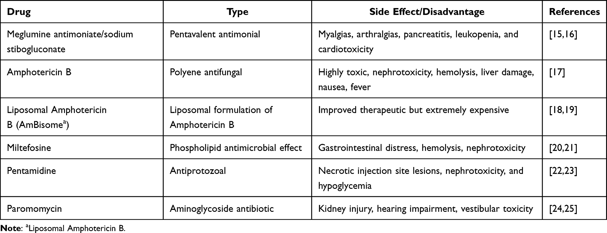 |
Table 1 Current Drugs Used in the Treatment of VL, CL, and MCL with Their Side Effects and Disadvantages |
Nanotechnology and Green Synthesis of Nanoparticles in Treatment of Leishmaniasis
In the recent years, developments in nanotechnology using nanoparticles have begun to address the limitations of existing therapeutic approaches in the treatment of leishmaniasis. Nanoparticles often refer to particles with a diameter of 10–1000 nm. In leishmaniasis, macrophages engulf the nanoparticles as foreign bodies, which results in a specific delivery system against Leishmania parasites residing inside macrophages.26,27 This targeted delivery allows for reduced dosing which could lower the potential toxicity of the administered drug.28
NPs have special features like high ratio of surface to volume, nanometers size, and deep tissue penetration which effect enhanced permeability and retention.29 The stability of NPs depends on their ability to make metastable aqueous suspensions or aerosols in environmental liquids. This affects the movement of released NP. The colloidal stability (rate of dissolution) is controlled by size and surface capping.30 Capping agents include surfactants, small ligands, polymers, dendrimers, cyclodextrins, and polysaccharides. They are essentially amphiphilic molecules with a polar head group and a non-polar hydrocarbon tail, with the polar head interacting with the metal of the metallic NP.31 Surface capping also prevents further growth and agglomeration30 while also providing electrostatic, steric, or electrosteric interactions to encourage dispersion of NP in suspensions.32,33
Metals like silver, gold, antimony, iron, zinc, selenium, and others can form metal/metal oxide nanoparticles (NP).30 Metallic nanoparticles, especially silver and antimony-based ones, have shown promise since the early 2010s in vitro and in vivo, but have limited clinical translation to date. Several studies showed that metal oxide nanoparticles show significant promise as chemotherapeutics since the current evidence indicates that they promote the generation of intracellular reactive oxygen species (ROS) which leads to parasite DNA damage, protein and lipid oxidation, and ultimately death via apoptosis.34,35
Metal NP synthesis typically occurs in one of two ways: Physical/chemical or biological. Physical/chemical synthesis is known to be costly and produce toxic by-products. These processes can include radiation, precipitations, depositions, explosions, and impregnation.36 Therefore, biological processes, ie green synthesis, are now of interest because they utilize biological starting materials, such as bacteria, fungi, algae, and plant extracts (leaves, roots, flowers, and fruits) which are more readily available components. Additionally, they are less-toxic, eco-friendly, and use a universally recognized solvent like water.37 These benefits address the concerns of both cost and toxicity.30,36,38
Each biological material provides unique characteristics that affect the formation of metallic NP. Bacteria provide distinct shapes and sizes and can be easily manipulated whereas fungi possess enzymes, proteins, and reducing agents on their cell surfaces that allow production of larger amounts of metallic NP, making them a superior biological material. Yeast offers 1500 identified species that could be further utilized in green synthesis. Plants, however, are especially unique due to the phytochemicals found in their extracts.30 These extracts can also include a broad range of additional therapeutic constituents such as triterpenoids, phenolic compounds, coumarins, alkaloids, tannins, quinines, glycosides, steroids, anthocyanins, and specific flavonoids like luteolin, all of which have been shown to possess some anti-leishmanial properties.39
Biogenic NPs, those synthesized by green methods, show significant potential in combating parasitic infections by directly targeting parasites, interfering with their cellular functions, and stimulating host immune defenses. Evidence of their effectiveness has been reported against other pathogens such as Schistosoma mansoni, Toxoplasma gondii, Echinococcus granulosus, as well as Leishmania major (L. major); primarily through mechanisms like membrane disruption, mitochondrial impairment, and induction of oxidative stress. Compared to conventional chemical synthesis, biogenic production methods offer reduced toxicity and environmental impact, positioning biogenic NPs as a sustainable and eco-friendly approach for developing novel antiparasitic treatments.40–43 Although several studies have explored chemically synthesized nanoparticles for leishmaniasis, a consolidated review focusing on biogenic/green-synthesized nanoparticles remains lacking. This review addresses this gap and analyzed relevant studies from January 1, 2010, to April 30, 2025, which encompasses a broad range of metal-based biogenic nanoparticles (antimony sulfide, silver, gold, zinc, iron, copper, selenium, nickel, cobalt, magnesium, and barium), covering both plant-derived and microorganism-derived synthesis methods. The articles were analyzed by the specific search criteria such as nanometal type, source of green synthesis starting material (plant or microorganism), size of NPs produced, if any in vivo study was done, and which Leishmania sp. and life cycle stage was used. In vitro studies also compared cytotoxicity on Leishmania parasite and host cells.
Biogenic Antimony Sulfide Nanoparticles
In many countries, pentavalent antimony (SbV) has been the drug of choice for the treatment of all clinical forms of leishmaniasis.44 Although the purely metallic form of antimony is not soluble or reactive under most physiological conditions, two forms (sodium stibogluconate and meglumine antimoniate) readily react in the body and have been used therapeutically.45,46 The mode of action of pentavalent antimonial against leishmaniasis still is not well understood.47,48 It is not even clear which form of pentavalent antimonial (Sb(V) or Sb (III)) is the activated form in humans and animals. Several studies suggest that axenic amastigotes showed Sb(V) susceptibility, whereas promastigotes did not. This suggests some life cycle stage-specific reduction of Sb(V) to Sb(III) may happen49 (Figure 2). Regardless of the exact mechanism of action, both antimony drugs are given as injections which have several side effects, and are relatively easy for Leishmania to become resistant to.44
 |
Figure 2 Proposed mechanism of antimony in Leishmania parasites. SbV is reduced to SbIII in the amastigote in order to exert antileishmanial activity. SbIII uptake can be proceeded by an aquaglycoporin (AQP1). Ornithine decarboxylase (ODC) and ɣ-glutamylcysteine synthetase (ɣ-GCS) biosynthesize spermidine and glutathione (GSH), respectively in order to make trypanothione. Conjugation of SbIII forms a trypanothione and glutathione stibogluconate complex, which results in apoptosis in amastigotes in the macrophages through two ways: 1- Binding to trypanothione reductase results in inhibition of the formation of trypanothione (T(SH)2) and makes cells susceptible to oxidative stress, 2- Binding to Zn finger protein results in inhibition of the expression of essential genes.49,50 |
The use of biogenic antimony sulfide nanoparticles may offer some advantages over injectables. Aside from the benefits of any nanoparticle mentioned above, antimony sulfide nanoparticles (Sb2S5) can be naturally synthesized in Serratia marcescens (S. marcescens) bacteria that were isolated from seawater samples of the Caspian Sea and extracted using two-solvent phase partitioning systems.51 Microorganisms can accumulate antimony as a toxic metal in their cytoplasm and other cell compartments such as the cell well.51 The Sb2S5 NPs had a size of less than 35 nm and are composed of sulfur and antimony atoms with the ratio of 84/16. During incubation or extraction time, the component may be decomposed to free sulfur groups and Sb2S351 which would be more toxic for Leishmania parasites. One study showed that BALB/c mice inoculated with L. major had significantly smaller and less populated amastigote lesions over controls (IC50 = 70 μg/mL) when treated with the Sb2S5 NPs topically.52 This may also be partly due to the fact that Sb(V) nanoparticles also demonstrated antibacterial activity against S. aureus and E. coli,51 thus allowing for a faster rate of healing in the absence of some secondary opportunistic infections.53 Also, in vitro studies showed that antimony NPs significantly reduced the number of amastigotes in the infected peritoneal macrophages after 72 h (IC50 = 62.5 μg/mL)52 but there is no report about the cytotoxicity effect in macrophages. A second study tested the effect of these same S. marcescens generated biogenic antimony sulfide NPs on a different strain of Leishmania parasites, Leishmania infantum (L. infantum), and found that both the promastigotes and amastigotes responded to treatment by inducing apoptosis (IC50 = 50 μg/mL and 25 μg/mL, respectively).44 Together, these studies indicate that biological antimony sulfide NPs could be considered as an alternative in treatment of leishmaniasis. However, it is still at early stages, and further studies on pharmacokinetics and pharmacodynamic of antimony sulfide NPs are needed.
Biogenic Silver and Gold Nanoparticles
Silver is the most used metal for medicinal nanoparticles. It has been shown to have anticarcinogenic, antimicrobial, and specifically antileishmanial effects.54–56 Silver nanoparticles (Ag NPs) are relatively small in size and round. This spherical shape seems to be correlate with higher levels of phagocytosis and an ability to pass directly through parasitic cell membranes. Ag NPs undergo redox reactions creating free Ag+ ion which can then interact with thiols or phosphates, inhibit enzymes, and generate ROS.54–60 ROS can then interfere with Leishmania through DNA fragmentation, inhibition of trypanothione synthesis, and cell cycle arrest.34
Numerous green synthesis methods have looked at the bio-fabrication of silver with extracts used to reduce silver into silver NPs. Plant secondary metabolites are thought to be responsible for the reduction, chelation, and stabilization of silver. Starting with a silver salt, the metabolites form silver ions which are then chelated into the metallic core of the plant molecule. This chelation creates an NP with longer stability.61
Awad et al tested Commiphora molmol (myrrh) to create biogenic Ag NP from silver nitrate.62 These Bio-AgNP were found to be approximately 49.06 nm and were tested against L. major both in vitro and in vivo. Results were compared to chemical synthesized NPs and Pentostam, a commercial drug. The biogenic Ag NPs (100, 150 μL/100 μL) showed significantly higher inhibition of parasites over both chemical synthesized Ag NP and drug controls. In the murine model, lesions were found to have healed completely by day 21 with the control groups preforming to a lesser extent.62
Pirestani et al used dried root powder from Zingiber officinale (ginger) to create AgNP. They then screened the NP against both L. infantum and L. tropica finding similar IC50 values of 4.54ppm and 4.22 ppm, respectively. They also reported a higher IC50 value closer to the 20–40ppm range63 via MTT assays done on Raw 264.7 cells treated with the same AgNP.
Using the aerial parts of Astragalus spinsus as a catalyst, Majeed et al created AgNP’s in the 30–40nm range. They screened these particles with or without the addition of meglumine antimoniate in L. major amastigotes as well as in J774-A1 cells. Both AgNP’s and MA alone showed similar IC50 values (59.3 µg/mL and 51.2 µg/mL respectfully) against the amastigotes but were far less effective with the J774 cells (612.5 µg/mL and 789.8 µg/mL). A possibly synergistic affect was also tested in the amastigotes with a combination of both AgNPs+MA resulting in an IC50 of 18.6 µg/mL.64
The potentially beneficial generation of ROS from silver NPs has been tested by Bilal Javed et al.61 In this study, an aqueous extract from leaves of Mentha longofolia was used to create Ag NPs. The Ag NPs were found to be between 10 and 100 nm in size with an IC50 of 8.73 µg/mL against L. tropica promastigote. Further studies showed that neither the plant aqueous extract alone nor the biogenic Ag NPs induced apoptosis in HCT116 colon cancer cells when they were exposed. Additionally, the NPs were shown to generate free radicals against Leishmania.61
Fungi have also been studied for their ability to produce biogenic Ag NPs. Fusarium oxysporium generated NPs with spherical shapes and a size around 57.6 nm. These were screened again both promastigotes and amastigotes of Leishmania amazonensis (L. amazonensis). At the 24-hour treatment time, induction of a death mechanism was observed at 0.25 and 0.5 µg/mL in promastigotes and 0.5 µg/mL in amastigotes with no toxic affects noted in peritoneal macrophages.55 In the other study, these biogenic NPs also showed 3.3-fold higher activity compared to chemically synthetized Ag NPs and a similar parasitemia inhibition in infected BALB/C mice but at a 300-fold lower concentration when compared to amphotericin B.65
Gold nanoparticles (Au NPs) have many properties that make them a useful tool in both genes and drug delivery systems.66,67 From a green synthesis standpoint, they can be prepared by employing bioactive phytochemicals from aqueous plant extracts as reducing and stabilizing agents.67 Au NPs (30–60 nm) generated from extracts of Rhazya stricta decne were shown to not only have activity against L. tropica intracellular amastigotes (IC50 = 43 mg/mL after 48 h) but also showed inhibitory effects against E. coli and bacillus subtilis.67 It was also noted that there was no observed cytotoxicity in THP-1 cells after a 24-hour treatment.67 Similarly, 4 mg/mL of Au NPs from Maytenus royleanus were screened against promastigotes of L. tropica with 75% inhibitory effect after 72-hour exposure.66
Another study reported that both Ag NPs and Au NPs nanoparticles had IC50 values of 4.37 and 5.29 μg/mL for promastigotes and amastigotes, respectively, without any notable cytotoxic effects in J774 cells under concentration of 30 μg/mL.68
Combination NP have also begun to be explored. Silver and gold bimetallic NP (Ag-Au NPs) have begun to show some promise.69 Gold−silver bimetallic nanoparticles (Au−Ag BNPs) were synthesized in other study through a single-step reduction process using fenugreek, coriander, and soybean leaf extracts.70 These Au−AgNPs demonstrated high antileishmanial effects against Leishmania donovani (L. donovani) promastigotes (IC50 = 0.03−0.035 μg/mL) which was about 300-fold lower than that of the drug miltefosine. This effect was shown to be through a ROS-mediated apoptosis-like death in the promastigotes. In addition, treatment potentiated the antileishmanial activity of macrophages, however, intracellular amastigotes numbers were reduced by only a modest 31−46%.70
The cytotoxicity of Ag NPs is usually from of the presence of a positive charge on silver ions that interacts with the negatively charged plasma cell membrane and disrupts the ionic balance and finally the membrane structure. The small size Bio-NPs also have abilities to interact with the nucleic acids such as DNA and RNA because of the presence of a negatively charged phosphate backbone. This interaction results in the destabilization of the DNA and RNA structures which affects cell proliferation and finally leads to cell death.61 Monometal NPs require higher concentrations, resulting in toxicity to host cells.70
AuNP have also been coated with curcumin as a possible combination. S. M. Amini et al synthesized nanoparticles using DMSO, tetrachloroauric acid trihydrate, and curcumin. These curcumin coated gold nanoparticles were tested against both promastigote and amastigote forms of L. major as well as J774A.1 cells and infected BALB/C mice. While various concentrations were tested, IC50s of 64.79 (24 h) and 29.89 (48 h) μg/mL in promastigotes and 63.29 (24 h) and 54.04 (48 h) μg/mL in amastigotes were reported. The combination also showed significant improvement in mouse models without negative affect on J774A.1 cells.71
All these findings suggest that biosynthesized silver and gold nanoparticles are biocompatible nanomaterials with a broad-spectrum antimicrobial activity as they have more antileishmanial effects with less cytotoxicity and could be used as a vehicle for the delivery of bioactive natural products. Table 2 summarizes the antileishmanial activity of metal nanoparticles prepared by green methods.
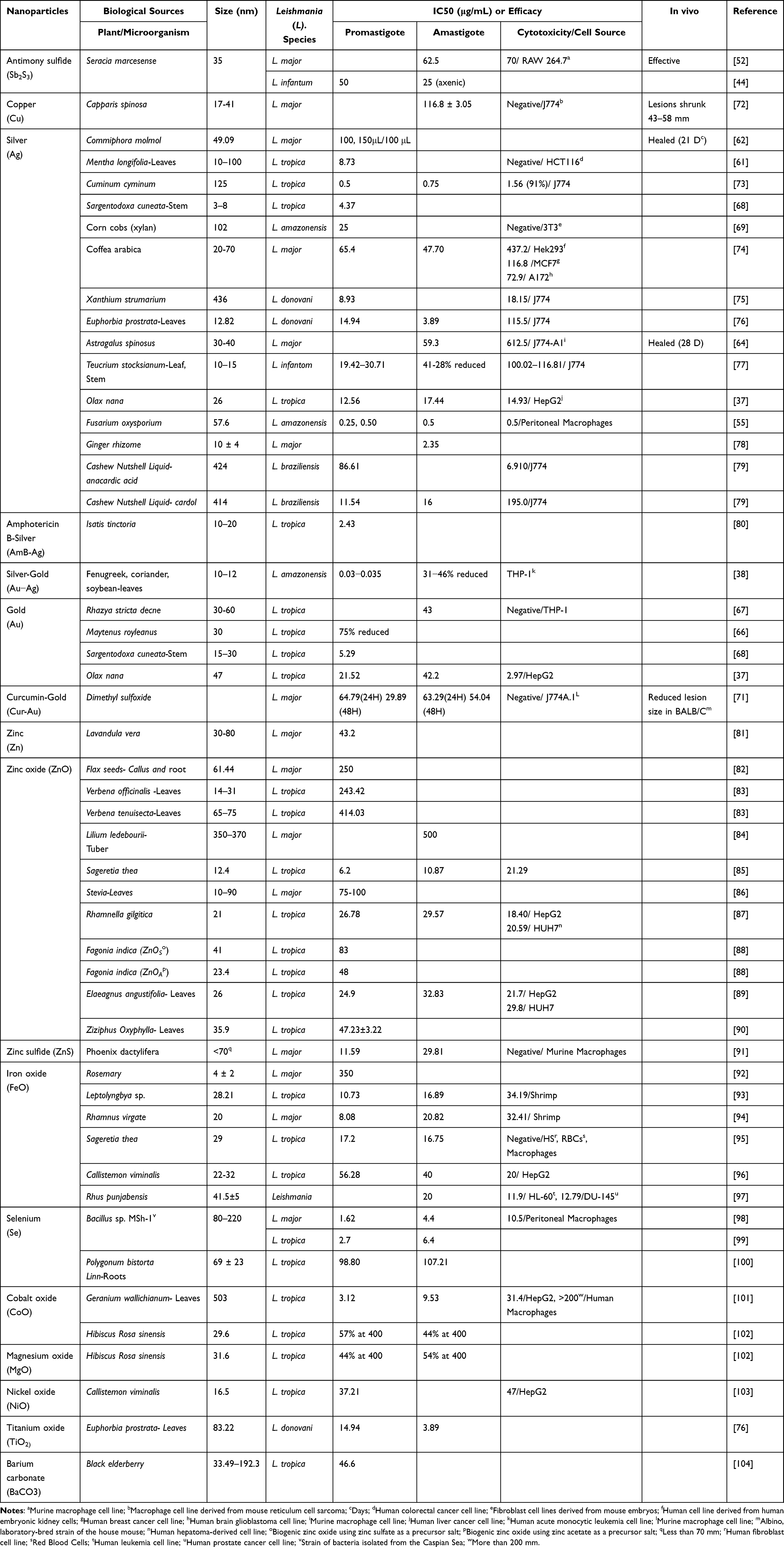 |
Table 2 Different Nanomaterial Types are Used for Treatment of Leishmaniasis Studies |
Biogenic Iron Oxide Nanoparticles
The applications of iron oxide (Fe2O3) nanoparticles (FeO NPs) in biomedicine are growing exponentially.93 While FeO NPs have a higher surface area and good stability, most methods of synthesis showed poor dispersion, a lack of uniformity in particle size and distribution,105 and are often costly because of intense energy requirements during manufacturing.106 In contrast, FeO NPs synthesized from aqueous leaf extracts showed a surface area four times bigger relative to the commercial preparations.93,105 Furthermore, biogenic FeO NPs were found to have impressive antimicrobial properties while also being nontoxic to humans compared to their chemically produced counterparts, thus suggesting higher biocompatibility.95,107–111
To test its anti-leishmaniasis effects, Mehrdad Khatami et al biosynthesized FeONPs using Rosemary.92 The results showed the fabrication of monodispersed spherical shaped FeONPs with a size 4 ± 2 nm and IC50 of 350 µg/mL against L. major promastigotes.92 Similarly, Sageretia thea (Osbeck) generated FeO NPs with a size of 29 nm which were tested against promastigote and amastigote forms of L. tropica (IC50 = 17.2 and 16.75 µg/mL respectively) and showed no cytotoxicity for human red blood cells (RBCs) or macrophages.95 The authors disclosed that these particles are also effective agents against bacterial strains and had some degree of antioxidant activities.95,107–113 Ahsan Abbasi et al biosynthesized FeO NPs using leaves extract of Rhamnus virgate.94 FeO NPs with a crystallite size of ~20 nm were seen to be effective agents in inhibiting the promastigote and amastigote forms of L. major after 72 h with an additional anticancer, antibacterial, and antifungal activity. Cytotoxic and potentially significant antioxidant activities were also reported.94 Minhas et al used L-2 extracts of Cyanobacterium Leptolyngbya sp. to synthesize FEONP’s from iron chloride hexahydrate. They were screened against both promastigotes and amastigotes from L. tropica (IC50 = 10.73 and 16.98 µg/mL respectively).114
Clear variations in the antileishmanial efficacies of the iron-based particles could be linked to the phytochemical constituents that were used to stabilize the NPs. The surface-capped biomolecules in biogenic metal-based particles also play an important role in the antimicrobial property of the prepared materials.34
Biogenic Zinc Oxide and Zinc Sulfide Nanoparticles
Zinc oxide nanoparticles (ZnO NPs) are novel antimicrobial and antileishmanial agents showing promise due to their high surface area to volume ratio and ability to generate ROS inside cells.82 ZnO NPs can cause DNA, lipid and protein damages in the Leishmania species.34 For synthesis of ZnO NPs, numerous physical and chemical approaches have been reported with most of these approaches showing similar drawbacks to other nanoparticles.82,115 For green biosynthesis, leaf extracts have been used.83 Recently, Lilium ledebourii tuber extract was used with both rod-shaped and spherical ZnO NPs being explored. IC50 about 0.012 mg/mL of biogenic ZnO NPs revealed a higher toxicity effect on L. major intracellular amastigotes compared to Meglumine antimoniate (glucantime) as positive control.84 Another study compared leaf extracts of Verbena officinalis (V. officinalis) and Verbena tenuisecta (V. tenuisecta) in the production of biogenic ZnO NPs. Both rod and flower shaped ZnO NPs were found but V. officinalis had a smaller size and increased phenolics, leading to more potent leishmanicidal activity (IC50 = 250 μg/mL) against L. tropica promastigotes.83
Zinc sulphide nanoparticles (ZnS NPs) were prepared using a similar green approach. ZnSNPs with a size less than 70 nm showed no cytotoxicity when tested against murine macrophages but showed leishmanicidal activity with the IC50 values of 29.81 and 11.59 μg/mL against L. major promastigotes and amastigotes, respectively. This result is about a 2–2.5-fold stronger effect when compared to the glucantime control.91
Overall, owing to their small size and electrostatic interactions, Zn NPs enter into leishmanial cells and lower the parasite’s metabolic activity and proliferation values. They also induce intracellular depletion via ROS production which in turn causes cell membrane disruption, cytoplasmic leakage, and finally protozoa death.83
Biogenic Selenium Nanoparticles
Selenium (Se) is an essential micro-mineral element whose functions include regulating metabolism, improving immunity, enhancing reproductive performance, and preventing cancer.116 Biogenic Selenium NPs (Se NPs) have been shown to have lower toxicity than selenium oxyanions,117–119 and there is some evidence that Se NPs have successful antibacterial and antiparasitic effects.42,120,121 Se NPs generated by Shakibaie et al using Bacillus sp. MSh-1, were spherical in shape and 80–220 nm in size.122 They were tested against L. major and antileishmanial effects were shown after 72 hours with an IC50 against promastigotes (1.62 ± 0.6 μg/mL) and amastigotes (4.4 ± 0.6 μg/mL) vs peritoneal macrophages which was 10.5 ± 0.6 μg/mL. Se NPs also delayed the development of localized cutaneous lesions in a mouse model.98
Mahmoudvand et al examined SeNPs, separately or in combination with meglumine antimonate, against sensitive and drug resistant L. tropica.123 They reported that the combination of drugs had higher efficiency against promastigotes of both sensitive and resistant strains (IC50 = 1.5 and 2.8 μg/mL, respectively) and significantly reduced proliferation of amastigotes in the macrophages at a concentration of more than 2.5 μg/mL. Pre-incubation of macrophages with SeNPs significantly reduced the infectivity of macrophages by parasites.123 In general, Se NPs were effective against cutaneous leishmaniasis121 and they have less toxicity against mammalian macrophages, but more studies in animal models are needed.
Other Biogenic Nanoparticles
Copper nanoparticles (Cu NPs), likely due to their high surface-to-volume ratio, are very reactive and interact with other particles.124,125 Cu NPs synthesized by Capparis spinosa fruit methanolic extract had a spherical shape, and a particle size of 17 to 41 nm.72 A combination therapy of Cu NPs and glucantime was found to significantly inhibit the growth rate of L. major intracellular amastigotes and triggered the production of nitric oxide (NO) in a dose-dependent manner. NO is considered to be one of the main mediators of immunity produced in macrophages, which plays a critical role in the control of Leishmania parasites.126 Also, this combination reduced amastigotes in the L. major infected mice while having no significant cytotoxicity in J774 cells.126 Previous studies have shown Cu NPs have potent antiparasitic effects against other protozoan parasites such Giardia lamblia cysts, Entamoeba histolytica, and Cryptosporidum parvum.127,128 Antimicrobial studies suggest that Cu NPs destroy proteins in bacteria via interaction with thiol groups129 as well as cause degradation of DNA, lipid peroxidation, and production of ROS, causing cell wall destruction and death.130
Nickel oxide NPs (NiO NPs) have a unique nature and interesting properties like stability, conductance, catalysis, electron transferability and wider band gap (3.7–4.0 meV). Their cytotoxicity has been previously demonstrated via ROS and Ni++ ions which can lead to the oxidative damage.131–133 Biogenic NiO NPs recently synthesized by Ayesha Sani et al using floral extracts of Callistemon viminalis (C. viminalis) have spherical morphology are 16.5 nm.103 They reported that NiO NPs had anti-leishmanial activity on L. tropica promastigotes (IC50 = 37.21 µg/mL) and additionally anticancer and antibacterial activity. Overall good antioxidant nature and biocompatibility were revealed.103
Cobalt based NPs (Co NPs) have gained the attention of the scientific community because of their ecofriendly nature, easy handling, low cost and strong electro potential.134 Co NPs have already been shown to have a significant role as a cofactor in vitamin B12 and have been effectively used in sensing of amino acids, nitrates, glucose, arsenic and methanol.134 Biogenic Co NPs were created using aqueous leaf extract of Geranium wallichianum Oliv. and have shown a dose-dependent cytotoxicity against Leishmania promastigotes (IC50 = 3.12 µg/mL) and amastigotes (IC50 = 9.53 µg/mL) with additional anticancer, antibacterial, and antifungal activity being reported.101 Cobalt oxide particles (Co3O4-NPs) have also been generated using aqueous extracts of Hibiscus Rosa sinensis.102 These NP (29.6–31.6nm) showed activity against both forms of L. tropica (57.32% at 400 ug/mL for promastigotes and 48% at 400 ug/mL for amastigotes).102
Nanocomposites built of different metal nanoparticles have also been explored. Amini et al used ethanol extracts from the seaweed Laurencia dendroidea to create AgNP and CuONP but also polymeric PVP-Ag-CuO nanocomposite. These were screened against L. amazonensis promastigotes to show that the PVP-Ag-CuO nanocomposite was the most effective (IC50 = 17.48 µg/mL) followed closely by the AnNPs with a IC50 of 18.75 µg/mL. The CuONPs were found to be much less active with an IC50 of greater than 25 µg/mL.135
Pharmacokinetics (PK)
While green synthesis and nanoparticle usage are the central focus of this paper, there is the equally important question of NP safety. Other manuscripts have sought out the ADME properties of metal-based nanoparticles. These papers, while currently extremely limited, suggest that NP made of silver, gold, or zinc, tend to show that the specific route of administration has a significant effect on absorbance and distribution of the metallic particles, with toxicological effects like that which would be expected from those metals.136 At the time of this writing, no pharmacokinetic data was available relating for NP made using selenium, cobalt, nickel, titanium, or barium. Some PK data is noted for antimony, but it is not specific to nanoparticle formulations. This limitation is one that will need to be addressed in the future.
Equally important to note in the future will be the need to understand not just how metallic ions from nanoparticles are expelled from the body, but what impacts their synthesis, even being green, would have. Many of the current methods of green NP synthesis rely on the use of weeds, grasses, moss, or other biologic material. On the small scale, much of this is starting material is readily available and often viewed as a nuisance. Should any of these materials prove to be a truly beneficial treatment, then the harvesting of these materials on a large scale could cause shifts within whatever ecosystems they exist. Work will need to be done to ensure that the effects of harvesting these biologic materials are offset by other grown.
Current Limitations and Future Directions of Green Synthesis of NPs
Green synthesis of biogenic nanoparticles is an emerging field with a lot of potential for growth, but as the field itself is fairly new, there are significant gaps in knowledge that will need to be addressed in the near future. To date, most reporting surrounding the green synthesis of NPs is limited to describing how a synthesis was done with almost no comment on the actual chemical pathways for generation. Additionally, little is currently known or speculated on regarding the transition from lab to industrial scale productions such as maintaining consistent reaction conditions (including uniform temperature, concentration, and pH), batch-to-batch variation, and economic and sourcing challenges as certain plant species used for extracts may only be available during specific flowering or harvesting periods. With how complex some of the preparations were (everything from brewing roots to long term fermentations and sun exposure), there is a distinct lack of explanation for how the researcher came to these methods. Based on the literature that there are many avenues of improvement and refinement such as scalability, reproducibility, safety, and the desired properties of the final product. As reported, different starting materials do show differences in the size, shape, and relative abundance of NP produced. Better understanding of the actual chemical processes occurring during synthesis would allow for optimization and ideal selections of starting materials based on the desired outcomes.
In its current form, green synthesis being done on a laboratory scale is relatively easy to deal with from the production side. This situation would drastically change once the production was scaled to a larger, more industrial size. The production of large bioreactors running green synthesis would require larger amounts of starting materials and biological waste that could no longer be supported by the gathering methods used in a research setting. Once better understood, the possibility of resource sharing or recycling with other production sectors could become a reality. The use of bioreactors, particularly in the case of microorganisms, could also post safety risks as they are concentrated population of what are otherwise benign organisms.
All in all, green synthesized NP hold great promise as a new synthesizing process which is not only more environmentally but potentially more customizable and targeted treatment approach. All the above-mentioned areas will still need to be explored before these systems could really move into complex biological models or preclinical trials. At that point, better regulations and globally driven benchmarking would also still need to be developed in order to more completely understandings of production chemistry, parasite biology, and ecological impacts surrounding biogenic NPs use in the treatment of Leishmania infections.
Conclusion
The use of nanoparticles has continuously increased due to their having a smaller size and larger surface area to volume ratio and can be more efficiently taken up by the infected macrophages than free drugs. This may make nanoparticles a promising and novel treatment option for leishmaniasis. Biogenic synthesis of nanoparticles relies on plant extracts or microorganisms rather than traditional chemicals thus reducing many of the hazardous and ecological impacts associated with nanoparticle products and waste. This method increases accessibility of the drugs and reduces the cost. Treatment of Leishmania is already difficult because of their genetic plasticity, ease of treatment resistance development, and a general lack of understanding of all the ways they evade host immune responses. Biological materials show promise for the treatment of leishmaniasis, especially plant extracts because they are easily accessible and generally safe. These nanoparticles can be incorporated into treatment as a monotherapy or in multifaceted therapy. Therefore, investing in green synthesis of NPs may help resolve treatment efficacy, accessibility, and cost, especially in low- and middle-income areas of the world. Different biogenic nanoparticles had been preliminary evaluated on Leishmania parasites such as antimony sulfide, silver, gold, zinc, iron, copper, selenium, nickel, and cobalt. Because of the previous known antimicrobial activity of silver, most studies on antileishmanial activity have been performed with silver nanoparticles. Among biogenic NPs, silver and selenium NPs demonstrated the most potent antileishmanial activity with low host cytotoxicity, while antimony sulfide NPs showed promise in early in vivo models. Although the mode of action of nanoparticles is complex, several studies suggest that they induced apoptosis in parasites by generation of intracellular ROS which leads to Leishmania DNA damage, protein and lipid oxidation. There was only minimal and inconsistent reporting on the process for determining EC50 values, not enough reporting on the mechanism of actions or types of cell death, and no work on understanding biodistribution of the NP. Once understood, NP could be created with additional features or to specific criteria that would allow for targeting of infected macrophages and parasites with no off-target effects that could outweigh their treatment potential. Moreover, current studies are in the early phases of testing, focused more on cutaneous strains with and animal models showing less progression toward clinical trials. In addition, more studies on visceralizing strains are needed. Furthermore, methods of green synthesis of nanoparticles need to be optimized by standardization of choice of plant parts, growth and purification requirements, and optimizing microorganisms’ choice by avoiding unintended cytotoxicity and secondary infections. Moreover, potential drawbacks-such as scalability, batch variability, environmental cost of green synthesis, or safety in long-term exposure need to be studied.
Data Sharing Statement
All data generated or analyzed in the current study are included in the published article. Any other information is available from the corresponding author on reasonable request.
Acknowledgments
The authors would like to thank Alireza Borjian Boroujeni and Mitra Eimanzadeh for their collaboration. The authors are pleased to use Canva.com and Servier Medical Art (https://smart.servier.com/), licensed under CC BY 4.0 (https://creativecommons.org/licenses/by/4.0/) for designing some figures.
Funding
There is no funding to report.
Disclosure
The authors declare that they have no known competing financial interests or personal relationships that could have appeared to influence the work reported in this article.
References
1. Alvar J, Vélez ID, Bern C, et al. Leishmaniasis worldwide and global estimates of its incidence. PLoS One. 2012;7(5):e35671. doi:10.1371/journal.pone.0035671
2. Guery R, Walker SL, Harms G, et al. Clinical diversity and treatment results in tegumentary Leishmaniasis: a European clinical report in 459 patients. PLoS Negl Trop Dis. 2021;15(10):e0009863. doi:10.1371/journal.pntd.0009863
3. Serafim TD, Coutinho-Abreu IV, Dey R, et al. Leishmaniasis: the act of transmission. Trends Parasitol. 2021;37(11):976–987. doi:10.1016/j.pt.2021.07.003
4. Ngere I, Gufu Boru W, Isack A, et al. Burden and risk factors of cutaneous leishmaniasis in a peri-urban settlement in Kenya, 2016. PLoS One. 2020;15(1):e0227697. doi:10.1371/journal.pone.0227697
5. Georgiadou SP, Makaritsis KP, Dalekos GN. Leishmaniasis revisited: current aspects on epidemiology, diagnosis and treatment. J Transl Int Med. 2015;3(2):43–50. doi:10.1515/jtim-2015-0002
6. Gossage SM, Rogers ME, Bates PA. Two separate growth phases during the development of Leishmania in sand flies: implications for understanding the life cycle. Int J Parasitol. 2003;33(10):1027–1034. doi:10.1016/s0020-7519(03)00142-5
7. Bhattacharya P, Bhattacharjee S, Gupta G, et al. Arabinosylated lipoarabinomannan-mediated protection in visceral leishmaniasis through up-regulation of toll-like receptor 2 signaling: an immunoprophylactic approach. J Infect Dis. 2010;202(1):145–155. doi:10.1086/653210
8. Gupta G, Majumdar S, Adhikari A, et al. Treatment with IP-10 induces host-protective immune response by regulating the T regulatory cell functioning in Leishmania donovani-infected mice. Med Microbiol Immunol. 2011;200(4). doi:10.1007/s00430-011-0197-y
9. Wilson J, Huynh C, Kennedy KA, et al. Control of parasitophorous vacuole expansion by LYST/Beige restricts the intracellular growth of Leishmania amazonensis. PLoS Pathog. 2008;4(10):e1000179. doi:10.1371/journal.ppat.1000179
10. Gupta G, Oghumu S, Satoskar AR. Mechanisms of immune evasion in leishmaniasis. Adv Appl Microbiol. 2013;82:155–184. doi:10.1016/B978-0-12-407679-2.00005-3
11. Malvolti S, Malhame M, Mantel CF, Le Rutte EA, Kaye PM. Human leishmaniasis vaccines: use cases, target population and potential global demand. PLoS Negl Trop Dis. 2021;15(9):e0009742. doi:10.1371/journal.pntd.0009742
12. Sasidharan S, Saudagar P. Leishmaniasis: where are we and where are we heading? Parasitol Res. 2021;120(5):1541–1554. doi:10.1007/s00436-021-07139-2
13. Maltezou HC. Visceral leishmaniasis: advances in treatment. Recent Pat Antiinfect Drug Discov. 2008;3(3):192–198. doi:10.2174/157489108786242341
14. Zhao S, Sun W, Sun J, Peng L, Wang C. Clinical features, treatment, and outcomes of nivolumab induced psoriasis. Invest New Drugs. 2025;43(1):42–49. doi:10.1007/s10637-024-01494-4
15. López L, Robayo M, Vargas M, Vélez ID. Thermotherapy. An alternative for the treatment of American cutaneous leishmaniasis. Trials. 2012;13:58. doi:10.1186/1745-6215-13-58
16. González U, Pinart M, Rengifo-Pardo M, Macaya A, Alvar J, Tweed JA. Interventions for American cutaneous and mucocutaneous leishmaniasis. Cochrane Database Syst Rev. 2009;(2):CD004834. doi:10.1002/14651858.CD004834.pub2
17. Ribeiro TG, Franca JR, Fuscaldi LL, et al. An optimized nanoparticle delivery system based on chitosan and chondroitin sulfate molecules reduces the toxicity of amphotericin B and is effective in treating tegumentary leishmaniasis. Int J Nanomed. 2014;9:5341–5353. doi:10.2147/IJN.S68966
18. Bern C, Adler-Moore J, Berenguer J, et al. Liposomal amphotericin B for the treatment of visceral leishmaniasis. Clin Infect Dis. 2006;43(7):917–924. doi:10.1086/507530
19. Egger SS, Meier S, Leu C, et al. Drug interactions and adverse events associated with antimycotic drugs used for invasive aspergillosis in hematopoietic SCT. Bone Marrow Transplant. 2010;45(7):1197–1203. doi:10.1038/bmt.2009.325
20. Sundar S, Olliaro PL. Miltefosine in the treatment of leishmaniasis: clinical evidence for informed clinical risk management. Ther Clin Risk Manag. 2007;3(5):733–740.
21. Jamshaid H, ud DF, Khan GM. Nanotechnology based solutions for anti-leishmanial impediments: a detailed insight. J Nanobiotechnol. 2021;19(1):106. doi:10.1186/s12951-021-00853-0
22. Omarch G, Kippie Y, Mentor S, et al. Comparative in vitro transportation of pentamidine across the blood-brain barrier using polycaprolactone nanoparticles and phosphatidylcholine liposomes. Artif Cells Nanomed Biotechnol. 2019;47(1):1428–1436. doi:10.1080/21691401.2019.1596923
23. O’Brien JG, Dong BJ, Coleman RL, Gee L, Balano KB. A 5-year retrospective review of adverse drug reactions and their risk factors in human immunodeficiency virus—infected patients who were receiving intravenous pentamidine therapy for Pneumocystis carinii Pneumonia. Clinl Infect Dis. 1997;24(5):854–859. doi:10.1093/clinids/24.5.854
24. Wiwanitkit V. Interest in paromomycin for the treatment of visceral leishmaniasis (kala-azar). Ther Clin Risk Manag. 2012;8:323–328. doi:10.2147/TCRM.S30139
25. Prayle A, Watson A, Fortnum H, Smyth A. Side effects of aminoglycosides on the kidney, ear and balance in cystic fibrosis. Thorax. 2010;65(7):654–658. doi:10.1136/thx.2009.131532
26. Müller RH, Jacobs C, Kayser O. Nanosuspensions as particulate drug formulations in therapy: rationale for development and what we can expect for the future. Adv Drug Delivery Rev. 2001;47(1):3–19. doi:10.1016/S0169-409X(00)00118-6
27. Kumar R, Singh N, Gautam S, et al. Leishmania specific CD4 T cells release IFNγ that limits parasite replication in patients with visceral Leishmaniasis. PLoS Negl Trop Dis. 2014;8(10):e3198. doi:10.1371/journal.pntd.0003198
28. Gustafson HH, Holt-Casper D, Grainger DW, Ghandehari H. Nanoparticle uptake: the phagocyte problem. Nano Today. 2015;10(4):487–510. doi:10.1016/j.nantod.2015.06.006
29. Gavas S, Quazi S, Karpiński TM. Nanoparticles for cancer therapy: current progress and challenges. Nanoscale Res Lett. 2021;16:173. doi:10.1186/s11671-021-03628-6
30. Singh J, Dutta T, Kim KH, Rawat M, Samddar P, Kumar P. ‘Green’ synthesis of metals and their oxide nanoparticles: applications for environmental remediation. J Nanobiotechnol. 2018;16(1):84. doi:10.1186/s12951-018-0408-4
31. Javed R, Zia M, Naz S, Aisida SO, Ain NU, Ao Q. Role of capping agents in the application of nanoparticles in biomedicine and environmental remediation: recent trends and future prospects. J Nanobiotechnol. 2020;18(1):172. doi:10.1186/s12951-020-00704-4
32. Restrepo CV, Villa CC. Synthesis of silver nanoparticles, influence of capping agents, and dependence on size and shape: a review. Environ Nanotechnol Monit Manage. 2021;15:100428. doi:10.1016/j.enmm.2021.100428
33. Li CC, Chang SJ, Su FJ, Lin SW, Chou YC. Effects of capping agents on the dispersion of silver nanoparticles. Colloids Surf A. 2013;419:209–215. doi:10.1016/j.colsurfa.2012.11.077
34. Ahmad A, Ullah S, Syed F, Tahir K, Khan AU, Yuan Q. Biogenic metal nanoparticles as a potential class of antileishmanial agents: mechanisms and molecular targets. Nanomedicine. 2020;15(8):809–828. doi:10.2217/nnm-2019-0413
35. Allahverdiyev AM, Abamor ES, Bagirova M, et al. Antileishmanial effect of silver nanoparticles and their enhanced antiparasitic activity under ultraviolet light. Int J Nanomed. 2011;6:2705–2714. doi:10.2147/IJN.S23883
36. Islam NU, Amin R, Shahid M, Amin M. Gummy gold and silver nanoparticles of apricot (Prunus armeniaca) confer high stability and biological activity. Arabian J Chem. 2019;12(8):3977–3992. doi:10.1016/j.arabjc.2016.02.017
37. Ovais M, Khalil AT, Raza A, et al. Multifunctional theranostic applications of biocompatible green-synthesized colloidal nanoparticles. Appl Microbiol Biotechnol. 2018;102(10):4393–4408. doi:10.1007/s00253-018-8928-2
38. Alti D, Veeramohan Rao M, Rao DN, Maurya R, Kalangi SK. Gold-Silver Bimetallic Nanoparticles Reduced with Herbal Leaf Extracts Induce ROS-Mediated Death in Both Promastigote and Amastigote Stages of Leishmania donovani. ACS Omega. 2020;5(26):16238–16245. doi:10.1021/acsomega.0c02032
39. Shah NA, Khan MR, Nadhman A. Antileishmanial, toxicity, and phytochemical evaluation of medicinal plants collected from Pakistan. Biomed Res Int. 2014;2014:384204. doi:10.1155/2014/384204
40. Detoni MB, da Silva Bortoleti BT, Tomiotto-Pellissier F, et al. Biogenic silver nanoparticle exhibits schistosomicidal activity in vitro and reduces the parasitic burden in experimental schistosomiasis mansoni. Microb Infect. 2023;25(7):105145. doi:10.1016/j.micinf.2023.105145
41. Arruda da da Silva Sanfelice R, Silva TF, Tomiotto-Pellissier F, et al. Biogenic silver nanoparticles reduce Toxoplasma gondii infection and proliferation in RAW 264.7 macrophages by inducing tumor necrosis factor-alpha and reactive oxygen species production in the cells. Microb Infect. 2022;24(5):104971. doi:10.1016/j.micinf.2022.104971
42. Mahmoudvand H, Fasihi Harandi M, Shakibaie M, et al. Scolicidal effects of biogenic selenium nanoparticles against protoscolices of hydatid cysts. Int J Surg. 2014;12(5):399–403. doi:10.1016/j.ijsu.2014.03.017
43. Mahmoud EHM, Abdulsalam L, Moustafa EH, Alhanbali IA, Ullah S, Ahmad I. Anti-protozoan applications of the biogenic nanoparticles and their mechanism of action. In: Khalil AT, Islam A editors. Expanding Nanobiotechnology. CRC Press; 2025:241–284. doi:10.1201/9781003378563-12
44. Soflaei S, Dalimi A, Ghaffarifar F, Shakibaie M, Shahverdi AR, Shafiepour M. In vitro antiparasitic and apoptotic effects of antimony sulfide nanoparticles on Leishmania infantum. J Parasitol Res. 2012;2012:756568. doi:10.1155/2012/756568
45. Filella M, Belzile N, Chen YW. Antimony in the environment: a review focused on natural waters: II. Relevant solution chemistry. Earth Sci Rev. 2002;59(1–4):265–285. doi:10.1016/S0012-8252(02)00089-2
46. Filella M, Belzile N, Chen YW. Antimony in the environment: a review focused on natural waters: I. Occurrence. Earth-Sci Rev. 2002;57(1–2):125–176. doi:10.1016/S0012-8252(01)00070-8
47. Duffin J, René P. “anti–moine; anti–biotique”: the public fortunes of the secret properties of antimony potassium tartrate (Tartar Emetic). J Hist Med Allied Sci. 1991;46(4):440–456. doi:10.1093/jhmas/46.4.440
48. Estes JW. The Medical Skills of Ancient Egypt. Science History Publications/USA; 1993.
49. Croft SL, Sundar S, Fairlamb AH. Drug resistance in leishmaniasis. Clin Microbiol Rev. 2006;19(1):111–126. doi:10.1128/CMR.19.1.111-126.2006
50. Frézard F, Demicheli C, Ribeiro RR. Pentavalent antimonials: new perspectives for old drugs. Molecules. 2009;14(7):2317–2336. doi:10.3390/molecules14072317
51. Bahrami K, Nazari P, Sepehrizadeh Z, Zarea B, Shahverdi AR. Microbial synthesis of antimony sulfide nanoparticles and their characterization. Ann Microbiol. 2012;62(4):1419–1425. doi:10.1007/s13213-011-0392-5
52. Mohtasebi S, Mohebali M, Elikaee S, et al. In vitro and in vivo anti-parasitic activity of biogenic antimony sulfide nanoparticles on Leishmania major (MRHO/IR/75/ER). Parasitol Res. 2019;118(9):2669–2678. doi:10.1007/s00436-019-06382-y
53. Mohebali M, Rezayat MM, Gilani K, et al. Nanosilver in the treatment of localized cutaneous leishmaniasis caused by Leishmania major (MRHO/IR/75/ER): an in vitro and in vivo study. DARU J Pharma Sci. 2015;17(4):285–289.
54. Behra R, Sigg L, Clift MJ, et al. Bioavailability of silver nanoparticles and ions: from a chemical and biochemical perspective. J Royal Soc Interface. 2013;10(87):20130396. doi:10.1098/rsif.2013.0396
55. Fanti JR, Tomiotto-Pellissier F, Miranda-Sapla MM, et al. Biogenic silver nanoparticles inducing Leishmania amazonensis promastigote and amastigote death in vitro. Acta Trop. 2018;178:46–54.
56. Chen M, von Mikecz A. Formation of nucleoplasmic protein aggregates impairs nuclear function in response to SiO2 nanoparticles. Exp Cell Res. 2005;305(1):51–62. doi:10.1016/j.yexcr.2004.12.021
57. Durán N, Durán M, De Jesus MB, Seabra AB, Fávaro WJ, Nakazato G. Silver nanoparticles: a new view on mechanistic aspects on antimicrobial activity. Nanomed Nanotechnol Biol Med. 2016;12(3):789–799.
58. Geiser M, Rothen-Rutishauser B, Kapp N, et al. Ultrafine particles cross cellular membranes by nonphagocytic mechanisms in lungs and in cultured cells. Environ Health Perspect. 2005;113(11):1555–1560. doi:10.1289/ehp.8006
59. Chithrani BD, Ghazani AA, Chan WC. Determining the size and shape dependence of gold nanoparticle uptake into mammalian cells. Nano Lett. 2006;6(4):662–668. doi:10.1021/nl052396o
60. Long TC, Saleh N, Tilton RD, Lowry GV, Veronesi B. Titanium dioxide (P25) produces reactive oxygen species in immortalized brain microglia (BV2): implications for nanoparticle neurotoxicity. Environ Sci Technol. 2006;40(14):4346–4352. doi:10.1021/es060589n
61. Javed B, Mashwani ZUR, Sarwer A, Raja NI, Nadhman A. Synergistic response of physicochemical reaction parameters on biogenesis of silver nanoparticles and their action against colon cancer and leishmanial cells. Artif Cells Nanomed Biotechnol. 2020;48(1):1340–1353. doi:10.1080/21691401.2020.1850467
62. Awad MA, Al Olayan EM, Siddiqui MI, Merghani NM, Alsaif SSAL, Aloufi AS. Antileishmanial effect of silver nanoparticles: green synthesis, characterization, in vivo and in vitro assessment. Biomed Pharmacother. 2021;137:111294. doi:10.1016/j.biopha.2021.111294
63. KarimipourI Saryazdi A, Dalimi A, Sadeghi SH, Moghadamizad Z, Pirestani M. Effects of silver nanoparticle based on ginger extract on Leishmania infantum and Leishmania tropica parasites: in vitro. Arch Razi Inst. 2024;79(2):335–344. doi:10.32592/ARI.2024.79.2.335
64. Majeed QA, Shater AF, Alanazi AD. Green synthesis, characterization, and antileishmanial activity of the silver nanoparticles alone and along with meglumine antimoniate against Leishmania major Infection. Iran J Parasitol. 2023;18(4):535–545. doi:10.18502/ijpa.v18i4.14262
65. Rossi-Bergmann B, Pacienza-Lima W, Marcato PD, De Conti R, Durán N. Therapeutic potential of biogenic silver nanoparticles in murine cutaneous leishmaniasis. J Nano Res. 2012;20:89–97.
66. Ahmad A, Syed F, Imran M, et al. Phytosynthesis and antileishmanial activity of gold nanoparticles by M aytenus Royleanus. J Food Biochem. 2016;40(4):420–427. doi:10.1111/jfbc.12232
67. Ahmad A, Wei Y, Ullah S, et al. Synthesis of phytochemicals-stabilized gold nanoparticles and their biological activities against bacteria and Leishmania. Microb Pathog. 2017;110:304–312. doi:10.1016/j.micpath.2017.07.009
68. Ahmad A, Syed F, Shah A, et al. Silver and gold nanoparticles from Sargentodoxa cuneata: synthesis, characterization and antileishmanial activity. RSC Adv. 2015;5(90):73793–73806.
69. Silva Viana RL, Pereira Fidelis G, Jane Campos Medeiros M, et al. Green synthesis of antileishmanial and antifungal silver nanoparticles using corn cob xylan as a reducing and stabilizing agent. Biomolecules. 2020;10(9):1235. doi:10.3390/biom10091235
70. Alti D, Veeramohan Rao M, Rao DN, Maurya R, Kalangi SK. Gold-silver bimetallic nanoparticles reduced with herbal leaf extracts induce ROS-mediated death in both promastigote and amastigote stages of Leishmania donovani. ACS Omega. 2020;5(26):16238–16245. doi:10.1021/acsomega.0c02032
71. Amini SM, Hadighi R, Najm M, et al. The therapeutic effects of curcumin-coated gold nanoparticle against Leishmania Major causative agent of Zoonotic Cutaneous Leishmaniasis (ZCL): an In vitro and in vivo study. Curr Microbiol. 2023;80(4):104. doi:10.1007/s00284-022-03172-1
72. Albalawi AE, Abdel-Shafy S, Khudair Khalaf A, et al. Therapeutic potential of green synthesized copper nanoparticles alone or combined with meglumine antimoniate (Glucantime®) in cutaneous leishmaniasis. Nanomaterials. 2021;11(4):891. doi:10.3390/nano11040891
73. Bagirova M, Dinparvar S, Allahverdiyev AM, Unal K, Abamor ES, Novruzova M. Investigation of antileshmanial activities of Cuminum cyminum based green silver nanoparticles on L. tropica promastigotes and amastigotes in vitro. Acta Trop. 2020;208:105498. doi:10.1016/j.actatropica.2020.105498
74. Sharifi F, Mohamadi N, Tavakoli Oliaee R, et al. The potential effect of silver nanoparticles synthesized with Coffea arabica green seeds on Leishmania major proliferation, cytotoxicity activity, and cytokines expression level. J Parasit Dis. 2023;47(1):131–139. doi:10.1007/s12639-022-01549-4
75. Kumar V, Gundampati RK, Singh DK, Jagannadham MV, Sundar S, Hasan SH. Photo-induced rapid biosynthesis of silver nanoparticle using aqueous extract of Xanthium strumarium and its antibacterial and antileishmanial activity. J Ind Eng Chem. 2016;37:224–236. doi:10.1016/j.jiec.2016.03.032
76. Zahir AA, Chauhan IS, Bagavan A, et al. Green synthesis of silver and titanium dioxide nanoparticles using euphorbia prostrata extract shows shift from apoptosis to G0/G1 arrest followed by necrotic cell death in Leishmania donovani. Antimicrob Agents Chemother. 2015;59(8):4782–4799. doi:10.1128/AAC.00098-15
77. Ullah I, Cosar G, Abamor ES, Bagirova M, Shinwari ZK, Allahverdiyev AM. Comparative study on the antileishmanial activities of chemically and biologically synthesized silver nanoparticles (AgNPs). 3 Biotech. 2018;8(2). doi:10.1007/s13205-018-1121-6
78. Mohammadi M, Zaki L, KarimiPourSaryazdi A, et al. Efficacy of green synthesized silver nanoparticles via ginger rhizome extract against Leishmania major in vitro. PLoS One. 2021;16(8):e0255571. doi:10.1371/journal.pone.0255571
79. Teixeira Bezerra T, Oliveira de Almeida M, Maria de Amorim Lima N, et al. In vitro antileishmanial activity of sustainable anacardic acid and cardol based silver nanoparticles on L. braziliensis. Int J Pharm. 2022;619:121698. doi:10.1016/j.ijpharm.2022.121698
80. Ahmad A, Wei Y, Syed F, et al. Isatis tinctoria mediated synthesis of amphotericin B-bound silver nanoparticles with enhanced photoinduced antileishmanial activity: a novel green approach. J Photochem Photobiol B. 2016;161:17–24. doi:10.1016/j.jphotobiol.2016.05.003
81. Ghasemian Yadegari J, Khudair Khalaf A, Ezzatkhah F, Shakibaie M, Mohammadi HR, Mahmoudvand H. Antileishmanial, cellular mechanisms, and cytotoxic effects of green synthesized zinc nanoparticles alone and in combined with glucantime against Leishmania major infection. Biomed Pharmacother. 2023;164:114984. doi:10.1016/j.biopha.2023.114984
82. Abbasi BH, Anjum S, Hano C. Differential effects of in vitro cultures of Linum usitatissimum L. (Flax) on biosynthesis, stability, antibacterial and antileishmanial activities of zinc oxide nanoparticles: a mechanistic approach. RSC Adv. 2017;7(26):15931–15943. doi:10.1039/C7RA02070H
83. Siddique Afridi M, Salman Hashmi S, Ali GS, Zia M, Haider Abbasi B, Haider Abbasi B. Comparative antileishmanial efficacy of the biosynthesised ZnO NPs from genus Verbena. IET Nanobiotechnol. 2018;12(8):1067–1073. doi:10.1049/iet-nbt.2018.5076
84. Khatami M, Khatami S, Mosazade F, et al. Greener synthesis of Rod shaped zinc oxide nanoparticles using Lilium ledebourii tuber and evaluation of their Leishmanicidal activity. Iran J Biotechnol. 2020;18(1):e2196. doi:10.30498/IJB.2020.119481.2196
85. Khalil AH, Ovais M, Ullah I, et al. Sageretia thea (Osbeck) mediated synthesis of zinc oxide nanoparticles and its biological applications. Nanomedicine (Lond). 2017;12(15):1767–1789. doi:10.2217/nnm-2017-0124
86. Khatami M, Alijani HQ, Heli H, Sharifi I Rectangular shaped zinc oxide nanoparticles: green synthesis by Stevia and its biomedical efficiency. Ceram Int. 2018;44(13):15596–15602.
87. Abbasi BA, Iqbal J, Israr M, et al. Rhamnella gilgitica functionalized green synthesis of ZnONPs and their multiple therapeutic properties. Microsc Res Tech. 2022;85(6):2338–2350. doi:10.1002/jemt.24090
88. Hameed S, Khalil T, Ali M, et al. Precursor effects on the physical, biological, and catalytic properties of Fagonia indica Burm.f. mediated zinc oxide nanoparticles. Microsc Res Tech. 2021;84(12):3087–3103. doi:10.1002/jemt.23867
89. Iqbal J, Abbasi BA, Yaseen T, et al. Green synthesis of zinc oxide nanoparticles using Elaeagnus angustifolia L. leaf extracts and their multiple in vitro biological applications. Sci Rep. 2021;11(1):20988. doi:10.1038/s41598-021-99839-z
90. Syed S, Islam A, Shabeer M, et al. Biomedical applications of green synthesized zinc oxide and magnesium-doped zinc oxide nanoparticles using aqueous extract of Ziziphus Oxyphylla leaves. IEEE Trans NanoBiosci. 2024;23(3):418–427. doi:10.1109/TNB.2024.3373777
91. Sharifi F, Sharififar F, Sharifi I, Alijani HQ, Khatami M. Cytotoxicity, leishmanicidal, and antioxidant activity of biosynthesised zinc sulphide nanoparticles using Phoenix dactylifera. IET Nanobiotechnol. 2018;12(3):264–269. doi:10.1049/iet-nbt.2017.0204
92. Khatami M, Alijani H, Sharifi I, et al. Leishmanicidal activity of biogenic Fe₃O₄ Nanoparticles. Sci Pharm. 2017;85(4):36. doi:10.3390/scipharm85040036
93. Minhas LA, Kaleem M, Minhas MAH, et al. Biogenic fabrication of iron oxide nanoparticles from Leptolyngbya sp. L-2 and multiple in vitro pharmacogenetic properties. Toxics. 2023;11(7):561. doi:10.3390/toxics11070561
94. Abbasi BA, Iqbal J, Mahmood T, Qyyum A, Kanwal S. Biofabrication of iron oxide nanoparticles by leaf extract of Rhamnus virgata: characterization and evaluation of cytotoxic, antimicrobial and antioxidant potentials. Appl Organomet Chem. 2019;33(7):e4947. doi:10.1002/aoc.4947
95. Khalil AT, Ovais M, Ullah I, Ali M, Shinwari ZK, Maaza M. Biosynthesis of iron oxide (Fe2O3) nanoparticles via aqueous extracts of Sageretia thea (Osbeck.) and their pharmacognostic properties. Green Chem Lett Rev. 2017;10(4):186–201. doi:10.1080/17518253.2017.1339831
96. Hassan D, Khalil AT, Saleem J, et al. Biosynthesis of pure hematite phase magnetic iron oxide nanoparticles using floral extracts of callistemon viminalis (bottlebrush): their physical properties and novel biological applications. artif cells, Nanomed Biotechnol. 2018;46(sup1). doi:10.1080/21691401.2018.1434534
97. Naz S, Islam M, Tabassum S, Fernandes NF, de Blanco Ej C, Zia M Green synthesis of hematite (α-Fe2O3) nanoparticles using Rhus punjabensis extract and their biomedical prospect in pathogenic diseases and cancer. J Mol Struct. 2019;1185:1–7. doi:10.1016/j.molstruc.2019.02.088
98. Beheshti N, Soflaei S, Shakibaie M, et al. Efficacy of biogenic selenium nanoparticles against Leishmania major: in vitro and in vivo studies. J Trace Elem Med Biol. 2013;27(3):203–207. doi:10.1016/j.jtemb.2012.11.002
99. Mahmoudvand H, Shakibaie M, Tavakoli R, Jahanbakhsh S, Sharifi I In vitro study of leishmanicidal activity of biogenic selenium nanoparticles against Iranian isolate of sensitive and glucantime-resistant leishmania tropica. Iran J Parasitol. 2014;9(4):452–460. 605. doi:10.1016/j.ijsu.2014.03.017
100. Haseeb HA, Khan MA, Rasheed H, et al. Polygonum bistorta Linn. as a green source for synthesis of biocompatible selenium nanoparticles with potent antimicrobial and antioxidant properties. Biometals. 2024;37(6):1511–1527. doi:10.1007/s10534-024-00622-0
101. Iqbal J, Abbasi BA, Batool R, et al. Biogenic synthesis of green and cost effective cobalt oxide nanoparticles using Geranium wallichianum leaves extract and evaluation of in vitro antioxidant, antimicrobial, cytotoxic and enzyme inhibition properties. Mater Res Express. 2019;6(11):115407. doi:10.1088/2053-1591/ab4f04
102. Khan MA, Ali F, Ali F, et al. Exploring the therapeutic potential of Hibiscus rosa sinensis synthesized cobalt oxide (Co3O4-NPs) and magnesium oxide nanoparticles (MgO-NPs). Saudi J Biol Sci. 2021;28(9):5157–5167. doi:10.1016/j.sjbs.2021.05.035
103. Sani A, Hassan D, Khalil AT, et al. Floral extracts-mediated green synthesis of NiO nanoparticles and their diverse pharmacological evaluations. J Biomol Struct Dyn. 2021;39(11):4133–4147. doi:10.1080/07391102.2020.1775120
104. Hashemi N, Alijani HQ, Mousazadeh F, et al. Leishmanicidal activities of biosynthesized BaCO3 (witherite) nanoparticles and their biocompatibility with macrophages. Bioprocess Biosyst Eng. 2021;44(9):1957–1964. doi:10.1007/s00449-021-02576-w
105. Nidhin M, Indumathy R, Sreeram KJ, Nair BU. Synthesis of iron oxide nanoparticles of narrow size distribution on polysaccharide templates. Bull Mater Sci. 2008;31:93–96. doi:10.1007/s12034-008-0016-2
106. Diallo A, Ngom BD, Park E, Maaza M. Green synthesis of ZnO nanoparticles by Aspalathus linearis: structural & optical properties. J Alloys Compd. 2015;646:425–430. doi:10.1016/j.jallcom.2015.05.242
107. Naseem T, Farrukh MA. Antibacterial activity of green synthesis of iron nanoparticles using lawsonia inermis and gardenia jasminoides leaves extract. J Chem. 2015;2015:1–7. doi:10.1155/2015/912342
108. Khan I, Ahmad K, Khalil AT, et al. Evaluation of antileishmanial, antibacterial and brine shrimp cytotoxic potential of crude methanolic extract of herb Ocimum basilicum (Lamiacea). J Traditional Chin Med. 2015;35(3):316–322. doi:10.1016/S0254-6272(15)30104-7
109. Behera SS, Patra JK, Pramanik K, Panda N, Thatoi H. Characterization and evaluation of antibacterial activities of chemically synthesized iron oxide nanoparticles. World J Nano Sci Eng. 2012;02(04):196–200. doi:10.4236/wjnse.2012.24026
110. Gasmalla HB, Idris AM, Shinger MI, Qin D, Shan D, Lu X. Balanites aegyptiaca oil synthesized iron oxide nanoparticles: characterization and antibacterial activity. J Biomater Nanobiotechnol. 2016;7(3):154–165. doi:10.4236/jbnb.2016.73016
111. Li Y, Zhang W, Niu J, Chen Y. Mechanism of photogenerated reactive oxygen species and correlation with the antibacterial properties of engineered metal-oxide nanoparticles. ACS Nano. 2012;6(6):5164–5173. doi:10.1021/nn300934k
112. Chung SK, Kim YC, Takaya Y, Terashima K, Niwa M. Novel flavonol glycoside, 7-O-methyl mearnsitrin, from Sageretia theezans and its antioxidant effect. J Agric Food Chem. 2004;52(15):4664–4668. doi:10.1021/jf049526j
113. Jafri L, Saleem S, Ullah N, Mirza B, Mirza B. In vitro assessment of antioxidant potential and determination of polyphenolic compounds of Hedera nepalensis K. Koch. Arabian J Chem. 2017;10:S3699–S3706. doi:10.1016/j.arabjc.2014.05.002
114. Minhas LA, Kaleem M, Minhas MAH, et al. Biogenic fabrication of iron oxide nanoparticles from Leptolyngbya sp. L-2 and multiple in vitro pharmacogenetic properties. Toxics. 2023;11(7):561. doi:10.3390/toxics11070561
115. Zak AK, Razali R, Majid WHA, Darroudi M. Synthesis and characterization of a narrow size distribution of zinc oxide nanoparticles. Int J Nanomed. 2011;6:1399–1403. doi:10.2147/IJN.S19693
116. Mater DDG, Bretigny L, Firmesse O, et al. Streptococcus thermophilus and Lactobacillus delbrueckii subsp. bulgaricus survive gastrointestinal transit of healthy volunteers consuming yogurt. FEMS Microbiol Lett. 2005;250(2):185–187. doi:10.1016/j.femsle.2005.07.006
117. Mohanpuria P, Rana NK, Yadav SK. Biosynthesis of nanoparticles: technological concepts and future applications. J Nanopart Res. 2008;10:507–517. doi:10.1007/s11051-007-9275-x
118. Zhang J, Wang H, Yan X, Zhang L. Comparison of short-term toxicity between Nano-Se and selenite in mice. Life Sci. 2005;76(10):1099–1109. doi:10.1016/j.lfs.2004.08.015
119. Zhang J, Wang X, Xu T. Elemental selenium at nano size (Nano-Se) as a potential chemopreventive agent with reduced risk of selenium toxicity: comparison with se-methylselenocysteine in mice. Toxicol Sci. 2008;101(1):22–31. doi:10.1093/toxsci/kfm221
120. Yang J, Huang K, Qin S, Wu X, Zhao Z, Chen F. Antibacterial action of selenium-enriched probiotics against pathogenic Escherichia coli. Dig Dis Sci. 2009;54:246–254. doi:10.1007/s10620-008-0361-4
121. Tran PA, Webster TJ. Selenium nanoparticles inhibit Staphylococcus aureus growth. Int J Nanomed. 2011;6:1553–1558. doi:10.2147/IJN.S21729
122. Shakibaie M, Khorramizadeh MR, Faramarzi MA, Sabzevari O, Shahverdi AR. Biosynthesis and recovery of selenium nanoparticles and the effects on matrix metalloproteinase-2 expression. Biotechnol Appl Biochem. 2010;56(1):7–15. doi:10.1042/BA20100042
123. Mahmoudvand H, Shakibaie M, Tavakoli R, Jahanbakhsh S, Sharifi I. In vitro study of leishmanicidal activity of biogenic selenium nanoparticles against iranian isolate of sensitive and glucantime-resistant Leishmania tropica. Iran J Parasitol. 2014;9(4):452–460. doi:10.1016/j.ijsu.2014.03.017
124. Ingle AP, Duran N, Rai M. Bioactivity, mechanism of action, and cytotoxicity of copper-based nanoparticles: a review. Appl Microbiol Biotechnol. 2014;98(3):1001–1009. doi:10.1007/s00253-013-5422-8
125. Ingle A, Gade A, Pierrat S, Sonnichsen C, Rai M. Mycosynthesis of silver nanoparticles using the fungus Fusarium acuminatum and its activity against some human pathogenic bacteria. Curr Nanosci. 2008;4(2):141–144. doi:10.2174/157341308784340804
126. Horta MF, Mendes BP, Roma EH, et al. Reactive oxygen species and nitric oxide in cutaneous leishmaniasis. J Parasitol Res. 2012;2012:203818. doi:10.1155/2012/203818
127. Malekifard F, Tavassoli M, Vaziri K. In vitro assessment antiparasitic effect of selenium and copper nanoparticles on Giardia deodenalis cyst. Iran J Parasitol. 2020;15(3):411. doi:10.18502/ijpa.v15i3.4206
128. Saad HA, Soliman MI, Azzam AM, Mostafa B. Antiparasitic activity of silver and copper oxide nanoparticles against Entamoeba histolytica and Cryptosporidium parvum cysts. J Egypt Soc Parasitol. 2015;45(3):593–602. doi:10.12816/0017920
129. Mahmoodi S, Elmi A, Hallaj-Nezhadi S. Copper nanoparticles as antibacterial agents. J Mol Pharm Org Process Res. 2018;6(1):1–7. doi:10.4172/2329-9053.1000140
130. Chatterjee AK, Chakraborty R, Basu T. Mechanism of antibacterial activity of copper nanoparticles. Nanotechnology. 2014;25(13):135101. doi:10.1088/0957-4484/25/13/135101
131. Gong N, Shao K, Feng W, Lin Z, Liang C, Sun Y. Biotoxicity of nickel oxide nanoparticles and bio-remediation by microalgae Chlorella vulgaris. Chemosphere. 2011;83(4):510–516. doi:10.1016/j.chemosphere.2010.12.059
132. Sudhasree S, Shakila Banu A, Brindha P, Kurian GA. Synthesis of nickel nanoparticles by chemical and green route and their comparison in respect to biological effect and toxicity. Toxicol Environ Chem. 2014;96(5):743–754. doi:10.1080/02772248.2014.923148
133. Pandian CJ, Palanivel R, Dhananasekaran S. Green synthesis of nickel nanoparticles using Ocimum sanctum and their application in dye and pollutant adsorption. Chin J Chem Eng. 2015;23(8):1307–1315. doi:10.1016/j.cjche.2015.05.012
134. Caballero AB, Salas JM, Sánchez-Moreno M. Metal-based therapeutics for leishmaniasis. In: Leishmaniasis: Trends in Epidemiology, Diagnosis, and Treatment. London: InTechOpen; 2014:465–493.
135. Amina M, Al Musayeib NM, Alterary S, El-Tohamy MF, Alhwaiti SA. Advanced polymeric metal/metal oxide bionanocomposite using seaweed Laurencia dendroidea extract for antiprotozoal, anticancer, and photocatalytic applications. PeerJ. 2023;11:e15004. doi:10.7717/peerj.15004
136. Sánchez-López E, Gomes D, Esteruelas G, et al. Metal-based nanoparticles as antimicrobial agents: an overview. Nanomaterials. 2020;10(2):292. doi:10.3390/nano10020292
 © 2025 The Author(s). This work is published and licensed by Dove Medical Press Limited. The
full terms of this license are available at https://www.dovepress.com/terms
and incorporate the Creative Commons Attribution
- Non Commercial (unported, 4.0) License.
By accessing the work you hereby accept the Terms. Non-commercial uses of the work are permitted
without any further permission from Dove Medical Press Limited, provided the work is properly
attributed. For permission for commercial use of this work, please see paragraphs 4.2 and 5 of our Terms.
© 2025 The Author(s). This work is published and licensed by Dove Medical Press Limited. The
full terms of this license are available at https://www.dovepress.com/terms
and incorporate the Creative Commons Attribution
- Non Commercial (unported, 4.0) License.
By accessing the work you hereby accept the Terms. Non-commercial uses of the work are permitted
without any further permission from Dove Medical Press Limited, provided the work is properly
attributed. For permission for commercial use of this work, please see paragraphs 4.2 and 5 of our Terms.