
Back to Journals » International Journal of Nanomedicine » Volume 20
Biogenic Metal Transformations from Plectranthus amboinicus: Complexes, Nanoparticles, and Therapeutic Applications
Authors Mohsen EMA, Saeed AAM  , Bin Yahia ARA
Received 26 May 2025
Accepted for publication 23 August 2025
Published 5 September 2025 Volume 2025:20 Pages 10933—10962
DOI https://doi.org/10.2147/IJN.S538209
Checked for plagiarism Yes
Review by Single anonymous peer review
Peer reviewer comments 2
Editor who approved publication: Dr Sachin Mali
Eishah Mohammed Ali Mohsen,1 Adel AM Saeed,2 Abdul-Rahman Alawi Bin Yahia3
1Department of Chemistry, Faculty of Education, University of Aden, Aden, Yemen; 2Department of Chemistry, Faculty of Science, University of Aden, Aden, Yemen; 3Department of Pharmaceutical Chemistry, Faculty of Pharmacy, University of Aden, Aden, Yemen
Correspondence: Adel AM Saeed, Email [email protected]
Introduction: Green synthesis of metal complexes and metal oxide nanoparticles offers promising antimicrobial, antioxidant, and anticancer properties. Their efficacy hinges on precise control of preparation methods and reaction conditions, which influence particle size and shape. Due to their nanoscale dimensions, these materials can penetrate and destroy target cells, positioning them as potential future therapeutics. Using plant extracts reduces costs and enhances efficiency, though many green methods remain experimental and require strict control for optimal results.
Methods: Copper, zinc, and manganese complexes were synthesised by reacting these metals with flavonoids extracted from Plectranthus amboinicus, identified via LC-MS/MS. Corresponding metal oxide nanoparticles were produced from methanolic extracts. Characterisation utilised UV-Vis, FT-IR, XRD, 1H NMR, and FESEM. Antimicrobial activity was assessed against four bacteria and one fungus using agar diffusion; antioxidant activity via DPPH and ABTS assays; and anticancer effects on AML (WTS-1) and Calu-3 lung cancer cells (SRB assay). Flow cytometry analysed apoptosis mechanisms.
Results: Diagnostic analyses confirmed successful synthesis of metal complexes and oxide nanoparticles. UV-Vis spectra showed characteristic peaks; FT-IR identified M–O bonds. XRD and FESEM revealed nanoscale sizes and diverse morphologies. EDX indicated elemental composition differences; higher carbon content was observed in complexes compared to nanoparticles. Copper complexes were most effective antimicrobial agents, outperforming antibiotics like clindamycin and ampicillin. Conversely, copper oxide nanoparticles exhibited superior antioxidant and anticancer activities, inducing apoptosis in cancer cells.
Discussion: Green synthesis using Plectranthus amboinicus effectively produced nanoscale metal complexes and oxide nanoparticles suitable for therapeutic applications. Their demonstrated biological activities—antimicrobial, antioxidant, and anticancer—alongside apoptosis induction, underscore their potential as promising drug candidates. Further research is warranted to optimise these green methods for clinical use.
Keywords: biogenic synthesis, metal complexes, oxide nanoparticles, Plectranthus amboinicus, therapeutic potentials
Introduction
Copper, zinc, and manganese are essential trace elements with diverse biological roles, including enzymatic activity, antioxidant functions, and maintenance of bone and skin health.1–3 Metal complexes and nanoparticles of these elements have garnered significant interest for their therapeutic applications, including their anti-inflammatory and antibacterial properties4–6 for applications in biosensors and drug delivery systems.7–9 The biological activity of metal-based materials often surpasses that of their individual components, making them promising candidates for medical applications.10,11
Flavonoids are widely distributed in medicinal plants and are potent antioxidants with remarkable metal-binding capacity. Their proficiency in coordinating metal ions often enhances biological activity,12,13 leading to improved therapeutic effects. Plectranthus amboinicus, a medicinal plant traditionally used across tropical regions,14,15 contains rich deposits of polyphenolic compounds. These compounds not only serve as excellent ligands for metal complexation but also act as biological reducing agents in the creation of nanoparticles.16,17
The study explores a unique approach that leverages P. amboinicus extract to synthesise both metal complexes and oxide nanoparticles. This dual-pathway method demonstrates how plant metabolites can serve different roles, such as coordinating ligands for complex formation and reducing agents for nanoparticle synthesis. This approach addresses the growing need for sustainable material synthesis methods while providing products suitable for biological applications.
Experimental
Materials and Chemicals
All reagents, mineral salts, and solvents were of analytical grade, procured from reputable suppliers such as Merck, BDH, and Aldrich. Solutions were prepared using ultrapure water (resistivity >18.2 MΩ·cm).
Plant Material Collection and Extraction
Leaves of P. amboinicus were collected from Yafae and Yemen, and their identity was confirmed by Professor Dr. Abdul-Nasir Al-Gefri (voucher specimen number PA-2020-01). The plant material was thoroughly rinsed with tap water, followed by sterilised water, then air-dried naturally over a period of four weeks. The dried leaves were ground into a fine powder using a mechanical grinder.
For extraction, 200 g of the powdered plant material was macerated in 2000 mL of 80% methanol at room temperature (~25°C) for 48 hours with intermittent stirring. The mixture was filtered through Whatman No. 1 filter paper, and the filtrate was concentrated under reduced pressure at 60°C using a rotary evaporator (Heidolph, model R-200). The resultant extract was stored at 4°C for subsequent use.18
Preparation of the n-Butanol Fraction
The segmentation process, adapted from an established method and illustrated in Figure 1, involved macerating 10 g of dried, ground plant material in 100 mL of distilled water for 20 hours, followed by four filtrations. The aqueous phase was first extracted three times with chloroform to remove sugars and monoglycosides, then concentrated. Subsequent extraction with ethyl acetate (three times) targeted phenolic compounds, with the ethyl acetate fractions concentrated. Finally, the remaining aqueous phase was extracted four times with n-butanol to recover additional phenolics, and concentrated. Multiple n-butanol extractions ensured maximum recovery.19
 |
Figure 1 Polyphenolic extraction process. |
Analysis of n-Butanol Fraction Extract by LC-MS/MS
The polyphenolic constituents of the n-butanol extract were analysed using an Orbitrap Elite LC-MS/MS system (Thermo Fisher Scientific) fitted with a C18 column (2.1 × 50 mm, 1.7 μm).
- Injection volume: 3 μL
- Mobile phase: Solvent A (0.1% formic acid in water), Solvent B (acetonitrile)
- Gradient programme: from 5% B to 95% B over 20 minutes
- Flow rate: 0.3 mL/min
- Ionisation mode: Electrospray ionisation (ESI) in negative mode
- MS parameters: capillary voltage 3.0 kV, sheath gas at 40 psi, auxiliary gas at 10 psi, spray temperature 350°C.
Compounds were identified by comparing fragmentation patterns, retention times, and spectral data with authentic standards and spectral databases such as MassBank and METLIN. Fragmentation analysis focused on characteristic ions of flavonoids and phenolic compounds, aiding in structural confirmation.
Synthesis Methods for Metal-Based Materials
Two distinct synthetic routes have been developed to obtain different metal-based materials from P. amboinicus: metal complexes through coordination chemistry, and metal oxide nanoparticles through green synthesis. Each approach utilises specific extraction methods and reaction conditions to obtain the desired products.
Synthesis of Metal Complexes
Following the methodology described by Bamigboi et al,20 n-butanol plant extract (0.15 g) was combined with metal salts (0.27 g), including CuSO4·5H2O, ZnSO4·7H2O, and Mn(CH3COO)2·4H2O, in methanol (10 mL). NaOH was used to bring the solution pH to between 9 and 10, followed by a 2-hour incubation at 60 °C. The resulting precipitate was collected upon cooling, purified by ethanol recrystallisation, and stored in sealed containers.
Synthesis of Oxide Nanoparticles
Metal oxide nanoparticles were synthesised based on the procedure outlined by Singh et al21 with certain adjustments. The process involved mixing methanolic extract (0.2 g) with 80% methanol (20 mL) and metal salts (0.1 g each of CuSO4·5H2O, ZnSO4·7H2O, and Mn(CH3COO)2·4H2O). NaOH (1 M) was used to bring the pH of the solution to 9, followed by 2 hours of magnetic stirring at 50 °C. Nanoparticle formation is indicated by immediate colour changes. The products were isolated by centrifugation (5000 rpm, 20 min), rinsed with distilled water, followed by drying at 80 °C for a duration of 3 hours.
Characterisation of Metal Complexes and Metal Oxide Nanoparticles
Spectroscopic Analysis Using UV-Vis
Absorption spectra were recorded using a SCINCO S-3100 UV-Vis spectrophotometer (scanning range 200–800 nm). Samples were prepared by diluting in a mixture of DMSO and methanol (2:1 v/v). Spectra were obtained at room temperature using quartz cuvettes with 1 cm path length.
FT-IR Spectroscopy
FTIR spectra were collected with a PerkinElmer FT-IR spectrometer (model C116189) (range 4000–400 cm−1). Samples were prepared as KBr pellets or as thin films on the ATR accessory. Spectral resolution was set to 4 cm−1, with an average of 32 scans.
X-Ray Diffraction
XRD analyses were performed on a Bruker D8 Advance diffractometer (Cu Kα, λ = 1.5406 Å) operating at 40 kV and 45 mA. Data were collected over a 2θ range of 5–100°, with a step size of 0.02°, and a scan rate of 1° per minute. Crystallite sizes were estimated using the Scherrer equation.
1H NMR Spectroscopy
1H NMR spectra were acquired on a Bruker Avance Neo 500 MHz spectrometer at 25°C, using DMSO-d6 as the solvent. Chemical shifts (δ) are reported in parts per million (ppm) relative to residual solvent signals.
FESEM-EDX Analysis
Morphological and elemental characterisation was performed using a Carl Zeiss Supra 55 scanning electron microscopy (FESEM) system equipped with energy-dispersive X-ray (EDX) capability. This instrument, with a resolution of 1 nm, enabled detailed analysis of the nanostructure morphology while simultaneously providing elemental composition data through EDX spectroscopy.
Antimicrobial Screening
Flavonoid extracts, their metal complexes, the methanolic plant extract, and nanoparticle oxides were tested against four bacteria—Staphylococcus aureus (NCTC 12493), Klebsiella pneumoniae (ATCC 700603), Pseudomonas aeruginosa (ATCC 27853), Escherichia coli (ATCC 25922)—and Candida albicans, sourced from Aden Central Laboratories.
Antimicrobial activity was assessed via agar diffusion22 at concentrations of 40, 80, 120, and 160 mg/mL. Antibiotics—clindamycin (2 µg), ofloxacin (5 µg), ampicillin (10 µg)—served as positive controls, with DMSO as negative control.
Antioxidant Tests
Samples were dissolved in DMSO (10 mg/mL) and tested across various concentrations, compared to ascorbic acid.23
DPPH Assay
Using 517 nm spectrophotometry, samples (25–150 μg/mL) were evaluated for free radical scavenging.24 The percentage inhibition was calculated as:

where As is the absorbance of the sample, and Ac is the absorbance of the control (only DPPH solution).
In addition, the IC50 was estimated graphically.
ABTS Assay
ABTS radical cation, prepared by reacting ABTS with potassium persulfate, was diluted to 0.70 ± 0.02 at 734 nm.25 Sample solutions (diluted appropriately) were mixed with ABTS, and absorbance was measured after 6 minutes to assess antioxidant activity.
Anticancer Assay
Cytotoxicity was evaluated against AML (THP-1) and Calu-3 lung cancer cells using WST-1 and SRB assays, respectively.
Cell Culture
THP-1 cells were maintained in RPMI; Calu-3 in DMEM, both supplemented with antibiotics and serum, incubated at 37°C in 5% CO2.
Cytotoxicity Assays
THP-1 viability was assessed with WST-1; 3×10³ cells per well, treated with test compounds for 48 hours, then measured at 450 nm.
Calu-3 viability was measured via SRB; 5×10³ cells per well, treated for 72 hours, fixed with TCA, stained with SRB, and read at 540 nm. Cisplatin served as positive control.26,27
Flow Cytometry
Post-treatment, cells (105) were stained with Annexin V-FITC and PI, incubated in the dark, and analysed using flow cytometry. Data acquisition involved 12,000 events per sample, with analysis of apoptotic and necrotic populations performed via quadrag.28,29
Statistical Analysis
All experiments were performed in triplicate (n = 3), and the data are presented as mean ± standard deviation. Statistical significance was determined using one-way ANOVA followed by Tukey’s post-hoc test, with p < 0.05 considered statistically significant. Size distribution analysis was performed on a minimum of 200 particles per sample.
Results and Discussion
Preparation of Metal Complexes and Metal Oxide Nanoparticles
The synthesis of metal complexes and metal oxide nanoparticles was achieved through environmentally friendly methods using P. amboinicus extract. The formation of these materials was evidenced by distinct colour changes during the synthesis process.30 As shown in Figure 2, the reactions between copper (II) ions and polyphenol extract produced green complexes (50 ± 1.2 nm), whereas their interaction with methanolic extract yielded blackish-brown nanoparticles (39 ± 1.6 nm).31 The zinc-based materials exhibit different colourations, with yellow complex (43 ± 2.1 nm) and white nanoparticles (38 ± 1.3 nm).32 In contrast, both manganese complex (60 ± 1.2 nm) and nanoparticles (55 ± 1.2 nm) exhibit a dark brown colouration.33,34 These colour variations result from specific interactions between the plant phytochemicals and metal ions during the formation process.
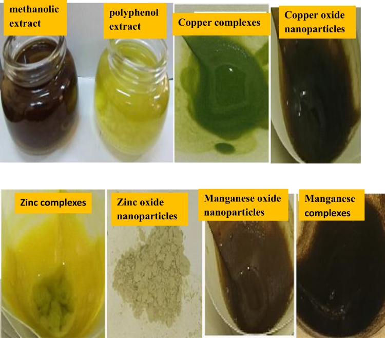 |
Figure 2 Construction of metal complexes and metal oxide nanoparticles. |
Mechanisms of Metal Complex and Nanoparticle Formation Using Plectranthus Amboinicus Extracts
Fabrication of metal complexes and nanoparticles employing P. amboinicus extracts involves distinct pathways governed by the phytochemical composition and reaction conditions of the extract (Table S1). The following sections in detail:
Metal Complex Formation
1. Ligand coordination: Flavonoids isolated from the n-butanol fraction (eg, kaempferol and apigenin) act as polydentate ligands. The hydroxyl (-OH) and carbonyl (C=O) groups coordinate with the metal ions (Cu2+, Zn2+, and Mn2+) to form stable complexes:

This chelation is favoured by the deprotonation of phenolic -OH groups at alkaline pH (9–10).
2. Hydroxide incorporation: The addition of NaOH introduces hydroxide ions (OH−), further stabilising the complexes by occupying vacant coordination sites:

This step is crucial for achieving octahedral geometries, as evidenced by the UV-Vis spectra (eg Jahn-Teller distortion in Cu2+ complex at 530 nm).
3. Precipitation and crystallisation: Upon cooling, the complexes precipitated owing to their reduced solubility. Recrystallisation with ethanol removed the unreacted ions, yielding pure complexes with distinct colours (green for Cu, yellow for Zn, and brown for Mn).
Metal Oxide Nanoparticle Formation
1. Reduction of metal ions: Polyphenols and flavonoids in the crude methanolic extract acted as reducing agents. For example, genistein donates electrons to reduce Cu2+ into Cu0.

2. Nucleation and growth: Reduced metal atoms aggregate to form nuclei, which then grow into nanoparticles. The reaction pH (9.0) and temperature (50 °C) optimised particle size control.

Immediate colour changes (eg black-brown for CuO nanoparticles) indicate the formation of nuclei.
Spherical morphologies (observed via FESEM) are a result of isotropic growth.
3. Stabilisation by Phytochemicals: Residual flavonoids and proteins are adsorbed onto nanoparticle surfaces, preventing aggregation. FTIR peaks at 593 cm−1 (M-O bonds) and EDX metal-to-oxygen ratios (~1:1) confirm oxide formation.
Analytical Validation
UV-Vis spectroscopy: Complexes exhibit ligand-to-metal charge transfer bands (eg, Cu2+ complexes at 340 nm). Nanoparticles show characteristic absorption bands in the UV-Vis region (eg, ZnO at 350 nm) attributed to band gap transitions.
FT-IR spectroscopy: Shifts from 1629 cm−1 (C=O in complexes) to 1558 cm−1 (M-O in nanoparticles) confirm distinct bonding environments.
X-Ray diffraction (XRD): Sharp peaks in complexes (eg Zn at 2θ = 22°) versus broader peaks in nanoparticles (eg MnO at 2θ = 29°) reflect differences in crystallinity.
This dual-pathway mechanism highlights the versatility of P. amboinicus in synthesising both coordination compounds and nanoparticles, which are suitable for medical uses like antimicrobial agents or drug transport mechanisms.
Analysis of n-Butanol Fraction Extract by LC-MS/MS
LC-MS/MS analysis of polyphenolic compounds (Figure 3) identified several biologically significant flavonoids (Table S2) (Figure 4), including kaempferol, apigenin, naringin, and genistein. These compounds are recognised for their antioxidant properties and therapeutic potential in the treatment of Alzheimer’s and other neurodegenerative diseases.35,36 P. amboinicus contains coumarins and flavonoid glycosides, which represent a valuable source of these typically expensive and hard-to-obtain compounds. These flavonoids have diverse biological activities, including anticancer properties through tumour growth inhibition, cardioprotective effects,37,38 and antibacterial and anti-inflammatory properties,39,40 thus supporting traditional medicinal applications. The identified flavonoids exhibit strong metal-binding capabilities through their hydroxyl or carbonyl groups, forming complexes that often exhibit enhanced antioxidant properties compared with free flavonoids.10
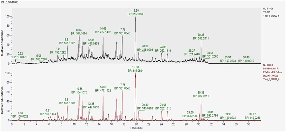 |
Figure 3 LC-MS/MS of n-butanol fraction extract. |
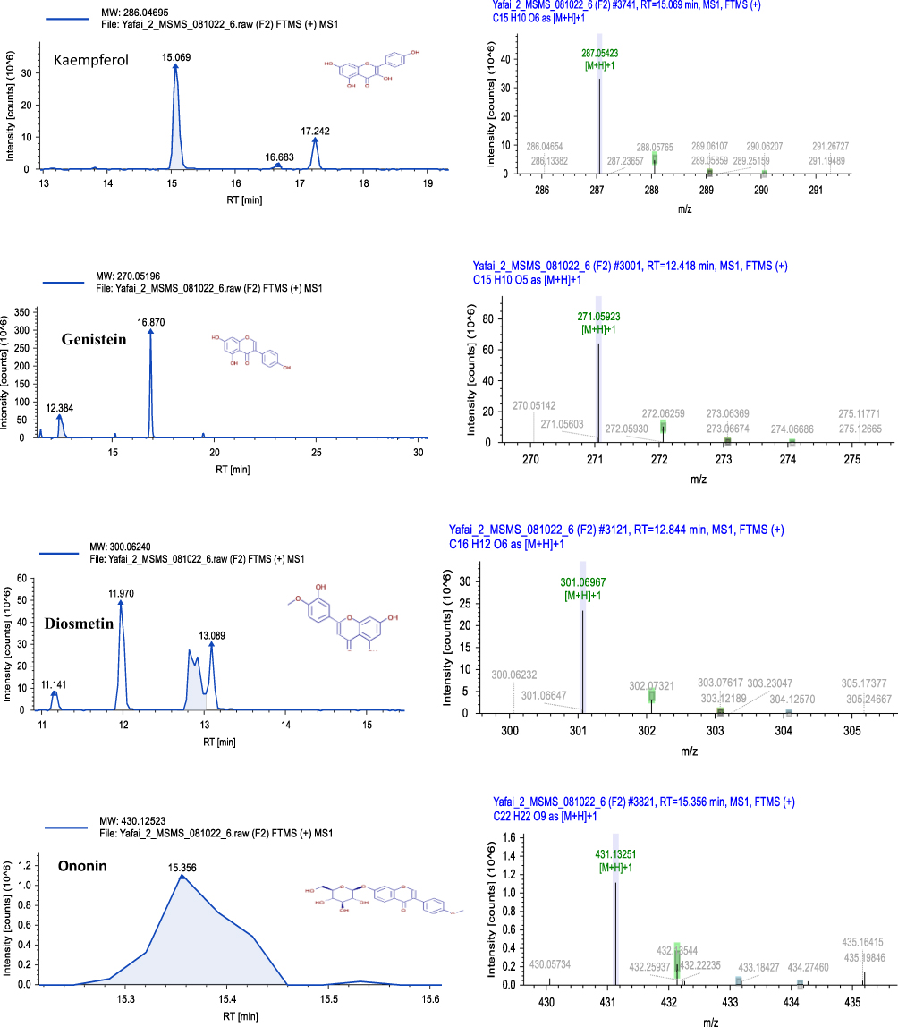 |
Figure 4 Some flavonoids detected by LC-MS/MS analysis. |
UV-Visible Spectroscopic Analysis
The UV-vis spectrum of P. amboinicus n-butanol extract exhibited characteristic flavonoid absorption peaks at 230 and 290 nm, attributed to aromatic π–π * transitions (Figure 5a), whereas the methanolic extract showed a broad absorption band spanning 200–390 nm (Figure 5b). The copper complexes showed shifts to 220, 340, and 530 nm (Figure 5a),41,42 with a 530 nm band suggesting a distorted octahedral structure due to the Jahn-Teller effect, whereas the copper oxide nanoparticles exhibited a single peak at 250 nm (Figure 5b).43,44 Zinc complexes demonstrate absorption peaks at 260 and 340 nm, reflecting ligand-based transitions, whereas zinc oxide nanoparticles show a characteristic peak at 350 nm.45,46 The manganese complexes exhibited shifted peaks at 250 and 420 nm, with a weak 420 nm band indicating octahedral geometry for the high-spin Mn2+ complexes, and the corresponding oxide nanoparticles displayed peaks at 270 and 330 nm.47,48
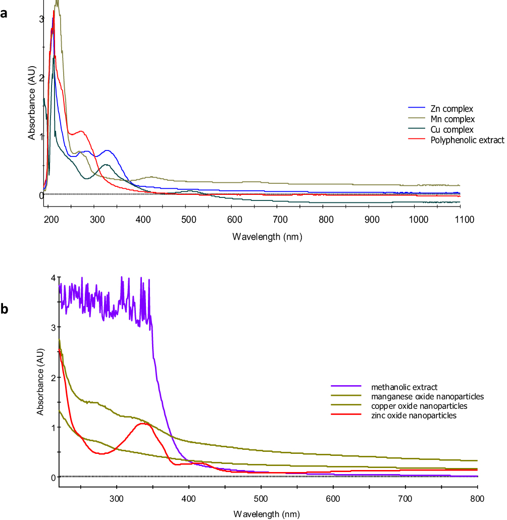 |
Figure 5 UV‒vis absorption spectra of the polyphenolic extract and the metal complexes (a), methanolic extract, and metal oxide nanoparticles (b). |
All the metal complexes retained shifted phenolic compound absorption peaks, whereas the metal oxide nanoparticle spectra showed distinct patterns without methanolic extract absorption features. This spectral evidence suggests the role of the methanolic extract as a reducing agent for the formation of metal oxide nanoparticles.
Fourier Transform Infrared Spectroscopy (FT-IR)
The FT-IR spectra revealed distinct structural characteristics of the synthesised materials (Figure 6a and b). The n-butanol extract exhibited characteristic bands at 3421 cm−1 (O-H stretching), 2924 cm−1 (C-H stretching), 1629 cm−1 (C=O stretching), and 1384 cm−1 (C-O stretching).49,50
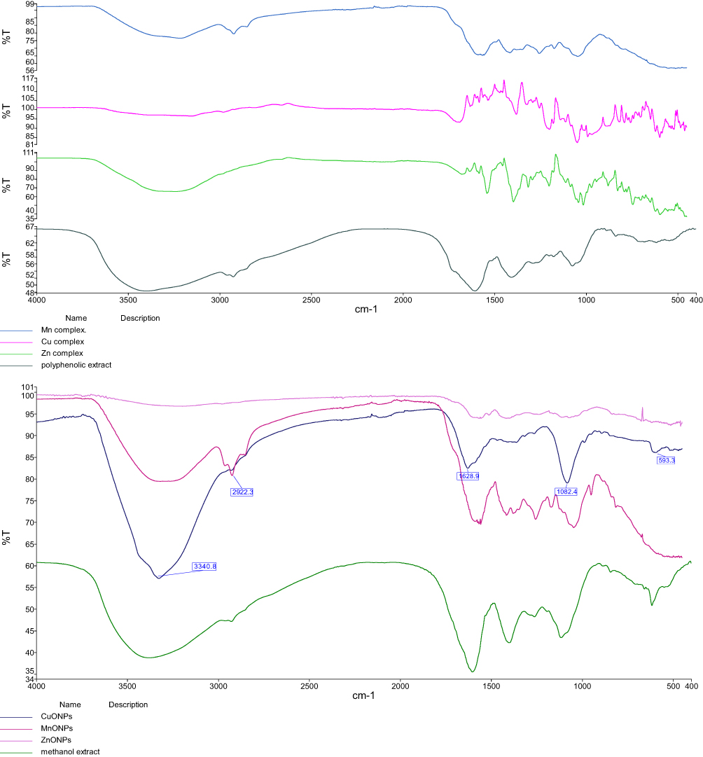 |
Figure 6 FT-IR spectra of the polyphenolic extract and metal-complex: (a) methanolic extract and (b) metal oxide nanoparticle. |
Metal Complexes (Figure 6a):
- Copper complex showed shifts to 3398 cm−1 (O-H), 1558 cm−1 (C=O), and new bands at 593 cm−1 (Cu-O)
- Zinc complex displayed bands at 3412 cm−1 (O-H), 1562 cm−1 (C=O), and 545 cm−1 (Zn-O)
- Manganese complex exhibited shifts to 3405 cm−1 (O-H), 1565 cm−1 (C=O), and 528 cm−1 (Mn-O)
Metal Oxide Nanoparticles (Figure 6b):
- CuO nanoparticles: characteristic peaks at 535 cm−1 and 590 cm−1. This is consistent with what is stated in the literature.51
- ZnO nanoparticles: strong band at 450–500 cm−1
- MnO nanoparticles: distinctive peaks at 520 cm−1 and 580 cm−1
These spectral changes confirm successful complex formation and nanoparticle synthesis.52,53
XRD Analysis
X-ray diffraction demonstrated the successful creation of both metal complexes and nanoparticles in the powder samples. The diffraction patterns exhibit distinctive characteristics for each material.
Methanolic and Polyphenol Extracts
- Methanolic extract (Figure 7a): minimal peaks at 2θ = 25°, 28°, 30°, and 45°
- Polyphenol mixture (Figure 7b): crystalline properties with peaks at 2θ = 21°,29°, 32°, 39°, 40°, and 43°
 |
Figure 7 XRD for: (a) methanolic extract, (b) polyphenol extract, (c) copper complex, (d) copper oxide nanoparticles (CuONPs), (e) zinc complex, (f) zinc oxide nanoparticles (ZnONPs), (g) manganese complex, and (h) manganese oxide nanoparticles (MnONPs). |
Copper-Based Materials
- Copper complex (50 ± 1.2 nm) (Figure 7c): distinctive peaks at 2θ = 26°, 37°, 39°, 55°, 59, 63°, and 70°.
- Copper oxide nanoparticles (39 ± 1.6 nm) (Figure 7d): peaks at 2θ = 18°, 20°, 22°, 24°, 29°, 30°, 32°, 35°, 42°, 53°, and 60°, aligning with reference data54
Zinc-Based Materials
- Zinc complex (43 ± 2.1 nm) (Figure 7e): peaks at 2θ = 23°, 25°, 28°, 31°, 33°, 39°, 40°, 45°, and 56°.
- ZnO nanoparticles (38 ± 1.3 nm) (Figure 7f): peaks at 2θ = 29°, 30°, 32°, 34°, 47°, 52°, 60°, and 68°, consistent with literature findings.55,56
Manganese-Based Materials
- Mn complex (60 ± 1.2 nm) (Figure 7g): absorption peaks at 2θ = 30°, 36°, 39°, 40°, 42°.
The manganese oxide nanoparticles (55 ± 1.2 nm) (Figure 7h) showed peaks at 2θ = 19°, 29°, 35°, 41°, 45°, and 50°. The more pronounced and intense peaks in the zinc complex suggest their enhanced crystalline nature compared to that of the ZnO nanoparticles. Plant-synthesised MnO nanoparticles typically exhibit a weakly crystalline nature.57 The crystallite sizes were determined using the Debye-Scherrer equation. Research indicates that plants of the Lamiaceae family can produce nanoparticles of varying sizes and shapes, which are influenced by plant extract concentration.58
1H NMR Analysis
1H NMR spectroscopic analysis was conducted using the samples dissolved in DMSO-d6. The methanolic extract (Figure 8a) showed less intense peaks in the aromatic region, whereas the polyphenol mixture spectrum (Figure 8b) exhibited signals at 6–8 and 9–13 ppm, corresponding to the benzene ring protons of phenolic compounds.59,60 Signals around 6.5 ppm indicated mono- and di-substituted phenyl rings in the plant extracts, whereas peaks at 9–13 ppm confirmed the presence of flavonoids in the polyphenol mixture. The signal at 1.21 ppm was assigned to methyl protons (-CH3), and the signals between 3.2–3.8 ppm corresponded to methylene (-CH2-) and methoxy (O-CH3) protons, respectively.
Figure 8 Continued. Figure 8 Continued. Figure 8 Continued. Figure 8 1H NMR for: (a) methanolic extract, (b) polyphenol extract, (c) copper complex, and (d) copper oxide nanoparticles (CuONPs), (e) zinc complex, and (f) zinc oxide nanoparticles (ZnONPs), (g) manganese complex, and (h) manganese oxide nanoparticles (MnONPs).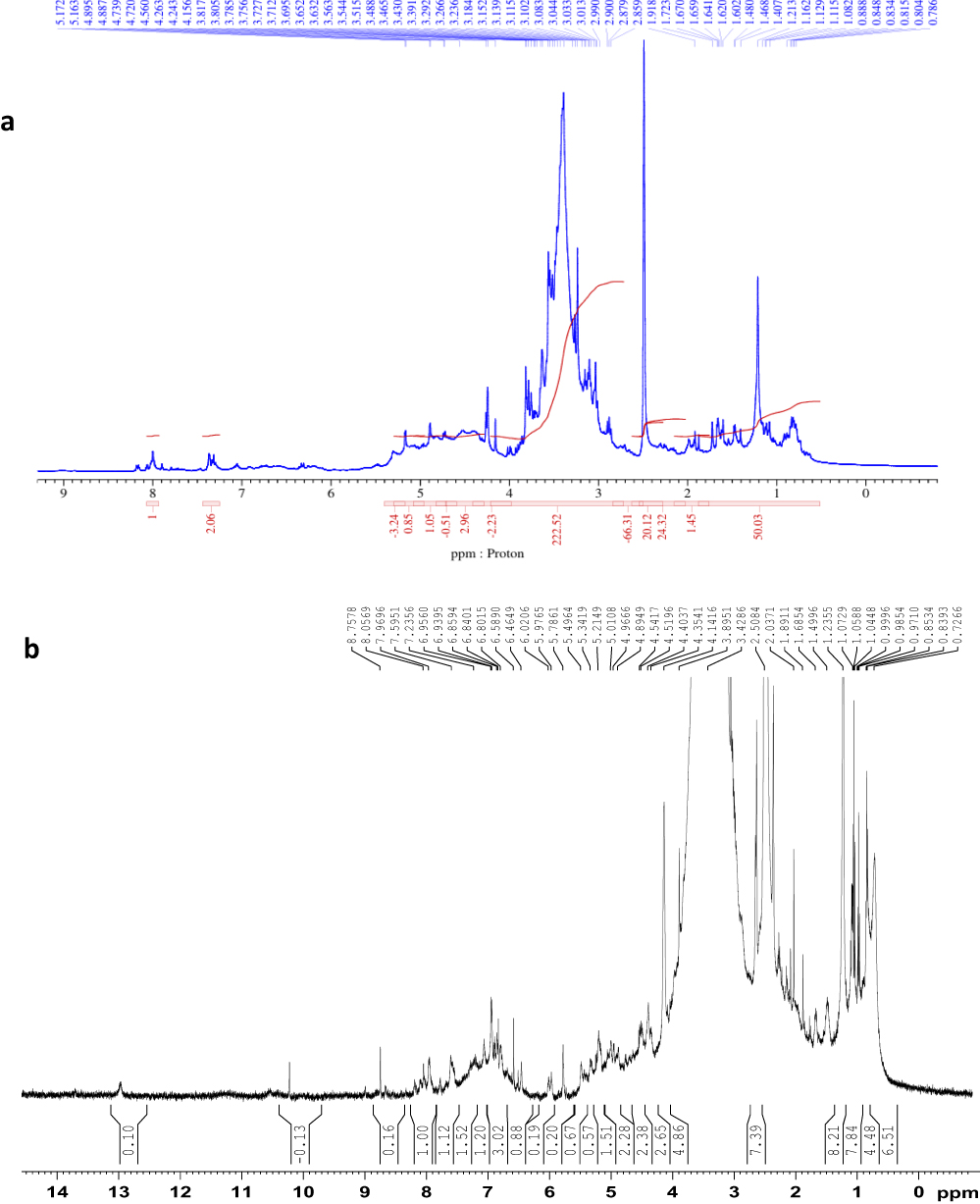
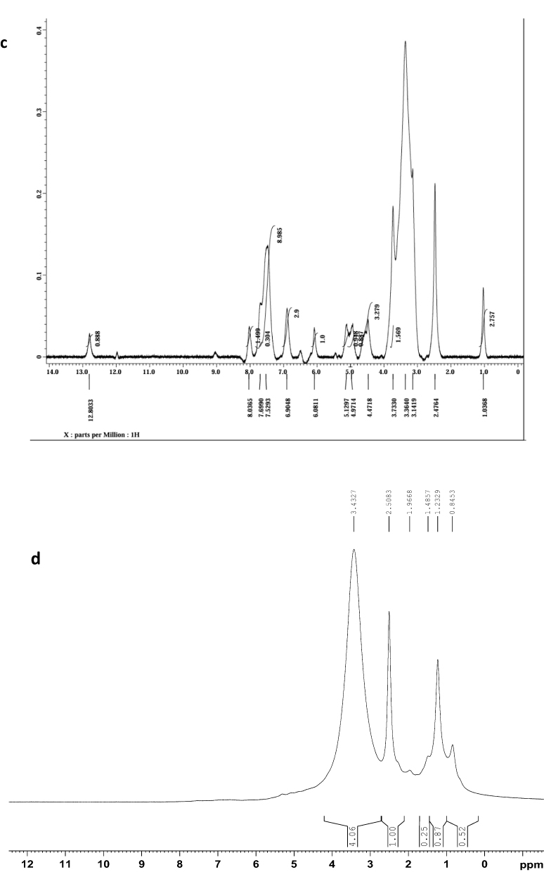
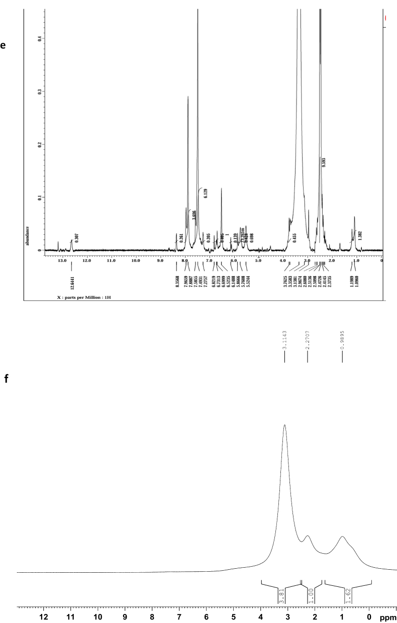
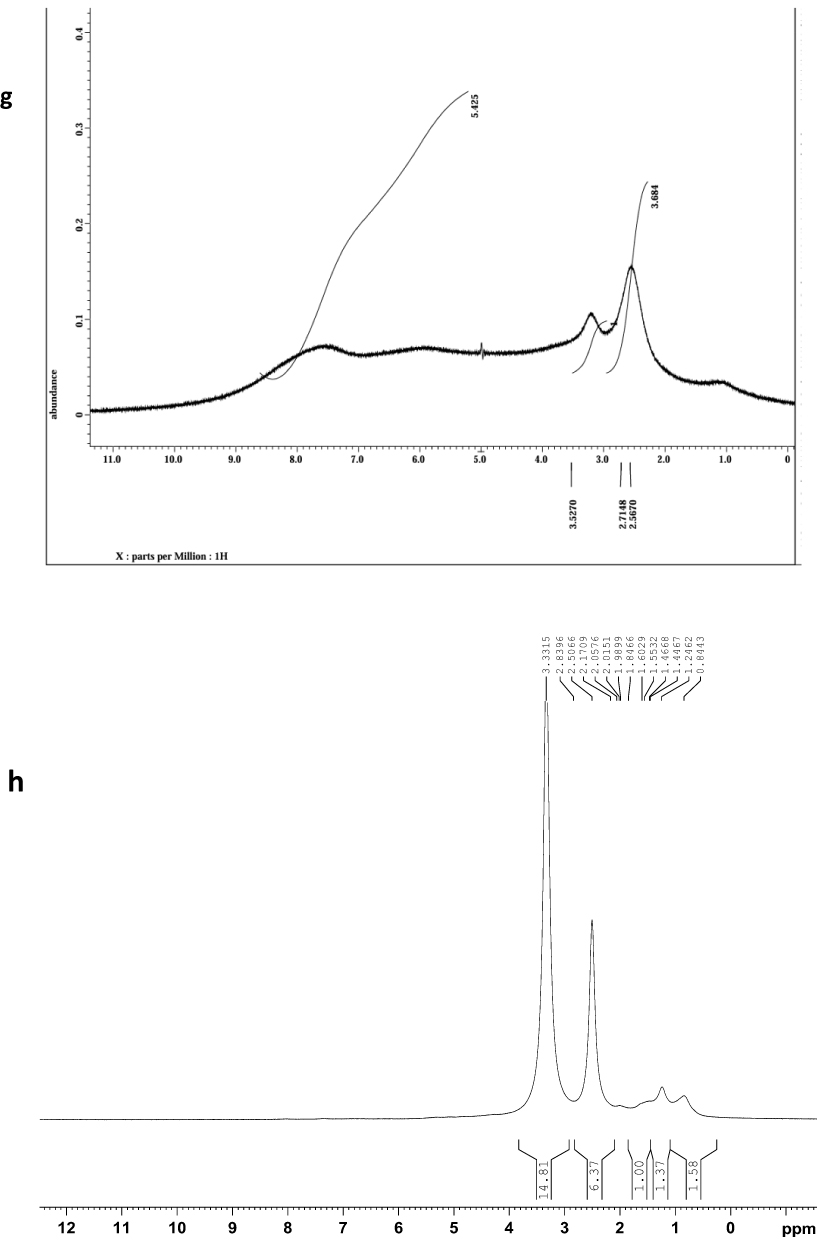
The metal complexes displayed distinct peak shifts at 6–13 ppm, as evidenced by the copper complex (Figure 8c), while copper oxide nanoparticles (CuONPs) (Figure 8d) showed no signals in the aromatic region. Similarly, the zinc complex (Figure 8e) exhibits characteristic peaks, whereas the zinc oxide nanoparticles (ZnONPs) (Figure 8f) lack aromatic signals. Manganese complex (Figure 8g) showed a broad peak, indicating high spin characteristics,61,62 whereas manganese oxide nanoparticles (MnONPs) (Figure 8h) displayed no aromatic region.
All metal oxide nanoparticles spectra confirmed the absence of phenolic compounds, with signals at 2.50 ppm and 3.12–3.39 ppm attributed to DMSO-d6 solvent and water, respectively.63 The 1H NMR data demonstrate clear spectral differences between the metal complexes and their corresponding metal-oxide nanoparticles.
EDX Analysis
The elemental makeup of the sample was analyzed with Energy-dispersive X-ray spectroscopy (EDX) for the metallic complexes and metal oxide nanoparticles synthesised using Plectranthus amboinicus. EDX (Table S3) of the copper (Figure 9a), zinc (Figure 9b), and manganese complex (Figure 9c) revealed varying elemental compositions. The copper complex showed carbon (46.21 wt%), oxygen (33.58 wt%), and copper (20.21 wt%).
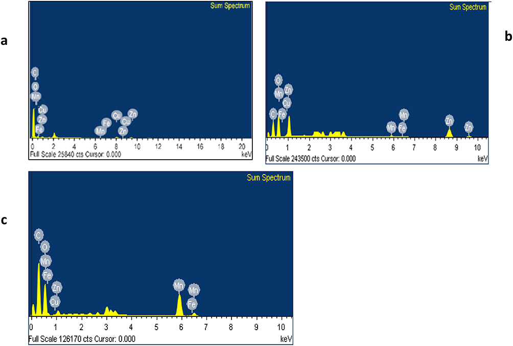 |
Figure 9 EDX for: (a) copper complex, (b) zinc complex and (c) manganese complex. |
As shown in Table S4 and Figure 10, the analyses of the copper oxide nanoparticles (Figure 10a), zinc oxide nanoparticles (Figure 10b), and manganese oxide nanoparticles (Figure 10c) demonstrate different compositions. CuONPs contained oxygen (33.04 wt%), copper (43.34 wt%), and carbon (22.95 wt%). A strong characteristic peak at 8 keV indicated metallic copper in the CuONPs, consistent with previous studies,51,64 whereas the copper complex showed a weaker peak at this position. The oxygen peak appeared at 0.5 keV in the metal oxide nanoparticles and at 0.2 keV in the complexes, confirming distinct formation pathways.
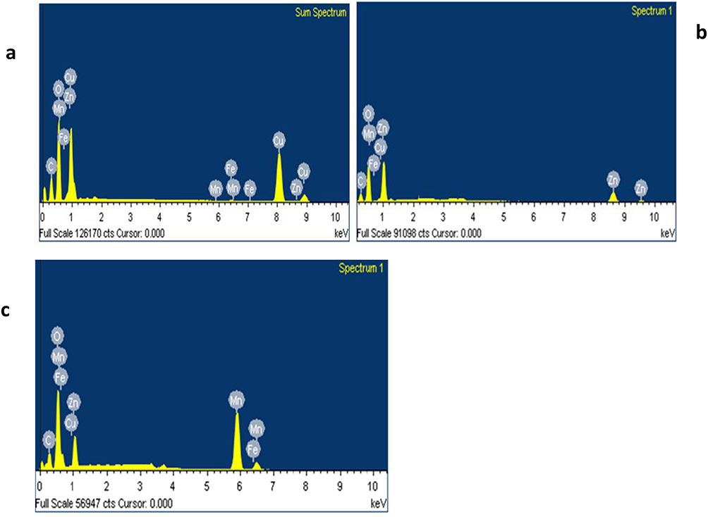 |
Figure 10 EDX for: (a) copper oxide nanoparticles (CuONPs), (b) zinc oxide nanoparticles (ZnONPs), and (c) manganese oxide nanoparticles (MnONPs). |
The zinc complex displayed peaks similar to those of copper complex for oxygen and carbon, with distinctive zinc peaks at 1.5 and 8.8 keV. ZnONPs showed a higher zinc content than their complex,65 whereas zinc complex contained more carbon owing to their structural composition.
Manganese oxide nanoparticles exhibited less pronounced carbon peaks but sharper manganese peaks than their complex, with a higher oxygen content in the nanoparticles. The carbon signal at 0.4 keV in metal oxide nanoparticles may originate from the sample carrier, atmospheric carbon species, or residual plant extract.66,67
According to EDX data, carbon was the dominant element in the complexes, whereas the metal oxide nanoparticles showed the highest peaks for metal and oxygen, confirming their formation. A metal-to-oxygen ratio (M: O) of approximately 1:1 in all nanoparticles indicated the formation of CuO, ZnO, and MnO.
Spectrum processing identified the following deletable peaks: copper complex (1.600, 2.077, 3.340, 11.089 keV), CuONPs (1.257–3.591 keV), zinc complex (1.269–4.335 keV), ZnONPs (1.260–3.600 keV), manganese complex (1.493–3.708 keV), and MnONPs (1.259–3.168 keV).
FESEM Analysis
Surface morphology analysis using scanning electron microscopy (FESEM) revealed distinct characteristics for each compound (Figure 11a–h):
 |
Figure 11 FESEM analysis; (a) Copper complex, (b) Copper oxide nanoparticles (CuONPs), (c) zinc complex, (d) zinc oxide nanoparticles (ZnONPs), (e) manganese complex, and (f) manganese oxide nanoparticles (MnONPs). |
Copper-Based Materials
- Copper complex (50 ± 1.2 nm) (Figure 11a): flat sheet-like structures with agglomerates, attributed to the preparation method and pH conditions68,69
- Copper oxide nanoparticles (39 ± 1.6 nm) (Figure 11b): uniform spherical shapes with even distribution70,71
Zinc-Based Materials
- Zinc complex (43 ± 2.1 nm) (Figure 11c): ellipsoidal morphology with consistent size distribution72
- ZnO nanoparticles (38 ± 1.3 nm) (Figure 11d): Hierarchical flower-like structures with distinct petals73
Manganese-Based Materials
- Mn complex (60 ± 1.2 nm) (Figure 11e and f): sheet-like structures with a layered arrangement
- Manganese oxide nanoparticles (55 ± 1.2 nm) (Figure 11g and h): spherical with minimal agglomeration74
The FESEM micrographs confirmed the uniform particle distribution and distinct morphological features of each compound. These size distributions, particularly those spanning 10 nm to 50 nm, are deemed ideal for medical applications owing to their unique therapeutic properties.75 Higher magnification images (Figure 11f and h) further revealed the detailed surface characteristics and crystalline nature of the synthesised materials.
Antimicrobial Screening
The metal complexes demonstrated greater antimicrobial activity than the metal oxide nanoparticles (Figure 12). The flavonoid extract was more effective than the methanolic extract, with copper complexes showing the highest activity—25.47 cm against Staphylococcus aureus, 24 cm against Klebsiella pneumoniae and E. coli, and 26.17 cm against Pseudomonas aeruginosa (Table S5)—surpassing previous reports.76 Zinc complexes displayed similar activity to copper against E. coli (22 cm), while manganese complexes exhibited antimicrobial effects, though less potent than copper-zinc complexes. All complexes outperformed the flavonoid extract, aligning with existing literature.77 They also inhibited Candida albicans, albeit less effectively. The methanolic extract showed the weakest activity. Copper and zinc oxide nanoparticles demonstrated higher antimicrobial efficacy than manganese oxide, with copper complexes outperforming antibiotics against all microbes except Candida albicans.78
 |
Figure 12 Antimicrobial activity of metal complexes and metal oxide nanoparticles. |
Antioxidant Results
Copper oxide nanoparticles exhibited the strongest antioxidant activity in both DPPH and ABTS assays. The scavenging capacity followed the order: copper oxide nanoparticles > copper complexes > zinc complexes > zinc oxide nanoparticles > manganese oxide nanoparticles > manganese complexes > flavonoid extract > methanolic extract (Table S6). The IC50 values for radical scavenging of these nanoparticles were higher than in previous studies.79,80 In the DPPH test, IC50 values were 28.32 μg/mL for copper complexes and 17.12 μg/mL for copper oxide nanoparticles; zinc complexes and zinc oxide nanoparticles showed IC50 values of 37.46 and 32.97 μg/mL, respectively (Figure 13). In the ABTS assay (Figure 14), IC50 values were 11.03 μg/mL for copper nanoparticles and 22.5 μg/mL for their complexes, exceeding previous findings,81 with ascorbic acid at 10.65 μg/mL (Table S6). Manganese complexes and manganese oxide nanoparticles showed IC50 values of 55.04 and 46.83 μg/mL (DPPH), and 49.53 and 43.67 μg/mL (ABTS). The methanolic extract was the least effective antioxidant.
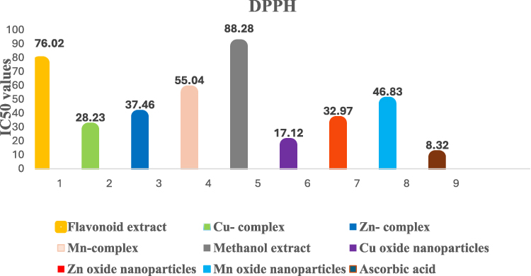 |
Figure 13 DPPH scavenging capacity of metal complexes and metal oxide nanoparticles. |
 |
Figure 14 ABTS scavenging capacity of ascorbic acid, metal complexes, and metal oxide nanoparticles. |
Anticancer Results
The WST-1 assay on THP-1 leukaemia cells and the SRB assay on Calu-3 lung cancer cells (Figures 15, 16 and Table S7) demonstrated significant anticancer activity, with copper oxide nanoparticles showing the highest efficacy, followed by zinc oxide nanoparticles. Copper oxide nanoparticles exhibited IC50 values of 13.63 μg/mL (THP-1) and 26.67 μg/mL (Calu-3), surpassing previous reports.82,83 Zinc oxide nanoparticles had IC50 values of 37.23 μg/mL and 58.42 μg/mL, respectively, also higher than prior data.84 Manganese oxide nanoparticles showed IC50s of 60.8 μg/mL and 80.46 μg/mL. The methanolic extract was less active. Although metal complexes displayed anticancer effects, they were less effective than nanoparticles, likely due to the superior tissue penetration of nanoscale materials. The order of anticancer activity was: copper complexes > zinc complexes > manganese complexes.
 |
Figure 15 IC50 values metal complexes and metal oxide nanoparticlesin THP-1 cells. |
 |
Figure 16 IC50 of metal complexes and metal oxide nanoparticlesin Calu-3 cells. |
Flow Cytometry Results
Flow cytometry revealed that all studied metal complexes and metal oxide nanoparticles induced apoptosis in Calu-3 cells (Figure 17A–C). Nanoparticles caused greater apoptosis than complexes, consistent with their higher cytotoxicity, likely due to their size and composition. The flavonoid extract induced more apoptosis than the methanolic extract. Over time, apoptosis increased: late apoptosis (Q2) rose after 72 hours, while early apoptosis (Q4) peaked within the first 24 hours. Early apoptosis (Q4) indicates initial cell death with membrane integrity; late apoptosis (Q2) reflects advanced cell death with membrane perforation; necrosis (Q1) involves non-apoptotic cell death, and healthy cells are represented in Q3. The increase in apoptosis over time is consistent with previous studies.85 Copper oxide nanoparticles induced the highest late apoptosis (28.6%) after 72 hours, with total apoptosis reaching 39%. Zinc and manganese oxide nanoparticles showed lower rates, at 26.06% and 23.93%. Metal complexes exhibited higher apoptosis than the metal oxides, with total rates of 31.72%. The apoptotic effects of these materials enhance their therapeutic potential, as apoptosis analysis provides insight into cell death stages and drug efficacy.86
Figure 17 Continued. Figure 17 Continued. Figure 17 Mechanism of cell death in Calu-3 lung cancer cell line after treatment with metal complexes and metal oxide nanoparticles, (A) After 24 hours, (B) After 48 hours, (C) After 72 hours.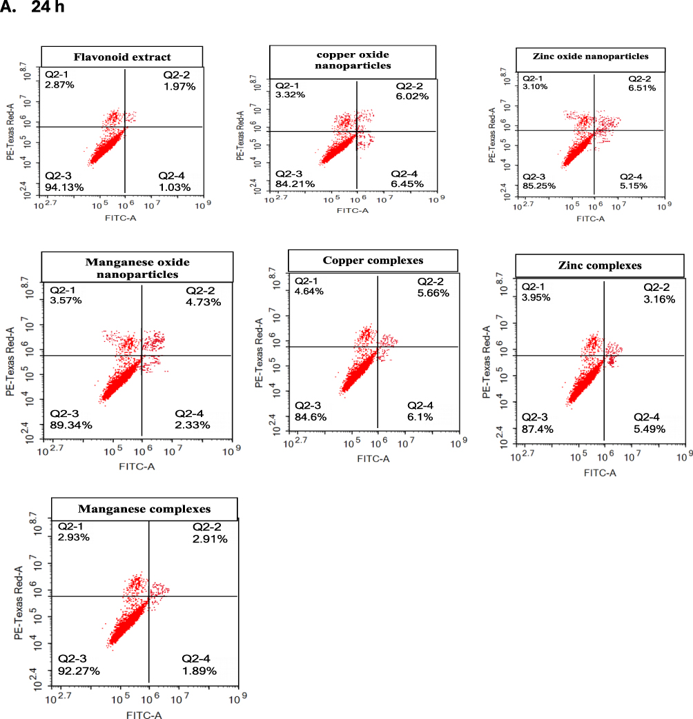


Conclusion
This study presents an eco-friendly method for synthesising metal-based materials using Plectranthus amboinicus extracts. The dual-pathway synthesis successfully produced well-defined metal complexes and oxide nanoparticles, with sizes of approximately 50 nm (copper), 43 nm (zinc), 60 nm (manganese), and their corresponding oxides—39 nm, 38 nm, and 55 nm respectively. Spectroscopic and microscopic analyses confirmed the formation of sheet-like complexes and spherical, crystalline nanoparticles, as evidenced by XRD patterns.
The controlled synthesis and distinctive properties of these materials underpin their significant biological activity, often surpassing standard antibiotics in antimicrobial tests. Their notable antioxidant capacity was linked to their ability to inhibit cancer cell growth in leukaemia and lung cancer models. Both complexes and nanoparticles induced apoptosis, further enhancing their therapeutic potential.
The utilisation of plant metabolites as natural reducing and stabilising agents underscores the potential for sustainable and eco-friendly biomedical material production. Looking ahead, further research is warranted to evaluate the biocompatibility and toxicity profiles of these materials, alongside surface functionalisation strategies to optimise their therapeutic efficacy. Developing scalable manufacturing protocols and exploring their applications against resistant microbes, as well as elucidating drug delivery mechanisms, are essential steps towards translating these materials from laboratory research to clinical use.
This green synthesis approach marks an important stride towards sustainable nanomedicine production, fostering advances across multidisciplinary biomedical research and opening new avenues for the development of novel, eco-friendly therapeutic agents.
Abbreviations
CuONPs, Copper oxide nanoparticles; DMSO, Dimethyl sulfoxide; EDX, Energy-dispersive X-ray; FESEM, Field emission scanning electron microscopy; FT-IR, Fourier-transform infrared; 1H NMR, Proton nuclear magnetic resonance; LC-MS/MS, Liquid chromatography-mass spectrometry; MnONPs, Manganese oxide nanoparticles; UV-Vis, Ultraviolet-visible spectroscopy; XRD, X-ray diffraction; ZnONPs, Zinc oxide nanoparticles.
Data Sharing Statement
Data are available upon request from the corresponding author, Adel AM Saeed.
Acknowledgments
We extend our heartfelt appreciation to the Department of Chemistry, College of Aden Education, University of Aden, Yemen, and the Supreme Authority for Drugs and Metrology, Aden Branch, Yemen, for offering the essential resources and assistance needed to carry out this investigation.
Author Contributions
All authors contributed significantly to the work reported, whether through the conception, design, execution, data acquisition, analysis, or interpretation. They participated in drafting, revising, or critically reviewing the manuscript, and all provided final approval of the version to be published. Furthermore, they have collectively agreed on the journal to which the manuscript has been submitted and affirm their accountability for all aspects of the work.
Funding
This work received no external funding.
Disclosure
The authors state that they have no conflicts of interest concerning this research.
References
1. Waris A, Din M, Ali A, et al. A comprehensive review of green synthesis of copper oxide nanoparticles and their diverse biomedical applications. Inorg Chem Commun. 2021;123:108369. doi:10.1016/j.inoche.2020.108369
2. Hussain A, Alajmi MF, Rehman MT, et al. Copper(II) complexes as potential anticancer and nonsteroidal anti-inflammatory agents: in vitro and in vivo studies. Sci Rep. 2019;9:5237. doi:10.1038/s41598-019-41063-x
3. Chen BH, Pan ZY, Feng WW, Liu QY, Liu Y, He L. Synthesis, characterization, and biological activity of novel copper complexes containing a β-carboline derivative and amino acids. Dalton Trans. 2023;52:8040–8050. doi:10.1039/d3dt01088k
4. Wani AL, Parveen N, Ansari MO, Ahmad MF, Jameel S, Shadab GGHA. Zinc: an element of extensive medical importance. Curr Med Res Pract. 2017;7:90–98. doi:10.1016/j.cmrp.2017.02.006
5. Sun Y, Zhang W, Li Q, Liu H, Wang X. Preparations and applications of zinc oxide based photocatalytic materials. Adv Sens Energy Mater. 2023;2:100069. doi:10.1016/j.asems.2023.100069
6. Palanisamy GV, Kaya SS, Ahmed SB, Sumathi P, Premkumar R, Kamal C. Synthesis and spectroscopic characterization of Schiff base metal complexes, biological activity, and molecular docking studies. ACS Omega. 2024;9:8123–8138. doi:10.1021/acsomega.3c08526
7. Hassan AM, Said AO, Heakal BH, Younis A, Aboulthana WM, Mady MF. Green synthesis, characterization, antimicrobial and anticancer screening of new metal complexes incorporating Schiff base. ACS Omega. 2022;7:32418–32433. doi:10.1021/acsomega.2c03911
8. Li Z, Wang Y, Xu M, Liu H, Li L, Xu D. Plant flavonoids in heavy metal stress tolerance. Plant Physiol Biochem. 2023;204:108125. doi:10.1016/j.plaphy.2023.108125
9. Shen N, Wang T, Gan Q, Liu S, Wang L, Jin B. Plant flavonoids: classification, distribution, biosynthesis, and antioxidant activity. Food Chem. 2022;383:132531. doi:10.1016/j.foodchem.2022.132531
10. Kejík Z, Kaplánek R, Masařík M, et al. Iron complexes of flavonoids-antioxidant capacity and beyond. Int J Mol Sci. 2021;22:646. doi:10.3390/ijms22020646
11. Eker F, Duman H, Akdaşçi E, et al. A comprehensive review of nanoparticles: from classification to application and toxicity. Molecules. 2024;29:3482. doi:10.3390/molecules29153482
12. Pourmadad M, Rahdar A, Holghoomi R, Pandey S. Copper nanoparticles from chemical, physical, and green synthesis to medical application: a review. Plant Nano Biol. 2024;8:100070. doi:10.1016/j.plana.2024.100070
13. Najahi-Missaoui W, Arnold RD, Cummings BS. Safe nanoparticles: are we there yet? Int J Mol Sci. 2021;22:385. doi:10.3390/ijms22010385
14. Arumugam G, Swamy MK, Sinniah UR. Plectranthus amboinicus: botanical, phytochemical, pharmacological and nutritional significance. Molecules. 2016;21:369. doi:10.3390/molecules21040369
15. Barbosa MO, Wilairatana P, Leite GML, et al. Plectranthus species with anti-inflammatory and analgesic potential: a systematic review on ethnobotanical and pharmacological findings. Molecules. 2023;28:5653. doi:10.3390/molecules28155653
16. Rajalakshmi U, Esaivani C, Kumar TA, et al. Green synthesis of iron oxide nanoparticles from spermacoce ocymoides Burm. F. Plant extracts for targeted lung cancer A549 cell therapy. Bull Chem Soc Ethiop. 2024;38:123–134. doi:10.4314/bcse.v38i1.10
17. Dai Q, Shu T, Yang H, Su L, Li X, Zhang X. One-pot synthesis of stable antioxidant metal-ornamented polyphenol supramolecular assemblies for material engineering. NPG Asia Mater. 2022;14:97. doi:10.1038/s41427-022-00449-6
18. Ali AM, Saeed AAM, Fdhel TA. Phytochemical analysis and antimicrobial screening of selected Yemeni folk medicinal plants. J Med Plants Stud. 2019;7:108–114.
19. Chanah NB. Extraction of Phenols and Flavonoids from Seeds of Pronus armenica Plant, and Study of Antioxidant Activity [M.Sc. thesis]. University Kasdi Merbah-Ouargla; 2020.
20. Bamigboye O, Ejidike I, Ahmed R, Lawal M, Nnabuike G, Medubi K. Chelation, characterization, and antibacterial activities of some mixed isonicotinic acid hydrazide-paracetamol metal drug complexes. Suranaree J Sci Technol. 2020;27:030026–030029.
21. Singh K, Chopra DS, Singh D, Singh N. Green synthesis and characterization of iron oxide nanoparticles using Coriandrum sativum L. leaf extract. Indian J Biochem Biophys. 2022;59:450–454. doi:10.56042/ijbb.v59i4.61913
22. Bamigboye O, Ejidik I, Lawal M. Synthesis, characterization, and antimicrobial potentials of some flavonoid-metal complexes from Chromolaena Odorata. Iraqi J Sci. 2020;61:2440–2447. doi:10.24996/ijs.2020.61.10.1
23. Saeed N, Khan R, Shabbir M. Antioxidant activity, total phenolic and total flavonoid contents of whole plant extracts Torilis leptophylla L. BMC Complement Altern Med. 2012;12:3. doi:10.1186/1472-6882-12-221
24. Wong F, Chai T, Hoo Y. Antioxidant and cytotoxic activities of selected medicinal herbs used in Malaysia. J Med Plants Res. 2012;6:3169–3175. doi:10.5897/jmpr11.1631
25. Manurung K, Sulastri D, Zubir N, Ilyas S. In silico anticancer activity and in vitro antioxidant activity of flavonoids in Plectranthus amboinicus. Pharmacogn J. 2020;12:1573–1577. doi:10.5530/pj.2020.12.215
26. Skehan P, Toreng R, Scudiero D, et al. New colorimetric cytotoxicity assay for anticancer-drug screening. J Natl Cancer Inst. 1990;82:1107–1112. doi:10.1093/jnci/82.13.1107
27. Allam R, Al-Abd A, Khedr A, Sharaf O. Fingolimod interrupts the cross-talk between estrogen metabolism and sphingolipid metabolism within prostate cancer cells. Toxicol Lett. 2018;11:77–85. doi:10.1016/j.toxlet.2018.04.008
28. Fekry M, Ezzat S, Salama M, Alshehri O, Al-Abd A. Bioactive glycoalkaloides isolated from solanum melongena fruit peels with potential anticancer properties against hepatocellular carcinoma cells. Sci Rep. 2019;9(1):1746. doi:10.1038/s41598-018-36089-6
29. Bashmail H, Alamoudi A, Noorwali A, et al. Thymoquinone synergizes gemcitabine anti-breast cancer activity via modulating its apoptotic and autophagic activities. Sci Rep. 2018;8(1):11674. doi:10.1038/s41598-018-30046-z
30. Halevas E, Mavroidi B, Zahariou G, Pelecanou M, Hatzidimitriou AG. Structurally characterized copper complexes of flavonoid naringenin with enhanced radical scavenging activity. Inorg Chim Acta. 2023;546:121325. doi:10.1016/j.ica.2022.121325
31. Andualem WW, Sabir FK, Mohammed ET, Belay HH, Gonfa BA. Synthesis of copper oxide nanoparticles using plant leaf extract of catha edulis and its antibacterial activity. Nanotechnology. 2020;2020:1–20. doi:10.1155/2020/2932434
32. Masoudiasl A, Montazerozohori M, Naghiha R, Assoud A, McArdle P, Shalamzar MS. Synthesis, X-ray crystal structures and thermal analyses of some new antimicrobial zinc complexes: new configurations and nano-size structures. Mater Sci Eng C. 2016;61:809–823. doi:10.1016/j.msec.2016.01.017
33. Brindhadevi K, Vasantharaj S, Le QH, Devanesan S, Farhat K, Liu X. Fabrication and characterization of Manganese Dioxide (MnO2) nanoparticles and its degradation potential of benzene and pyrene. Chemosphere. 2023;343:140123. doi:10.1016/j.chemosphere.2023.140123
34. Mounika T, Belagali SL, Vadiraj KT. Manganese oxide nanoparticles synthesis route, characterization, and optical properties. Mater Today Proc. 2023;75:72–76. doi:10.1016/j.matpr.2022.11.017
35. Chen J, Zhong H, Huang Z, Chen X, You J, Zou T. A critical review of kaempferol in intestinal health and diseases. Antioxidants. 2023;12:1642. doi:10.3390/antiox12081642
36. Nguyen-Thi PT, Vo TK, Pham THT, Nguyen TT, Van Vo G. Natural flavonoids as potential therapeutics in the management of Alzheimer’s disease: a review. 3 Biotech. 2024;14:68. doi:10.1007/s13205-024-03925-8
37. Grosso G, Godos J, Lamuela-Raventos R, et al. A comprehensive meta-analysis on dietary flavonoid and lignan intake and cancer risk: level of evidence and limitations. Mol Nutr Food Res. 2016;61:1600930. doi:10.1002/mnfr.201600930
38. Kim Y, Je Y. Flavonoid intake and mortality from cardiovascular disease and all causes: a meta-analysis of prospective cohort studies. Clin Nutr ESPEN. 2017;20:68–77. doi:10.1016/j.clnesp.2017.03.004
39. Chagas MSS, Behrens MD, Moragas-Tellis CJ, Penedo GXM, Silva AR, Gonçalves-de-Albuquerque CF. Flavonols and flavones as potential anti-inflammatory, antioxidant, and antibacterial compounds. Oxid Med Cell Longev. 2022;2022:9966750. doi:10.1155/2022/9966750
40. Rakha A, Umar N, Rabail R, et al. Anti-inflammatory and anti-allergic potential of dietary flavonoids: a review. Biomed Pharmacother. 2022;156:113945. doi:10.1016/j.biopha.2022.113945
41. Mohsen EMA, Saeed AAM, Yahia ARAB. Synthesis, characterization, and bioactivity evaluation of several divalent transition metal ion-bioflavonoid complexes. Results Chem. 2024;7:101535. doi:10.1016/j.rechem.2024.101535
42. Ejidike IP, Bamigboye MO. Spectral, in vitro antiradical and antimicrobial assessment of copper complexes containing tridentate Schiff base derived from dihydroxybenzene functionality with diaminoethylene bridge. Spectrosc Lett. 2021;54:212–230. doi:10.1080/00387010.2021.1893191
43. Nzilu DM, Madivoli ES, Makhanu DS, et al. Green synthesis of copper oxide nanoparticles and its efficiency in degradation of rifampicin antibiotic. Sci Rep. 2023;13:14030. doi:10.1038/s41598-023-41119-z
44. Anwaar S, Altaf F, Anwar T, et al. Biogenic Synthesis of Copper Oxide Nanoparticles Using Eucalyptus globulus Leaf Extract and Its Impact on Germination and Phytochemical Composition of Lactuca sativa. Sci Rep. 2024;14:30203. doi:10.1038/s41598-024-78956-5
45. Damena T, Alem MB, Zeleke D, Desalegn T, Eswaramoorthy R, Demissie TB. Synthesis, characterization, and biological activities of Zinc(II), Copper(II) and Nickel(II) complexes of an aminoquinoline derivative. Front Chem. 2022;10:1–21. doi:10.3389/fchem.2022.1053532
46. Ahmad W, Kalra D. Green synthesis, characterization and antimicrobial activities of ZnO nanoparticles using euphorbia hirta leaf extract. J King Saud Univ Sci. 2020;32:2358–2364. doi:10.1016/j.jksus.2020.03.014
47. Anguraj G, Kumar RA, Inmozhi C, et al. MnO2 doped with Ag nanoparticles and their application in antimicrobial and photocatalytic reactions. Catalysts. 2023;13:397. doi:10.3390/catal13020397
48. Ermakova EA, Eremina JA, Smirnova KS, et al. Mixed-Ligand Manganese(II) complexes with 5-phenyltetrazole and polypyridine derivatives: synthesis, crystal structures and biological activity. Results Chem. 2021;3:100239. doi:10.1016/j.rechem.2021.100239
49. Mohamed A, Atta RR, Kotp AA, et al. Green synthesis and characterization of iron oxide nanoparticles for the removal of heavy metals (Cd2+ and Ni2+) from aqueous solutions with antimicrobial investigation. Sci Rep. 2023;13:1–30. doi:10.1038/s41598-023-31704-7
50. Amjad R, Parveen A, Tauseef I, et al. Green synthesis and characterization of copper nanoparticles using fortunella margarita leaves. Polymers. 2021;13:1–12. doi:10.3390/polym13244364
51. Renuga D, Jeyasundari J, Athithan ASS, Jacob YBA. Synthesis and characterization of copper oxide nanoparticles using Brassica oleracea var. italic extract for its antifungal application. Mater Res Express. 2020;7:045007. doi:10.1088/2053-1591/ab7b94
52. Ikhuoria EU, Uwidia IE, Okojie RO, Ifijen IH, Chikaodili ID, Fatiqin A. Advancing green nanotechnology: harnessing the bio-reducing properties of Musa paradisiaca peel extract for sustainable synthesis of iron oxide nanoparticles. J Multidiscip Appl Nat Sci. 2024;4:108–119. doi:10.47352/jmans.2774-3047.194
53. Faisal S, Al-Radadi NS, Jan H, et al. Curcuma longa mediated synthesis of copper oxide, nickel oxide and Cu-Ni bimetallic hybrid nanoparticles: characterization and evaluation for antimicrobial, anti-parasitic and cytotoxic potentials. Coatings. 2021;11:849. doi:10.3390/coatings11070849
54. Kavith G, Kumar V, Nirami A, Arulmozhi R, Siranjeevi R, Satish R. Green synthesis of copper oxide nanoparticles decorated with graphene oxide for anticancer activity and catalytic applications. Arab J Chem. 2020;8:6802–6814. doi:10.1016/j.arabjc.2020.06.033
55. Singh J, Kumar S, Alok A, et al. The potential of green synthesized zinc oxide nanoparticles as nutrients source for plant growth. J Clean Prod. 2019;214:1061–1070. doi:10.1016/j.jclepro.2019.01.018
56. Mohammadi A, Hashemi N, Ghassabzadeh M, Sharafi A, Yazdinezhad A, Danafar H. Green synthesis and toxicological evaluation of zinc oxide nanoparticles utilizing Punica granatum fruit Peel extract: an eco-friendly approach. Sci Rep. 2025;15:20853. doi:10.1038/s41598-025-05544-6
57. Manjula M, Thenmozhi S, Thilagavathi R, Srinivasan A, Kathirvel A. Green synthesis and characterization of manganese oxide nanoparticles from gardenia resinifera leaves. Mater Today Proc. 2020;26:3559–3563. doi:10.1016/j.matpr.2019.07.396
58. Agarwal H, Kumar SV, Rajeshkumar S. A review on green synthesis of zinc oxide nanoparticles - an eco-friendly approach. Resour-Effic Technol. 2017;3:406–413. doi:10.1016/j.reffit.2017.03.002
59. Hêmily M, Jéssica M, Artemiza S, et al. Insights into interactions of flavanones with target human respiratory syncytial virus M2-1 protein from STD-NMR, fluorescence spectroscopy, and computational simulations. Int J Mol Sci. 2020;21:224. doi:10.3390/ijms21062241
60. Kapoor P, Masamitsu M, Katsuhiko M, Derek T, Kento K. Structural investigation of Hesperidin-7-O-glucoside inclusion complex with cyclodextrin: a spectroscopic assessment. Molecules. 2022;27:5395. doi:10.3390/molecules27175395
61. Martini P, Boschi A, Marvelli L, et al. Synthesis and characterization of manganese dithiocarbamate complexes: new evidence of dioxygen activation. Molecules. 2021;26:5954. doi:10.3390/molecules26195954
62. Walencik PK, Choińska R, Gołębiewska E, Kalinowska M. Metal-flavonoid interactions-from simple complexes to advanced systems. Molecules. 2024;29:2573. doi:10.3390/molecules29112573
63. Sádaba N, Salsamendi M, Casado N, et al. Catechol end-functionalized polylactide by organocatalyzed ring-opening polymerization. Polymers. 2018;10:1–11. doi:10.3390/polym10020155
64. Ali K, Muhammad MS, Rashid Z, Bakar SA, Younus A, Ali H. Synthesis of Copper Oxide (CuO) via coprecipitation method: tailoring structural and optical properties of CuO Nanoparticles for optoelectronic device applications. Hybrid Adv. 2024;6:100250. doi:10.1016/j.hybadv.2024.100250
65. Udayagiri H, Sana SS, Dogiparthi LK, et al. Phytochemical fabrication of ZnO nanoparticles and their antibacterial and anti-biofilm activity. Sci Rep. 2024;14:19714. doi:10.1038/s41598-024-69044-9
66. Üstün E, Önbaş SC, Çelik SK, Ayvaz MÇ, Şahin N. Green synthesis of iron oxide nanoparticles by using ficus carica leaf extract and its antioxidant activity. Biointerface Res Appl Chem. 2021;12:2108–2116. doi:10.33263/BRIAC122.21082116
67. Yassin MT, Mostafa AAF, Al-Askar AAA, Al-Otibi FO. Facile green synthesis of zinc oxide nanoparticles with potential synergistic activity with common antifungal agents against multidrug-resistant candidal strains. Crystals. 2022;12:774. doi:10.3390/cryst12060774
68. Gopichand K, Mahipal V, Rao NN, Ganai AM, Rao PV. Co(II), Ni(II), Cu(II), and Zn(II) complexes with Benzothiazole Schiff base ligand: preparation, spectral characterization, DNA binding, and in vitro cytotoxic activities. Results Chem. 2023;5:100868. doi:10.1016/j.rechem.2023.100868
69. Majumdar D, Philip JE, Dubey A, Tufail A, Roy S. Synthesis, spectroscopic findings, SEM/EDX, DFT, and single-crystal structure of Hg/Pb/Cu-SCN complexes: in silico ADME/T profiling and promising antibacterial activities. Heliyon. 2023;9:e16103. doi:10.1016/j.heliyon.2023.e16103
70. Yan-Peng L, Chan Yu B, Mohammod A, Mohammod A. Green synthesis and characterization of copper oxide nanoparticles from Durian (Durio zibethinus) Husk for environmental applications. Catalysts. 2025;15(3):275–291. doi:10.3390/catal15030275
71. Amin F, Haq I, Malana MA, et al. Green synthesis of copper oxide nanoparticles using aerva javanica leaf extract and their characterization and investigation of in vitro antimicrobial potential and cytotoxic activities. Evid Based Complement Altern Med. 2021;2021:1–12. doi:10.1155/2021/5589703
72. Leung AHM, Pike SD, Clancy AJ, Yau HC, Lee WJ. Layered zinc hydroxide monolayers by hydrolysis of organozincs. Chem Sci. 2018;9:2135–2146. doi:10.1039/C7SC04256F
73. Dey S, Mohanty D, Divya N, et al. A critical review on zinc oxide nanoparticles: synthesis, properties and biomedical applications. Intell Pharm. 2024;8. doi:10.1016/j.ipha.2024.08.004
74. Pani S, Singh SK, Mohapatra BK. Synthesis and characterization of MnO nanoparticles using thermal plasma technique. Trans Indian Inst Met. 2019;72:65–71. doi:10.1007/s12666-018-1461-2
75. Öztürk K, Kaplan M, Çalış S. Effects of nanoparticle size, shape, and zeta potential on drug delivery. Int J Pharm. 2024;666:124799. doi:10.1016/j.ijpharm.2024.124799
76. Adriana C, Corina H, Roxana L, et al. New copper complexes with antibacterial and cytotoxic activity. Int J Mol Sci. 2023;24(18):13819. doi:10.3390/ijms241813819
77. Santos NE, Braga SS. Redesigning nature: ruthenium flavonoid complexes with antitumour, antimicrobial and cardioprotective activities. Molecules. 2021;26(15):4544. doi:10.3390/molecules26154544
78. Gudkov SV, Burmistrov DE, Fomina PA, Validov SZ, Kozlov VA. Antibacterial properties of copper oxide nanoparticles (Review). Int J Mol Sci. 2024;25(21):11563. doi:10.3390/ijms252111563
79. Xuan T, Thanh B, Thuan T, Thuy N. Biosynthesis of ZnO nanoparticles using aqueous extracts of Eclipta prostrata and Piper longum: characterization and assessment of their antioxidant, antibacterial, and photocatalytic properties. Nanoscale Adv. 2024;6:4885–4899. doi:10.1039/d4na00326h
80. Sonam M, Bishnu P, Shuchita D, Mayank J, Ravindra D. Green synthesis of amorphous and crystalline copper oxide nanoparticles and their photocatalytic and antioxidant activities. J Alloys Compd. 2025;1033:181210. doi:10.1016/j.jallcom.2025.181210
81. Rajkumar M, Presley SID, Govindaraj P, et al. Synthesis of chitosan/PVA/copper oxide nanocomposite using Anacardium occidentale extract and evaluating its antioxidant, antibacterial, anti-inflammatory and cytotoxic activities. Sci Rep. 2025;15:3931. doi:10.1038/s41598-025-87932-6
82. Yemane T, Fisseha G. Green and cost-effective biofabrication of copper oxide nanoparticles: exploring antimicrobial and anticancer applications. Biotechnol Rep. 2024;41:e00828. doi:10.1016/j.btre.2024.e00828
83. Elizabeth M, Rafael V, Karla J. Exploring the cytotoxicity mechanisms of copper ions and copper oxide nanoparticles in cells from the excretory system. Chemosphere. 2024;347:140713. doi:10.1016/j.chemosphere.2023.140713
84. Nahed H, Maha K, Schagerl M, Sameh A. Green synthesis of zinc oxide nanoparticles as a promising nanomedicine approach for anticancer, antibacterial, and anti-inflammatory therapies. Int J Nanomed. 2025;20:4299–4317. doi:10.2147/IJN.S507214
85. Saeed AAM, Mohsen EMA, Bin Yahia ARA. Eco-friendly synthesis, characterization, and anticancer potential of iron oxide nanoparticles derived from Plectranthus amboinicus and Dorstenia foetida plant extracts. Green Chem Lett Rev. 2025;18(1):1–28. doi:10.1080/17518253.2025.2525090
86. Mohammad H, Nasrin S, Miguel de la G. Early stage diagnosis of programmed cell death (apoptosis) using electroanalysis: nanomaterial and methods overview. TrAC Trends Anal Chem. 2017;93:199–211. doi:10.1016/j.trac.2017.06.007
 © 2025 The Author(s). This work is published and licensed by Dove Medical Press Limited. The
full terms of this license are available at https://www.dovepress.com/terms
and incorporate the Creative Commons Attribution
- Non Commercial (unported, 4.0) License.
By accessing the work you hereby accept the Terms. Non-commercial uses of the work are permitted
without any further permission from Dove Medical Press Limited, provided the work is properly
attributed. For permission for commercial use of this work, please see paragraphs 4.2 and 5 of our Terms.
© 2025 The Author(s). This work is published and licensed by Dove Medical Press Limited. The
full terms of this license are available at https://www.dovepress.com/terms
and incorporate the Creative Commons Attribution
- Non Commercial (unported, 4.0) License.
By accessing the work you hereby accept the Terms. Non-commercial uses of the work are permitted
without any further permission from Dove Medical Press Limited, provided the work is properly
attributed. For permission for commercial use of this work, please see paragraphs 4.2 and 5 of our Terms.