Back to Journals » International Journal of Women's Health » Volume 17
Bidirectional Two-Sample Mendelian Randomization Study Reveals Causal Associations Between Aging and Endometriosis
Received 1 November 2024
Accepted for publication 28 March 2025
Published 13 April 2025 Volume 2025:17 Pages 1027—1037
DOI https://doi.org/10.2147/IJWH.S504181
Checked for plagiarism Yes
Review by Single anonymous peer review
Peer reviewer comments 2
Editor who approved publication: Dr Everett Magann
Limei Chen,1,* Han Yan,2,* Jichan Nie2
1Hysteroscoy Center, Shanghai Obstetrics and Gynecology Hospital, Fudan University, Shanghai, 200011, People’s Republic of China; 2Department of General Gynecology, Shanghai Obstetrics and Gynecology Hospital, Fudan University, Shanghai, 200011, People’s Republic of China
*These authors contributed equally to this work
Correspondence: Jichan Nie, Shanghai Obstetrics and Gynecology Hospital, Fudan University, 419 Fangxie Road, Shanghai, 200011, People’s Republic of China, Email [email protected]
Background: Previous studies have suggested that aging may influence reproductive functions of female. Nonetheless, the causal relationship between aging and endometriosis has yet to be completely understood.
Objective: This study aims to determine whether aging had a causal association with the incidence of endometriosis.
Methods: We conducted bidirectional MR analyses to evaluate the causal relationship between aging biomarkers, particularly leukocyte telomere length (LTL), and endometriosis risk. Instrumental variables for LTL were derived from the UK Biobank GWAS, while endometriosis-associated variants were obtained from the FinnGen GWAS dataset. Subgroup analyses were performed to investigate the association between LTL and endometriosis subtypes. Additionally, validation was performed using independent GWAS meta-analysis datasets.
Results: Inverse variance-weighted (IVW) analysis revealed a significant association between longer LTL and an increased risk of endometriosis (OR-IVW = 1.276, 95% CI: 1.143 to 1.424, FDR-adjusted P = 7.00E-5), with consistent findings across multiple MR methods. Sensitivity analysis using an independent GWAS meta-analysis dataset did not confirm the LTL-endometriosis association (OR-IVW = 1.128, 95% CI: 0.140 to 9.115, P = 0.910). Bidirectional MR analysis found no causal relationship between endometriosis and LTL. Subgroup analyses indicated that longer LTL was significantly associated with endometriosis of the ovary (OR-IVW = 1.343, 95% CI: 1.143 to 1.577, P = 3.00E-4) and endometriosis of the rectovaginal septum and vagina (OR-IVW = 1.336, 95% CI: 1.064 to 1.676, P = 0.013), while no significant association was found with endometriosis of the pelvic peritoneum.
Conclusion: Our findings suggest that longer LTL may contribute to an increased risk of endometriosis, particularly in ovarian and rectovaginal subtypes. However, no causal effect of endometriosis on aging was observed. The lack of replication in independent datasets highlights the potential influence of population heterogeneity and dataset-specific factors, warranting further validation in diverse cohorts.
Keywords: Mendelian randomization analysis, causal association, endometriosis, aging
Introduction
Endometriosis occurs when tissue similar to the endometrium grows outside the uterus, potentially causing persistent pain, infertility, and various other gynecological problems.1 Endometriosis, impacting around 5–10% of women in their reproductive years, is a multifaceted condition with a complicated origin that includes hormonal, inflammatory, genetic, epigenetic, and environmental influences.1,2 Although significant studies have been conducted, the exact processes that lead to the onset and advancement of endometriosis are still not fully understood.
The aging process is a core biological phenomenon that affects multiple facets of human well-being, such as reproductive health.3 As women grow older, the aging process can lead to cellular aging, hormonal shifts, and changes in immune system function, which may influence female reproductive health, including the development of endometriosis.4,5 Nonetheless, the potential relationship between aging and endometriosis has yet to be completely understood.
Mendelian randomization (MR) is a novel approach that employs genetic variants as tools to explore causal links between exposures and outcomes, minimizing confounding bias and reverse causation.6,7 MR analysis utilizes genetic information to offer understanding of how aging causally influences the incidence of endometriosis. This research seeks to use a bidirectional two-sample MR method to investigate possible causal relationships between aging and endometriosis, employing summary-level genome-wide association study (GWAS) data from extensive cohorts. For example, we chose GWAS data for five aging indicators, such as leukocyte telomere length (LTL), HannumAge, IEAA, PhenoAge, and GrimAge.8,9 This study seeks to illuminate the biological processes connecting aging and endometriosis, providing fresh insights for preventive and therapeutic approaches.
Materials and Methods
Study Design
A bidirectional two-sample Mendelian randomization analysis was performed to explore the causal link between aging and endometriosis. As shown in Figure 1, the research is based on three main premises: firstly, a robust association between the exposure and the instrumental variables (IVs) is essential; secondly, the IVs must be free from any confounding influences; and thirdly, the IVs should impact the outcomes only through their effect on the exposure.
 |
Figure 1 Concepts and consumptions of Mendelian Randomization. |
Data Source
We chose instrumental variables associated with endometriosis and its subtypes from the FinnGen GWAS datasets (R12 release), including overall endometriosis (20,190 cases, 130,160 controls, endometriosis of the ovary (7878 cases, 130,160 controls), endometriosis of the pelvic peritoneum (7617 cases, 130,160 controls), and endometriosis of the rectovaginal septum and vagina (3226 cases, 130,160 controls).10 Additionally, we employed aging indicators such as LTL from the UK Biobank (N = 464,716),8 along with HannumAge, IEAA, PhenoAge, and GrimAge, which were obtained from a recent GWAS meta-analysis (N = 34,710).9 Then, we also chose other GWAS datasets of endometriosis, and telomere length as the validation.11,12
Selection of Instrumental Variables
Genetic variants significantly linked to the exposure (P < 5×10⁻⁸) were selected as instrumental variables. We implemented various quality assurance steps to guarantee the choice of competent IVs. Initially, the independence of SNPs was evaluated using rigorous standards (r2 < 0.001; clumping window > 10,000 kb).13 Secondly, the PhenoScanner tool was employed to confirm that none of the chosen SNPs were associated with possible confounding factors. Third, proxy SNPs were omitted if they were absent from the 1000G reference dataset.14 Furthermore, to prevent possible bias from low confidence in the initial GWAS, SNPs with a minor allele frequency (MAF) less than 0.01 were omitted. We also calculated the R2 and F statistics to identify and exclude weak instruments, removing SNPs with F statistics less than 10.15 Specifically, after screening for correlations and eliminating linkage disequilibrium, the final set of SNPs associated with aging and endometriosis was selected as instrumental variables and was provided in Supplementary Data 1–9.
MR Analysis for the Association between Aging and Endometriosis
MR is a technique employed to assess the causal impact of a risk factor on an outcome by leveraging genetic variants obtained from observational studies.16 In this research, we utilized bidirectional two-sample MR, a technique that estimates SNP-exposure and SNP-outcome relationships using GWAS summary data from separate investigations. The study utilized the “TwoSampleMR” package in R, containing the necessary source codes for the analysis.13 We specifically investigated the causality of aging on endometriosis using this method. To determine the relationship between aging and endometriosis, the traditional MR approach known as the inverse-variance weighted (IVW) method was employed to calculate a weighted mean of the effect estimates.
Sensitivity Analysis
To tackle possible pleiotropy, which breaches the MR assumption, we performed Q-tests in both the IVW and MR-Egger regression analyses to assess potential heterogeneity. The MR-Egger-intercept was utilized to determine if the included SNPs exhibited horizontal pleiotropy. Details could be seen in Table S1. To address different assumptions regarding horizontal pleiotropy, several MR analysis techniques such as MR-Egger regression, weighted median estimation, and weighted mode estimation were utilized.17–19 Leave-one-out permutation analyses were conducted to determine if individual SNPs influenced the IVW model results. Random effects IVW was conducted when heterogeneity was detected.20
Additionally, we performed a global test using the “MR-PRESSO” software to evaluate potential biases in the MR analyses. Conversely, we also performed reverse-direction MR analysis to infer the causality of endometriosis on aging.
Odds ratios (ORs) with 95% confidence intervals (CI) were calculated for the risk of endometriosis. False discovery rate (FDR) adjustment for multiple comparisons was performed using the Benjamini-Hochberg procedure at a significance level of 0.05 in R software.21
Results
The Casual Effect of Aging on Endometriosis
IVW analysis indicated a notable association between extended LTL and an increased likelihood of endometriosis (OR-IVW = 1.276, 95% CI = 1.143 to 1.424, P-value adjusted for FDR = 7.00E-5) (see Figures 2, 3 and Table 1). Consistent results were obtained using MR-Egger, weighted median, and weighted mode methods (see Table 1 and Figure 2).
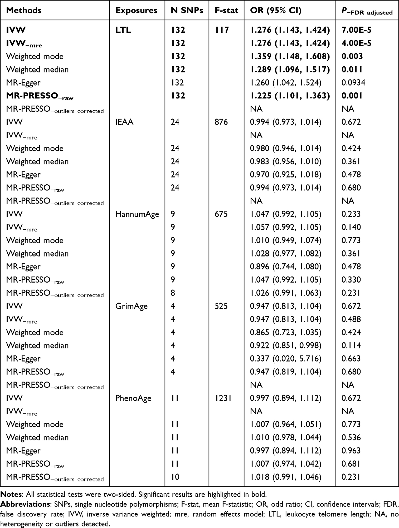 |
Table 1 The Casual Effect of Aging on Endometriosis by Mendelian Randomization |
 |
Figure 2 Scatter plots on the association between leukocyte telomere length and endometriosis. |
 |
Figure 3 Forest plots on the association between leukocyte telomere length and endometriosis. |
Nonetheless, no statistically significant associations were found between IEAA, HannumAge, GrimAge, PhenoAge, and endometriosis (see Table 1). Furthermore, sensitivity analysis using an independent dataset of endometriosis and LTL from meta-analyses yielded no significant results (OR-IVW = 1.128, 95% CI: 0.140 to 9.115, P = 0.910).
The Casual Effect of Endometriosis on Aging
According to Table 2, the IVW analysis indicated no causal association between endometriosis and LTL (β-IVW = 0.002, 95% CI = −0.007 to 0.012, P-value = 0.904). The remaining outcomes were consistent with the IVW findings.
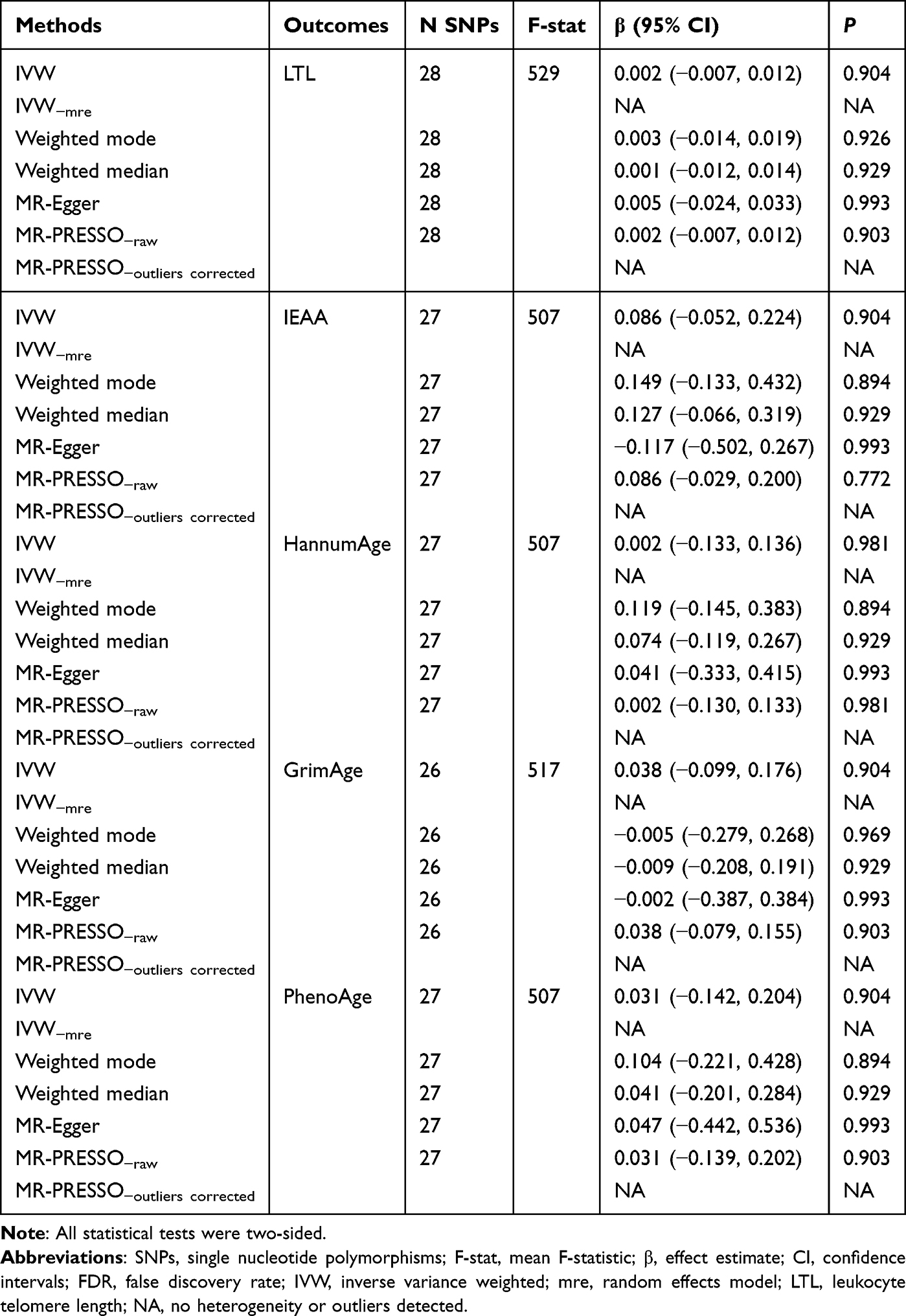 |
Table 2 The Casual Effect of Endometriosis on Aging by Mendelian Randomization |
The Casual Effect of LTL on Endometriosis Subtypes
The results of the MR analyses examining the association between LTL and the risk of endometriosis subtypes are presented in Table 3. In the subgroup analyses, a positive association was observed between LTL and endometriosis of the ovary (OR-IVW = 1.343, 95% CI = 1.143 to 1.577, P-value = 3.00E-4). Similarly, a significant positive association was found between LTL and endometriosis of the rectovaginal septum and vagina (OR-IVW = 1.336, 95% CI = 1.064 to 1.676, P-value = 0.013). However, no significant association was identified between LTL and endometriosis of the pelvic peritoneum (OR-IVW = 1.114, 95% CI = 0.966 to 1.284, P-value = 0.139). Consistent estimates were also observed using the weighted median, MR-Egger, and weighted mode methods.
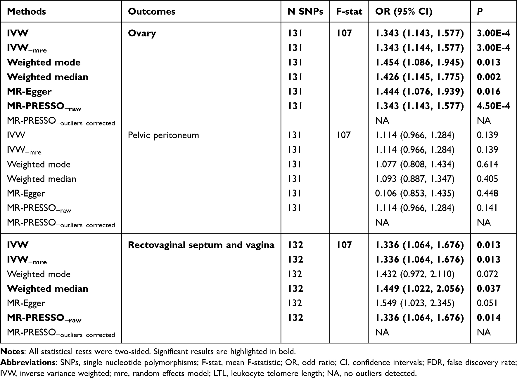 |
Table 3 The Casual Effect of Leukocyte Telomere Length on Endometriosis Subtype by Mendelian Randomization |
The Casual Effect of Endometriosis Subtypes on LTL
As presented in Table S2, the MR analysis found no evidence of a causal association between endometriosis subtypes and LTL.
Discussion
In this research, we employed bidirectional two-sample MR analysis to investigate the causal impact of aging on endometriosis risk, as well as its subtypes. Our findings provide novel insights into the potential role of biological aging in the development of endometriosis and its subtypes while addressing the reverse causality between these traits.
An encouraging discovery in this research is the causal relationship between aging and endometriosis, shedding new light on the disease’s development. In particular, the IVW technique indicated that the hallmark of aging, LTL, was causally and positively associated with endometriosis. This outcome was uniform across multiple MR techniques, such as MR-Egger, weighted median, and weighted mode analyses. Even after correcting for horizontal pleiotropy using the MR-PRESSO method, the association remained statistically significant. The results indicate that aging could contribute to the development of endometriosis. Telomeres, which protect chromosome ends, shorten with each cell division, and their length is considered a marker of aging.22 Longer telomeres in leukocytes might indicate a cellular environment that is more conducive to the development of endometriosis, possibly through mechanisms involving cellular senescence, immune response, or hormonal regulation.23–25 Interestingly, no significant associations were found between other aging biomarkers, such as IEAA, HannumAge, GrimAge, and PhenoAge, and the risk of endometriosis. This suggests that not all aging processes are equally relevant to the development of endometriosis. The lack of association with these other markers might reflect different biological pathways or mechanisms of aging that do not directly influence endometriosis.
To further validate the robustness of our findings, we conducted a sensitivity analysis using an independent dataset from a meta-analysis of endometriosis and LTL. Notably, this analysis did not yield significant results, suggesting that the observed association between LTL and endometriosis may be dataset-dependent and requires further investigation in larger and more diverse populations.
In contrast to the observed association between LTL and endometriosis risk, reverse MR analysis indicated no evidence of a causal relationship between endometriosis and LTL. These findings suggest that while aging-related biological processes may contribute to the development of endometriosis, the disease itself does not appear to influence systemic aging, at least in terms of telomere length dynamics.
Given the heterogeneity of endometriosis, we further explored its subtypes to assess whether specific forms of the disease exhibit stronger associations with LTL. Subgroup analyses revealed that longer LTL was significantly associated with an increased risk of ovarian endometriosis and endometriosis of the rectovaginal septum and vagina. However, no significant association was found between LTL and endometriosis of the pelvic peritoneum. These findings suggest that aging-related biological mechanisms may differentially influence distinct subtypes of endometriosis, particularly those affecting the ovary and rectovaginal regions. Consistency across different MR methods further supports the robustness of these results. Finally, to evaluate the potential impact of endometriosis subtypes on aging, we conducted reverse MR analyses, which demonstrated no significant causal effect of any endometriosis subtype on LTL. This further supports the hypothesis that aging-related processes, particularly telomere length maintenance, may contribute to the risk of developing endometriosis rather than the other way around.
The findings from our study suggest several potential mechanisms by which aging, as indicated by LTL, could influence the risk of endometriosis. Cellular senescence, a hallmark of aging, may contribute to the chronic inflammatory environment that promotes the development of endometriotic lesions.1,26 Additionally, the immune dysregulation associated with aging could impair the body’s ability to clear ectopic endometrial cells,27 facilitating their growth and proliferation.28 Besides, hormonal changes, such as decreased levels of ovarian hormones, could also play a role in this process.29 Fascinatingly, current research indicates that immune cells like macrophages, natural killer cells, dendritic cells, neutrophils, T cells, and B cells may enhance the vascularization and fibrogenesis of endometriotic lesions, thereby aiding the implantation and growth of ectopic endometrial tissue.30 Moreover, extended telomere might prevent immune cells from aging,16 potentially clarifying the link between LTL and endometriosis.
This research is the first to establish a causal relationship between aging and endometriosis, paving the way for further studies into the pathophysiological mechanisms connecting aging and endometriosis. Further studies could explore the specific biological pathways through which telomere length influences endometriosis risk, potentially identifying novel therapeutic targets. Clinically, these findings highlight the importance of considering aging in the management of endometriosis. Patients with endometriosis might benefit from interventions aimed at modulating telomere dynamics or addressing the broader impacts of biological aging. For instance, lifestyle interventions known to preserve telomere length, such as regular physical activity, a healthy diet, and stress reduction techniques, could be beneficial for patients with endometriosis.31,32 Upcoming studies ought to confirm our results in broader and more varied groups, while also delving deeper into the fundamental biological processes. Investigating the interactions between genetic, epigenetic, and environmental factors in the context of aging and endometriosis could provide a more comprehensive understanding of the disease. Additionally, longitudinal studies tracking aging and endometriosis progression over time would be valuable in establishing temporal relationships and causality.
While our study provides robust evidence for a causal relationship between aging and endometriosis, several limitations should be acknowledged. First, MR analyses rely on the availability of large, well-characterized GWAS datasets. Any biases or inaccuracies in these datasets could affect our findings. Secondly, the LTL measurements used in GWAS studies may not fully capture telomere dynamics in reproductive tissues, limiting their direct biological relevance to endometriosis. Thirdly, the lack of association observed in the independent dataset underscores the potential influence of population heterogeneity and dataset-specific factors, warranting further validation in diverse cohorts.
To sum up, this bidirectional two-sample MR analysis offers compelling proof of a causal relationship between aging and a heightened risk of endometriosis, particularly ovarian and rectovaginal subtypes. The results imply that aging, reflected by telomere length, significantly contributes to the incidence of endometriosis. Although other aging indicators did not exhibit the same results, the association between LTL and endometriosis underscores the intricate nature of aging and its effects on reproductive well-being. Additional studies are required to investigate the fundamental processes and possible treatment applications of these results.
Data Sharing Statement
All GWAS datasets used in this study were obtained from online publicly available summary statistics.
Ethics Statement
Since the GWAS data used in this study are derived entirely from publicly available summary-level statistics, ethical approval is not required. According to Article 32 of China’s Measures for the Ethical Review of Biomedical Research Involving Humans, which took effect on February 18, 2023, biomedical research involving human participants may be exempt from ethical review under the following conditions: 1. Item 1: Research based on public databases that does not involve the identification or use of personal information. 2. Item 2: Analysis of collected data that does not involve the collection of new data or direct intervention in participants. Based on these provisions, this study qualifies for an exemption from ethical review, as it exclusively utilizes publicly accessible data and involves no direct interaction with human subjects or collection of new personal information.
Author Contributions
All authors have made substantial contributions to this work, including but not limited to the conception, study design, execution, data acquisition, analysis, and interpretation. They have actively participated in drafting, revising, or critically reviewing the manuscript, provided final approval for the version to be published, and consented to the journal submission. All authors take full accountability for the integrity and accuracy of the work in all its aspects.
Funding
This research has received funding by Clinical research project of Shanghai Municipal Health Commission (Jichan Nie, 202440057).
Disclosure
The authors have no conflicts of interest to declare in this work.
References
1. Taylor HS, Kotlyar AM, Flores VA. Endometriosis is a chronic systemic disease: clinical challenges and novel innovations. Lancet. 2021;397(10276):839–852. doi:10.1016/S0140-6736(21)00389-5
2. Chapron C, Marcellin L, Borghese B, Santulli P. Rethinking mechanisms, diagnosis and management of endometriosis. Nat Rev Endocrinol. 2019;15(11):666–682. doi:10.1038/s41574-019-0245-z
3. Chiang JL, Shukla P, Pagidas K, et al. Mitochondria in ovarian aging and reproductive longevity. Ageing Res Rev. 2020;63:101168. doi:10.1016/j.arr.2020.101168
4. Wang L, Tang J, Wang L, et al. Oxidative stress in oocyte aging and female reproduction. J Cell Physiol. 2021;236(12):7966–7983. doi:10.1002/jcp.30468
5. Orisaka M, Mizutani T, Miyazaki Y, et al. Chronic low-grade inflammation and ovarian dysfunction in women with polycystic ovarian syndrome, endometriosis, and aging. Front Endocrinol. 2023;14:1324429.
6. Sekula P, Del Greco MF, Pattaro C, Köttgen A. Mendelian randomization as an approach to assess causality using observational data. J Am Soc Nephrol. 2016;27(11):3253–3265. doi:10.1681/ASN.2016010098
7. Birney E. Mendelian Randomization. Cold Spring Harb Perspect Med. 2022;12(4):a041302. doi:10.1101/cshperspect.a041302
8. Codd V, Wang Q, Allara E, et al. Polygenic basis and biomedical consequences of telomere length variation. Nat Genet. 2021;53(10):1425–1433. doi:10.1038/s41588-021-00944-6
9. McCartney DL, Min JL, Richmond RC, et al. Genome-wide association studies identify 137 genetic loci for DNA methylation biomarkers of aging. Genome Biol. 2021;22(1):194. doi:10.1186/s13059-021-02398-9
10. Kurki MI, Karjalainen J, Palta P, et al. FinnGen provides genetic insights from a well-phenotyped isolated population. Nature. 2023;613(7944):508–518. doi:10.1038/s41586-022-05473-8
11. Rahmioglu N, Mortlock S, Ghiasi M, et al. The genetic basis of endometriosis and comorbidity with other pain and inflammatory conditions. Nat Genet. 2023;55(3):423–436. doi:10.1038/s41588-023-01323-z
12. Keener R, Chhetri SB, Connelly CJ, et al. Validation of human telomere length multi-ancestry meta-analysis association signals identifies POP5 and KBTBD6 as human telomere length regulation genes. Nat Commun. 2024;15(1):4417.
13. Hemani G, Zheng J, Elsworth B, et al. The MR-Base platform supports systematic causal inference across the human phenome. Elife. 2018;7:e34408. doi:10.7554/eLife.34408
14. Auton A, Brooks LD, Durbin RM, et al.; 1000 Genomes Project Consortium. A global reference for human genetic variation. Nature. 2015;526(7571):68–74. doi:10.1038/nature15393
15. Park JH, Wacholder S, Gail MH, et al. Estimation of effect size distribution from genome-wide association studies and implications for future discoveries. Nat Genet. 2010;42(7):570–575. doi:10.1038/ng.610
16. Lawlor DA, Harbord RM, Sterne JA, Timpson N, Davey Smith G. Mendelian randomization: using genes as instruments for making causal inferences in epidemiology. Stat Med. 2008;27(8):1133–1163. doi:10.1002/sim.3034
17. Bowden J, Davey Smith G, Burgess S. Mendelian randomization with invalid instruments: effect estimation and bias detection through Egger regression. Int J Epidemiol. 2015;44(2):512–525. doi:10.1093/ije/dyv080
18. Bowden J, Davey Smith G, Haycock PC, Burgess S. Consistent estimation in Mendelian randomization with some invalid instruments using a weighted median estimator. Genet Epidemiol. 2016;40(4):304–314. doi:10.1002/gepi.21965
19. Huang D, Lin S, He J, Wang Q, Zhan Y. Association between COVID-19 and telomere length: a bidirectional Mendelian randomization study. J Med Virol. 2022;94(11):5345–5353. doi:10.1002/jmv.28008
20. Luo Q, Chen J, Qin L, et al. Psoriasis may increase the risk of lung cancer: a two-sample Mendelian randomization study. J Eur Acad Dermatol Venereol. 2022;36(11):2113–2119. doi:10.1111/jdv.18437
21. Rodríguez-Fernández B, Vilor-Tejedor N, Arenaza-Urquijo EM, et al. Genetically predicted telomere length and Alzheimer’s disease endophenotypes: a Mendelian randomization study. Alzheimers Res Ther. 2022;14(1):167. doi:10.1186/s13195-022-01101-9
22. Rossiello F, Jurk D, Passos JF, d’Adda Di Fagagna F. Telomere dysfunction in ageing and age-related diseases. Nat Cell Biol. 2022;24(2):135–147. doi:10.1038/s41556-022-00842-x
23. Liu J, Wang L, Wang Z, Liu JP. Roles of telomere biology in cell senescence, replicative and chronological ageing. Cells. 2019;8(1):54. doi:10.3390/cells8010054
24. Lanna A, Vaz B, D’Ambra C, et al. An intercellular transfer of telomeres rescues T cells from senescence and promotes long-term immunological memory. Nat Cell Biol. 2022;24(10):1461–1474. doi:10.1038/s41556-022-00991-z
25. Taheri M, Ghafouri-Fard S, Najafi S, et al. Hormonal regulation of telomerase activity and hTERT expression in steroid-regulated tissues and cancer. Cancer Cell Int. 2022;22(1):258. doi:10.1186/s12935-022-02678-9
26. Lin X, Dai Y, Tong X, et al. Excessive oxidative stress in cumulus granulosa cells induced cell senescence contributes to endometriosis-associated infertility. Redox Biol. 2020;30:101431. doi:10.1016/j.redox.2020.101431
27. Cheung P, Vallania F, Warsinske HC, et al. Single-cell chromatin modification profiling reveals increased epigenetic variations with aging. Cell. 2018;173(6):1385–1397.e14. doi:10.1016/j.cell.2018.03.079
28. Symons LK, Miller JE, Kay VR, et al. The immunopathophysiology of endometriosis. Trends Mol Med. 2018;24(9):748–762. doi:10.1016/j.molmed.2018.07.004
29. Koninckx PR, Ussia A, Adamyan L, Gomel V, Martin DC. Peritoneal fluid progesterone and progesterone resistance in superficial endometriosis lesions. Hum Reprod. 2022;37(2):203–211. doi:10.1093/humrep/deab258
30. Chen S, Liu Y, Zhong Z, Wei C, Liu Y, Zhu X. Peritoneal immune microenvironment of endometriosis: role and therapeutic perspectives. Front Immunol. 2023;14:1134663. doi:10.3389/fimmu.2023.1134663
31. Mundstock E, Zatti H, Louzada FM, et al. Effects of physical activity in telomere length: systematic review and meta-analysis. Ageing Res Rev. 2015;22:72–80. doi:10.1016/j.arr.2015.02.004
32. Canudas S, Becerra-Tomás N, Hernández-Alonso P, et al. Mediterranean diet and telomere length: a systematic review and meta-analysis. Adv Nutr. 2020;11(6):1544–1554. doi:10.1093/advances/nmaa079
 © 2025 The Author(s). This work is published and licensed by Dove Medical Press Limited. The
full terms of this license are available at https://www.dovepress.com/terms
and incorporate the Creative Commons Attribution
- Non Commercial (unported, 4.0) License.
By accessing the work you hereby accept the Terms. Non-commercial uses of the work are permitted
without any further permission from Dove Medical Press Limited, provided the work is properly
attributed. For permission for commercial use of this work, please see paragraphs 4.2 and 5 of our Terms.
© 2025 The Author(s). This work is published and licensed by Dove Medical Press Limited. The
full terms of this license are available at https://www.dovepress.com/terms
and incorporate the Creative Commons Attribution
- Non Commercial (unported, 4.0) License.
By accessing the work you hereby accept the Terms. Non-commercial uses of the work are permitted
without any further permission from Dove Medical Press Limited, provided the work is properly
attributed. For permission for commercial use of this work, please see paragraphs 4.2 and 5 of our Terms.