
Back to Journals » Biologics: Targets and Therapy » Volume 19
Berberine Intervention Mitigates Myocardial Ischemia-Reperfusion Injury in a Rat Model: Mechanistic Insights via miR-184 Signaling
Authors Yang H, Cao G, Li X, Zhao Z, Wang Y, Xu F
Received 3 September 2024
Accepted for publication 31 December 2024
Published 25 February 2025 Volume 2025:19 Pages 31—42
DOI https://doi.org/10.2147/BTT.S479430
Checked for plagiarism Yes
Review by Single anonymous peer review
Peer reviewer comments 2
Editor who approved publication: Dr Shein-Chung Chow
Haichen Yang,1,* Gang Cao,2,* Xia Li,3 Zhikun Zhao,4 Yong Wang,5 Fei Xu6
1Department of Emergency, The Affiliated Huai’an Hospital of Xuzhou Medical University and Huai’an second People’s Hospital, Huai’an, People’s Republic of China; 2Department of Respiratory Medicine, Hongze District People’s Hospital, Hongze, Jiangsu, People’s Republic of China; 3Department of Geriatric, The Affiliated Huai’an Hospital of Xuzhou Medical University and Huai’an Second People’s Hospital, Huai’an, People’s Republic of China; 4Department of Intensive Care Unit, The Affiliated Huai’an Hospital of Xuzhou Medical University and Huai’an Second People’s Hospital, Huai’an, People’s Republic of China; 5Department of Cardiology, The Affiliated Huai’an Hospital of Xuzhou Medical University and Huai’an Second People’s Hospital, Huai’an, People’s Republic of China; 6Department of Intensive Care Unit, Lianshui County People’s Hospital, Huai’an, People’s Republic of China
*These authors contributed equally to this work
Correspondence: Yong Wang, Department of Cardiology, The Affiliated Huai’an Hospital of Xuzhou Medical University and Huai’an Second People’s Hospital, Huai’an, People’s Republic of China, Email [email protected] Fei Xu, Department of Intensive Care Unit, Lianshui County People’s Hospital, Huai’an, People’s Republic of China, Email [email protected]
Background: Ischemia-reperfusion (I/R) injury is a major contributor to myocardial dysfunction and tissue damage. A natural alkaloid-Berberine having a wide range of pharmacological properties, has garnered interest for its potential cardioprotective properties. This study aimed to investigate the protective effects of berberine on myocardial tissue in a rat model of myocardial ischemia-reperfusion (I/R) injury. Additionally, the study explored the role of the miR-184/NOTCH1 signaling pathway in mediating these effects.
Methods: Male Wistar rats were randomly assigned to five groups: sham-operated control, I/R injury, I/R treated with berberine, I/R treated with inhibitor NC and I/R treated with a miR-184 inhibitor. The I/R injury was induced by ligating the left anterior descending (LAD) coronary artery for 30 minutes, followed by 2 hours of reperfusion. Berberine was administered orally at 100 mg/kg/day for 2 weeks, and the miR-184 inhibitor was administered via intraperitoneal injection. Hemodynamic parameters were recorded using a pressure sensor connected to a catheter inserted into the left ventricle. Myocardial infarct size was assessed using TTC staining, while histological and molecular changes were evaluated through H&E staining, TUNEL assay, and Western blotting. The expression levels of target genes were analyzed using quantitative real-time PCR (qRT-PCR).
Results: Berberine significantly reduced myocardial infarct size and improved hemodynamic parameters compared to the untreated I/R group. Additionally, berberine treatment attenuated apoptosis as evidenced by decreased TUNEL-positive cells. The miR-184 inhibitor also demonstrated protective effects by modulating key signaling pathways involved in myocardial injury. Western blot analysis revealed downregulation of NOTCH1 and HES1 expression in treated groups, indicating a potential mechanism for the observed cardio protection.
Conclusion: Berberine and miR-184 inhibition offer significant protection against myocardial ischemia-reperfusion injury. These findings suggest that targeting miR-184 and associated pathways may be a promising therapeutic strategy for reducing cardiac damage following ischemia-reperfusion.
Keywords: berberine, myocardial ischemia-reperfusion, cardioprotective effects, miR-184, NOTCH1 signaling pathway
Introduction
Coronary heart disease (CHD), also known as coronary artery disease, is a major public health concern globally.1 It results from the accumulation of atherosclerotic plaques in the coronary arteries, leading to reduced blood flow to the heart muscle. This condition often manifests as acute myocardial infarction (AMI) or angina pectoris, both of which can have severe health implications.2 Globally, CHD is the leading cause of death, with an estimated 9.4 million deaths attributed to it in 2019, according to the World Health Organization (WHO).3 The disease’s prevalence is linked to various risk factors, including hypertension, diabetes, smoking, and high cholesterol levels.4 The global burden of CHD is particularly concerning in high-income countries where lifestyle factors such as poor diet, lack of physical activity, and high rates of smoking contribute significantly to its prevalence.5 However, as socioeconomic conditions improve in low- and middle-income countries, the incidence of CHD is also rising in these regions.6 This shift underscores the need for effective prevention and treatment strategies on a global scale. In China, CHD has become a pressing health issue, with its incidence steadily increasing over recent decades. According to recent epidemiological studies, CHD is now a leading cause of morbidity and mortality in the country.7 Due to its high mortality rate, AMI is often referred to as the “number one killer” of human health.8 The advancements in coronary artery bypass surgery, percutaneous coronary intervention (PCI), and other revascularization techniques have significantly improved the outcomes for AMI patients by restoring blood flow to the ischemic myocardium.9 However, while these procedures are essential for salvaging acutely ischemic myocardium, the process of reestablishing blood flow can paradoxically lead to myocardial damage, a phenomenon known as ischemia-reperfusion (I/R) injury. Mitigating I/R injury while maximizing the benefits of reperfusion therapy remains a critical challenge in the treatment of AMI.10 Berberine, an isoquinoline alkaloid extracted from Coptidis rhizoma, is widely recognized in traditional Chinese medicine for its heat-clearing, detoxifying, and antibacterial properties.11 The molecular structure of berberine, with a formula of C20H18NO4, has been extensively studied for its pharmacological effects, particularly in the cardiovascular system. Beyond its traditional uses, berberine has demonstrated a wide range of cardiovascular benefits, including positive inotropic effects, antiarrhythmic properties, vasodilation, blood pressure reduction, and myocardial protection against ischemia.12,13 Additionally, berberine has shown potential in managing metabolic conditions such as type-II diabetes and obesity. Its cardiovascular benefits have led to its clinical application in treating heart failure, arrhythmias, and hypertension, as documented in various studies.14 Given berberine’s extensive cardiovascular effects, it is hypothesized that it may also play a protective role in myocardial ischemia-reperfusion injury.13 Previous study indicated that berberine protected Kawasaki disease-induced human coronary artery endothelial cells dysfunction by inhibiting of oxidative and endoplasmic reticulum stress.15 The Notch1 signaling pathway, an evolutionarily conserved pathway, plays a crucial role in various cellular processes, including cell growth, proliferation, differentiation, and organ development. Recent studies suggested that berberine regulates many oncogenic and tumor suppressor miRNAs implicated in different phases of cancer and stable coronary heart disease.16 Notch1 signaling is also implicated in pathological processes related to oxidative stress and inflammation, both of which are key components of ischemia-reperfusion injury. Recent research suggests that microRNAs (miRNAs) may exert cardioprotective effects by modulating the Notch1 signaling pathway.17 Specifically, certain miRNAs have been found to activate the Notch1 pathway, thereby contributing to cardio-protection.18 Despite these findings, the involvement of the miRNA/Notch1 signaling pathway in the cardioprotective effects of berberine has not been thoroughly investigated.19 In light of these considerations, the present study aims to explore the potential cardioprotective effects of berberine in a rat model of ischemia-reperfusion injury.20 The study focuses on assessing berberine’s impact on the miRNA/Notch1/Hes1 signaling pathway within the myocardium. By elucidating the mechanisms through which berberine confers protection against ischemia-reperfusion injury, this research seeks to advance our understanding of berberine’s therapeutic potential in the context of acute myocardial infarction and broader cardiovascular diseases. The findings of this study could pave the way for the development of novel therapeutic strategies targeting the miRNA/Notch1 pathway, ultimately improving outcomes for patients suffering from ischemia-reperfusion injuries.
Materials and Methods
All animal experiments conducted in this study were performed in accordance with the Declaration of Helsinki. The study was approved by the Animal Ethics Committee of Huai’an Second People’s Hospital, which ensured that all procedures involving animal subjects were carried out with the highest ethical standards. The study adhered to all relevant regulations regarding the use of laboratory animals, including obtaining prior approval from the Institutional Animal Care and Use Committee (IACUC) and following the 3Rs principles—Replacement, Reduction, and Refinement. This commitment to ethical standards was integral to ensuring the validity of the research findings while upholding the highest standards of animal welfare.
Agents
Berberine (C20H18NO4) and triphenyl tetrazolium chloride (TTC) were both obtained from Sigma-Aldrich (St. Louis, USA). Berberine was prepared by dissolving in saline for oral administration, while TTC was dissolved in saline for use in infarct size determination through staining.
Myocardial Ischemia-Reperfusion Rat Model and Various Interventions
Male Wistar rats, aged 12 weeks and weighing between 220–250 grams, were selected for the myocardial ischemia-reperfusion (I/R) model. Rats were maintained under a standard 12 h light: 12 h darkness cycle, and were housed in a controlled temperature (24±2 °C) and humidity (50±5%) environment. Rats were fed with standard rat laboratory chow and water ad libitum. For the induction of myocardial ischemia-reperfusion, the rats were first anesthetized using intraperitoneal injections of ketamine (85 mg/kg) and xylazine (9 mg/kg). Following anesthesia, a 20 G catheter was carefully inserted into the trachea and connected to a ventilator to maintain respiration. A midline incision was made to expose the heart by carefully cutting the pericardium with blunt-ended scissors. The left anterior descending (LAD) coronary artery, located approximately 2 mm below the left auricle, was temporarily ligated using a 6–0 polypropylene suture and a tapered needle. This ligation induced myocardial ischemia by blocking blood flow for 30 minutes. After the ischemic period, the suture was loosened to allow reperfusion of the myocardium for 2 hours. The rats were randomly divided into four experimental groups (n=5 per group): (1) the sham surgery group, where rats underwent thoracotomy without ligation of the LAD artery; (2) the I/R group, where rats underwent LAD artery ligation for 30 minutes followed by 2 hours of reperfusion without any additional treatment; (3) the I/R + Berberine group, where rats received oral administration of berberine at a dose of 100 mg/kg daily for 2 weeks prior to LAD artery ligation and reperfusion; (4) the I/R + inhibitor NC group, where rats were administered inhibitor NC (Riobio, Guangzhou, China) via intraperitoneal injection at 200nmoL daily for 2 weeks prior to LAD artery ligation and reperfusion; and (5) the I/R + miR-184 Inhibitor group, where rats were administered miR-184 inhibitor (Riobio, Guangzhou, China) via intraperitoneal injection at 200nmoL daily for 2 weeks prior to LAD artery ligation and reperfusion. Using anesthetics or euthanasia methods during the experimental process to alleviate the pain of animals. At the conclusion of the reperfusion period, the rats were euthanized with a high dose of sodium pentobarbital (75 mg/kg, intraperitoneally). The chest cavity was reopened, and the heart was carefully excised. The heart was then divided into two sections: the apex was reserved for histological analysis, and the base was used for biochemical assays to assess tissue parameters.
Hemodynamic Detection
To assess the cardiac dysfunction induced by ischemia-reperfusion (I/R) injury, comprehensive hemodynamic measurements were conducted on the experimental rats. The procedure began with the careful isolation of the right common carotid artery (CCA). A small incision was made on the exposed right CCA to allow the insertion of a polyethylene catheter. This catheter was carefully advanced into the left ventricle to enable accurate measurement of cardiac parameters. Once the catheter was properly positioned within the left ventricle, it was connected to a high-precision pressure sensor capable of capturing detailed hemodynamic data. The sensor was interfaced with LabChart-7 software, provided by ADI Instruments, to facilitate the real-time recording and analysis of various cardiac function variables. Several key hemodynamic parameters were continuously monitored throughout the procedure. These included the left ventricular systolic pressure (LVSP), which reflects the pressure generated during ventricular contraction; the maximum rate of rise of left ventricular pressure (+dp/dtmax), an indicator of the contractile strength of the heart; and the maximum rate of decline of left ventricular pressure (-dp/dtmax), which provides insight into the rate of ventricular relaxation. The electrophysiological recording device used for this purpose was sourced from BioPAC Systems, USA, ensuring high accuracy and reliability of the recorded data. These measurements were crucial for evaluating the extent of cardiac dysfunction caused by the I/R injury, providing detailed insights into the heart’s mechanical performance under the experimental conditions.
Quantitative Real-Time PCR (qRT-PCR) Analysis
qRT-PCR assay was employed to quantify the expression levels of target mRNAs in myocardial tissues. A total of RNA was extracted from the myocardial tissues using the TRIzol® (Takara Inc). The quality and quantity of RNA accepted with absorbance ratios (A260/A280) between 1.8 and 2.0, indicating pure RNA samples. The reverse transcription was carried out in a thermal cycler under the following conditions: 37°C for 15 minutes, 85°C for 5 seconds, and then held at 4°C. The cDNA was quantified using a spectrophotometer to ensure accurate input for subsequent qPCR analysis. A total of 4 µg of the extracted RNA was reverse transcribed into cDNA using the PrimeScript SYBR Premix Ex Taq™ II kit, also supplied by Takara Inc. The reverse transcription was carried out in a thermal cycler under the following conditions: 37°C for 15 minutes, 85°C for 5 seconds, and then held at 4°C. The cDNA was quantified using a spectrophotometer to ensure accurate input for subsequent qPCR analysis. The PCR reaction mixture consisted of 10 µL of PrimeScript SYBR Premix Ex Taq™ II (2x), 0.8 µL of each forward and reverse primer (10 µM), 2 µL of cDNA template, and 6.4 µL of RNase-free water, making a total volume of 20 µL per reaction. The specific primers for the target mRNAs, as shown in Table 1, were designed and supplied by Takara Inc. to ensure specificity and optimal amplification efficiency for each gene analyzed. The qRT-PCR was performed under the following cycling conditions: initial denaturation at 95°C for 30 seconds, followed by 40 cycles of 95°C for 5 seconds and 60°C for 34 seconds. Melt curve analysis was conducted at the end of the amplification to verify the specificity of the PCR products. During the amplification process, the expression levels of the target mRNAs were normalized to GAPDH, a commonly used housekeeping gene, to account for any variations in RNA input. The relative expression levels of the mRNAs were then calculated using the 2(-ΔΔCq) method, a widely accepted approach for quantifying gene expression changes relative to a control or baseline condition. This method provided a reliable quantification of the gene expression levels, facilitating the analysis of the impact of various treatments on the myocardial tissues at the molecular level.
 |
Table 1 Specific Primers for Target mRNAs Used in qRT-PCR Analysis |
TTC Staining
The hearts were sliced into 4-mm sections and promptly submerged in a 1% TTC solution dissolved in phosphate buffer for a duration of 20 mins at room temperature. Following three washes, the slices were captured in photographs, and ImageJ (Image-Pro Plus 6.0) was employed to compare infarct area with total area.21
Hematoxylin and Eosin (H&E) Staining
The apex of the heart was promptly excised, washed with ice-cold 0.9% saline solution, and then fixed in a 4% paraformaldehyde solution at pH 7.4. Subsequently, the fixed tissues were embedded in paraffin wax. Sections of the paraffin-embedded tissues, with a thickness of 4 μm, were prepared and subjected to H&E staining.22
TUNEL Assay
The heart was cut in half, and the other half was fixed in a 4% paraformaldehyde solution for 60 minutes, followed by permeabilization with 0.1% Triton X-100 for 10 minutes. Subsequently, the hearts were washed three times with PBS. The In Situ Cell Death Detection Kit from Beyotime was used for the experimental procedure.23 Images were captured using an Olympus fluorescence microscope at a magnification of 400×.
Western Blotting Assay
We extracted total protein from rat heart tissues using 1% PMSF and determined the protein sample concentrations using the BCA method. Each sample’s protein (30μg) underwent 10% SDS-PAGE at 80V for 2.5 hours and was subsequently transferred onto PVDF membranes. After blocking the membranes with skimmed milk solution for 1 hour, we first incubated the membranes with primary antibodies against NOTCH1 (Abcam, ab52627, 1:1000) and HES1 (CST,#11988, 1:1000) at 4°C overnight, followed by incubation with secondary antibodies (thermo, 31460, 1:10000) for 120 minutes at 37°C. Protein bands were developed using ECLPlus reagent, and images were captured in gel imaging system. The relative expression levels of proteins were calculated with GAPDH as the internal reference protein and compared to the control group.
Statistical Analysis
All data were presented as mean ± standard deviation. One-way analysis of variance was performed followed by multiple comparisons using Tukey’s method. GraphPad Prism version 6.01 (GraphPad Software, La Jolla, CA) was used for all statistical analysis and graphical construction, with a significance level set at 0.05.
Results
Effects of Berberine Intervention on Myocardial Tissue in Myocardial Ischemia-Reperfusion Rats
The myocardial ischemia-reperfusion model in rats demonstrated a substantial increase in the area of ischemic myocardial tissue, as visualized by the TTC staining (Figure 1A). In the sham group, where no ischemia was induced, the myocardial tissue sections showed no signs of ischemia, appearing uniformly stained. In contrast, the MIR group displayed large, pale, non-stained areas indicative of ischemic damage, which was significantly more extensive compared to the sham group. Quantitative analysis of the ischemic area (Figure 1B) revealed a marked increase in the ischemic zone in the MIR group compared to the sham group (p < 0.001). However, in the group treated with berberine (MIR+BBR), there was a significant reduction in the ischemic area. The TTC staining in these rats showed a smaller pale region, indicating that berberine effectively reduced myocardial ischemic damage. Quantification of the ischemic area in the MIR+BBR group confirmed this observation, with a significant decrease compared to the untreated MIR group (Figure 1B). The body weight of rats was measured over the course of the experiment. The body weights of the rats in the sham, MIR, and MIR+BBR groups were monitored over 14 days (Figure 1C). No significant differences were observed in the body weight trajectories among the groups, indicating that the berberine treatment did not adversely affect the overall growth or health of the animals during the experimental period. Histological analysis of myocardial tissue sections stained with Hematoxylin and Eosin (H&E) further supported the protective effects of berberine. Myocardial tissue in the sham group exhibited normal architecture with well-organized muscle fibers. In contrast, the MIR group displayed significant structural disorganization, including extensive fiber disruption and increased interstitial space, indicative of severe myocardial injury. However, the MIR+BBR group showed a notable preservation of myocardial structure, with reduced fiber disruption and interstitial expansion compared to the MIR group (Figure 1D). This suggests that berberine treatment mitigated the histological damage associated with myocardial ischemia-reperfusion. The present results demonstrate that berberine significantly reduces myocardial ischemic injury in the MIR rat model, as evidenced by decreased ischemic area and preserved myocardial architecture, without affecting the overall body weight of the animals.
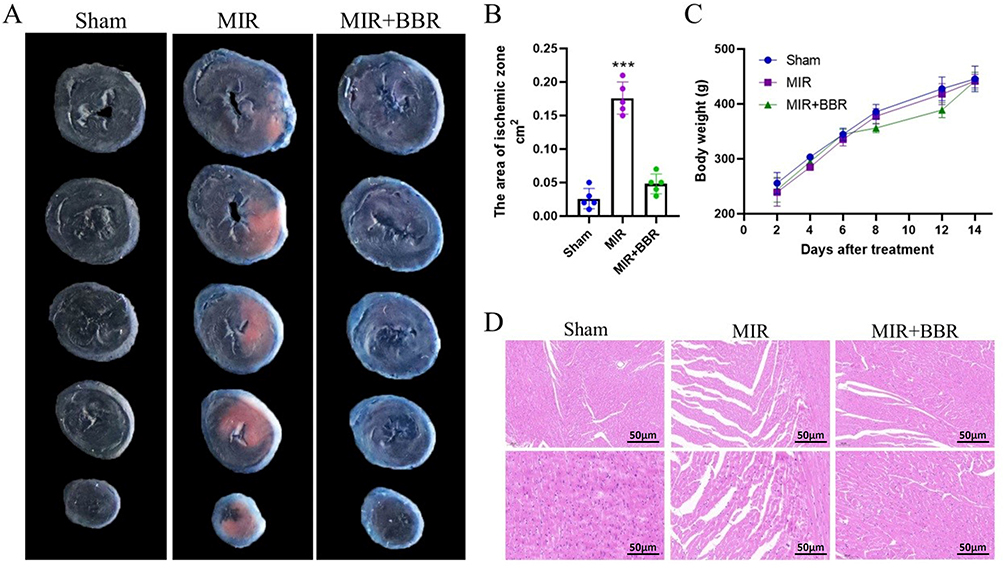 |
Figure 1 Effects of Berberine intervention on myocardial tissue in a rat model of myocardial ischemia-reperfusion. (A) TTC staining of the heart tissues of control rats, the myocardial ischemia-reperfusion rats with and without berberine intervention. (B) Bar graph illustrating the myocardial ischemia-reperfusion area in the heart tissues of control rats, the myocardial ischemia-reperfusion rats with and without berberine intervention. (C) Body weight of control rats, and myocardial ischemia-reperfusion rats with and without berberine intervention. (D) H&E staining of the heart tissues of control rats, the myocardial ischemia-reperfusion rats with and without berberine intervention. N = 5. ***P< 0.001. |
Berberine Intervention Restore Various Myocardial Functional Indicators
The MIR is a condition characterized by a temporary reduction in blood flow to the heart muscle (ischemia) followed by the restoration of blood flow (reperfusion). This process often leads to significant cardiac dysfunction, as indicated by changes in several key myocardial functional indicators. Following MIR in rats, a marked increase in left ventricular end-diastolic pressure (LVEDP) is observed (Figure 2A). LVEDP is a critical measure of the heart’s diastolic function, and an elevated LVEDP reflects increased pressure in the left ventricle at the end of diastole, suggesting that the heart is struggling to fill properly due to the damage caused by ischemia-reperfusion. In contrast, other essential myocardial functional indicators decrease following MIR, including the maximal rate of pressure increase (+dp/dt), heart rate (HR), left ventricular systolic pressure (LVSP), maximal rate of pressure decrease (-dp/dt), and coronary flow (CF): +dp/dt (Figure 2B) measures the heart’s contractile strength, and its reduction indicates weakened contractions post-MIR. HR reflects the number of heart beats per minute, and a decrease in HR suggests impaired cardiac function due to MIR (Figure 2C) while LVSP results represents the pressure generated by the left ventricle during systole, and its reduction signifies compromised pumping ability (Figure 2D). Additional, -dp/dt measures the heart’s rate of relaxation, with a lower value indicating impaired diastolic function (Figure 2E) and CF is indicative of the blood flow through the coronary arteries, and a decrease suggests reduced blood supply to the heart muscle, which is vital for recovery post-MIR (Figure 2F). Berberine (BBR) intervention has demonstrated promising results in mitigating the adverse effects of MIR in this rat model. The administration of Berberine significantly restores these myocardial functional indicators. These findings highlight Berberine’s potential as a therapeutic agent in restoring myocardial function after ischemia-reperfusion injury, positioning it as a promising candidate for further research and potential clinical application.
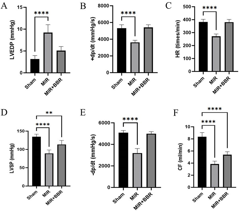 |
Figure 2 Berberine intervention can restore various myocardial functional indicators in a rat model of myocardial ischemia-reperfusion. (A) LVEDP, (B) +dp/dt, (C) HR, (D) VSP, (E) -dp/dt, and (F) CF in control rats, the myocardial ischemia-reperfusion rats with and without berberine intervention. N = 5. **P < 0.01 and ****P < 0.0001. |
Effects of miR-184 Expression Inhibition on Myocardial
Transcriptome analysis has revealed that miR-184 is highly expressed in ischemic myocardial tissue following MIR (Figure 3A). This elevated expression suggests that miR-184 plays a significant role in the pathological processes associated with ischemia-reperfusion injury in the heart. Pre-treatment with berberine has been shown to effectively reduce the expression level of miR-184 in ischemic myocardial tissue (Figure 3B). This downregulation of miR-184, facilitated by berberine, indicates that the compound may exert its cardioprotective effects by modulating the expression of specific miRNAs involved in myocardial injury. Further corroborating these findings, the results of TTC staining, demonstrate that the downregulation of miR-184 significantly reduces the ischemic area in the myocardial tissue of the MIR rat model (Figure 3C). Quantitative analysis of the ischemic zone size confirms this observation, with a marked reduction in the ischemic area following miR-184 inhibition (Figure 3D). These results suggest that miR-184 contributes to the extent of ischemic damage in myocardial tissue and that its inhibition, particularly through berberine treatment, can mitigate the effects of myocardial ischemia-reperfusion injury. This highlights the therapeutic potential of targeting miR-184 in the treatment of ischemic heart disease.
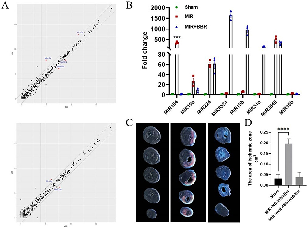 |
Figure 3 Effects of miR-184 expression inhibition on myocardial tissue in a rat model of myocardial ischemia-reperfusion. (A) Transcriptomic analysis in the heart tissues between control rats and myocardial ischemia-reperfusion rats. (B) Validation of miRNA expression levels in heart tissues of control rats, the myocardial ischemia-reperfusion rats with and without miR_184 inhibitor intervention. (C) TTC staining of the heart tissues of control rats, the myocardial ischemia-reperfusion rats with and without miR-184 inhibitor intervention. (D) Bar graph illustrating the myocardial ischemia-reperfusion area in the heart tissues of control rats, the myocardial ischemia-reperfusion rats with and without miR-184 inhibitor intervention. N = 3. ***P< 0.001 and ****P < 0.0001. |
Effects of the miR-184/NOTCH1 Signaling Pathway on Myocardial Cell Apoptosis
Myocardial cell apoptosis is significantly increased in rats subjected to myocardial ischemia reperfusion (MIR), as shown by the increased incidence of apoptosis in the MIR+NC-inhibitor group compared to the sham group (Figure 4A and B). This increase in apoptosis suggests that MIR induces substantial cell death in myocardial tissue, contributing to cardiac dysfunction. However, inhibition of miR-184 expression effectively reduces the level of myocardial cell apoptosis in these rats. The reduction in apoptosis observed in the MIR+miR-184-inhibitor group (Figure 4A and B) highlights the potential protective role of miR-184 inhibition against MIR-induced myocardial injury. Additionally, the protein expression levels of NOTCH1 and HES1, two critical components of the NOTCH1 signaling pathway, are upregulated in the myocardial cells of MIR rats, indicating activation of this pathway in response to ischemia-reperfusion injury (Figure 4C and D). Interestingly, the inhibition of miR-184 expression reverses the upregulation of NOTCH1 and HES1 protein levels, as evidenced by the lower expression levels in the MIR+miR-184-inhibitor group compared to the MIR+NC-inhibitor group (Figure 4C and D) (Supplementary Figure 1). These findings suggest that miR-184 may contribute to myocardial cell apoptosis through the NOTCH1 signaling pathway and that targeting miR-184 could be a therapeutic strategy to mitigate myocardial apoptosis and the associated cardiac damage in myocardial ischemia-reperfusion injury.
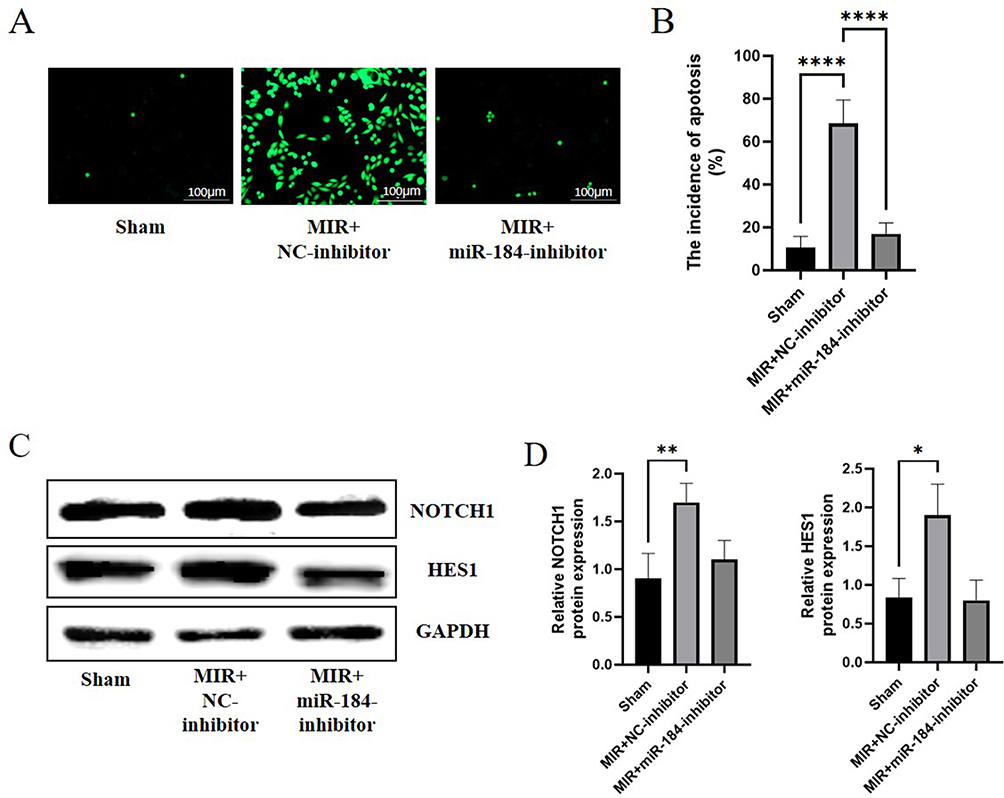 |
Figure 4 Effects of the miR-184/NOTCH1 signaling pathway on myocardial cell apoptosis. (A) TUNEL assay analysis of heart tissues of control rats, the myocardial ischemia-reperfusion rats with inhibitor NC and with miR_184 inhibitor intervention at 100µm. (B) Bar graph illustrating the apoptosis in the heart tissues of control rats, the myocardial ischemia-reperfusion rats with and without miR-184 inhibitor intervention. (C) Western blot analysis of NOTCH1 and HES1 proteins in the heart tissues of control rats, the myocardial ischemia-reperfusion rats with and without miR-184 inhibitor intervention. (D) Bar graph illustrating the protein levels of NOTCH1 and HES1 in the heart tissues of control rats, the myocardial ischemia-reperfusion rats with and without miR-184 inhibitor intervention. N = 3. *P< 0.05, **P< 0.01 and ****P < 0.0001. |
Discussion
This study delves into the cardioprotective effects of berberine in a rat model of myocardial ischemia-reperfusion (I/R) injury, emphasizing the role of the miR-184/NOTCH1 signaling pathway in mediating these effects. The findings contribute to a growing body of evidence that highlights berberine’s potential as a therapeutic agent for myocardial injury across various experimental models. Previous research has consistently supported berberine’s efficacy in mitigating myocardial ischemia-reperfusion injury through diverse mechanisms. Almowallad S et al indicated the potential treatment strategy of berberine in cardiovascular diseases via targeting SIGMAR1, GRP78, and CASP3 using in silico prospective screening approaches.24 In addition, BBR could repress HMGB1-mediated TLR4/NF-κB signalling pathway through miR-340-5p to suppress cardiomyocyte apoptosis and inflammation.25 Jia et al demonstrated that berberine alleviates myocardial injury by attenuating inflammatory responses and oxidative stress.26 This aligns with evidence showing that oxidative stress and inflammation are critical contributors to myocardial damage during ischemia-reperfusion events. Zhu et al further elaborated on berberine’s protective effects by revealing its role in inhibiting the PI3K/AKT signaling pathway.27 This pathway is pivotal for cell survival and metabolic processes, suggesting that berberine’s inhibition of this pathway helps preserve myocardial integrity during stress. Moreover, Wang et al provided evidence that berberine protects against cardiac injury by modulating apoptosis through the activation of Smad7, a protein involved in cell death regulation.28 Additionally, Zhu et al highlighted berberine’s role in modulating the mitophagy-mediated HIF-1α/BNIP3 pathway, further underscoring its multifaceted cardioprotective properties.29 A natural compound berberine, known for its diverse pharmacological activities, has been recognized for its potential cardioprotective effects.
In our study, berberine treatment significantly reduced the myocardial ischemic area and alleviated tissue damage resulting from I/R injury. These findings corroborate previous research documenting berberine’s ability to mitigate myocardial injury and enhance cardiac function in various ischemic heart disease models.30 Notably, berberine intervention restored critical myocardial functional indicators disrupted by I/R, such as left ventricular end-diastolic pressure (LVEDP), +dp/dt, HR, ventricular systolic pressure (VSP), -dp/dt, and coronary flow (CF). The improvement in these parameters suggests that berberine not only prevents myocardial injury but also supports recovery of cardiac function following ischemic events. Previous studies have indicated that berberine can counteract sunitinib-induced cardiac dysfunction by normalizing calcium regulation through SGK1 activation.31 Beyond its direct effects on myocardial tissue, berberine also influences the miR-184/NOTCH1 signaling pathway, which plays a significant role in the pathophysiology of I/R injury. Our study found that berberine led to a downregulation of miR-184 expression. This reduction was associated with a decreased ischemic area and diminished myocardial cell apoptosis. miR-184 is known to contribute to I/R injury by modulating target genes involved in crucial cellular processes, such as apoptosis and inflammation. Consistent with these studies, Chang et al demonstrated that berberine treatment protects against cardiac dysfunction and remodeling in type-2 diabetic rats by activating 5’-adenosine monophosphate-activated protein kinase.32 One notable target of miR-184 is the NOTCH1 receptor, which is involved in a range of cellular functions including development, differentiation, and tissue homeostasis.33,34 In multiple myeloma, miR-184 expression is increased and promotes MM cell proliferation and colony formation by regulating Notch1 expression.35 In the context of myocardial ischemia-reperfusion injury, NOTCH1 signaling plays a critical role in the underlying pathophysiological mechanisms.36 Studies have shown that the activation of NOTCH1 signaling is significantly altered during myocardial ischemia-reperfusion,37 with its activation being associated with cardioprotective effects, including the regulation of cell survival, inflammation, and angiogenesis. Specifically, NOTCH1 activation has been shown to mitigate myocardial ischemia-reperfusion injury by reducing cell death, inflammation, and oxidative stress.38 Conversely, dysregulation of NOTCH1 signaling has been linked to increased myocardial injury,39 inflammation, and apoptosis, suggesting that miR-184/NOTCH1 signaling could serve as a valuable biomarker for assessing myocardial injury severity and predicting patient outcomes.40 Our findings align with previous research that implicates the dysregulation of miR-184 and aberrant NOTCH1 signaling in the pathogenesis of myocardial ischemia-reperfusion injury. This study adds to the existing body of literature by further elucidating the cardioprotective effects of berberine, particularly through its modulation of the miR-184/NOTCH1 signaling pathway. The observed restoration of myocardial function, reduction in tissue damage, and regulation of this critical pathway highlight the therapeutic potential of berberine in managing ischemic heart disease. Nevertheless, further investigations, including clinical trials, are warranted to fully understand the underlying mechanisms and establish the translational value of berberine-based interventions in the treatment of myocardial ischemia-reperfusion injury. While the results of this study are promising, future research will address several important areas. First, the findings will be extended to human models to better understand how they translate to the complex pathophysiology of myocardial ischemia-reperfusion injury in clinical settings. Additionally, future studies will explore the long-term effects of berberine, including its impact on chronic heart failure. Finally, optimizing the dosage and administration regimen of berberine will be a focus to ensure its efficacy and safety for human use. Comprehensive clinical trials will be essential for fully elucidating the mechanisms involved and confirming the clinical benefits of berberine-based therapies for myocardial ischemia-reperfusion injury.
Conclusion
Our study demonstrates that berberine significantly mitigates myocardial ischemia-reperfusion injury in a rat model, and the relationship between berberine and miR-184/NOTCH1 signaling pathway needs further research. The observed improvements in myocardial function, reduction in tissue damage, and modulation of critical signaling pathways highlight berberine’s potential as a therapeutic agent for myocardial ischemia-reperfusion injury. However, to fully understand its clinical applicability and efficacy, further research, including well-designed clinical trials, is essential. These studies should address the limitations identified, explore additional mechanisms of action, and determine the optimal therapeutic strategies for berberine in the context of ischemic heart disease.
Data Sharing Statement
The data and materials used in this study are available upon request. Researchers interested in accessing the dataset or related materials for academic and non-commercial purposes can contact the corresponding author for further information.
Ethics Approval
Present study approved by the Animal Ethics Committee of Huai’an Second People’s Hospital AUP-230615-DSW-0596-01, which ensured that all procedures involving animal subjects were carried out with the highest ethical standards.
Author Contributions
All authors made a significant contribution to the work reported, whether that is in the conception, study design, execution, acquisition of data, analysis and interpretation, or in all these areas; took part in drafting, revising or critically reviewing the article; gave final approval of the version to be published; have agreed on the journal to which the article has been submitted; and agree to be accountable for all aspects of the work.
Funding
There is no funding to report.
Disclosure
The authors declare no competing interests.
References
1. Grimes DS. An epidemic of coronary heart disease. QJM: Int J Med. 2012;105(6):509–518. doi:10.1093/qjmed/hcr265
2. Makover ME, Shapiro MD, Toth PP. There is urgent need to treat atherosclerotic cardiovascular disease risk earlier, more intensively, and with greater precision: a review of current practice and recommendations for improved effectiveness. Am J Preventive Cardiol. 2022;12:100371. doi:10.1016/j.ajpc.2022.100371
3. Dai Z, Zhang X. Pathophysiology and clinical impacts of chronic kidney disease on coronary artery calcification. J Cardiovasc Dev Dis. 2023;10(5):207. doi:10.3390/jcdd10050207
4. Nakamura M, Yamamoto Y, Imaoka W, et al. Relationships between smoking status, cardiovascular risk factors, and lipoproteins in a large Japanese population. J Atheroscler Thromb. 2021;28(9):942–953. doi:10.5551/jat.56838
5. Ghodeshwar GK, Dube A, Khobragade D. Impact of lifestyle modifications on cardiovascular health: a narrative review. Cureus. 2023;15(7):e42616. doi:10.7759/cureus.42616
6. Roth GA, Mensah GA, Johnson CO, et al. Global burden of cardiovascular diseases and risk factors, 1990-2019: update from the GBD 2019 study. J Am Coll Cardiol. 2020;76(25):2982–3021. doi:10.1016/j.jacc.2020.11.010
7. Liang X, He H, Zeng H, et al. The relationship between polycystic ovary syndrome and coronary heart disease: a bibliometric analysis. Front Endocrinol. 2023;14:1172750. doi:10.3389/fendo.2023.1172750
8. Liu Q, Shi RJ, Zhang YM, et al. Risk factors, clinical features, and outcomes of premature acute myocardial infarction. Front Cardiovasc Med. 2022;9:1012095. doi:10.3389/fcvm.2022.1012095
9. Abubakar M, Javed I, Rasool HF, et al. Advancements in percutaneous coronary intervention techniques: a comprehensive literature review of mixed studies and practice guidelines. Cureus. 2023. 15(7):e41311. doi:10.7759/cureus.41311
10. Zhao Q, Xu H, Zhang X, et al. Current status and hospital-level differences in care and outcomes of patients with acute non-st-segment elevation myocardial infarction in China: insights from China acute myocardial infarction registry. Front Cardiovasc Med. 2021;8:800222. doi:10.3389/fcvm.2021.800222
11. Wang J, Wang L, Lou GH, et al. Coptidis Rhizoma: a comprehensive review of its traditional uses, botany, phytochemistry, pharmacology and toxicology. Pharm Biol. 2019;57(1):193–225. doi:10.1080/13880209.2019.1577466
12. Lau CW, Yao XQ, Chen ZY, Ko WH, Huang Y. Cardiovascular actions of berberine. Cardiovasc Drug Rev. 2001;19(3):234–244. doi:10.1111/j.1527-3466.2001.tb00068.x
13. Ai X, Yu P, Peng L, et al. Berberine: a review of its pharmacokinetics properties and therapeutic potentials in diverse vascular diseases. Front Pharmacol. 2021;12. doi:10.3389/fphar.2021.762654
14. Cai Y, Xin Q, Lu J, et al. A new therapeutic candidate for cardiovascular diseases: berberine. Front Pharmacol. 2021;12. doi:10.3389/fphar.2021.631100
15. Xu M, Qi Q, Men L, et al. Berberine protects Kawasaki disease-induced human coronary artery endothelial cells dysfunction by inhibiting of oxidative and endoplasmic reticulum stress. Vascul Pharmacol. 2020;127:106660. doi:10.1016/j.vph.2020.106660
16. Ali M, Mishra D, Singh RP. Cancer pathways targeted by berberine: role of microRNAs. Curr Med Chem. 2024;31(32):5178–5198. doi:10.2174/0109298673275121231228124031
17. Shi Q, Xue C, Zeng Y, et al. Notch signaling pathway in cancer: from mechanistic insights to targeted therapies. Sig Transduct Target Ther. 2024;9(1):1–37. doi:10.1038/s41392-024-01828-x
18. Marracino L, Fortini F, Bouhamida E, et al. Adding a “Notch” to cardiovascular disease therapeutics: a MicroRNA-based approach. Front Cell Dev Biol. 2021;9:695114. doi:10.3389/fcell.2021.695114
19. Peng X, Wang S, Chen H, Chen M. Role of the Notch1 signaling pathway in ischemic heart disease (Review). Int J Mol Med. 2023;51(3):27. doi:10.3892/ijmm.2023.5230
20. Cipriano GL, Schepici G, Mazzon E, Anchesi I. Multiple sclerosis: roles of miRNA, lcnRNA, and circRNA and their implications in cellular pathways. Int J mol Sci. 2024;25(4):2255. doi:10.3390/ijms25042255
21. Yang T, Liu H, Yang C, et al. Galangin attenuates myocardial ischemic reperfusion-induced ferroptosis by targeting Nrf2/Gpx4 signaling pathway. Drug Des Devel Ther. 2023;17:2495–2511. doi:10.2147/DDDT.S409232
22. Hristu R, Stanciu SG, Dumitru A, et al. Influence of hematoxylin and eosin staining on the quantitative analysis of second harmonic generation imaging of fixed tissue sections. Biomed Opt Express. 2021;12(9):5829–5843. doi:10.1364/BOE.428701
23. Zhang GY, Lu D, Duan SF, et al. Hydrogen sulfide alleviates lipopolysaccharide-induced diaphragm dysfunction in rats by reducing apoptosis and inflammation through ROS/MAPK and TLR4/NF- κB signaling pathways. Oxid Med Cell Longev. 2018;2018(1):9647809. doi:10.1155/2018/9647809
24. Almowallad S, Al-Massabi R. Berberine modulates cardiovascular diseases as a multitarget-mediated alkaloid with insights into its downstream signals using in silico prospective screening approaches. Saudi J Biol Sci. 2024;31(5):103977. doi:10.1016/j.sjbs.2024.103977
25. Long T, Pan W, Li F, Sheikh SA, Xie Q, Zhang C. Berberine up-regulates miR-340-5p to protect myocardial ischaemia/reperfusion from HMGB1-mediated inflammatory injury. ESC Heart Fail. 2023;10(2):931–942. doi:10.1002/ehf2.14235
26. Jia X, Shao W, Tian S. Berberine alleviates myocardial ischemia-reperfusion injury by inhibiting inflammatory response and oxidative stress: the key function of miR-26b-5p-mediated PTGS2/MAPK signal transduction. Pharm Biol. 2022;60(1):652–663. doi:10.1080/13880209.2022.2048029
27. Qin-Wei Z, Yong-Guang LI. Berberine attenuates myocardial ischemia reperfusion injury by suppressing the activation of PI3K/AKT signaling. Exp Ther Med. 2016;11(3):978–984. doi:10.3892/etm.2016.3018
28. Yao W, Wang X, Xiao K. Protective effect of berberine against cardiac ischemia/reperfusion injury by inhibiting apoptosis through the activation of Smad7. mol Cell Probes. 2018;38:38–44. doi:10.1016/j.mcp.2017.12.002
29. Zhu N, Li J, Li Y, et al. Berberine protects against simulated ischemia/reperfusion injury-induced H9C2 cardiomyocytes apoptosis in vitro and myocardial ischemia/reperfusion-induced apoptosis in vivo by regulating the mitophagy-mediated HIF-1α/BNIP3 Pathway. Front Pharmacol. 2020;11:367. doi:10.3389/fphar.2020.00367
30. Chen C, Lin Q, Zhu XY, et al. Pre-clinical evidence: berberine as a promising cardioprotective candidate for myocardial ischemia/reperfusion injury, a systematic review, and meta-analysis. Front Cardiovasc Med. 2021;8:646306. doi:10.3389/fcvm.2021.646306
31. Li C, Wu W, Xing J, et al. Berberine attenuates sunitinib-induced cardiac dysfunction by normalizing calcium regulation disorder via SGK1 activation. Food Chem Toxicol. 2023;175:113743. doi:10.1016/j.fct.2023.113743
32. Chang W, Zhang M, Meng Z, et al. Berberine treatment prevents cardiac dysfunction and remodeling through activation of 5’-adenosine monophosphate-activated protein kinase in type 2 diabetic rats and in palmitate-induced hypertrophic H9c2 cells. Eur J Pharmacol. 2015;769:55–63. doi:10.1016/j.ejphar.2015.10.043
33. Yang H, Zhang Y, Chen H, et al. Mir-184 contributes to brain injury through targeting PPAP2B following ischemic stroke in male rats. Front Mol Neurosci. 2021;14:613887. doi:10.3389/fnmol.2021.613887
34. Kumar V, Vashishta M, Kong L, et al. The role of notch, hedgehog, and wnt signaling pathways in the resistance of tumors to anticancer therapies. Front Cell Dev Biol. 2021;9:650772. doi:10.3389/fcell.2021.650772
35. Li L, Yang B, Wu C. Effect of miR-184 on multiple myeloma by targeting Notch1. Ann Clin Lab Sci. 2024;54(4):533–538.
36. Xu Q, Liu S, Gong Q, et al. Notch1 protects against ischemic-reperfusion injury by suppressing PTEN-Pink1-mediated mitochondrial dysfunction and mitophagy. Cells. 2022;12(1):137. doi:10.3390/cells12010137
37. He J, Liu D, Zhao L, et al. Myocardial ischemia/reperfusion injury: mechanisms of injury and implications for management (Review). Exp Ther Med. 2022;23(6):430. doi:10.3892/etm.2022.11357
38. Chen C, Yu LT, Cheng BR, et al. Promising therapeutic candidate for myocardial ischemia/reperfusion injury: What are the possible mechanisms and roles of phytochemicals? Front Cardiovasc Med. 2021;8:792592. doi:10.3389/fcvm.2021.792592
39. Dai SH, Wu QC, Zhu RR, Wan XM, Zhou XL. Notch1 protects against myocardial ischaemia-reperfusion injury via regulating mitochondrial fusion and function. J Cell Mol Med. 2020;24(5):3183–3191. doi:10.1111/jcmm.14992
40. Urbanek K, Cabral-da-Silva MC, Ide-Iwata N, et al. Inhibition of notch1-dependent cardiomyogenesis leads to a dilated myopathy in the neonatal heart. Circ Res. 2010;107(3):429–441. doi:10.1161/CIRCRESAHA.110.218487
 © 2025 The Author(s). This work is published and licensed by Dove Medical Press Limited. The
full terms of this license are available at https://www.dovepress.com/terms
and incorporate the Creative Commons Attribution
- Non Commercial (unported, 3.0) License.
By accessing the work you hereby accept the Terms. Non-commercial uses of the work are permitted
without any further permission from Dove Medical Press Limited, provided the work is properly
attributed. For permission for commercial use of this work, please see paragraphs 4.2 and 5 of our Terms.
© 2025 The Author(s). This work is published and licensed by Dove Medical Press Limited. The
full terms of this license are available at https://www.dovepress.com/terms
and incorporate the Creative Commons Attribution
- Non Commercial (unported, 3.0) License.
By accessing the work you hereby accept the Terms. Non-commercial uses of the work are permitted
without any further permission from Dove Medical Press Limited, provided the work is properly
attributed. For permission for commercial use of this work, please see paragraphs 4.2 and 5 of our Terms.