Back to Journals » Clinical Interventions in Aging » Volume 14
Association Between Glucose Metabolism And Vascular Aging In Chinese Adults: A Cross-Sectional Analysis In The Tianning Cohort Study
Authors Yu J ![]() , Sun H, Shang F, Wu H, Shi H, Ren L, He Y, Zhang M
, Sun H, Shang F, Wu H, Shi H, Ren L, He Y, Zhang M ![]() , Peng H
, Peng H ![]()
Received 18 July 2019
Accepted for publication 28 October 2019
Published 6 November 2019 Volume 2019:14 Pages 1937—1946
DOI https://doi.org/10.2147/CIA.S223690
Checked for plagiarism Yes
Review by Single anonymous peer review
Peer reviewer comments 2
Editor who approved publication: Dr Zhi-Ying Wu
Jia Yu,1,* Hongyan Sun,2,* Fei Shang,2,* Haishu Wu,2 Hongfei Shi,2 Liyun Ren,1 Yan He,1 Mingzhi Zhang,1 Hao Peng1
1Department of Epidemiology, School of Public Health and Jiangsu Key Laboratory of Preventive and Translational Medicine for Geriatric Diseases, Medical College of Soochow University, Suzhou, People’s Republic of China; 2Center for Disease Prevention and Control of Tianning District, Changzhou, People’s Republic of China
*These authors contributed equally to this work
Correspondence: Hao Peng
Department of Epidemiology, School of Public Health And Jiangsu Key Laboratory of Preventive and Translational Medicine for Geriatric Diseases, Medical College of Soochow University, 199 Renai Road, Industrial Park, Suzhou 215123, People’s Republic of China
Tel +86 512 6588 0078
Fax +86 512 6588 0052
Email [email protected]
Mingzhi Zhang
Department of Biostatistics, School of Public Health and Jiangsu Key Laboratory of Preventive and Translational Medicine for Geriatric Diseases, Medical College of Soochow University, 199 Renai Road, Industrial Park, Suzhou 215123, People’s Republic of China
Tel +86 512 6588 0079
Fax +86 512 6588 0052
Email [email protected]
Aim: Fasting glucose has been associated with vascular aging, but the association between HbA1c and vascular aging has been limitedly studied in Chinese and other ethnic populations. We aimed to examine this association in a large sample of Chinese adults.
Methods: In the Tianning Cohort (N=5142), fasting glucose, HbA1c, carotid-femoral pulse wave velocity (cfPWV), and pulse pressure (PP) were measured. Vascular aging was defined as having the highest quartile level of cfPWV or PP. We applied quantile regression models to examine the association between glucose metabolism and vascular aging.
Results: The median cfPWV was significantly increased as increasing quintiles of fasting glucose (β=0.14, P<0.001) and HbA1c (β=0.07, P=0.0056), respectively. Per 1-mmol/L increment of fasting glucose was significantly associated with a higher risk of having vascular aging defined by cfPWV (OR=1.05, P=0.022), PP (OR=1.06, P=0.048), or either (OR=1.08, P=0.002). Similarly, per 1% increment of HbA1c was significantly associated with a higher risk of having vascular aging defined by cfPWV (OR=1.06, P=0.044), PP (OR=1.10, P=0.012), or either (OR=1.12, P=0.042).
Conclusion: Glucose metabolism was significantly and positively associated with vascular aging in Chinese adults, but the causality is uncertain.
Keywords: vascular aging, glucose metabolism, Chinese, fasting glucose
Introduction
Vascular aging, a latent and continuous physiological process, is an established risk factor of cardiovascular disease (CVD) morbidity and mortality1–3 but can be detectable and modifiable in the whole life cycle. One of the hallmarks of vascular aging is an increase in arterial stiffness, which can be measured in various ways,4 including the most common noninvasive methods - pulse wave velocity (PWV)5,6 and pulse pressure (PP). These indices have been established to be independent predictors of CVD risks.7,8 Identification of potential biomarkers of vascular aging may help move forward the window of primary prevention and management of CVD.9
A wide range of mechanisms has been found to accelerate the process of vascular aging, such as oxidative stress, inflammation, and endothelial dysfunction.4,10–12 These pathologic responses also emerged in the conditions of glucose metabolic disorders, e.g., insulin resistance, hyperglycemia, and diabetes mellitus,13–15 indicating a potential role of glucose metabolism in vascular aging.16 This hypothesis has also been suggested by prior studies. For example, an in vitro experiment found an increased excretion of inflammatory biomarkers in human aortic endothelial cells exposed to high glucose.17 This phenomenon was also demonstrated in an animal experiment.16 In humans, genetic polymorphisms at several genes involved in glucose metabolism have been associated with vascular disorders even in non-diabetic individuals.18 To date, the association between fasting glucose and arterial stiffness has been studied in populations,2,19–21 but these studies were mainly conducted in individuals with European ancestry and there appeared an ethnic-difference in the magnitude of this association.22 Besides, the association between hemoglobin A1c (HbA1c), another index reflecting long-term glycemic control,23 and vascular aging is not well studied in Chinese, as well as other ethnic populations. Moreover, some novel behavioral factors, e.g., physical inactivity24–26 and sleep inefficiency,27,28 were not well controlled in previous studies. The present study aimed to intensively investigate the association between glucose metabolism (assessed by fasting glucose and HbA1c) and vascular aging independent of behavioral factors in more than five thousand Chinese adults in the Tianning Cohort. Considering the potential time-dependent vascular responses to high glucose in the body,29,30 we also examined the association between glycemic status and vascular aging.
Methods
Study Participants
The Tianning Cohort is an on-going multi-center community-based prospective longitudinal study designed to search for new risk factors and potential therapeutic targets of CVD and related risk factors in Chinese adults. This study was initiated on May 25, 2018, and recruited participants aged over 18 years in conformity with the principle of voluntariness from 9 communities residing in a traditional but economically developed district (Tianning) in the center of Changzhou, China. After giving a signed written informed consent, a total of 5,199 participants have received a face-to-face interview, physical examination, urine and blood drawn and would be followed by phone every two years to collect CVD events and repeatedly examined every five years to monitor their cardiovascular health. The protocols of this study were approved by the Ethics Committee of Soochow University (approval No. ECSU-201800051) and this study was conducted following the Declaration of Helsinki. After excluding participants with missing data on either glucose metabolism or vascular aging (N=57), a total of 5,142 participants were included in the final analysis.
Measurement Of Glucose Metabolic Indices
Fasting glucose and HbA1c were determined in overnight fasting blood samples by Hexokinase methods (Siemens Healthcare Diagnostic Inc., Co Antrim, UK) and latex agglutination test methods (Siemens Healthcare Diagnostic Inc., Newark, USA), respectively. All tests were performed by the Simens ADVIA Chemistry XPT system in the Jiangsu Key Laboratory of Preventive and Translational Medicine for Geriatric Diseases. The laboratory staff was blinded to the health conditions of study participants. The criteria of diabetes diagnosis were to meet one of the following: (1) self-reported history of diabetes, (2) measured fasting glucose ≥7.0 mmol/L in this examination, (3) measured HbA1c ≥6.5% in this examination.
Measurement Of Vascular Aging
Arterial stiffness, assessed by carotid-femoral PWV (cfPWV) and PP, was used to evaluate vascular aging in our study. Measurement of cfPWV was performed by the use of cuffs placed around the carotid and femoral to record the time of arrival of the pulse wave oscillometrically applying a validated device AS-2000 (Hong Kong Biomedical Holdings LTD., Hong Kong, China). Briefly, after the participants had rested for at least 15 mins, trained technicians and physicians placed the pressure cuffs on carotid and femoral and performed the measurement in the supine position. The device simultaneously measures pulse waves of the carotid and femoral arteries using an oscillometric cuff technique. The device estimates the travel path by subtracting the carotid-sternal notch distance from the femoral-sternal notch distance and automatically computes cfPWV as the ratio of the travel path divided by the time difference between the pulse waves that are transmitted to the carotid and femoral arteries. Blood pressure was measured 3 times by trained staff using a standard mercury sphygmomanometer and a cuff of appropriate size, according to a standard protocol,31 after the participants had been resting for at least 5 mins in a relaxed, sitting position. The first and fifth Korotkoff sounds were recorded as systolic blood pressure (SBP) and diastolic blood pressure (DBP), respectively. The mean of the 3 measurements was used in statistical analyses. PP was calculated as the difference between SBP and DBP. Vascular aging was defined as individuals with a cfPWV in the highest quartile (≥8.84 m/s), a PP in the highest quartile (≥54 mmHg), or either.
Assessment Of Confounding Factors
Cigarette smoking was classified as current smoking, past smoking, and never smoking. Current smoking was defined as having smoked cigarettes regularly and smoking currently. Past smoking was defined as having smoked cigarettes regularly in the past but not smoking currently. Never smoking was defined as never smoked cigarettes. Alcohol consumption was classified as current drinkers or not. Current drinkers were those who had consumed any alcohol during the past year. Education level was estimated as years of under education. Bodyweight (kg) and height (cm) were measured when participants wore light clothes and no shoes by trained staff. Body mass index (BMI) was calculated by dividing weight in kilograms by the square of height in meters (kg/m2). Physical activity was determined using the Global Physical Activity Questionnaire (GPAQ) which was developed by the WHO for physical activity surveillance in developing countries.32 It collects information on physical activity at work, commuting, and recreational activities as well as sedentary behavior. The measured data were processed according to the GPAQ Analysis Guide, and MET-minutes per week values were calculated and used in data analysis. Sleep was assessed by a total score of the Pittsburgh Sleep Quality Index (PSQI), a widely used and well-validated measure of sleep quality.33 Blood lipids including low-density lipoprotein cholesterol (LDL-C) and high-density lipoprotein cholesterol (HDL-C) were measured by the Simens ADVIA Chemistry XPT system using commercial reagents (Siemens Healthcare Diagnostic Inc., Co Antrim, UK).
Statistical Analysis
Characteristics of study participants were presented in diabetic and non-diabetic participants, respectively. To examine the association between fasting glucose and cfPWV, we constructed a median regression model in which cfPWV was the dependent variable and fasting glucose (continuous or categorized as quintiles) was the independent variable, adjusting for age, sex, education level, cigarette smoking, alcohol consumption, BMI, LDL-C, HDL-C, and SBP. The median regression model was used here to account for the skewed distribution of cfPWV. The associations between fasting glucose and PP, between HbA1c and cfPWV, between HbA1c and PP, were similarly examined. To facilitate data interpretation, we further constructed logistic regression models to examine whether fasting glucose and HbA1c were associated with advanced vascular aging defined as cfPWV or PP values within the highest quartile. A two-tailed P-value less than 0.05 was considered statistically significant. All statistical analyses were conducted using SAS statistical software, version 9.4 (SAS Institute, Cary, NC).
Sensitivity Analysis
To examine whether physical activity and sleep, two behavioral factors ignored by prior studies, affect the association between glucose metabolism and vascular aging, physical activity, sedentary behavior, and PSQI score were additionally adjusted. To examine whether hypertension, diabetes, and prevalent CVD influent our results, participants with prevalent hypertension, diabetes and CVD were excluded.
Results
Baseline Characteristics
A total of 5,142 participants (mean age 50.97±15.63 years, 58.52% of women) were included in the current study. Of them, 579 (11.26%) participants had prevalent diabetes including 462 under hypoglycemic treatments, and 1043 (20.28%) participants had prediabetes. 2203 (42.84%) participants were classified as having vascular aging referring to individual cfPWV (N=1291), individual PP (N=1282), or either. The baseline characteristics of study participants are shown in Table 1. Diabetic participants had significantly higher levels of PP (mean: 54.7 vs 46.6 mmHg, P<0.001) and cfPWV (median: 7.4 vs 6.7 m/s, P=0.003) than those without diabetes. Compared to non-diabetic participants, those with diabetes were more likely to be older, male, past smoking and to have higher levels of blood pressure, lipids and more hypertension, dyslipidemia, and CVD (all P<0.05). Sleep quality, sedentary behavior, and education level seemed to be lower in participants with diabetes than those without (all P<0.05). No significant difference in other listed variables was identified between these two groups.
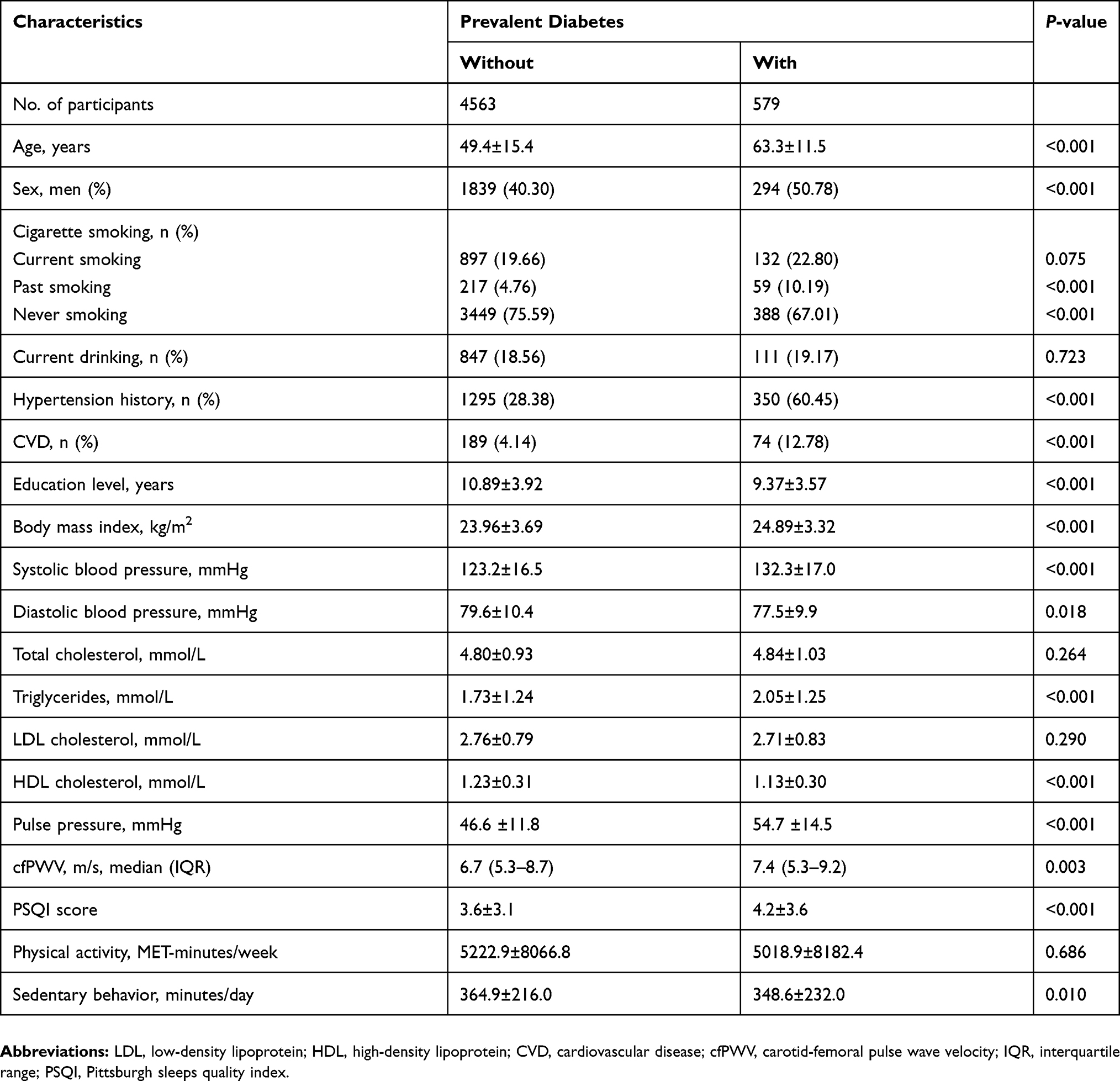 |
Table 1 Baseline Characteristics Of Study Participants |
Association Between Fasting Glucose And Vascular Aging
The median level of cfPWV was significantly increased as increasing quintile levels of fasting glucose (Figure 1, β=0.14, P<0.0001). Table 2 shows the associations of fasting glucose with vascular aging. Regression using continuous fasting glucose as the independent variable showed that per 1-mmol/L increment of fasting glucose was significantly associated with a higher risk of having vascular aging defined by individual cfPWV (OR=1.05, P=0.022), individual PP (OR=1.06, P=0.048), or either (OR=1.08, P=0.002). Regression using quintiles of fasting glucose as the independent variable revealed similar associations in the same direction. Compared to participants with the lowest quintile of fasting glucose, those with the highest quintile had a 31%, 49%, and 45% higher risk of having vascular aging defined by cfPWV (OR=1.31, P=0.023), PP (OR=1.49, P=0.016), or either (OR=1.45, P=0.001), respectively.
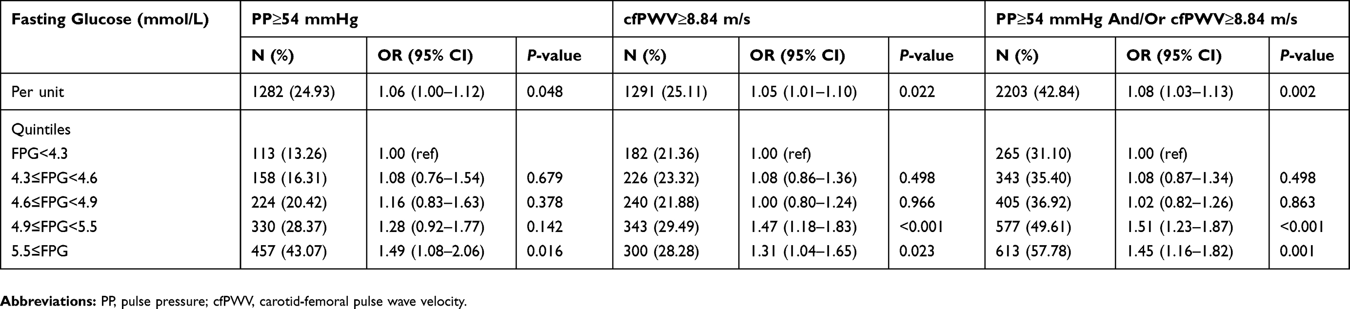 |
Table 2 Multivariate *Adjusted Associations Of Fasting Glucose With Vascular Aging |
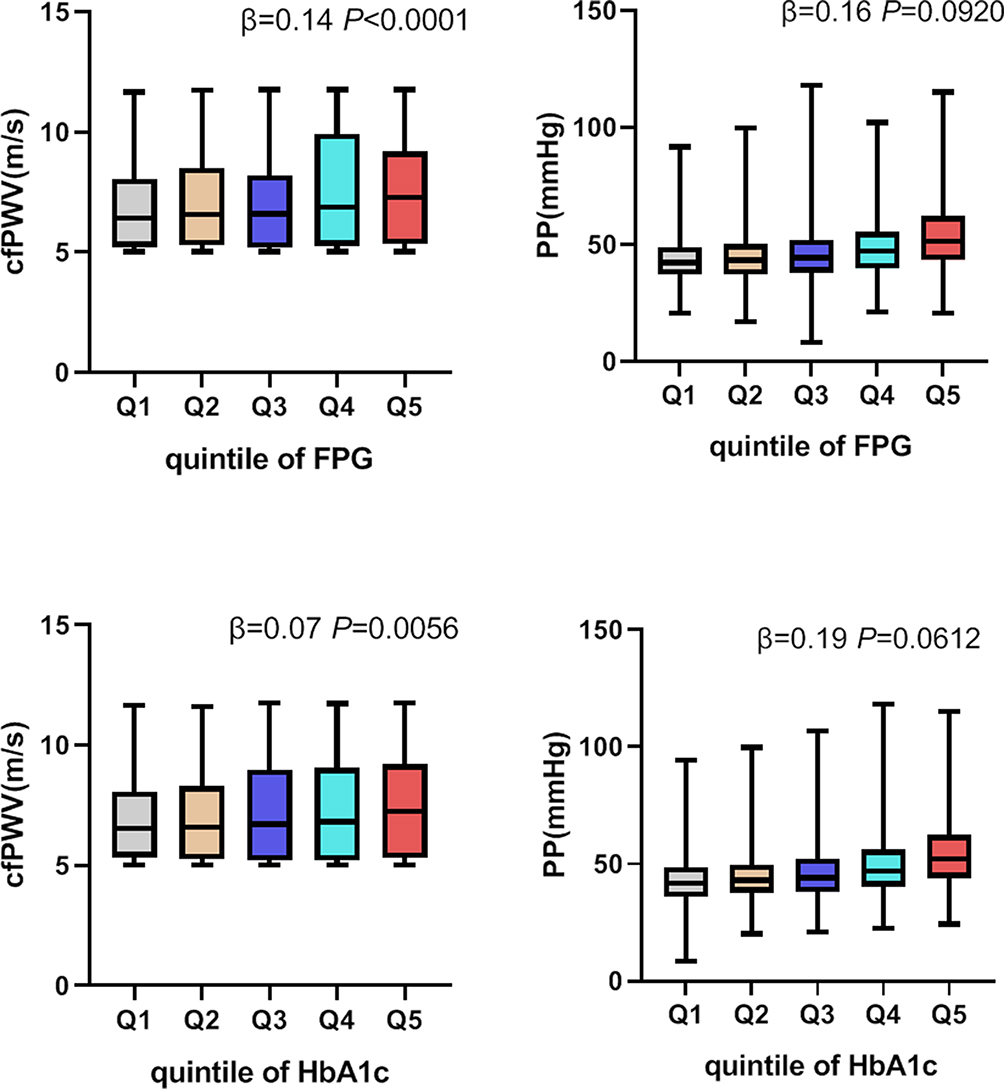 |
Figure 1 An illustration of the median levels of cfPWV and PP according to quintiles of fasting glucose and HbA1c. β indicates the median increase in cfPWV and PP associated with the increasing quintiles of fasting glucose and HbA1c, independent of age, sex, education level, cigarette smoking, alcohol drinking, body mass index, low- and high-density lipoprotein cholesterol, and systolic blood pressure. |
Association Between HbA1c And Vascular Aging
The median level of cfPWV was significantly increased as increasing quintile levels of HbA1c (Figure 1, β=0.07, P=0.0056). Table 3 shows the associations of HbA1c with vascular aging. Regression using continuous HbA1c as the independent variable showed that per 1% increment of HbA1c was significantly associated with a higher risk of having vascular aging defined by individual cfPWV (OR=1.06, P=0.044), individual PP (OR=1.10, P=0.012), or either (OR=1.12, P=0.042). Regression using quintiles of HbA1c as the independent variable revealed similar associations in the same direction. Compared to participants with the lowest quintile of HbA1c, those with the highest quintile had a 52% higher risk of having vascular aging defined by PP (OR=1.52, P=0.015). We failed to observe significantly increased risks of having vascular aging defined by cfPWV for the highest quintile of HbA1c compared to those with the lowest quintile.
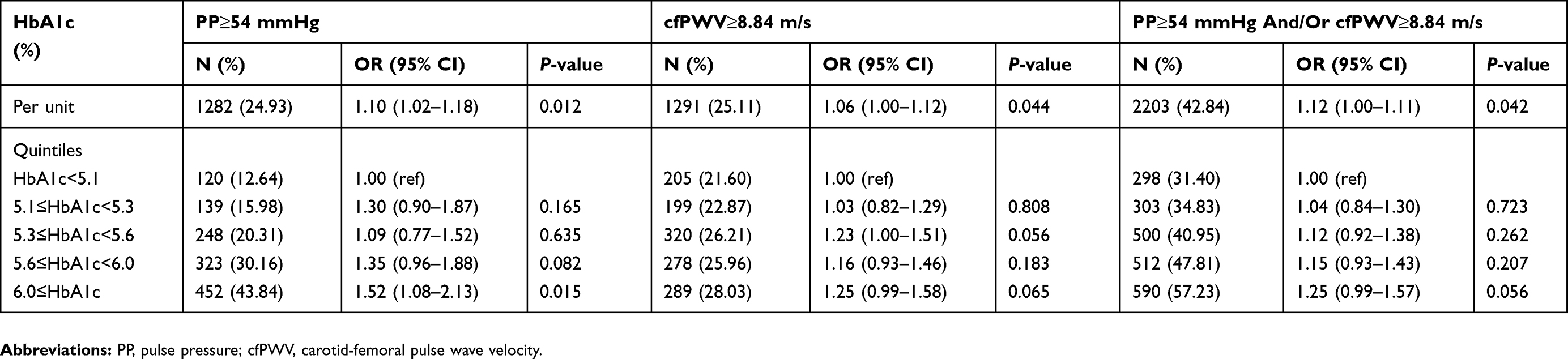 |
Table 3 Multivariate * Adjusted Associations Of HbA1c With Vascular Aging |
Association Between Diabetic Status And Vascular Aging
Compared to participants with a normal glucose metabolic condition, participants with diabetes had a 35% higher risk of having vascular aging defined by PP (OR=1.35, P=0.014). We failed to observe significantly increased risks of having vascular aging defined by cfPWV for diabetic participants compared to those with normal glucose (Table 4).
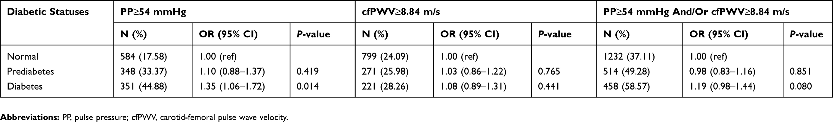 |
Table 4 Multivariate *Adjusted Associations Between Diabetic Status And Vascular Aging |
Result Of Sensitivity Analysis
After additionally adjusting for physical activity and sleep quality, the association between glucose metabolism and vascular aging did not change (Supplementary Table S1). This result indicated that the association between glucose metabolism and vascular aging may not be affected by physical activity and sleep efficiency. Further, excluding participants with prevalent hypertension, diabetes and CVD did not change our results a lot, indicating that the association between glucose metabolism and vascular aging may be unlikely to be driven by these conditions (Supplementary Tables S2 and S3).
Discussion
In a cross-sectional analysis of Chinese adults in the Tianning Cohort, we found that higher levels of fasting glucose and HbA1c were significantly associated with advanced vascular aging, a subclinical condition of CVD. These associations persisted after adjusting for behaviors and other metabolic factors such as blood pressure and lipids. These findings suggested that glucose metabolic disorders may exert harmful effects on cardiovascular health through advancing vascular aging. Noninvasive measurements of vascular aging, e.g., PP and cfPWV, may be of great usefulness in primary prevention of CVD among community residents, diabetic patients in particular.
The average levels of cfPWV and PP in our study participants were similar to the levels reported in previous studies. For example, an average cfPWV of 5.7 m/s and 8.3 m/s was reported in a young population34 and a sample of participants aged over 60 years,35 respectively, which was 6.7 m/s in our study participants with a mean age of 51 years. The average PP level was 47.45 mmHg in our study participants, similar to 45.9 mmHg reported in a Spanish adult population.36 These results suggested that cfPWV and PP, both are noninvasive easy operating methods, could be good screening methods for the identification of advanced vascular aging.
In line with our study, the association of glucose metabolism with vascular aging has also been identified in other studies. For example, an animal study found that a glucose-riched diet could result in microvascular aging in mice.37 In humans, a study including 697 Korea non-diabetic participants found that fasting glucose was significantly associated with vascular aging assessed by brachial-ankle PWV.2 This phenomenon was also observed in another study including 1,312 participants,22 where the ethnic difference in the contribution of fasting glucose to vascular aging appeared to exist. However, the association between fasting glucose and vascular aging is limited studied in Chinese. To our knowledge, only one study including more than 5000 participants found that fasting glucose was not only cross-sectionally but also prospectively associated with vascular stiffness measured by baPWV in Chinese.19,21 Our study provided another evidence that fasting glucose may accelerate vascular aging in Chinese, not limited to white populations. Most of these results including ours were generated from observational study designs that cannot elucidate the causality between fasting glucose and vascular aging. The prospective association between glucose metabolism and vascular aging has been reported in very few studies with mixed results and most studies did not focus on glucose metabolism.19,38–41 However, a Mendelian Randomization study conducted in 2,853 individuals from Malmö Diet and Cancer study found that genetically elevated fasting glucose was associated with vascular aging assessed by cfPWV.20 Together with prior studies, our study added the possibility that fasting glucose may be a risk factor of vascular aging. In addition to fasting glucose, HbA1c, another indicator of glucose metabolism, may also be associated with vascular aging, but this hypothesis, to the best of our knowledge, has been rarely studied. We also examined and found that HbA1c was also significantly associated with vascular aging. All these results suggested that glucose metabolism, not limited to the specific indicator, may play a role in vascular aging. As expected, abnormal glucose metabolic conditions, e.g., diabetes and prediabetes, have been consistently associated with advanced vascular aging in our and other studies.42–44
To examine whether hypertension, diabetes, and CVD affect the association between glucose metabolism and vascular aging, we applied sensitivity analysis and found that excluding participants with hypertension, diabetes, and CVD did not change our results. This finding indicated that the association between glucose metabolism and vascular aging may not be driven by these conditions. Two important behavioral factors, e.g., sleep and physical activity, that may play confounding effects on the association between glucose metabolism and vascular aging have been ignored by prior studies. We additionally adjusted for these two factors and found that the association between glucose metabolism and vascular aging persisted. Our findings added to the literature that the vascular toxicity of glucose may be independent of sleep and physical activity. Moreover, these sensitivity analyses revealed a robust association between glucose metabolism and vascular aging.
The mechanisms underlying the identified association between glucose metabolism and vascular aging needed to be discussed to deepen our understanding of the vascular toxicity of glucose. Increased arterial stiffness is a clinically important phenotype associated with vascular aging in humans and generally associated with vascular oxidative stress and endothelial dysfunction.45 Researchers have found that hyperglycemia, particularly the fluctuation of glucose levels, caused a significant degree of oxidative stress through decreasing endothelial nitric oxide synthase (eNOS) expression, reducing nitric oxide (NO) bioavailability, and impairing NO metabolism,46,47 and thereby leading to endothelial dysfunction. Glucose-induced increased vascular oxidative stress and endothelial dysfunction would ultimately accelerate vascular aging which is strongly linked to cardiovascular disease. Vascular aging could serve as an intermediate marker of the link between hyperglycemia and vascular complications.48 Therefore, strict glycemic control might achieve positive long-term protection against cardiovascular disease, at least partially through delay and reverse vascular aging.
Our study provides initial evidence that glucose metabolism may play a potential role in vascular aging, independent of lifestyle and metabolic factors. There are some strengths in our study including comprehensive adjustment for lifestyle and metabolic factors, application of fasting glucose and HbA1c as glucose metabolic indicators and cfPWV and PP as vascular aging measurements, and relatively large sample size. Several limitations of this study should be considered. First, in our study, all the participants were Han Chinese. The generalizability of our findings to other ethnic populations is unclear. Second, based on its cross-sectional design, we are unable to determine the causal association between glucose metabolism and vascular aging. Further studies are needed to confirm causality. Third, our results showed that glucose and HbA1c were consistently associated with cfPWV in both quantile and logistic regression models, whereas the association between glucose metabolism and the other vascular aging index PP was not statistically significant in the quantile regression model (Figure 1). The inconsistent results should be interpreted by cautious.
In conclusion, our results indicate that higher levels of HbA1c and fasting glucose were significantly associated with advanced vascular aging in Chinese adults, independent of behavioral and metabolic factors. These findings may suggest that dysregulated glucose metabolism may embed adverse effects on cardiovascular health through accelerating vascular aging. The causality between glucose metabolism and vascular aging and the underlined mechanisms warranted further investigation.
Abbreviations
BMI, body mass index; CVD, cardiovascular disease; fPWV: carotid-femoral pulse wave velocity; DBP, diastolic blood pressure; GPAQ, the Global Physical Activity Questionnaire; HDL-C, high-density lipoprotein cholesterol; LDL-C, low-density lipoprotein cholesterol; PWV, pulse wave velocity; PSQI, the Pittsburgh Sleep Quality Index; SBP, systolic blood pressure.
Data Availability
The data are available on request from the corresponding author at [email protected].
Acknowledgments
We are deeply appreciative of the participants in this study and thank all staff for their support and assistance. Especially, we thank the Center for Disease Prevention and Control of Tianning District for their support in the recruitment of participants.
Disclosure
The authors report no conflicts of interest in this work.
References
1. Urbina EM, Dolan LM, Mccoy CE, et al. Relationship between elevated arterial stiffness and increased left ventricular mass in adolescents and young adults. J Pediatr. 2011;158(5):715–721. doi:10.1016/j.jpeds.2010.12.020
2. Shin JY, Lee HR, Lee DC. Increased arterial stiffness in healthy subjects with high-normal glucose levels and in subjects with pre-diabetes. Cardiovasc Diabetol. 2011;10:30. doi:10.1186/1475-2840-10-30
3. Nilsson PM. Impact of vascular aging on cardiovascular disease: the role of telomere biology. J Hypertens. 2012;30(Suppl):S9–S12. doi:10.1097/HJH.0b013e328353e512
4. El Assar M, Angulo J, Rodriguez-Manas L. Oxidative stress and vascular inflammation in aging. Free Radic Biol Med. 2013;65:380–401. doi:10.1016/j.freeradbiomed.2013.07.003
5. Townsend RR, Wilkinson IB, Schiffrin EL, et al. Recommendations for Improving and standardizing vascular research on arterial stiffness: a scientific statement from the american heart association. Hypertension. 2015;66(3):698–722. doi:10.1161/HYP.0000000000000033
6. Lu Y, Zhu M, Bai B, et al. Comparison of carotid-femoral and brachial-ankle pulse-wave velocity in association with target organ damage in the community-dwelling elderly chinese: the northern shanghai study. J Am Heart Assoc. 2017;6(2). doi:10.1161/JAHA.116.004168
7. Ohkuma T, Ninomiya T, Tomiyama H, et al. brachial-ankle pulse wave velocity and the risk prediction of cardiovascular disease: an individual participant data meta-analysis. Hypertension. 2017;69(6):1045–1052. doi:10.1161/HYPERTENSIONAHA.117.09097
8. Borrell LN, Samuel L. The effect of pulse pressure on all-cause and cardiovascular-specific mortality risks in US adults. Ethn Dis. 2015;25(2):152–156.
9. Brassard JA, Fekete N, Garnier A, et al. Hutchinson–gilford progeria syndrome as a model for vascular aging. Biogerontology. 2016;17(1):129–145. doi:10.1007/s10522-015-9602-z
10. Northcott JM, Czubryt MP, Wigle JT. Vascular senescence and ageing: a role for the MEOX proteins in promoting endothelial dysfunction. Can J Physiol Pharmacol. 2017;95(10):1067–1077. doi:10.1139/cjpp-2017-0149
11. Lakatta EG, Levy D. Arterial and cardiac aging: major shareholders in cardiovascular disease enterprises: part I: aging arteries: a “set up” for vascular disease. Circulation. 2003;107(1):139–146. doi:10.1161/01.CIR.0000048892.83521.58
12. Maloberti A, Vallerio P, Triglione N, et al. Vascular aging and disease of the large vessels: role of inflammation. High Blood Press Cardiovasc Prev. 2019;26(3):175–182. doi:10.1007/s40292-019-00318-4
13. Rask-Madsen C, Kahn CR. Tissue-specific insulin signaling, metabolic syndrome, and cardiovascular disease. Arterioscler Thromb Vasc Biol. 2012;32(9):2052–2059. doi:10.1161/ATVBAHA.111.241919
14. Kozakova M, Palombo C. Diabetes mellitus, arterial wall, and cardiovascular risk assessment. Int J Environ Res Public Health. 2016;13(2):201. doi:10.3390/ijerph13020201
15. Assar ME, Angulo J, Rodriguez-Manas L. Diabetes and ageing-induced vascular inflammation. J Physiol. 2016;594(8):2125–2146. doi:10.1113/JP270841
16. Fan LM, Cahill-Smith S, Geng L, Du J, Brooks G, Li JM. Aging-associated metabolic disorder induces Nox2 activation and oxidative damage of endothelial function. Free Radic Biol Med. 2017;108:940–951. doi:10.1016/j.freeradbiomed.2017.05.008
17. Piga R, Naito Y, Kokura S, et al. Short-term high glucose exposure induces monocyte-endothelial cells adhesion and transmigration by increasing VCAM-1 and MCP-1 expression in human aortic endothelial cells. Atherosclerosis. 2007;193(2):328–334. doi:10.1016/j.atherosclerosis.2006.09.016
18. Montesanto A, Bonfigli AR, Crocco P, et al. Genes associated with type 2 diabetes and vascular complications. Aging (Albany NY). 2018;10(2):178–196. doi:10.18632/aging.v10i2
19. Wu Y, Yu J, Jin C, et al. Longitudinal fasting blood glucose patterns and arterial stiffness risk in a population without diabetes. PLoS One. 2017;12(11):e0188423. doi:10.1371/journal.pone.0188423
20. Gottsäter M, Hindy G, Orho-Melander M, Nilsson PM, Melander O. A genetic risk score for fasting plasma glucose is independently associated with arterial stiffness: a Mendelian randomization study. J Hypertens. 2018;36(4):809–814. doi:10.1097/HJH.0000000000001646
21. Wang J, Liu L, Zhou Y, et al. Increased fasting glucose and the prevalence of arterial stiffness: a cross-sectional study in Chinese adults. Neurol Res. 2014;36(5):427–433. doi:10.1179/1743132814Y.0000000345
22. Park CM, Tillin T, March K, et al. Adverse effect of diabetes and hyperglycaemia on arterial stiffness in Europeans, South Asians, and African Caribbeans in the SABRE study. J Hypertens. 2016;34(2):282–289. doi:10.1097/HJH.0000000000000789
23. Vlachopoulos C, Xaplanteris P, Aboyans V, et al. The role of vascular biomarkers for primary and secondary prevention. A position paper from the European Society of Cardiology Working Group on peripheral circulation: endorsed by the Association for Research into Arterial Structure and Physiology (ARTERY) Society. Atherosclerosis. 2015;241(2):507–532. doi:10.1016/j.atherosclerosis.2015.05.007
24. Boss HM, van der Graaf Y, Visseren FLJ, et al. Physical activity and characteristics of the carotid artery wall in high-risk patients-the SMART (Second Manifestations of Arterial Disease) Study. J Am Heart Assoc. 2017;6(7). doi:10.1161/JAHA.116.005143
25. Endes S, Schaffner E, Caviezel S, et al. Physical activity is associated with lower arterial stiffness in older adults: results of the SAPALDIA 3 cohort study. Eur J Epidemiol. 2016;31(3):275–285. doi:10.1007/s10654-015-0076-8
26. Ahmadi‐Abhari S, Sabia S, Shipley MJ, et al. Physical activity, sedentary behavior, and long-term changes in aortic stiffness: the whitehall II study. J Am Heart Assoc. 2017;6(8). doi:10.1161/JAHA.117.005974
27. Osonoi Y, Mita T, Osonoi T, et al. Poor sleep quality is associated with increased arterial stiffness in Japanese patients with type 2 diabetes mellitus. BMC Endocr Disord. 2015;15:29. doi:10.1186/s12902-015-0026-1
28. Kim CW, Chang Y, Zhao D, et al. Sleep duration, sleep quality, and markers of subclinical arterial disease in healthy men and women. Arterioscler Thromb Vasc Biol. 2015;35(10):2238–2245. doi:10.1161/ATVBAHA.115.306110
29. Esposito C, Fasoli G, Plati AR, et al. Long-term exposure to high glucose up-regulates VCAM-induced endothelial cell adhesiveness to PBMC. Kidney Int. 2001;59(5):1842–1849. doi:10.1046/j.1523-1755.2001.0590051842.x
30. Bjarnegard N, Arnqvist HJ, Lindstrom T, et al. Long-term hyperglycaemia impairs vascular smooth muscle cell function in women with type 1 diabetes mellitus. Diab Vasc Dis Res. 2009;6(1):25–31. doi:10.3132/dvdr.2009.005
31. Chobanian AV, Bakris GL, Black HR, et al. The Seventh Report Of The Joint National Committee on prevention, detection, evaluation, and treatment of high blood pressure: the JNC 7 report. JAMA. 2003;289(19):2560–2572. doi:10.1001/jama.289.19.2560
32. Armstrong T, Bull F. Development of the World Health Organization Global Physical Activity Questionnaire (GPAQ). J Public Health (Bangkok). 2006;14(2):66–70. doi:10.1007/s10389-006-0024-x
33. Buysse DJ, Reynolds CF
34. Boardman H, Lewandowski AJ, Lazdam M, et al. Aortic stiffness and blood pressure variability in young people: a multimodality investigation of central and peripheral vasculature. J Hypertens. 2017;35(3):513–522. doi:10.1097/HJH.0000000000001192
35. McEniery CM, Wilkinson IB, Johansen NB, et al. Nondiabetic glucometabolic status and progression of aortic stiffness: the whitehall II study. Diabetes Care. 2017;40(4):599–606. doi:10.2337/dc16-1773
36. Garcia-Hermoso A, Notario-Pacheco B, Recio-Rodriguez JI, et al. Sedentary behaviour patterns and arterial stiffness in a Spanish adult population - The EVIDENT trial. Atherosclerosis. 2015;243(2):516–522. doi:10.1016/j.atherosclerosis.2015.10.004
37. Yamada S, Ohkubo C. The influence of frequent and excessive intake of glucose on microvascular aging in healthy mice. Microcirculation. 1999;6(1):55–62. doi:10.1080/713773927
38. Meani P, Maloberti A, Sormani P, et al. Determinants of carotid-femoral pulse wave velocity progression in hypertensive patients over a 3.7 years follow-up. Blood Press. 2018;27(1):32–40. doi:10.1080/08037051.2017.1378069
39. Benetos A, Adamopoulos C, Bureau JM, et al. Determinants of accelerated progression of arterial stiffness in normotensive subjects and in treated hypertensive subjects over a 6-year period. Circulation. 2002;105(10):1202–1207.
40. AlGhatrif M, Strait JB, Morrell CH, et al. Longitudinal trajectories of arterial stiffness and the role of blood pressure: the baltimore longitudinal study of aging. Hypertension. 2013;62(5):934–941. doi:10.1161/HYPERTENSIONAHA.113.01445
41. Safar ME, Thomas F, Blacher J, et al. Metabolic syndrome and age-related progression of aortic stiffness. J Am Coll Cardiol. 2006;47(1):72–75. doi:10.1016/j.jacc.2005.08.052
42. van Popele NM, Elizabeth HA, Mattace-Raso FU, et al. Impaired fasting glucose is associated with increased arterial stiffness in elderly people without diabetes mellitus: the Rotterdam Study. J Am Geriatr Soc. 2006;54(3):397–404. doi:10.1111/j.1532-5415.2005.00614.x
43. Lukich E, Matas Z, Boaz M, Shargorodsky M. Increasing derangement of glucose homeostasis is associated with increased arterial stiffness in patients with diabetes, impaired fasting glucose and normal controls. Diabetes Metab Res Rev. 2010;26(5):365–370. doi:10.1002/dmrr.v26:5
44. Cakar M, Balta S, Sarlak H, et al. Arterial stiffness and endothelial inflammation in prediabetes and newly diagnosed diabetes patients. Arch Endocrinol Metab. 2015;59(5):407–413. doi:10.1590/2359-3997000000061
45. Ungvari Z, Kaley G, de Cabo R, et al. Mechanisms of vascular aging: new perspectives. J Gerontol A Biol Sci Med Sci. 2010;65(10):1028–1041. doi:10.1093/gerona/glq113
46. Quagliaro L, Piconi L, Assaloni R, et al. Intermittent high glucose enhances ICAM-1, VCAM-1 and E-selectin expression in human umbilical vein endothelial cells in culture: the distinct role of protein kinase C and mitochondrial superoxide production. Atherosclerosis. 2005;183(2):259–267. doi:10.1016/j.atherosclerosis.2005.03.015
47. Pricci F, Leto G, Amadio L, et al. Oxidative stress in diabetes-induced endothelial dysfunction involvement of nitric oxide and protein kinase C. Free Radic Biol Med. 2003;35(6):683–694. doi:10.1016/S0891-5849(03)00401-5
48. Gordin D, Groop PH. Aspects of hyperglycemia contribution to arterial stiffness and cardiovascular complications in patients with type 1 diabetes. J Diabetes Sci Technol. 2016;10(5):1059–1064. doi:10.1177/1932296816636894
 © 2019 The Author(s). This work is published and licensed by Dove Medical Press Limited. The
full terms of this license are available at https://www.dovepress.com/terms
and incorporate the Creative Commons Attribution
- Non Commercial (unported, 3.0) License.
By accessing the work you hereby accept the Terms. Non-commercial uses of the work are permitted
without any further permission from Dove Medical Press Limited, provided the work is properly
attributed. For permission for commercial use of this work, please see paragraphs 4.2 and 5 of our Terms.
© 2019 The Author(s). This work is published and licensed by Dove Medical Press Limited. The
full terms of this license are available at https://www.dovepress.com/terms
and incorporate the Creative Commons Attribution
- Non Commercial (unported, 3.0) License.
By accessing the work you hereby accept the Terms. Non-commercial uses of the work are permitted
without any further permission from Dove Medical Press Limited, provided the work is properly
attributed. For permission for commercial use of this work, please see paragraphs 4.2 and 5 of our Terms.