Back to Journals » International Journal of Nanomedicine » Volume 20
Exploring the Potential of Non-Viral Nanocarriers for Improving Blood-Brain Barrier Permeability to Enhance the Treatment of Brain Diseases
Authors Yu Z ![]() , Sun Z, Yuan L, Wang C, Li W, Liu J, Zhao Q, Sun Y
, Sun Z, Yuan L, Wang C, Li W, Liu J, Zhao Q, Sun Y ![]() , Sun C
, Sun C
Received 9 June 2025
Accepted for publication 30 September 2025
Published 15 October 2025 Volume 2025:20 Pages 12593—12625
DOI https://doi.org/10.2147/IJN.S545696
Checked for plagiarism Yes
Review by Single anonymous peer review
Peer reviewer comments 2
Editor who approved publication: Dr Kamakhya Misra
Zijie Yu,1,* Zhihong Sun,1,2,* Lu Yuan,1 Cuicui Wang,1 Wei Li,2 Jie Liu,2 Qi Zhao,2 Yong Sun,1 Chengming Sun1,2
1Department of Pharmaceutics, School of Pharmacy, Qingdao Medical College, Qingdao University, Qingdao, Shandong, People’s Republic of China; 2The Affiliated Yantai Yuhuangding Hospital of Qingdao University, Yantai, Shandong, People’s Republic of China
*These authors contributed equally to this work
Correspondence: Yong Sun, Department of Pharmaceutics, School of Pharmacy, Qingdao Medical College, Qingdao University, Qingdao, Shandong, People’s Republic of China, Email [email protected] Chengming Sun, The Affiliated Yantai Yuhuangding Hospital of Qingdao University, Yantai, Shandong, People’s Republic of China, Email [email protected]
Abstract: Brain diseases have become an important health problem worldwide, especially in the aging population, and their incidence and prevalence continue to increase. Despite remarkable progress in medical technology, the treatment of brain diseases still faces many challenges, especially limitations caused by the blood-brain barrier (BBB), which significantly hinders the delivery of therapeutic drugs to the brain. In recent years, non-viral nanocarriers developed by nanotechnology have shown great potential for crossing the BBB, and have attracted much attention due to their low immunogenicity, high biocompatibility and good targeting. In this paper, we review the basic structure of the BBB, properties of nonviral vectors, and the mechanisms of crossing BBB. Moreover, this review summarizes the main types of non-viral vectors—liposomes, polymeric nanoparticles, biomimetic materials, and inorganic nanomaterials—while addressing the main translational barriers, including low BBB permeability, poor systemic stability, nonspecific peripheral accumulation, manufacturing challenges, and limited clinical validation, and suggests future research directions.
Keywords: brain diseases, blood-brain barrier, non-viral nanocarriers, brain drug delivery
Introduction
As the population ages and the environment deteriorates, death and disability due to brain diseases are increasingly being recognized as global public health challenges. Brain diseases affect the autonomic, peripheral, and central nervous system (CNS),1 and include cerebrovascular diseases, brain tumors, epilepsy, Alzheimer’s disease, Parkinson’s disease, and ischemic stroke. The main difficulty in the drug treatment of brain diseases is the difficulty in penetrating the blood-brain barrier (BBB) to reach the brain lesions. BBB is a protective interface composed of brain capillary endothelial cells, pericytes, astrocytes, neurons, and perivascular fibroblast-like cells, collectively referred to as the neurovascular unit.2 It restricts entry of most blood-derived molecules through two key mechanisms: physical barriers formed by tight and adherens junctions between endothelial cells, except for some small molecules and specialized cells (eg, monocytes, macrophages, and neutrophils) that can selectively enter the brain,3 and biochemical barriers mediated by active efflux pumps such as P-glycoprotein (ABCB1) and breast cancer resistance protein (BCRP/ABCG2).4 Owing the bidirectional protection of physical and biochemical barriers, the BBB can regulate the internal environment of the brain to maintain stability, so that neurons and synapses can function properly and prevent harmful substances or pathogenic microorganisms from entering the brain.5 Therefore, during drug delivery, approximately 98% of small-molecule drugs and almost all large molecule drugs cannot cross the BBB for treatment, making the treatment of CNS diseases extremely difficult.
With the rapid development of nanomaterials over the past 30 years, nanocarriers have entered the field of vision and have gradually become efficient drug delivery carriers. Nano-delivery vectors are divided into viral and non-viral nanocarriers. Viral vectors include adenoviruses, retroviruses, lentiviruses, and poxviruses. Although viral vectors have good transfection rates and sustained gene expression for gene delivery, there are still many biosafety concerns, poor cell targeting, complex production processes, high development costs, and susceptibility to severe inflammatory and immune responses.6 Non-viral nanocarriers are drug-loaded polymeric nanoparticles composed of natural polymers or synthetic chemicals, typically ranging in diameter from 1 to 1,000 nm. They possess favorable characteristics such as small particle size, high biocompatibility, low cytotoxicity, low immunogenicity, high stability, strong targeting ability, and effective penetration of the BBB, so as to treat brain diseases. Non-viral nanocarriers include organics (liposomes, micelles, nanogels, exosomes, etc) and inorganic substances (gold nanoparticles [AuNPs], carbon nanoparticles, mesoporous silica, etc), which have been extensively investigated in preclinical studies for targeted drug delivery (Figure 1). The vesicular structure of lipid-based nanocarriers has been approved by the FDA, as it enables the active loading of drugs and protects enzymes or small-molecule therapeutics from degradation, and these systems have even been utilized for various commercial purposes in clinical practice and the pharmaceutical industry.7 In the context of brain diseases, multiple clinical trials have investigated the application of LNPs and other nanocarriers in Alzheimer’s disease, brain tumors, and rare hereditary central nervous system disorders. Polymer nanoparticles (such as PLGA) can be used to deliver neurotrophic factors or anti-tumor gene drugs to enhance the therapeutic efficacy against malignant tumors like gliomas. In addition, inorganic nanocarriers, such as gold nanoparticles and silica nanoparticles, have entered early clinical trials for brain tumor imaging and targeted therapy, demonstrating promising prospects for integrating visualization with treatment.8
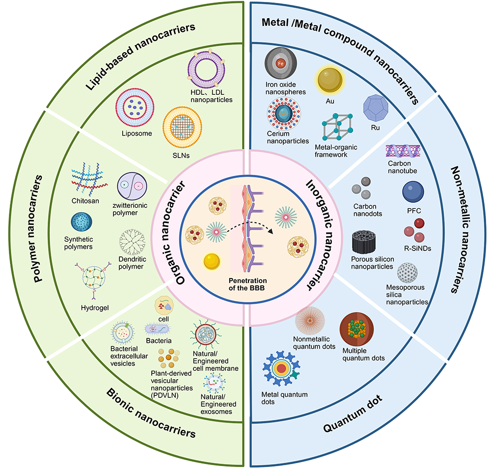 |
Figure 1 Different BBB penetrating nanocarriers. Created in BioRender. Yu, Z (2025) https://BioRender.com/d59s834. |
These nanocarriers deliver drugs to the brain in two ways: around or through the BBB. Bypassing the BBB mainly includes intranasal drug delivery, intrathecal drug delivery, intratumoral delivery (ITD), convection-enhanced delivery (CED), etc.9 Intranasal administration enables drug molecules to cross the nasal mucosa and access the olfactory bulb, thereby bypassing the BBB and directly diffusing into the cerebrospinal fluid (CSF) and brain interstitial fluid. This route also minimizes peripheral accumulation in off-target organs such as the liver and spleen, enhancing delivery specificity to the central nervous system.10,11 Although intranasal administration can bypass the BBB and offers a noninvasive advantage, its limited absorption, rapid mucociliary clearance, and significant interindividual variability still restrict its broad application. CED aims to enhance the convective flow of interstitial fluid to deliver drugs to the lesion, reduce the dilution of drug concentration owing to blood flow at the tumor site, and deliver paclitaxel with a significant effect. A limitation of CED is that it can lead to an uneven distribution of drugs and there are surgical risks involved, and it is not suitable for long-term treatment.12 The penetration of the BBB can be divided into endogenous and exogenous mechanisms (Figure 2). Endogenous mechanisms include passive diffusion, adsorption-mediated transcytosis (AMT), carrier-mediated transcytosis (CMT), receptor-mediated transcytosis (RMT), and down-regulation of the P-gp efflux pump.13 AMT is a transport pathway for chitosan, and cell-penetrating peptides (CPP, Angiopep-2, RVG).9,13 The CMT is an important pathway for nutrient and hormone transport. For example, GLUT1, LAT1, choline transporter, glutathione, etc.14 The RMT is a transport pathway for LDL, Tf, insulin, lactoferrin (Lf), bradykinin (BK), and RGD.9,13 Exogenous mechanisms include receptor-mediated endocytosis, external stimulation by focused ultrasound (FUS), electromagnetic fields, laser, etc.13,15 Immune and stem cells often pass through the BBB in the presence of transmembrane protein receptors or antibodies on the cell membrane surface, which can bind to the corresponding ligands.14 Focused ultrasound (FUS) with microbubbles is a non-invasive technique that reversibly activates the BBB instantaneously.16 FUS ablation has been used to treat AD, and PD,17,18 and for BBB during glioblastoma chemotherapy.19 FUS may induce a certain degree of sterile inflammatory response, creating a complex and unbalanced immune microenvironment, which poses some challenges to the treatment.20 FUS combined with microbubbles (FUS/MBs) has entered clinical trials for the treatment of Alzheimer’s and Parkinson’s diseases by transiently opening the BBB. Magnetic resonance imaging (MRI) confirmed successful BBB disruption following FUS irradiation, and clinical assessments reported no new neurological symptoms, such as headache or confusion, nor any significant adverse events.21 Magnetic nanoparticles (MNP) can disrupt endothelial cells-cell connections via an external magnetic field, or a low radio frequency field can instantaneously.22,23 Laser stimulation increases NP para-cellular diffusion in the BBB by enhancing permeability through TJ.24
 |
Figure 2 Schematic representation of BBB penetration comparison between conventional drugs and non-viral nanocarriers. (a) Passive transport: Liposomes and other modified nanoparticles (<4 nm) cross the BBB via permeation and diffusion. (b) Adsorptive-mediated transport (AMT): Cationic nanoparticles such as CPP- or chitosan-modified NPs interact with the endothelial cell membrane to enable BBB transcytosis. (c) Carrier-mediated transport (CMT): Nanoparticles modified with glutathione or GLUT1 ligands are transported across the BBB via specific carrier proteins. (d) Receptor-mediated transport (RMT): Nanoparticles conjugated with transferrin, insulin, or other receptor ligands cross the BBB through receptor-mediated endocytosis. Created in BioRender. Yu, Z (2025) https://BioRender.com/o8rh4c3. |
In this review, we summarize the research progress of non-viral nanocarriers for the treatment of brain diseases in recent years. Non-viral nanocarriers are divided into two categories: organic nanocarriers (lipid-based, polymer, and biomimetic nanocarriers) and inorganic nanocarriers (metal nanoparticles, non-metal nanoparticles, quantum dots, etc). In this review, we summarized the mechanisms of BBB penetration, and the advantages and disadvantages of various nanocarriers. Finally, we discuss the challenges faced by existing nanomaterials in clinical translation, including their delivery efficiency, cytotoxicity, and complex workflow. Future research should focus on optimizing the design of carriers to improve their selectivity and safety and achieve a more efficient treatment for brain diseases.
Organic Nanocarriers
Organic nanocarriers are nanoscale particles or systems composed of organic materials such as polymers, lipids, and proteins. Because of its biocompatibility and biodegradability, it is often used in drug delivery systems, gene therapy, and diagnostic imaging (Figure 3).
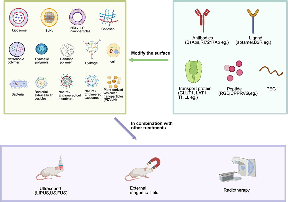 |
Figure 3 Organic nanocarriers with different molecular modifications are combined with other therapeutic methods to improve the therapeutic effect. Created in BioRender. Yu, Z (2025) https://BioRender.com/iim4l5i. |
Lipid-Based Nanocarriers
Compared to other materials, lipid-based nanoparticles have many advantages as non-viral nanocarriers. Its composition is simple, easy to modify, and has a long drug loading cycle, goc.
Liposomes
Liposomes were one of the earliest drug-carrying nanoparticles developed, and are a type of nanocarrier frequently used in clinical trials. Liposomes, as one of the most established nanocarriers, have already been validated by multiple FDA-approved formulations such as Doxil and AmBisome.25 Liposome particles are self-assembled ultra-spheroidal nanostructures with one or more lipid bilayer spherical vesicles and a smooth outer surface, in which hydrophilic or hydrophobic small molecule drugs can be encapsulated. The basic structure of the lipid bilayer of liposomes is similar to that of biofilms, which gives them good biocompatibility, high bioavailability, strong drug targeting and loading ability, low cytotoxicity, and low production costs.7 However, liposome are easily captured by immune phagocytes; therefore, their stability is poor, limiting their wide clinical application. However, liposomes have the disadvantages of short shelf life, low encapsulation efficiency, and difficult continuous release. Currently, the surface of improved versions of liposomes can be modified by a variety of proteins, peptides, polymers, etc., to form immune liposomes and long circulating liposomes, among others. Evaluation by reverse-phase high-performance liquid chromatography (HPLC) showed that the encapsulation efficiency of nanostructured lipid carriers (NLCs) for four levodopa (LD) combination drugs (PD) generally exceeded 70%, with good stability.26 Long-chain polyethylene glycol (PEG)-conjugated liposomes exhibit stronger BBB penetration, low immunogenicity, and prolonged blood circulation time, showing significantly higher accumulation in the brains and tumors of both normal and orthotopic glioblastoma (GBM) transplanted mice.27 Qu et al demonstrated that RVG29-modified PEGylated liposomes (RVG29-lip) exhibited a much higher efficiency in crossing the blood-brain barrier compared to PEGylated liposomes alone.28 Glutathione modified pegylated nanoliposomes (G-Technologys®) are excellent for drug delivery through the BBB for the treatment of brain cancer.7 Glutathione binding liposomes that deliver azithromycin can be used to treat Alzheimer’s disease.29 He et al30 incorporated phenodipine into liposomal nanoparticles (LNPS) to synthesize felodipine@LND nanomedicine, which penetrated the BBB using low-intensity pulsed ultrasound (LIPUS) and effectively treated AD in a 5xFAD mouse model (Figure 4). Neurotransmitter-derived liposomes have great potential for the treatment of CNS diseases and provide new ideas for the development of novel drug delivery systems. The main reason is that endogenous neurotransmitters can participate in the regulation of information between nerve cells and glandular cells, and can be used as a “guide” to cross the BBB. Ma et al31 found that NT1-Lipid derived from tryptamine can effectively promote BBB crossing, whereas phenethylamine and phenethanolamine derived liposomes can not. Doping NT1-Lipid into BBB-impermeable lipid nanoparticles (LNP) can effectively penetrate the BBB.
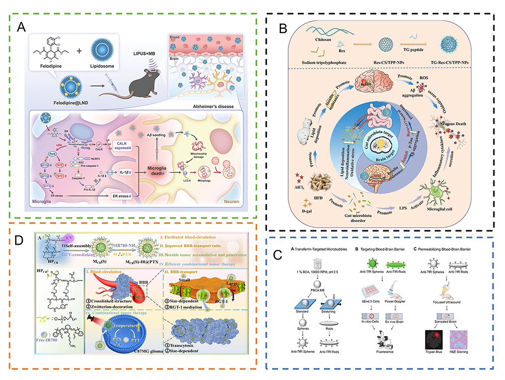 |
Figure 4 Relevant examples of lipid-based nanocarriers/polymeric nanocarriers for the treatment of brain diseases. (A) Lipus-felodipine nanomedicine preparation (felodipine@LND and mechanisms of crossing the BBB to reduce anxiety-likebehavior and cognitive impairment. Reproduced from He X, Peng Y, Huang S et al. Blood Brain Barrier-Crossing Delivery of Felodipine Nanodrug Ameliorates Anxiety-Like Behavior and Cognitive Impairment in Alzheimer’s Disease. Adv Sci (Weinh). 2024;11(34):e2401731.30 (B) The constructed TG-Res-CS/TPP-NPs effectively traversed the BBB, reduced cerebral insulin resistance and Aβ deposition, and regulated inflammation, lipid metabolism, and the intestinal microflora Reprinted from Carbohydrate Polymers, Vol 310, Yang L, Wang Y, Li Z et al, Brain targeted peptide-functionalized chitosan nanoparticles for resveratrol delivery: Impact on insulin resistance and gut microbiota in obesity-related Alzheimer’s disease, Pages No. 120714, Copyright (2023), with permission from Elsevier.32 (C) MB spheres and MB rods were first prepared and the anti-TFR MB cavitation signals recorded after transcranial 835 KHz ultrasound processing showed that MB rods produced more similar harmonics and higher wideband emission signals than MB balls, and more easily penetrated BBB. Reproduced from Dasgupta A, Sun T, Rama E et al. Transferrin Receptor-Targeted Nonspherical Microbubbles for Blood-Brain Barrier Sonopermeation. Adv Mater. 2023;35(52):e2308150.33 (D) Super-small zwitterionic micelle (MCB(S)) conjugated with free IR780 and loaded with PTX (MCB(S)-IR@PTX) to improve BBB-transport and enhance tumor penetration and accumulation for glioblastomas therapy. Reprinted from Journal of Controlled Release, Vol 364, Wang K, Zhao B, Ao Y, et al, Super-small zwitterionic micelles enable the improvement of blood-brain barrier crossing for efficient orthotopic glioblastoma combinational therapy, Pages No. 261–271, Copyright (2023), with permission from Elsevier.34 |
Solid Lipid Nanoparticles (SLNs)
Solid lipid nanoparticles are micellar spheres composed of a solid hydrophobic lipid core surrounded by triglycerides and fatty acids. Polysorbate 80 on the surface of the particles is adsorbed onto brain endothelial cells and enters the brain through transcytosis.35 Solid lipid nanoparticles (SLNs) feature a large hydrophobic core, providing higher loading efficiency for hydrophobic drugs than liposomes. Owing to their low toxicity, high stability, and good biocompatibility, they are suitable for large-scale production and have been used to treat neurological disorders such as Parkinson’s disease, Alzheimer’s disease, brain tumors, and epilepsy.7 Chitosan-modified SLNs deliver dopamine to Parkinson’s Chitosan-coated SLNs modified by CPP can deliver siRNA to reduce the number of amyloid plaques in the brain and thus treat Alzheimer’s disease.36 The αvβ3 and αvβ5 integrins are overexpressed on the EC of GBM, and cRGD is their classic binding peptide. Nano-micelles modified with internalized RGD (iRGD) can treat advanced glioma.37 iRGD -functionalized SLN-loaded PTX can crosses the BBB and targets GBM cells.38 Kuo et al designed solid lipid nanoparticles (SLNs) modified with tamoxifen (TX) and lactoferrin (Lf) ligands to deliver carmustine (BCNU) into the brain parenchyma, and found that, compared with bare BCNU-loaded SLNs, the TX- and Lf-ligand customized BCNU-loaded SLNs exhibited 10-fold higher BBB permeability.39 However, SLNs also have some disadvantages such as low encapsulation efficiency, unexpected gelation tendency, and decreased drug stability after the recrystallization of solid lipids.
High/Low-Density Lipoprotein Nanocarriers
High-density lipoprotein (HDL) and low-density lipoprotein (LDL) nanoparticles, composed of lipid precursors and apolipoproteins, are natural particles. Human glioblastomas have a large number of LDL receptors, and LDL-associated proteins may act as delivery transporters.40 Natural HDL nanoparticles are highly stable in the circulation, have a long half-life and good biocompatibility, and can deliver endogenous miRNAs, and cholesterol-coupled siRNAs.41,42 The principal apolipoprotein A1 (apoA1) of HDL can cross the BBB through transcytosis mediated by several receptors on brain endothelial cells, such as the scavenger receptor B-I (SR-B1), inducing efficient delivery of HDL mimicking nanoparticles without additional modification.43,44 It is also worth noting that recombinant HDL mimics nanoparticles themselves can be used as nanomedicines to treat brain diseases. For example, HDL nanoparticles with apoA1 / apoE3 reduced levels of brain amyloid beta (Aβ) and thus alleviated memory impairments.45 Apolipoprotein E (ApoE) efficiently crosses the BBB via the low -density lipoprotein receptor (LDLR) and transcytosis. Wei et al46 constructed a systemic nano delivery system for granzyme B and CpG mediated by ApoE. Targeting ApoE-PS-GrB by LDLRs not only increased the penetration of BBB and uptake rate of glioma cells, but also promoted the maturation of DC cells and enhanced the secretion of TNF-α, IFN-γ, and IL-6 cytokines, resulting in a wider immune response.
Polymer Nanocarriers
Compared with lipid-based nanoparticles, polymeric nanoparticles offer greater structural controllability and functional versatility, representing another important option for precise treatment of brain diseases. Polymer nanoparticles have a small particle size (size range 1–100 nm) and are easier to target specific therapeutic sites across the BBB. Currently, this is the most widely studied nanomedicine delivery system. The use of properly modified polymer nanoparticles can promote the targeted aggregation of drugs in the brain. In recent years, nanoparticles have been widely used in brain-targeted drug delivery systems, and drugs such as dalargin, loperamide, doxorubicin have been successfully delivered to the brain. Polymer nanoparticles are particularly helpful in the treatment of sporadic, rapidly growing malignant brain gliomas such as glioblastoma multiforme.
Natural High Polymer
Natural polymer nanoparticles are primarily derived from self-assembly of natural polysaccharide. Natural polysaccharides (glucan, angelica polysaccharide, hyaluronic acid, alginic acid, etc) exhibit excellent biodegradability, low immunogenicity, low toxicity, and biocompatibility.47 To treat brain diseases, chitosan (CS) is commonly used to assemble modified nano-systems. Chitosan is a natural polysaccharide derived from chitin, which is composed of β- (1–4) -n-acetyl-D-glucosamine monomer. Chitosan has been widely used to modify different nanomedical drug delivery systems to improve therapeutic efficacy because of biocompatibility, biodegradability, low toxicity, controlled release, mucous adhesion, modifiability,48,49 and with good mucoadhesive and permeation properties, they can be administered through various routes, particularly enabling noninvasive intranasal delivery of drugs directly to the brain.50 Chitosan can increase or restore the zeta potential of nanoparticles to be positive and has good adhesion, which enforces a higher biological interaction with the anionic cell barrier and enhances cell internalization.51 Many experiments have demonstrated the ability of chitosan to open tight junctions of epithelial cells, promote paracellular delivery of drugs across the epithelial barrier, and enhance penetration of the BBB.52 Almost all types of nanoparticles have been shown to meet the conditions for chitosan coating and have been widely used in brain diseases such as glioma, Alzheimer’s disease, Parkinson’s disease, spinocerebellar ataxia and cerebral ischemia.53–55 CS nanoparticles are synthesized by ion cross-linking, covalent cross-linking, emulsification, self-organization, and other methods in weakly acidic environments, and can effectively encapsulate many substances, such as small molecules, nucleotides, and proteins. Chitosan nanoparticles protect drugs from biological barriers, such as immune cells, PH, and enzymes. Bradykinin (BK) can down-regulate tight junctions and increase the permeability of the BBB, whereas the anti-bradykinin B2 receptor (B2R) and transferrin can co-modify CS to overcome the BBB.56 CS reacts with sodium tripolyphosphate (TPP) to form gel-like chitosan nanoparticles (CS/ TPP-NPS) through ionic interaction, surface modification targeting peptide TG,and then loads resveratrol (Res) to reduce cerebral insulin resistance and Aβ deposition to treat obesity-related Alzheimer’s disease (Figure 4).32
Synthesis of High Polymer
Poly (lactic acid) -glycolic acid copolymer (PLGA) is a random polymer of lactic and glycolic acids. Because the end products of PLGA are H2O, CO2, pyruvate, and other substances that can be metabolized in the body or directly excreted from the body, PEG, proteins, lipids and other modifications are now used to deliver drugs.57 Polyethylene glycol (PEG) -modified PLGA nanoparticles extend circulation time by escaping elimination by the reticuloendothelial system (RES), resulting in a higher degree of internalization of polymer nanoparticles in the brain, while the encapsulation of loperamide in PLGA nanocarriers can efficiently pass the BBB, thus treating CNS diseases.58,59 Nie et al developed an RVG29 peptide-conjugated monomethoxy-PEG (mPEG)-PLGA nanoparticle (RNP-DFO), which enhanced the in vivo stability of deferoxamine (DFO, an FDA-approved iron chelator for refractory anemia) while simultaneously improving drug delivery efficiency.60 Currently, aptamers are the most promising targeting ligands and substitutes for antibodies.61 Short-chain oligonucleotides, similar to antibodies, bind to small molecules by folding into “pockets” to form aptamers. This aptamer has high sensitivity, good tumor penetration, low immunogenicity, long circulation time in vivo, and improved BBB penetration and targeting.62 Polymeric nanoparticle PNP (PLGA-b-PEG) modified with anti-PDGFRβ aptamer (platelet-derived growth factor receptor β type) can enhance BBB penetration and target the treatment of brain glioma.61 Polybutylcyanoacrylate (PBCA) has good biodegradability and strong modifiability, and plays an important role in the research and development of CNS drugs. Polymer nanoparticles composed of PBCA coated with polysorbate 80 (PS80) often carry drugs to overcome the BBB.63 Anshuman et al designed a PBCA-synthesized amorphous microvesicle (MB) and modified it with the high-affinity antibody RI7217 (an anti-transferrin receptor antibody). The combination of this amorphous PBCA-MB with ultrasound (US) not only enhances drug delivery through the BBB, but also facilitates the binding of brain endothelial cells. In addition, antibody-modified amorphous MB can also target circulating cells, extending drug circulation time in vivo (Figure 4).33
Dendritic polymers have a small molecular weight, and their compact scaffold is suitable for delivering nucleic acid drugs, such as short interfering RNA (siRNA). Ligands and drug molecules are used to modify dendritic polymers to improve BBB permeability and overcome the limitations of traditional chemotherapy. Currently, poly-amidoamine (PAMAM), which is most commonly used, can effectively penetrate the BBB through clathrin-mediated transcytosis, selectively target damaged cells, and improve the solubility of drug delivery systems.64,65 Bhairavi et al modified fourth-generation PAMAM (G4-90/10 PAMAM) to carry the hSOX2 transcription factor and delivered it to the rat brain for the treatment of ischemic stroke.66 Hyperbranched polymers, similar to dendrimers, are three-dimensional nano molecules with high dispersion and low orderliness.67 Highly pegylated hyperbranched polymers (HBP) combined with BsAbs (a novel bi-specific antibody that drives drugs to target the EphA2 receptor in tumor cells) and doxorubicin (Dox) can enhance BBB penetration to treat brain glioma.68
Owing to their excellent hydrophilicity and biocompatibility, zwitterionic polymers can modify a variety of nanomedicine delivery systems, such as polycarboxybetaine (PCB), polysulfobetaine (PSB), and polyphosphoylcholine (PMPC). Zwitterionic polymers not only prolong drug circulation time but also overcome a variety of biological barriers, such as the BBB, cellular barrier, and lysosomal barrier.69 Hyperbranched polycarbonate functionalized with zwitterionic carboxybetaine (CB) is used to treat brain gliomas. CB zwitterions enhance drug permeability through the BBB through adsorption-mediated transferration (Figure 4).34
Nano-gels are both nano-materials and hydrogels, combined with the characteristics of solids and liquids. A hydrogel is a three -dimensional mesh polymer with high water or biological liquid content, which is similar to the natural extracellular matrix. Therefore, it exhibits good biocompatibility for improved drug delivery. Compared with ordinary hydrogels, nano-gels are smaller, their surface area is larger, their target is stronger, and their half -life is longer. Nano-gels can be broadly classified according to different synthetic methods and different types of NPs to perform drug delivery for a variety of diseases.70 Yu et al constructed a degradable zwitter-ion phosphate alkaline nano-gel HPMPC to treat GBM, which can cross the BBB with acetylcholine and choline transport proteins.71 Nano-micelles are nuclear -shell structures. It is a thermodynamically stable colloidal solution composed of synthetic hydrophilic embedded sections, and hydrophobic sections spontaneously composed of a hydraulic solution. Its core is hydrophobic and can encapsulate insoluble drugs, while its outer nucleus is hydrophilic and can separate the drug from water-soluble drugs. The rabies virus sugar -protein peptide derivative RVG is also a CPP, and RVG-modified nano-micelle, so that nanoparticles can pass through the BBB by targeting nicotine acetylcholine receptors.72–74
Biomimetic Nanocarriers
However, although polymeric nanoparticles exhibit good controllability and functionalization, their biocompatibility and immune evasion remain limited, and the emergence of biomimetic nanocarriers offers new strategies to overcome these challenges. In recent years, inspired by nature, researchers have extracted and purified different types of endogenous substances from humans, animals, and microorganisms to create nanospheres with good biodegradability, and then integrated them into nanoparticles to build new nanomaterial delivery systems (Figure 5). This system is designed to evade the immune system by simulating the unique biological properties and functions of natural organisms (eg, red blood cells, platelets, neutrophils, macrophages, stem cells, and cancer cells),75 which can reduce immunogenicity, prolong circulation time in the body, and enhance the targeting and biocompatibility of drug delivery.
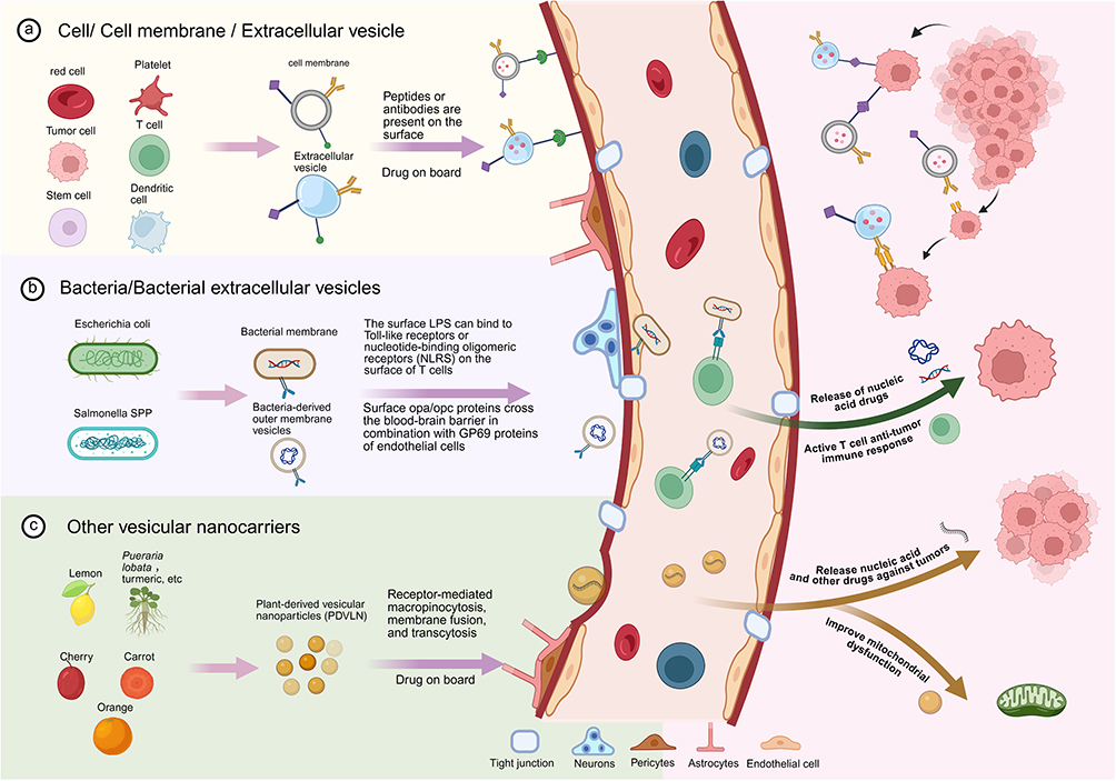 |
Figure 5 Biomimetic nanocarriers penetrate the BBB to treat brain tumors. (a) Cell/cell membrane or extracellular vesicle-based carriers, which can be engineered to present peptides or antibodies on their surface to enhance BBB crossing and tumor targeting. (b) Bacteria or bacteria-derived extracellular vesicles (eg, Escherichia coli and Salmonella spp.) utilize surface lipopolysaccharides (LPS) or outer membrane proteins to interact with endothelial receptors, facilitating vesicle transcytosis across the BBB. (c) Plant-derived vesicular nanoparticles (PDVLNs) can deliver therapeutic agents across the BBB via receptor-mediated macropinocytosis, membrane fusion, and transcytosis, contributing to effects such as mitochondrial protection and anti-tumor activity. Created in BioRender. Yu, Z (2025) https://BioRender.com/vs7rs9u. |
Cell-Derived Biomimetic Nanocarriers
Cell Membrane Bionic Nanocarriers
Mature red blood cells are abundant, lack nuclei and organelles, and their membranes are easy to extract and purify. In addition, red blood cells are completely degraded in the body, have good biodegradability, and do not produce toxic side effects. Owing to the rich hemoglobin in red blood cells, they can carry abundant oxygen to the tumor site, thus facilitating treatments related to tumor hypoxia (such as PDT therapy).76 The erythrocyte membrane expresses the CD47 transmembrane protein, which blocks uptake by macrophages by selectively binding to macrophage-expressed SIRP alpha,77,78 prolonging the half-life of coated drugs and reducing adverse reactions and immunogenicity. Gu et al modified erythrocyte membranes using TGN peptides as ligands to penetrate the BBB while delivering curcumin (Cur) to treat Alzheimer’s disease (Figure 6).79 Because red blood cells are highly hydrophilic, polysaccharides on the red blood cell membrane can increase the stability of coated nanoparticles. Platelets are key components of the blood circulatory system and are involved in hemostasis, thrombosis, and tumor metastasis; therefore, they can readily acquire inherent biocompatibility from the blood, becoming the most significant advantage of drug delivery. CD47 on the platelet membrane prevents clearance by macrophages, thereby extending its half-life.80 The interaction between P-selectin on platelets and CD44 receptors on tumor cells can improve the targeting of nanomedical drugs.81 Xu et al developed a bionic nanoparticle delivery system based on polydopamine by utilizing the inherent anti-ROS properties of the platelet membrane, which not only promotes vascular repair and hemostasis but also provides an antioxidant environment for treatment and promotes nerve recovery (Figure 6).82 Platelet membrane nanodrug-carrying systems are difficult to apply for various reasons, including damage to the body caused by allogeneic platelets and premature drug release due to platelet activation. Currently, the main research objective is to combine platelet membranes with other cell membranes to improve the efficiency of drug delivery. The membrane of tumor cells is obtained from tissues and cells extracted from patients and can be easily cultured and replicated in the laboratory.81 Natural tumor-associated antigens on the tumor cell membrane can stimulate a tumor-specific immune response and synergistically enhance anti-tumor immunotherapy when combined with immune adjuvants. Cancer cells also have the advantages of immune escape, anti-apoptosis, and the promotion of homologous tumor targeting;77,78 therefore, cancer cell membranes have been widely studied as drug delivery vehicles. Zou et al83 fused glioblastoma cells and mitochondrial membranes to design a novel nanodrug-carrying system that promotes the delivery of gboxin (a potent oxidative phosphorylation inhibitor), prolongs its half-life, and increases its availability. Simultaneously, the ability of the delivery system to penetrate the BBB and target tumor cells was significantly enhanced. Particularly in solid tumor cancers (such as glioma, breast cancer, lymphoma, and metastatic cancer), tumor cell membrane-mediated delivery systems show greater BBB penetration and tumor-homing capacity. Stem cells (SC), a type of cells with permanent self-renewal ability, are widely used in the field of therapy. SC may be inherently tumorophilica and driven by many tumor-associated chemokines.84 Neural stem cells (NSC) are commonly used to treat various brain diseases. NSC often express a variety of receptors related to inflammatory chemotaxis (eg, CXCR4); therefore, they have certain chemotaxis and low immunogenicity to brain inflammatory injury. The stem cell membrane (SCM) has a homing ability, and many peptides on its surface can play a targeted role and easily pass through the BBB, which helps in the treatment of glioblastoma. Studies have shown that SCM-coated nanodrug delivery systems can improve tumor cell uptake rates while improving the targeting of therapies under immune camouflage. Mesenchymal stem cell-derived neuron-like cell membranes coated with curcumin PLGA can treat PD, protect neurons, and reduce oxidative stress (Figure 6).85 However, there are still many unsolved problems in stem cell therapy, such as individual differences in clinical stem cell therapy doses, potential tumorigenicity of stem cells, stem cell retention in other tissues and organs, and other safety related issues.
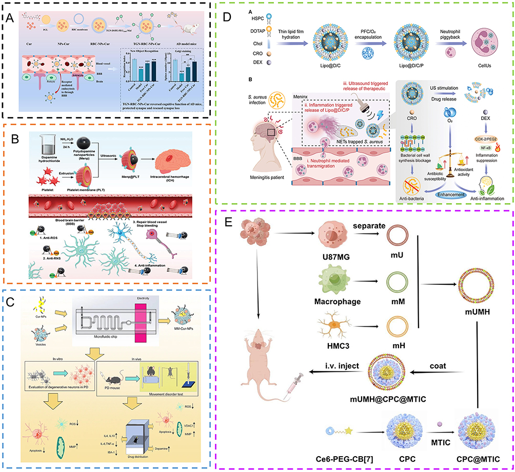 |
Figure 6 Relevant examples of cell/cell membrane biomimetic nanocarriers for the treatment of brain diseases. (A) Process of preparation of TGN-RBC-NPs, mechanism of penetration of BBB. TGN-RBC-NPs-Cur reversed the cognitive function of AD mice in vitro. New object recognition index of different mice (n = 10) and quantification of dendritic spine density in Golgi-stained brain sections (scale bar, 10 μm) are shown. Data are presented as mean ± SD. **P < 0.01, ***P < 0.001 compared with control; ##P < 0.01, ###P < 0.001. Reprinted from Journal of Controlled Release, Vol 366, Gu J, Yan C, Yin S, et al, Erythrocyte membrane-coated nanocarriers modified by TGN for Alzheimer’s disease, Pages No. 448–459, Copyright (2024), with permission from Elsevier.79 (B) Platelet (PLT) membrane-coated polydopamine (Menp) nanoparticles can significantly improve the neuroinflammation environment by scavenging ROS in ICH mice. Copyright ©2023. Wiley-VCH GmbH. Reproduced from Xu C, Pan Y, Zhang H et al. Platelet-Membrane-Coated Polydopamine Nanoparticles for Neuroprotection by Reducing Oxidative Stress and Repairing Damaged Vessels in Intracerebral Hemorrhage. Adv Healthc Mater. 1015 2023;12(26):e2300797.82 (C) Neuron-like cell envelope derived from mesenchymal stem cells promotes curcumin nanoparticles to improve dysactivity in Parkinson’s disease. Reproduced from Lei T, Li C, Liu Y et al. Microfluidics-enabled mesenchymal stem cell derived Neuron like cell membrane coated nanoparticles inhibit inflammation and apoptosis for Parkinson’s Disease. J Nanobiotechnology. 2024;22:370.85 (D) Production of smart responsive neutrophils (CellUs) for the treatment of meningitis. Copyright ©2024. Wiley-VCH GmbH. Reproduced from Zhou A, Kong D, Zhou X et al. Neutrophils for Smart Response in Brain Infection Management. Adv Mater. 2024;36(18):e2311661.86 (a) CellUs encapsulates HSPC, DOTAP, Chol, CRO, and DEX within living neutrophils. (b) CellUs crosses the BBB, targets inflamed meninges, and releases therapeutic drugs in response to inflammatory signals. (E): Supramolecular micelles coated with three types of fusion cell membranes (HMC3 membrane: macrophage cell membrane: U87MG membrane = 1:1:2) were used to target the tumor microenvironment of glioblastoma multiforme. Reprinted from Journal of Controlled Release, Vol 366, Huang X, Mu N, Ding Y, et al, Tumor microenvironment targeting for glioblastoma multiforme treatment via hybrid cell membrane coating supramolecular micelles, Pages No. 194–203, Copyright (2024), with permission from Elsevier.87 |
Immune cells, including neutrophils, macrophages, T cells, and dendritic cells, are increasingly becoming the focus of BDNS research owing to their excellent biocompatibility and unique characteristics of chemotactic recruitment. Immune cells cross the BBB via cell-mediated transcytosis. During brain tumor, brain infection and trauma, a large number of released inflammatory factors and chemokines destroy the integrity of the BBB, so neutrophils are more likely to accumulate and invade the damaged brain, interact with the inflammatory brain microvascular endothelium, and have a strong addiction to the injured site. Neutrophils can also efficiently cross the BBB,78 and neutrophil cell membranes modified by stroke-homing peptide (SHp) can treat ischemic stroke and clear excess ROS.88 In addition, neutrophils promote endothelialization to directly cover the damaged site by activating endothelial progenitor cells recruited by N-formylpeptide receptor 2 (FPR2), which plays an important role in promoting angiogenesis. Fan et al designed a hybrid nanoplatform (NPEOz) based on pH-responsive lipids and neutrophil membranes as a vehicle to deliver GW280264X (an ADAM17 inhibitor) and demosterol (an LXR agonist) across the BBB. Its mechanism of action is to promote erythrocyte phagocytosis and anti-neuroinflammation to treat intracerebral hemorrhage.89 Although the neutrophil cell membrane can enhance the immune escape ability of the nano-delivery system to target immune sites, its short life and excessive accumulation produce toxic substances in the reverse direction and further promote the occurrence and development of tumors. In contrast, macrophages are long-lived and exhibit highly specific targeting, allowing longer drug delivery and reducing the frequency of drug use. Macrophages are not easily degraded by enzymes that improve their stability. Tumor-associated macrophages (TAMs) are widely found in the tumor immune microenvironment and form a complex network closely related to tumor formation, progression, and invasion.90 Cell-penetrating peptides (CPPs) such as Angiopep-2 can cross the blood–brain barrier (BBB) via LRP-1–mediated transcytosis Cell-penetrating peptides (CPP) such as Angiopep-2 can penetrate the BBB91–93 through LRP-1-mediated transfer, thereby downregulating STAT3 to remodel the vascular system and immunosuppressive microenvironment. Furthermore, surface modification of macrophage membranes with Angiopep-2 enables efficient BBB penetration and glioma targeting,94 while simultaneously reducing clearance by the mononuclear phagocyte system and inducing ferroptosis through mitochondrial dysfunction and structural damage. Macrophage membrane modified molybdenum disulfide quantum dots (MoS2 QD) can eliminate ROS and resist the deposition of Aβ1-42 to treat AD.95 Macrophages also have many drawbacks, such as becoming vectors for pathogen transmission and triggering complex immune responses. T-lymphocytes are antigen-presenting cells that express a variety of receptors on the cell membrane to recognize tumor cells and release various lymphocytes and cytokines to enhance the immune response. T cell membrane-coated AIEgen nanocarriers in the treatment of GBM not only deliver drugs, destroy the TJ structure, and penetrate the BBB, but also enhance anti-tumor immune function, which can effectively improve the treatment effect and inhibit tumor recurrence.96
Cell Bionic Vector
Bionic cell drug delivery systems mainly involve the preparation of drug-carrying cells by co-incubating nanoparticles with living cells, which reduces the immunogenicity of nanomedical drugs and prolongs their cycle time. The use of living mammalian cells (eg, red blood cells, platelets, macrophages, monocytes, and neutrophils) results in better biocompatibility and safety. Red blood cells can last up to 120 days, avoid the attack and clearance of the reticuloendothelial system, function as an immune escape, and delay the rate of drug clearance from the body.97 Zhou et al developed a complex dual-response delivery system, CellUs, which uses genetic engineering of living neutrophils to deliver liposomal formulations of dexamethasone (DEX), ceftriaxone (CRO), and oxygen-rich perfluorinated carbon (Lipo@D/C/P) across the BBB to ameliorate brain infection (Figure 6).86 A nanodrug delivery system based on T-cell immunotherapy stimulates artificial antigen cells to present TSA98 while delivering the drug. It is difficult for cells to deliver drugs directly across the BBB; therefore, few experimental results have been reported.
In summary, different biofilms have different advantages in addition to good BBB penetration ability. Although the cell/cell membrane bionic nano drug loading method has strong advantages such as high biocompatibility, good targeting, and few toxic side effects, in order to meet clinical needs, researchers have also developed many hybrid membranes, such as macrophage cell membranes hybridized with neutrophil cell membranes, RAPA mixed to treat glioma, or red blood cell membrane mixed with platelet membrane.78 A hybrid membrane, mUMH (HMC3 membrane: macrophage membrane: U87MG membrane = 1:1:2), containing MITC-loaded nano-micelles could treat GBM (Figure 6).87 Second, cancer cell-mitochondrial hybrid membranes (HM-NPs@G) loaded with gboxin were developed for the treatment of GBM. HM-NPs@G not only facilitated the crossing of the BBB but also prolonged the half-life of the drug and increased its accumulation at the tumor site.99 However, there are still some difficulties associated with its clinical application. For example, immature technology leads to low cell membrane extraction rates, high costs, cumbersome processes, and poor reproducibility, among other challenges. Therefore, in future research, we should not only further improve the technology but also combine the pathological characteristics of different brain diseases to select the appropriate cell membrane bionic tools, that can provide more accurate treatment for brain diseases with different characteristics.
Extracellular Vesicle Nanocarriers
EVs, which are lipid vesicles naturally released by most cells and transported by a variety of body fluids, are increasingly being recognized as key mediators of intercellular communication and are also involved in the occurrence, development, and damage repair mechanisms of brain diseases. Extracellular vesicles (EVs) include exosomes (30~150 nm), microvesicles (50 nm~1 μm), and apoptotic bodies (50 nm~5 μm).100 EVs contain specific proteins, lipids, and genetic material, including messenger RNA, microRNAs, and other non-coding RNA, as well as genomic DNA101 from their progenitor cells. EV can cross the cerebral microvascular wall and the BBB. The interactions between exosomes and brain microvascular endothelial cells (BMEC) have recently been investigated. Chen et al showed that exosomes carrying luciferase could cross the BMEC monolayer under inflammatory conditions; under normal circumstances, they can bypass the BMEC monolayer through cellular pathways to endocytosis.102 In addition, intravenously injected infantile macrophage (Mφ) EV transport brain-derived neurotrophic factor proteins across the BBB.103 The cell-like membrane structure of EV can directly merge with the cell membrane through endocytosis to bypass the endosome-lysosome pathway entry mode, and may be protected by ribonucleases to avoid the endosomal pathway and lysosomal degradation, transporting its cargo directly into the cytoplasm, thus allowing more drug internalization than other carriers and extending its circulation time in the body.104–106 Nanosized extracellular vesicles (EVs) derived from the brain-seeking variant of TNBC cells MDA-MB-231 (Br-EVs) cross the BBB through transcytosis.107 Rab11fip2, promoting brain endothelial cells (BEC), is significantly downregulated and promotes vesicle recycling to the plasma membrane. Upregulation of Rab11fip3 promotes the structural stability of endosomal compartments, and upregulation of Rab11fip5 promotes vesicle recycling and transcytosis. EVs possess parental cell-specific characteristics, contain many endogenous marker molecules, inherit inherent targeting properties, and achieve good biocompatibility, low cytotoxicity, and low immunogenicity.102 In brain diseases, there is evidence that EVs can play a dual role. On the one hand, cells use EVs to remove substances such as toxic proteins from the cytoplasm; however, these EVs can interact with normal cells, deliver toxic substances, and trigger an immune response.101 For example, EVs stimulate the aggregation and degradation of amyloid beta (Aβ) peptides and tau proteins to treat Alzheimer’s disease.108,109 The application of DOX-loaded extracellular vesicles (EVs) for glioma therapy110 and miRNA-enriched EVs for stroke treatment has demonstrated the potential to promote brain remodeling.106 Wu et al used neural stem cell-derived bioresponsive vesicles (NSC-Lipo) to target the inflammatory BBB, and the delivery of metformin down-regulated the inflammatory response of BMECs and promoted BBB repair.111 Liang et al used adipose-derived stem cell-derived cellular vesicles (ADSC-EVs) to double modify RGD and Angiopep-2 peptides to enhance BBB penetration and target ischemic vessels for the treatment of ischemic stroke (Figure 7).112 Similarly, Silva et al also used ADSC-EVs to package donepezil (DNZ) for neuroprotective and anti-inflammatory treatment (Figure 7).113 ADMSC-EVs significantly reduced inflammatory mediators released by HMC3 microglia and reduced phagocytic activity and ROS levels in these cells. EV is rich in surface marker proteins (CD9, CD63, CD81, etc). and often contains misfolded proteins related to brain diseases, which is of great clinical significance for the early diagnosis and analysis of central nervous system diseases. Nowadays, artificial nanovesicles (ANVs) have become an alternative to EVs, which not only retain the structure and function of EVs or cell membranes, but also have the characteristics of simple synthesis, high yield, low cost, and high drug loading efficiency.114 Currently, several extracellular vesicle (EV)-based therapies have advanced into clinical translation for neurological disorders. Researchers at Shanghai Jiao Tong University School of Medicine have also initiated a clinical trial to evaluate the safety and efficacy of allogeneic adipose MSC-EVs for the treatment of mild-to-moderate Alzheimer’s disease (NCT04388982).115 Preliminary results indicated that intranasal administration was safe and identified a potentially effective dosage. In January 2024, the US FDA approved Aruna Bio’s investigational new drug AB126, an unmodified neural cell-derived EV, to enter clinical studies. AB126 has been shown to cross the BBB and exert anti-inflammatory and neuroprotective effects, highlighting its potential for treating a range of neurodegenerative diseases.116
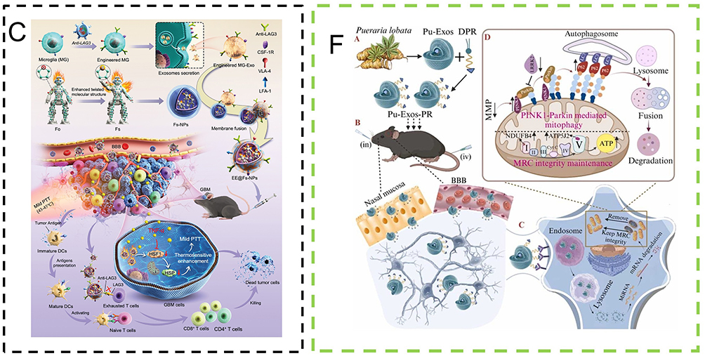
Figure 7 Continued. Figure 7 Relevant examples of vesicular nanocarriers for the treatment of brain diseases. (A) The cell penetration ability of RGD peptide and Angiopep-2 peptide will help ADSC-EVs to better penetrate the BBB and reach the brain parenchyma. Reprinted from Journal of Controlled Release, Vol 365, Liang HB, Chen X, Zhao R, et al, Simultaneous ischemic regions targeting and BBB crossing strategy to harness extracellular vesicles for therapeutic delivery in ischemic stroke, Pages No. 1037–1057, Copyright (2024), with permission from Elsevier.112 (B) Human plasma-derived EVs (pEVs) and adipose-derived mesenchymal stem cell EVs (ADMSC-EVs) loaded with donepezil (DNZ) attenuated lipopolysaccharide (LPS) -induced microglial inflammatory cytokines and chemokines. Reproduced from Silva RO, Haddad M, Counil H et al. Exploring the potential of plasma and adipose mesenchymal stem cell-derived extracellular vesicles as novel platforms for neuroinflammation therapy. J Control Release. 2024;365:1037–1057.113 (C) EE@Fs-NPs and EE@Fs-NPs Schematic of the preparation to penetrate the BBB and target the GBM to deliver anti-LAG3 and AIE photothermal agents in the GBM for mild PTT-ICB therapy. Copyright ©2023. Wiley-VCH GmbH. Reproduced from Lin X, Sun Z, Huang S et al. Engineered Microglia‐Exosomes Coated Highly Twisting AIE Photothermal Agents to Efficiently Cross Blood‐Brain‐Barrier for Mild Photothermal‐Immune Checkpoint Blockade Therapy in Glioblastoma. Adv Func Mater. 2023:1229.117 (D) Mechanism, preparation, and characterization of a hydrogel-based autolytic bacterial delivery system. Copyright ©2023. Wiley-VCH GmbH. Reproduced from Zhang Y, Xi K, Fu Z et al. Stimulation of tumoricidal immunity via bacteriotherapy inhibits glioblastoma relapse. Nat Commun. 2024:15:4241.118 (E) OMVs are extracted by differential centrifugation, and PGZ is loaded into OMVs by electroporation. OMV@PGZ is recognized and captured by TLR receptors on neutrophils and inhibits NLRP3 inflammasome assembly and ferroptosis. Reproduced from Pan J, Wang Z, Huang X et al. Bacteria-Derived Outer-Membrane Vesicles Hitchhike Neutrophils to Enhance Ischemic Stroke Therapy. Adv Mater. 2023;35(38): e2301779.119 (F) Pueraria lobata-derived exosomes (Pu-Exos) were modified with DSPE-PEG-RVG to remove dysfunctional mitochondria through the PINK1-Parkin pathway and promote the survival of DA neurons in PD. Reprinted from Nano Today, Vol 58, Xu Y, Yan G, Zhao J, et al, Plant-derived exosomes as cell homogeneous nanoplatforms for brain biomacromolecules delivery ameliorate mitochondrial dysfunction against Parkinson’s disease, Pages No. 102438, Copyright (2024), with permission from Elsevier.120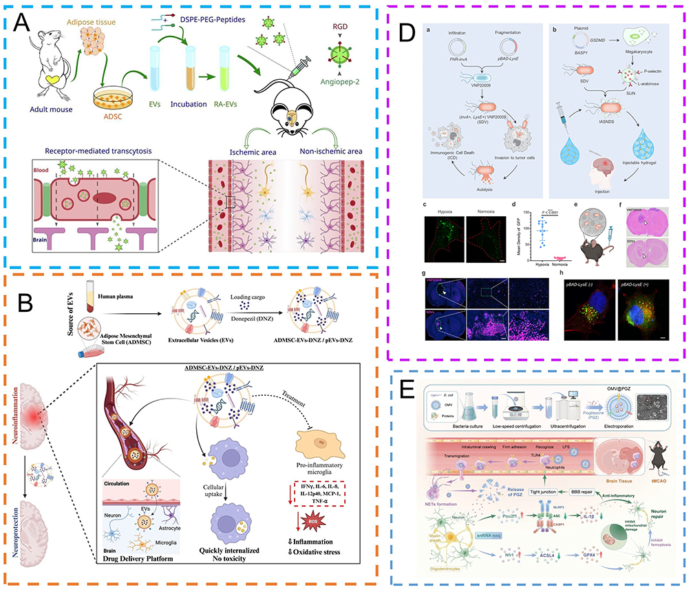
Compared to traditional biomarkers, exosomes have higher detection accuracy and sensitivity as biomarkers for clinical diagnosis. Exosomes are the smallest vesicles arising from the inward budding of multivesicular bodies. Exosomes have low immunogenicity, low toxicity, high biocompatibility and degradability. Exosomes often penetrate the BBB through RMT, and CD46 is the most important receptor for tumor cells to secrete exosomes that target the brain.121 TfR is overexpressed in the BBB and GBM cells and underexpressed in non-cancer cells. Therefore, exosomes can be modified with transferritin (Tf) or HAIYPRH (T7) peptides to deliver antisense miRNA oligonucleotides for miR-21 (AMO-21) in gliomas.122,123 Exosomes are often used to carry hydrophobic drugs, nucleic acids, genes, and small molecule drug, such as hydrogen peroxide for Parkinson’s disease, temozolomide (TMZ) for brain glioma, and BACE1 siRNA for AD.121 EXO can be engineered with the binding protein Fe65 to penetrate the BBB by interacting with APP and promote the uptake of corynoxine-B (Cory-B) carried by neuronal cells to improve AD.124 Lin et al genetically engineered microglia-exosome (MG-Exo) expressing immune checkpoint LAG3 inhibitory antibody (anti-LAG3), fused with AIE photothermal agent nanoparticles (Fs-NPs) through a “freeze-thaw cycle” membrane fusion method. Engineered microglia-exosome AIE nanoparticles (EE@Fs-NPs) were successfully prepared. EE@Fs-NPs crossed the BBB and actively targeted GBM, delivering anti-LAG3 and AIE photothermal agents to GBM, enhancing tumor immunotherapy and triggering mild PTT (Figure 7).117 Exosomes can be combined with methods such as electroporation and ultrasonic treatment to successfully incorporate small RNA and other substances, which can not only improve the drug loading rate of nanocarriers but also change the properties of cell membranes to enhance their permeability.121,125 Microvesicles are produced by the cell membrane towards the outgoing bud and usually contain rRNA and mRNA.125 Since folate receptors are present at low levels in most normal cells, they can be selectively overexpressed on the surface of brain tumor cells.126 Therefore, FA-modified microvesicles have a high affinity for folate receptors and can be internalized through receptor-mediated endocytosis. Magnetic nanovesicles (MNV), which are composed of nano-vesicles (NV) and magnetic nanoparticles (MNP), have recently been studied and are now widely proposed as a potential therapeutic and diagnostic methods. Magnetic nanoparticles are good contrast agents for magnetic resonance imaging (MRI),127 and nanovesicles are good carriers, the use of magnetic nanovesicles can provide guidance for drug delivery and allow the design of optimal drug delivery routes and drug doses using in vivo particle imaging. In addition, enhancing the external magnetic force induces more interactions between magnetic nanoparticles and endothelial cells, and the drug accumulates significantly at the tumor site, reducing off-target effects and increasing the uptake of nanoparticles by BBB endothelial cells.126 When magnetic nanoparticles are exposed to radiofrequency or microwave radiation, they produce medium or high temperatures and exert a thermal-killing effect on tumor cells. Although many studies have acknowledged the therapeutic effect of MNV in penetrating the BBB, clinical translation remains difficult, and its safety needs to be further evaluated. Apoptotic bodies are the largest class of EVs and are produced from characteristic bubbles and protrusions of apoptotic cell membranes.125 The apoptotic process is more controlled than the biogenesis of exosomes; therefore, the efficiency of apoptotic body production is higher.128 Apoptotic bodies retain some proteins on the cell membranes, deliver drugs (siRNAs or microRNAs) more efficiently, and have longer cycle times,128 although they have an uneven distribution of apoptotic body size, potential apoptosis-inducing activity, and susceptibility to phagocytosis.129
Bacterial-Derived Bionic Nanocarriers
Bacterial Biomimetic Nanocarriers
Compared to conventional therapies, bacteria have good targeting and tumor tissue penetration. Currently, bacteria are commonly loaded with genes that penetrate the BBB to treat brain diseases such as GBM. The most commonly used are gram-negative bacteria, which interact with toll-like receptors (TLRS) and nucleotide-binding oligomeric receptors (NLRS) on phagocytes to trigger antitumor immune responses.130,131 Escherichia coli K1 (EC-K1) with its highly expressed outer membrane protein A (Omp-A) binds to gp96 on the endothelial cells of the BBB and crosses BBB.132 Zhou et al extracted lipopolysaccharide (LPS) -free outer membrane protein (Omp) from non-toxic DH5α and coated with polylactic-glycolic acid copolymer nanoparticles (NPs) to target the biomimetic Omp@NC of GRP94.133 Omp@EMB can block the secretion of neuroserine protease inhibitors and restore plasmin mediated L1CAM inactivation and cytotoxicity, improving resistance to antiangiogenic therapy in brain metastases. Sun et al developed a “Trojan horse bacterium” (GP-ICG-SINP) to treat GBM.134 Glucose polymer (GP)-conjugated and indocyanine green (ICG)-loaded silicon nanoparticles were internalized into facultative anaerobic bacteria using bacteria-specific ATP-binding cassette (ABC) transporters. In addition, there is the “inactive Trojan horse EC-K1”, in which dead Escherichia coli K1 (EC-K1) retains both the ability of active bacteria to cross the BBB and the ability to reproduce and pathogenicity.135 Ni et al developed a bacterial-hydrogel to form an immune-stimulated autolytic Salmonella nanoparticle delivery system (IASNDS) by tethering Salmonella lysis- inducing nanocapsules (SLIN) to a Salmonella delivery vehicle (SDV). The hydrogel delivery of IASNDS into GBM cells releases bacterial components that activate antitumor immune responses (Figure 7).118
Bacterial Outer Membrane Vesicle Nanocarriers
Bacteria-derived outer membrane vesicles (OMV) are nanoscale, non-reproducible, spherical, double-layered vesicles that are released by gram-negative bacteria. OMV are rich in lipopolysaccharide (LPS) and can be specifically recognized by toll-like receptors (TLR) to trigger a series of anti-tumor immune responses (Figure 7).119 The use of OMVs to improve the efficiency of brain delivery of the neuroprotective agent pioglitazone (PGZ), OMV@PGZ, can hitch-ride neutrophils to inhibit the activation of the nucleotide oligomerization like receptor protein 3 (NLRP3) inflammasome and ferroptosis and reduce reperfusion injury for the treatment of ischemic stroke. DOX is assembled into an engineered Salmonella typhimurium OMV that induces neutrophils to penetrate the BBB and modulates the tumor microenvironment for targeted therapy in glioma.136
Other Vesicular Nanocarriers
Plant-derived vesicular nanoparticles (PDVLN) are vesicular nanostructured particles isolated from plants. As a therapeutic agent, PDVLN has low toxicity, no immunogenicity, high yield, simple preparation, low production cost, and environmental friendliness. It can pass through the BBB non-invasively by intranasal administration, and can also be administered by oral administration, intravenous injection, intraperitoneal injection, transdermal administration and other ways. Efficient isolation of PDVLNs from plant cells remains one of the challenges to be addressed in the field of plant EVs.137 Currently, PNVs can be used either directly for postisolation drug loading and delivery or modified to be plant-derived nanocarries derived from their lipids.138 As a superior drug delivery vector, PNV has the advantages of strong stability, low immunogenicity, low toxicity, high yield, and high application potential for the delivery of therapeutic drugs and gene drugs. The MiRNA encapsulated by acerola cherry PNVS are almost unaffected by ribonucleases, acids and bases, and can be incubated directly on ice without additional reagents.139 Extracellular vesicles derived from fruits (such as oranges, tangerines, grapefruit, lemons, etc) can also penetrate the BBB to deliver drugs to target brain diseases. Chen et al used cRGD-modified lemon-derived EVs to form extracellular vesicle (EV) engineered structural droplet drugs (ESDD) at the DOX@squalene-PBS interface to enhance the antitumor effect of glioma.140 The DOX@squalene-PBS interface enhances the softness of ESDD and its ability to cross the BBB through deformation-amplified macropinocytosis, membrane fusion, and transcytosis. Pueraria lobata-derived exosomes (Pu-Exos), a medicinal plant, restore ATP supply by removing dysfunctional mitochondria and preserving mitochondrial respiratory chain complex I and V activity through PINK1-Parkin-mediated mitophagy. DSPE-PEG-RVG can improve the mitochondrial dysfunction of SH-SY5Y cells and promote the penetration of nasal tissue and BBB to enhance the therapeutic effect of PD (Figure 7).120 Garlic-derived nanovesins were preferentially and selectively taken up by microglia in HFD mice and further inhibited cell activation.139 In terms of disadvantages, the source of PNV was affected by region, season, plant origin and location. The structure of PNV was damaged by low temperature storage. PNV is not derived from organisms and lacks specific markers, so its targeting ability is poor.139
Inorganic Nanocarrier
Although biomimetic nanocarriers show unique advantages in safety and immune evasion, their preparation remains complex and stability needs further improvement. In contrast, inorganic nanocarriers, with superior physicochemical stability, tunable structures, and the potential for theranostics, are emerging as another important platform in brain disease treatment. Inorganic nanocarriers are nanoscale structures made of inorganic materials, such as metals, silicon dioxide, carbon nanotubes, and quantum dots, which have unique physicochemical properties, such as high stability, tunable shape and size, and excellent optical and magnetic properties. They are often modified by surface functionalization to provide good biocompatibility and the targeting ability to deliver drugs specifically to the target site (Figure 8).
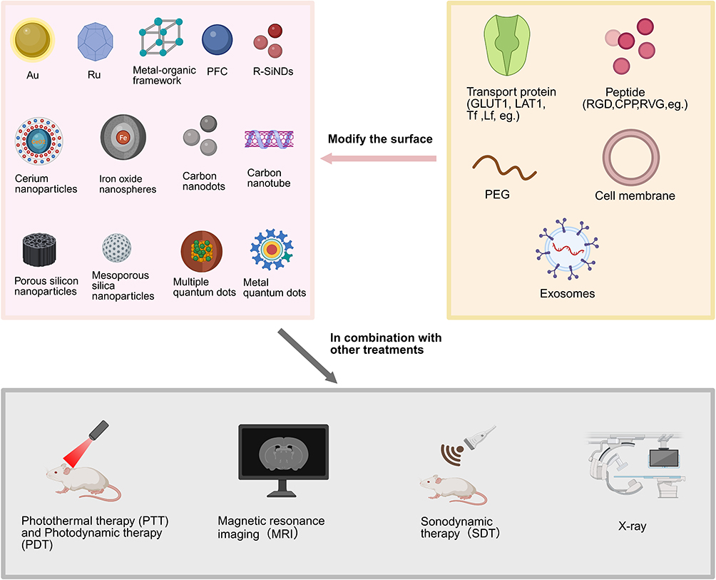 |
Figure 8 Inorganic nanocarriers with different molecular modifications are combined with other therapeutic methods to improve the therapeutic effect. Created in BioRender. Yu, Z (2025) https://BioRender.com/a7urxso. |
Metal Nanocarrier
Metal nanomaterials have a low molecular weight, easy surface modification, high stability, strong solubility, good biocompatibility and good cell penetration. Recent research has reported that metal nanomedicines have tremendous neuron-generating activity, causing damage to specific DNA in malignant tissues and making GBM tumor cells more sensitive to radiation. Metallic nanomaterials can be used as photothermal conversion agents for photothermal therapy, or as contrast agents for bioimaging to diagnose and treat tumors.141 Metal nanomaterials can directly induce DNA damage and amplify the damaging effect by blocking DNA repair to kill tumor cells. Metal nanoparticles can be used to design “personalized weapons” that target different types of DNA damage within tumor cells.142 Current research includes gold, manganese, ruthenium, silver, Prussian blue, and zinc-oxide nanoparticles. Although metal nanoparticles have good BBB-penetrating ability, their toxicity and ethical concerns remain a problem for clinical translation.
Metal-Based Nanocarriers
Nano-gold (AuNPs) has the advantages of controllable size, easy modification, good optical properties, and low toxicity and can be used as a drug delivery vehicle and tool for biological imaging.143 The size of the AuNPs has a strong effect on brain permeability. Sela et al showed that small-molecule AuNPs (2.5 nm) can spontaneously permeate the BBB and that the amount of AuNPs permeation can be controlled by an ion channel blocker.144 The appropriate size of the AuNPs as a drug was approximately 20 nm.145 Delivery carriers modified with TAT, Tf, PEG, angiopep-2, and other ligands have various advantages. For example, the surface modification of AuNPs by PEG could reduce the negative charge indicated by nanoparticles, improve the blood circulation time of AuNPs, and enhance their delivery to the brain.143 Angiopep-2 coupled AuNPs improve the efficiency of BBB crossing and increase the selectivity of administration. Ph-sensitive coupling complexes such as TAT-Au NP-Dox, improve drug targeting and enhance the effectiveness of glioma treatment.146 AuNPs have tunable SPR peaks in the near infrared region, and their good photothermal conversion function enables a combination of photothermal therapy (PTT) and photodynamic therapy (PDT) to enhance tumor-killing efficacy.147 However, AuNP-based drug delivery platforms have clinical issues that need to be addressed. Some targets for modifying AuNPs are also present in other organs or tissues; therefore, their specificity needs to be demonstrated. The elimination of AuNPs after entry into the brain, duration of residence in the spleen or liver, and systemic clearance of AuNPs need to be further explored. However, in general, AuNPs have important clinical significance in the diagnosis and treatment of AD, PD, brain tumors, and imaging-based diagnosis. Ruthenium nanoparticles have good biocompatibility and surface functionalization modification properties, and modified dual-targeted mesoporous ruthenium nanoparticles (MRN) can improve the loading capacity of anti-tumor drugs, prevent the premature release of drugs, and prolong the circulation time of drugs in the blood.148 Ruthenium nanomaterials have outstanding photothermal effect, and show high absorption rates and photothermal therapy conversion under near-infrared irradiation; therefore, they can be combined with photothermal therapy to treat brain tumors. Transferrin is commonly used to modify ruthenium nanoparticles, and Tf modification significantly enhances RuNP uptake by cells through fossa and clathrin-mediated endocytosis. In summary, Tf-RuNPs have low toxicity and high in vivo cell elimination ability and can be used as an effective PTT drug for tumor therapy.149 The combined application of silver nanoparticles (Ag NPs) and X-rays has a good killing effect on cancer cells, but accumulates in normal organs such as the kidney, liver, and lungs and producing toxicity. It can destroy the structure of the BBB and hinder the antioxidant mechanisms of astrocytes, inducing astrocytic toxicity.143
Metal Compound Nanocarriers
Magnetic iron oxide nanoparticles (MIONPs) are often used as imaging agents in magnetic resonance imaging (MRI) because of their small size and easy functionalization by biological ligands to enhance their penetration efficiency through the BBB.150 They can also cooperate with focused ultrasound to open the BBB.16,151 IONPs were functionalized with glucuronic acid to promote GLUT-mediated transcytosis across the BBB for targeted glioma therapy.152 Superparamagnetic iron oxide nanoparticle modified exosomes derived from human mesenchymal stem cells deliver miR-1228-5p across the BBB. Its mechanism of action involves the inhibit of TRAF6 expression and activation of the TRAF6-NADPH oxidase 1 (NOX1) pathway, thereby protecting against oxidative damage and treating stroke.153 Forgham et al developed a perfluoropolyether (PFPE) polymer-engineered iron oxide nanoparticle platform to enhance the ability to cross the BBB, combined with siRNA-binding agents to induce oncogene silencing and promote targeted apoptosis of tumor cells.154 Inspired by the rabies virus, a biomimetic nanobullet based on iron oxide nanorod was developed for the treatment of AD by modifying Fe NRs with a dopamine terminal (DA) and an RVG-coupled polymer (RVG-PBAC-PEG-DA, PBACR) to obtain PBACR@NRs.155 It can reduce Aβ oligomers, down-regulate the expression of NF-κB protein, reduce immune activation, protect neurons, and ultimately improve the memory and cognitive abilities of AD mice. Kang et al used injectable thermally responsive hydrogel nanocomposites of drug-loaded micelles and water-dispersible ferrous oxide nanocubes (wFION) to treat GBM.156 Cerium NPs (Ce NPs) can eliminate ROS. Currently, CeONP and CeO2NP are used to load with edaravone (a free radical scavenger) and other drugs. MnO2NP is another widely used MRI drug.143 Prussian blue nanoparticles (PBNPs) are widely used in the biomedical field because of their good magnetic properties, biocompatibility, and stability. BNPs exhibit excellent NIR laser absorption and high photothermal conversion efficiency, making them well-suited for the PTT of AD. Li et al used erythrocyte membrane encapsulation of PBNPs to effectively chelate Cu2+ reduction to reverse Aβ-Cu2+ plaque formation, restore steady-state levels of Cu2+, photothermally depolymerize Aβ fibrils, and reduce ROS levels (Figure 9).157 The PB/RBC multifunctional nano-system holds great promise for the treatment of AD. Metal-organic frameworks (MOF) are porous polymers formed by the coordination of metal clusters (SUB) with organic ligands. MIL-101-NH2 (Fe) is a nano-scale metal-organic framework with high drug loading and good biocompatibility. Chen et al158 incorporated copper ions into MIL-101-NH2 and encapsulated the neuroprotective agent rapamycin (RA) to form RA@M. Subsequently, the targeted molecular transporter, Tf was coated on RA@M to form a Tf@RA@MIL-101-NH multifunctional nanosystem (Figure 9). This system can treat ischemic stroke by achieving efficient delivery across the BBB, powerful ROS-scavenging effects, the regulation of oxidative stress, and the inhibition of neuroinflammation and neuronal apoptosis. In addition, modification of MIL-53 (Cr) nanoparticles with PEG and Angiopep-2 (ANG) improved BBB penetration of edaravone (Figure 9).159 EDA@MIL-53 (Cr) -P/A alleviates IR-induced brain injury by inhibiting oxidative stress, DNA damage, apoptosis and inflammation. For the treatment of Parkinson’s disease (PD), a Zr-FeP MOF was prepared using Fe-5,10,15, 20-four (4-carboxyphenyl) porphyrin (Fe-TCPP) and Zr encapsulated in mannitol (Man) liposomes (Figure 9).160 The MOF@Man nanoenzyme system effectively penetrates the BBB via a MAN-mediated mechanism. In addition, the nano-enzyme system effectively inhibited the NOD-like receptor protein 3 (NLRP3) inflammasome to alleviate glial cell activation and neuroinflammation.
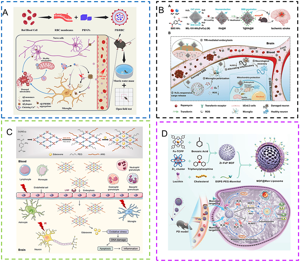 |
Figure 9 Relevant examples of metal compound nanocarriers for the treatment of brain diseases. (A) Red blood cell (RBC) membrane-encapsulated Prussian blue nanoparticles (PB/RBC) chelate Cu2+ and reduce reactive oxygen species (ROS). Reprinted from Journal of Controlled Release, Vol 375, Li L, Xiong Y, Zhang Y, et al, Biofilm-camouflaged Prussian blue synergistic mitochondrial mass enhancement for Alzheimer’s disease based on Cu2+ chelation and photothermal therapy, Pages No. 269–284, Copyright (2024), with permission from Elsevier.157 (B) Schematic representation of BBB penetrating MOF nanoenzymes for targeted IS therapy. Copyright ©2024. Wiley-VCH GmbH. Reproduced from Chen Q, Wang J, Xiong X et al. Blood-Brain Barrier-Penetrating Metal-Organic Framework Antioxidant Nanozymes for Targeted Ischemic Stroke Therapy. Adv Healthc Mater. 2024;7:e2402376.158 (C) MIL-53 (Cr) -P /A nanoparticles can enhance the ability of edaravone to cross the BBB and alleviate IR-induced brain injury by inhibiting oxidative stress, DNA damage, cell apoptosis and inflammatory response. Reprinted from Biomaterials, Vol 314, Li X, Hua S, Zhong D, et al, Metal-organic framework-edaravone nanoparticles for radiotherapy-induced brain injury treatment, Pages No. 122868, Copyright (2025), with permission from Elsevier.159 (D) The preparation process of MOF@Man liposome nanoenzyme system and its ability to alleviate oxidative stress and neuroinflammation by inhibiting the formation of NLRP3 inflammasome and the secretion of inflammatory cytokines. Copyright ©2024. Wiley-VCH GmbH. Reproduced from Li Q, Ding X, Chang Z et al. Metal–Organic Framework Based Nanozyme System for NLRP3 Inflammasome-Mediated Neuroinflammatory Regulation in Parkinson’s Disease. Adv Healthc Mater. 2024;13(10):e2303454.160 |
Non-Metal Nanocarrier
Carbon Nanocarrier
Carbon nanotubes are tubular nanostructures formed from graphene, with large surface areas, good biocompatibility, and extremely high drug loads.161 Carbon nanotubes are often divided into single-walled carbon nanotubes (SWCNTs) and multi-walled carbon nanotubes (MWCNTs). The key advantage of their delivery lies in the hollow tube structure of CNTs, in which drugs can be encased in a hydrophobic hollow core, whereas other molecules can modify the outer hydrophilic surface to make them dispersible and biocompatible for targeting purposes.162 Functionalized carbon nanotubes cross the BBB through RMT or passive transport.72 Carboxylated single-wall carbon nanotubes (SWCNT-COOH) can be used for the continuous delivery of levodopa (LD) in the treatment of Parkinson’s disease. Carbon nanomaterials also exhibit nerve regeneration activity, which has important implications in the treatment of Alzheimer’s and Parkinson’s diseases. Although carbon nanotubes have been widely used in drug delivery, they are hydrophobic and potentially toxic. They can be improved using a variety of methods, such as surface modification with polyethylene glycol, purification of nanotubes, and reasonable optimization of their length.161 Mesoporous carbon nanomaterials (MCBs) exhibit high biocompatibility, continuous drug release, easy surface modification, and high photosensitive conversion efficiency. Functionalized carbon nanodots are also an important tool for the treatment of the nervous system, with low toxicity and high biocompatibility,163 and can increase the affinity for target molecules. Cheng et al used Cu doping in carbon dots to form a novel sonosensitizer, Cu-Cds, for the treatment of GBM. Cu-Cds effectively bind to the mitochondrial enzyme lipoacylation and stimulate the aggregation of lipoacylated dihydrothiolane transacetylase (DLAT), leading to copper deposition and further amplifying the sonodynamic therapy (SDT) effect (Figure 10).164 Guo et al found that N-doped CD (CPL) not only eliminated ROS antioxidants but also released NO to reversibly open the BBB (Figure 10).165 Lf-modified CD (CPL) can further promote BBB permeability through receptor-mediated pathways and chelate free iron ions to inhibit the Fenton reaction in the treatment of PD. Perfluorinated carbon nanodroplets (PFC), a nano-delivery vector formed by combining focused ultrasound (FUS) and nanoemulsion, can help deliver hydrophobic drugs that penetrate the BBB and tumor barrier to improve the treatment of brain disease.166 Perfluorocarbon (PFC) nanodroplets exhibit extremely high stability in blood circulation and can permeate into and out of tissues owing to their small size (Figure 10).166 Because the liquid core of the PFC can be used for fluorine-19 magnetic resonance imaging (19F-MRI), it can be developed as a contrast agent for diagnostic therapy.167
Silicon Nanocarrier
The excellent ability of silica nanoparticles (SNPS) to cross the BBB may be due to the binding of silanol groups to active components in plasma (transferrin, low-density lipoprotein, and free nucleic acids, etc). These active components have the ability to pass through the BBB.168 The SNP exhibited good biocompatibility and biodegradability. It was modified with lactoferrin and PEG and loaded with TMZ to treat GBM.169 Internal pore rate of SNPs is high, and large pores can protect large protein and DNA from degrading.170 Mesoporous silica nanoparticles (MSNs) have good biocompatibility, large surface area, and high drug-loading capacity for low water-soluble drugs.171 MSNs have been shown to pass BBB by adsorptive⁃mediated transcytosis t (AMT) or by receptor⁃mediated transcytosis (RMT).172 LDL peptide-coupled PLA-coated mesoporous silica nanoparticles loaded with resveratrol (RSV) penetrate the BBB and reduce the production of ROS to treat neurological diseases caused by oxidative stress.172 Functional Mn-doped mesoporous silica nanoparticles (Mn-HMSNs) are sensitive to the tumor microenvironment and rapidly degrade to release drugs.173 Porous silicon nanoparticles (pSiNPs) loaded with 6-amino-2-naphthalenesulfonic acid (ANA) and modified with PEG have been used to effectively decompose amyloid plaques in the brain. The nanopreparation were biotin-cacl2-ANA-PsinPs (BCAP).174 Silicon nanodots (SiNDs) are a new type of nanomaterial with low toxicity and good optical properties. Liu et al175 prepared red luminescent silicon nanodots (R-SiNDs) using red luminescent fluorescent dyes. Based on the high luminous efficiency and stability of red dye, red luminous silicon nanodots are good targets for the dynamic visualization of mitochondria (Figure 10). Most importantly, R-SiNDs can penetrate the BBB without any modification.
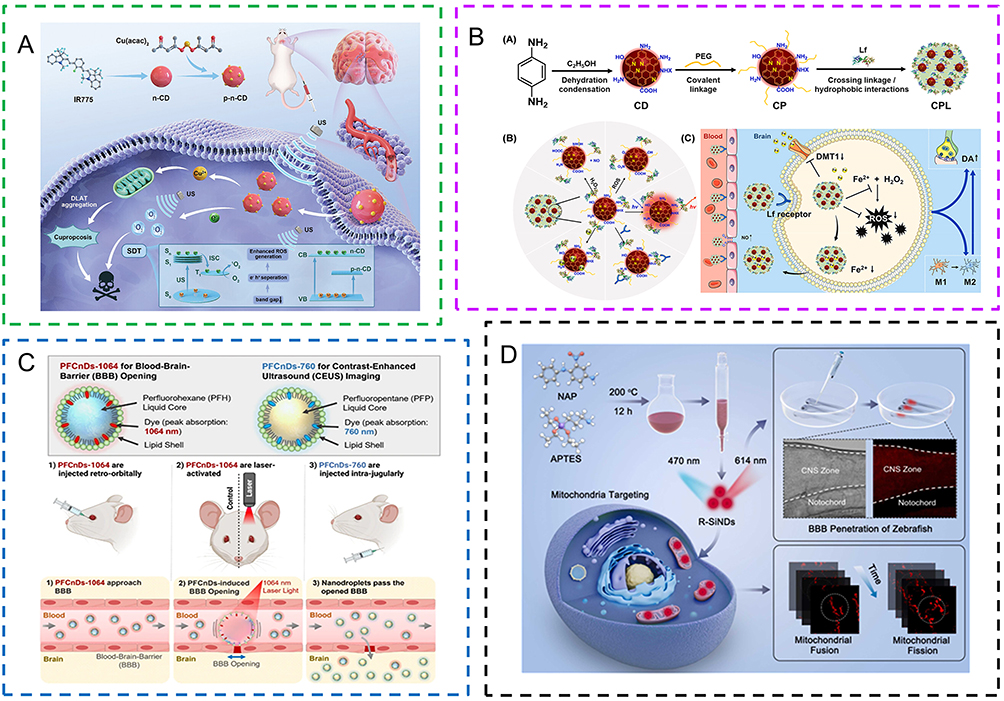 |
Figure 10 Relevant examples of carbon/ Silicon nanocarrier for the treatment of brain diseases. (A) Copper doped CD (Cu-CDs) binds to the mitochondrial enzyme of lipoacylation and stimulates the accumulation of lipoacylated dihydrothiolane transacetylase (DLAT), leading to copper deposition and inducing copper toxicity, which can well cross the BBB to treat GBM. Reproduced from Cheng M, Liu Y, You Q et al. Metal‐Doping Strategy for Carbon‐Based Sonosensitizer in Sonodynamic Therapy of Glioblastoma. Adv Sci (Weinh). 2024;;11(34):240423.164 (B) Synthesis of p-phenylene diamine (PPD) into CD by a simple solvothermal method not only eliminates ROS for antioxidation, but also generates nitric oxide (NO) to reversibly open the BBB, and chelates iron ions to inhibit the Fenton reaction. Reprinted from Biomaterials, Vol 309, Guo W, Ji M, Li Y, et al, Iron ions-sequestrable and antioxidative carbon dot-based nano-formulation with nitric oxide release for Parkinson’s disease treatment, Pages No. 122622, Copyright (2024), with permission from Elsevier.165 (C) PFCnD-1046 and PFCnD-760 were constructed and injected retroorbital and intravenously. The former approaches the BBB directly, while the latter opens the BBB into brain tissue. Reproduced from Hallam KA, Nikolai RJ, Jhunjhunwala A et al. Laser-activated perfluorocarbon nanodroplets for intracerebral delivery and imaging via blood–brain barrier opening and contrast-enhanced imaging. J Nanobiotechnology 2024;22:356.166 (D) Diagram of R-SiNDs synthesis. Reprinted from Sensors and Actuators B Chemical, Vol 408, Liu Y, Cao L, Zhu T, et al, Red-emissive silicon nanodots with highly biocompatible for mitochondrial dynamic tracking and blood-brain barrier penetration imaging, Pages No. 135523, Copyright (2024), with permission from Elsevier.175 |
Quantum Dot
Quantum dot (QD), also known as semiconductor nano-crystal, is composed of multi -functional biological puppets and surface-modified crystals.176 It is roughly divided into metal dots such as silver, crickets, and chromium, and non-metallic quantum dots, such as carbon and silicon. Its size is approximately 1 to 20 nm, with a small volume, large absorption spectrum, and high photobleaching. With excellent light, physical, and fluorescent characteristics, it occupies a place in the fields of biomedical imaging, nano-targeted drug delivery, and other fields177 Its excellent stability can enhance the cycle time of drugs in the body and the high-penetration, long retention effect.178 The quantum dot surface area is large and polyethylene glycol quantum dots can be combined with a variety of different functions of biomolecules (TF, NGR peptide, and RGD peptide) to penetrate BBB.143 The use of macrophage membrane coated molybdenum disulfide (MoS2) quantum dots to form a new nanomedicine MoS2QDs/MM can be combined with NIR irradiation to open the BBB and improve the efficiency of BBB drug delivery (Figure 11).179 MoS2QDs could not only eliminate reactive oxygen species (ROS) but also inhibit the deposition of Aβ1-42 to treat Alzheimer’s disease. Wu et al reported the encapsulation of ZnCdSe@ZnS quantum dots (QD) in exosomes (Exo). QD can cross the BBB, be internalized by macrophages (J774A.1) and tumor cells (HeLa), and then released into the extracellular environment along with Exo (Figure 11).180 However, the cytotoxicity of quantum dots can cause excessive amounts of active oxygen and mitochondrial disorders,which have yet to be resolved and lack a standard scheme for the assessment of cytotoxicity.181 At present, carbon quantum dots are less toxic because of the lack of metallic elements and are often regarded as more suitable materials. Amine-functionalized aspirin-derived carbon quantum dots (NACQD) can chelate iron, scavenge free radicals, reduce meningeal inflammation, promote neuronal repair, and ameliorate intracerebral hemorrhage in mice (Figure 11).182 Ternary I-III-VI QDs, such as silver indium selenide quantum dots, can be used for preoperative diagnosis and detection of combined traumatic brain injury (Figure 11).183 Silver indium selenide quantum dots have low toxicity and high biocompatibility, and their narrow bulk band-gap allows their luminescence wavelength to be tuned to the NIR spectral region, which is more conducive to penetrating biological tissues. Aluminum doped ZnS shells can effectively improve the photoluminescence quantum yield (PLQYs) and stability of ternary quantum dots.
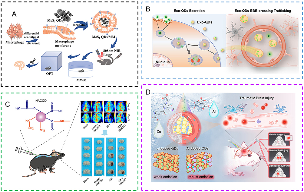 |
Figure 11 Relevant examples of Quantum dot for the treatment of brain diseases. (A) Macrophage membrane (MM) coated functionalized molybdenum disulfide quantum dots (MoS2QDs) can eliminate ROS and anti-Aβ1-42 deposition and play A role in NIR multi-target combined therapy. Copyright ©2024. Wiley-VCH GmbH. Reproduced from Qi X, Li L, Ye P et al. Macrophage Membrane-Modified MoS2 Quantum Dots as a Nanodrug for Combined Multi-Targeting of Alzheimer’s Disease. Adv Healthc Mater. 2024;(6):e2303211.179 (B): Distal transport and “barrier-crossing” pathways for Exo-QD. Reprinted with permission from Wu D, Sun H, Yang B et al. Exosome Heterogeneity Affects the Distal “Barrier-Crossing” Trafficking of Exosome Encapsulated Quantum Dots. ACS Nano. 2024;18(11):7907–7922. Copyright 2024 American Chemical Society.180 (C) Intrathecal administration of amino-functionalized aspirin-derived carbon quantum dot (NACQD) significantly reduced iron deposition and oxidative stress in brain tissue and improved neurological recovery in a mouse ICH model. Reprinted with permission from Yang X, Tang X, Jia G et al. Multifunctional Carbon Quantum Dots: Iron Clearance and Antioxidation for Neuroprotection in Intracerebral Hemorrhage Mice. ACS Appl Mater Interfaces. 2023;15(49):56820–56833. Copyright 2023 American Chemical Society.182 (D) The AISe@ZnS:Al nanostructure, consisting of a silver-indium-selenium (Ag-In-Se) core with near-infrared emission and an aluminum-doped zinc sulfide (ZnS) shell, demonstrates considerable potential for advancing the diagnosis and surgical intervention of traumatic brain injury (TBI) through near-infrared fluorescence imaging. Reprinted with permission from Jiao M, Li X, Liu H et al. Aqueous Grown Quantum Dots with Robust Near-Infrared Fluorescence for Integrated Traumatic Brain Injury Diagnosis and Surgical Monitoring. ACS Appl Mater Interfaces. 2024;18(29):19038–19053. Copyright 2024 American Chemical Society.18 |
Conclusion and Perspectives
The global aging population poses greater challenges for the treatment of brain diseases, making the development of new therapeutic strategies urgently needed. The advancement of nanomedicine offers the possibility to cross the blood-brain barrier (BBB) and achieve efficient targeted drug delivery. New nanocarriers should not only be able to effectively cross the BBB, but also have excellent biological characteristics (such as good biocompatibility, immune inertia, long half-life, high targeting, good stability, and low toxicity). By optimizing drug delivery and targeting capabilities, nanocarrier systems have the potential to directly improve patient outcomes, for example, by enhancing BBB permeability, increasing the effective intracerebral concentration of drugs in neurodegenerative diseases, or improving the efficacy and safety of brain tumor therapies.
This review focuses on various nanomaterials used for drug delivery across the BBB and targeted therapies for brain diseases. The detailed content is summarized in Table 1. At present, modified liposomes, solid lipid nanoparticles (SLNs), and certain functionalized high-density lipoprotein (HDL) mimetic nanoparticles have demonstrated promising clinical application prospects. Among them, multifunctional nanocarriers, one of the most promising tools currently available, are composed of many different types of nanomaterials with different properties in a single system, such as quantum dots, superparamagnetic iron oxide nanoparticles (SPION),184 thereby ensuring that the performance of the mixed material in a therapeutic system is enhanced.185 Many studies have already conducted preliminary monitoring and evaluation of issues such as oxidative stress, tissue inflammation, and potential DNA damage arising during drug delivery with nanocarriers.186
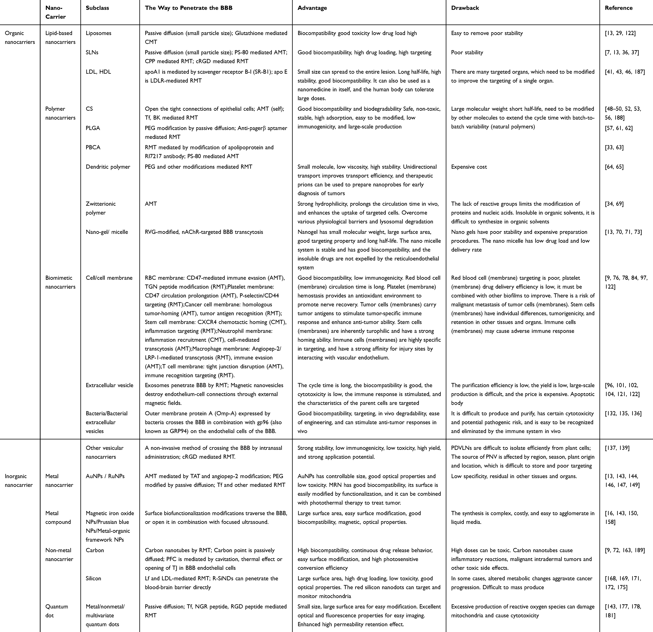 |
Table 1 Nanocarriers Penetrate the BBB Form |
Nevertheless, most nanodrug delivery systems are still at the preclinical or early clinical stage. Future research should focus on simplifying the preparation processes of nanocarriers, improving their stability and reproducibility, and optimizing targeting designs to enhance therapeutic specificity. Based on the concept of a “Two-stage targeted drug delivery system”, dual targeting functional groups are simultaneously modified on the surface of the delivery system to facilitate BBB penetration while enhancing localization to the lesion site, thereby reducing drug distribution throughout the brain and minimizing adverse effects.184 With the rapid advancement of genetic technologies, gene-editing tools such as CRISPR-Cas9 have opened new possibilities for the treatment of brain diseases. Combining CRISPR-Cas9 with nanocarriers not only enables drug delivery but also allows for gene regulation while targeting specific brain cells or regions—for instance, silencing pathogenic genes or activating protective genes—thereby improving therapeutic outcomes for brain disorders. With the continuous development and innovation of science and technology, it will be possible to develop BBB targets with higher selectivity, high-efficiency, and low-toxicity nanomedicines across the BBB in the future.
Acknowledgment
The graphical abstract was created in BioRender. Yu, Z. (2025) https://BioRender.com/gdqfteu.
Author Contributions
All authors made a significant contribution to the work reported, whether that is in the conception, study design, execution, acquisition of data, analysis and interpretation, or in all these areas; took part in drafting, revising or critically reviewing the article; gave final approval of the version to be published; have agreed on the journal to which the article has been submitted; and agree to be accountable for all aspects of the work.
Funding
This work was supported by the National Natural Science Foundation of China (32301176), Shandong Provincial Natural Science Foundation (ZR2023MH280, ZR2022QC133, ZR2022MH100).
Disclosure
The authors report no conflicts of interest in this work.
References
1. Liao J, Fan L, Li Y. et al. Recent advances in biomimetic nanodelivery systems: new brain-targeting strategies. Journal of Controlled Release: Official Journal of the Controlled Release Society. 2023;358:439–464. doi:10.1016/j.jconrel.2023.05.009
2. Schaeffer S, Iadecola C. Revisiting the neurovascular unit. Nature Neuroscience. 2021;24(9):1198–1209. doi:10.1038/s41593-021-00904-7
3. Xie J, Shen Z, Anraku Y, Kataoka K, Chen X. Nanomaterial-based blood-brain-barrier (BBB) crossing strategies. Biomaterials. 2019;224:119491. doi:10.1016/j.biomaterials.2019.119491
4. Sarkaria JN, Hu LS, Parney IF, et al. Is the blood-brain barrier really disrupted in all glioblastomas? A critical assessment of existing clinical data. Neuro-Oncology. 2018;20(2):184–191. doi:10.1093/neuonc/nox175
5. Sweeney MD, Sagare AP, Zlokovic BV. Blood-brain barrier breakdown in Alzheimer disease and other neurodegenerative disorders. Nature Reviews Neurology. 2018;14(3):133–150. doi:10.1038/nrneurol.2017.188
6. Butt MH, Zaman M, Ahmad A, et al. Appraisal for the Potential of Viral and Nonviral Vectors in Gene Therapy: a Review. Genes. 2022;13(8). doi:10.3390/genes13081370
7. Govindarajan K, Palanivel G, Shin‐Young P, Ho‐Won L, Dong‐Kug C. Lipid-based nanodelivery approaches for dopamine-replacement therapies in Parkinson’s disease: from preclinical to translational studies. Biomaterials. 2019. doi:10.1016/j.biomaterials.2019.119704
8. Rajeev T, Violina K, Sarathlal KC, et al. Nanocarrier mediated drug delivery as an impeccable therapeutic approach against Alzheimer’s disease. Journal of Controlled Release: Official Journal of the Controlled Release Society. 2022. doi:10.1016/j.jconrel.2022.01.044
9. Song X, Qian H, Yu Y. Nanoparticles Mediated the Diagnosis and Therapy of Glioblastoma: bypass or Cross the Blood-Brain Barrier. Small. 2023;19(45):e2302613. doi:10.1002/smll.202302613
10. Choi HY, Lee YH, Lim CH, et al. Assessment of respiratory and systemic toxicity of Benzalkonium chloride following a 14-day inhalation study in rats. Particle and Fibre Toxicology. 2020;17(1):5. doi:10.1186/s12989-020-0339-8
11. Sánchez Sofía V, Gabriela NO, Florence G, Amanda S, Javier M. Intranasal delivery of extracellular vesicles: a promising new approach for treating neurological and respiratory disorders. Journal of Controlled Release: Official Journal of the Controlled Release Society. 2025;0118. doi:10.1016/j.jconrel.2025.01.018
12. Zhan W, Wang CH. Convection enhanced delivery of chemotherapeutic drugs into brain tumour. Journal of Controlled Release: Official Journal of the Controlled Release Society. 2018;271:74–87. doi:10.1016/j.jconrel.2017.12.020
13. Zha S, Liu H, Li H, Li H, Wong KL, All AH. Functionalized Nanomaterials Capable of Crossing the Blood-Brain Barrier. ACS nano. 2024;18(3):1820–1845. doi:10.1021/acsnano.3c10674
14. Tang W, Fan W, Lau J, Deng L, Shen Z, Chen X. Emerging blood-brain-barrier-crossing nanotechnology for brain cancer theranostics. Chemical Society Reviews. 2019;48(11):2967–3014. doi:10.1039/c8cs00805a
15. Wu D, Chen Q, Chen X, Han F, Chen Z, Wang Y. The blood-brain barrier: structure, regulation, and drug delivery. Signal Transduction and Targeted Therapy. 2023;8(1):217. doi:10.1038/s41392-023-01481-w
16. He C, Wu Z, Zhuang M, et al. Focused ultrasound-mediated blood-brain barrier opening combined with magnetic targeting cytomembrane based biomimetic microbubbles for glioblastoma therapy. Journal of Nanobiotechnology. 2023;21(1):297. doi:10.1186/s12951-023-02074-z
17. Miller DB, O’Callaghan JP. New horizons for focused ultrasound (FUS) - therapeutic applications in neurodegenerative diseases. Metabolism: Clinical and Experimental. 2017;69S:S3–S7. doi:10.1016/j.metabol.2017.01.012
18. Rezai AR, Ranjan M, D’Haese PF, et al. Noninvasive hippocampal blood-brain barrier opening in Alzheimer’s disease with focused ultrasound. Proceedings of the National Academy of Sciences of the United States of America. 2020;117(17):9180–9182. doi:10.1073/pnas.2002571117
19. Carpentier A, Canney M, Vignot A, et al. Clinical trial of blood-brain barrier disruption by pulsed ultrasound. Science Translational Medicine. 2016;8(343):343re2. doi:10.1126/scitranslmed.aaf6086
20. Pengxuan Z, Tiantian W, Tian Y, Jia Y, Xin‐Wu C. Recent advances of focused ultrasound induced blood-brain barrier opening for clinical applications of neurodegenerative diseases. Advanced Drug Delivery Reviews. 2024. doi:10.1016/j.addr.2024.115323
21. Sohee P, Kyoungwon B, Seun J, Seok CW, Seok YB, Woo CJ. Extensive frontal focused ultrasound mediated blood-brain barrier opening for the treatment of Alzheimer’s disease: a proof-of-concept study. Translational Neurodegeneration. 2021. doi:10.1186/s40035-021-00269-8
22. Israel LL, Galstyan A, Holler E, Ljubimova JY. Magnetic iron oxide nanoparticles for imaging, targeting and treatment of primary and metastatic tumors of the brain. Journal of Controlled Release: Official Journal of the Controlled Release Society. 2020;320:45–62. doi:10.1016/j.jconrel.2020.01.009
23. Qiu Y, Tong S, Zhang L, et al. Magnetic forces enable controlled drug delivery by disrupting endothelial cell-cell junctions. Nature Communications. 2017;8:15594. doi:10.1038/ncomms15594
24. Li X, Vemireddy V, Cai Q, et al. Reversibly Modulating the Blood-Brain Barrier by Laser Stimulation of Molecular-Targeted Nanoparticles. Nano Letters. 2021;21(22):9805–9815. doi:10.1021/acs.nanolett.1c02996
25. Kia RA, Isaiah GR, Ali R, et al. Treating Alzheimer’s disease using nanoparticle-mediated drug delivery strategies/systems. Ageing Research Reviews. 2024. doi:10.1016/j.arr.2024.102291
26. Rita C, Elisabetta E, Markus D, et al. L-dopa co-drugs in nanostructured lipid carriers: a comparative study. Materials Science & Engineering C, Materials for Biological Applications. 2016. doi:10.1016/j.msec.2016.11.060
27. Qiuli D, Yan L, Mengyu F, Sijun W, Muhammad I, Meng Z. PEG length effect of peptide-functional liposome for blood brain barrier (BBB) penetration and brain targeting. Journal of Controlled Release: Official Journal of the Controlled Release Society. 2024. doi:10.1016/j.jconrel.2024.06.005
28. Mengke Q, Qing L, Shanshan H, et al. A brain targeting functionalized liposomes of the dopamine derivative N-3,4-bis(pivaloyloxy)-dopamine for treatment of Parkinson’s disease. Journal of Controlled Release: Official Journal of the Controlled Release Society. 2018. doi:10.1016/j.jconrel.2018.03.019
29. Rotman M, Welling MM, Bunschoten A, et al. Enhanced glutathione PEGylated liposomal brain delivery of an anti-amyloid single domain antibody fragment in a mouse model for Alzheimer’s disease. Journal of Controlled Release: Official Journal of the Controlled Release Society. 2015;203:40–50. doi:10.1016/j.jconrel.2015.02.012
30. He X, Peng Y, Huang S, et al. Blood Brain Barrier-Crossing Delivery of Felodipine Nanodrug Ameliorates Anxiety-Like Behavior and Cognitive Impairment in Alzheimer’s Disease. Advanced Science. 2024;11(34):e2401731. doi:10.1002/advs.202401731
31. Ma F, Yang L, Sun Z, et al. Neurotransmitter-derived lipidoids (NT-lipidoids) for enhanced brain delivery through intravenous injection. Science Advances. 2020;6(30):eabb4429. doi:10.1126/sciadv.abb4429
32. Yang L, Wang Y, Li Z, Wu X, Mei J, Zheng G. Brain targeted peptide-functionalized chitosan nanoparticles for resveratrol delivery: impact on insulin resistance and gut microbiota in obesity-related Alzheimer’s disease. Carbohydrate Polymers. 2023;310:120714. doi:10.1016/j.carbpol.2023.120714
33. Dasgupta A, Sun T, Rama E, et al. Transferrin Receptor-Targeted Nonspherical Microbubbles for Blood-Brain Barrier Sonopermeation. Advanced Materials (Deerfield Beach, Fla). 2023;35(52):e2308150. doi:10.1002/adma.202308150
34. Wang K, Zhao B, Ao Y, et al. Super-small zwitterionic micelles enable the improvement of blood-brain barrier crossing for efficient orthotopic glioblastoma combinational therapy. Journal of Controlled Release: Official Journal of the Controlled Release Society. 2023;364:261–271. doi:10.1016/j.jconrel.2023.10.019
35. Kim J, Ahn SI, Kim Y. Nanotherapeutics Engineered to Cross the Blood-Brain Barrier for Advanced Drug Delivery to the Central Nervous System. Journal of Industrial and Engineering Chemistry (Seoul, Korea). 2019;73:8–18. doi:10.1016/j.jiec.2019.01.021
36. Yonezawa S, Koide H, Asai T. Recent advances in siRNA delivery mediated by lipid-based nanoparticles. Advanced Drug Delivery Reviews. 2020;154-155:64–78. doi:10.1016/j.addr.2020.07.022
37. Lu L, Zhao X, Fu T, et al. An iRGD-conjugated prodrug micelle with blood-brain-barrier penetrability for anti-glioma therapy. Biomaterials. 2020;230:119666. doi:10.1016/j.biomaterials.2019.119666
38. Branco F, Cunha J, Mendes M, Vitorino C, Sousa JJ. Peptide-Hitchhiking for the Development of Nanosystems in Glioblastoma. ACS nano. 2024;18(26):16359–16394. doi:10.1021/acsnano.4c01790
39. Yung‐Chih K, Shih-Jue C. Brain targeted delivery of carmustine using solid lipid nanoparticles modified with tamoxifen and lactoferrin for antitumor proliferation. International Journal of Pharmaceutics. 2015. doi:10.1016/j.ijpharm.2015.12.054
40. Maletínská L, Blakely EA, Bjornstad KA, Deen DF, Knoff LJ, Forte TM. Human glioblastoma cell lines: levels of low-density lipoprotein receptor and low-density lipoprotein receptor-related protein. Cancer Res. 2000;60(8):2300–2303.
41. Tabet F, Vickers KC, Cuesta Torres LF, et al. HDL-transferred microRNA-223 regulates ICAM-1 expression in endothelial cells. Nature Communications. 2014;5:3292. doi:10.1038/ncomms4292
42. Mo ZC, Ren K, Liu X, Tang ZL, Yi GH. A high-density lipoprotein-mediated drug delivery system. Advanced Drug Delivery Reviews. 2016;106(Pt A):132–147. doi:10.1016/j.addr.2016.04.030
43. Fung KY, Wang C, Nyegaard S, Heit B, Fairn GD, Lee WL. SR-BI Mediated Transcytosis of HDL in Brain Microvascular Endothelial Cells Is Independent of Caveolin, Clathrin, and PDZK1. Frontiers in Physiology. 2017;8:841. doi:10.3389/fphys.2017.00841
44. Damiano MG, Mutharasan RK, Tripathy S, McMahon KM, Thaxton CS. Templated high density lipoprotein nanoparticles as potential therapies and for molecular delivery. Advanced Drug Delivery Reviews. 2013;65(5):649–662. doi:10.1016/j.addr.2012.07.013
45. Huang M, Hu M, Song Q, et al. GM1-Modified Lipoprotein-like Nanoparticle: multifunctional Nanoplatform for the Combination Therapy of Alzheimer’s Disease. ACS Nano. 2015;9(11):10801–10816. doi:10.1021/acsnano.5b03124
46. Wei J, Wu D, Shao Y, et al. ApoE-mediated systemic nanodelivery of granzyme B and CpG for enhanced glioma immunotherapy. J Control Release. 2022;347:68–77. doi:10.1016/j.jconrel.2022.04.048
47. Fan Y, Wu Y, Liu Y, et al. Natural polysaccharides based self-assembled nanoparticles for biomedical applications - A review. International Journal of Biological Macromolecules. 2021;192:1240–1255. doi:10.1016/j.ijbiomac.2021.10.074
48. Narmani A, Jafari SM. Chitosan-based nanodelivery systems for cancer therapy: recent advances. Carbohydrate Polymers. 2021;272:118464. doi:10.1016/j.carbpol.2021.118464
49. Ryu JH, Yoon HY, Sun IC, Kwon IC, Kim K. Tumor-Targeting Glycol Chitosan Nanoparticles for Cancer Heterogeneity. Advanced Materials (Deerfield Beach, Fla). 2020;32(51):e2002197. doi:10.1002/adma.202002197
50. Huang Q, Chen Y, Zhang W, et al. Nanotechnology for enhanced nose-to-brain drug delivery in treating neurological diseases. Journal of Controlled Release: Official Journal of the Controlled Release Society. 2024;366:519–534. doi:10.1016/j.jconrel.2023.12.054
51. Lang X, Wang T, Sun M, Chen X, Liu Y. Advances and applications of chitosan-based nanomaterials as oral delivery carriers: a review. International Journal of Biological Macromolecules. 2020;154:433–445. doi:10.1016/j.ijbiomac.2020.03.148
52. Elkomy MH, Ali AA, Eid HM. Chitosan on the surface of nanoparticles for enhanced drug delivery: a comprehensive review. Journal of Controlled Release: Official Journal of the Controlled Release Society. 2022;351:923–940. doi:10.1016/j.jconrel.2022.10.005
53. Kuo YC, Wang LJ, Rajesh R. Targeting human brain cancer stem cells by curcumin-loaded nanoparticles grafted with anti-aldehyde dehydrogenase and sialic acid: colocalization of ALDH and CD44. Materials Science & Engineering C, Materials for Biological Applications. 2019;102:362–372. doi:10.1016/j.msec.2019.04.065
54. Saleem Suraiya. Chitosan nanoparticles alleviate Alzheimer’s disease in zebrafish by adopting a dual strategy. Alzheimer’s & Dementia. 2020. doi:10.1002/alz.037238
55. Sathiyaseelan A, Saravanakumar K, Jayalakshmi J, et al. Trigonelline-loaded chitosan nanoparticles prompted antitumor activity on glioma cells and biocompatibility with pheochromocytoma cells. International Journal of Biological Macromolecules. 2020;163:36–43. doi:10.1016/j.ijbiomac.2020.06.165
56. Moreira R, Nóbrega C, de Almeida LP, Mendonça L. Brain-targeted drug delivery - nanovesicles directed to specific brain cells by brain-targeting ligands. Journal of Nanobiotechnology. 2024;22(1):260. doi:10.1186/s12951-024-02511-7
57. Martins C, Sousa F, Araújo F, Sarmento B. Functionalizing PLGA and PLGA Derivatives for Drug Delivery and Tissue Regeneration Applications. Advanced Healthcare Materials. 2018;7(1). doi:10.1002/adhm.201701035
58. Fornaguera C, Dols-Perez A, Calderó G, García-Celma MJ, Camarasa J, Solans C. PLGA nanoparticles prepared by nano-emulsion templating using low-energy methods as efficient nanocarriers for drug delivery across the blood-brain barrier. Journal of Controlled Release: Official Journal of the Controlled Release Society. 2015;211:134–143. doi:10.1016/j.jconrel.2015.06.002
59. Tosi G, Costantino L, Rivasi F, et al. Targeting the central nervous system: in vivo experiments with peptide-derivatized nanoparticles loaded with Loperamide and Rhodamine-123. Journal of Controlled Release: Official Journal of the Controlled Release Society. 2007;122(1):1–9. doi:10.1016/j.jconrel.2007.05.022
60. Linhao Y, Jing W, Tianqing L, et al. Targeted Brain Delivery of Rabies Virus Glycoprotein 29-Modified Deferoxamine-Loaded Nanoparticles Reverses Functional Deficits in Parkinsonian Mice. ACS nano. 2018. doi:10.1021/acsnano.7b08172
61. Monaco I, Camorani S, Colecchia D, et al. Aptamer Functionalization of Nanosystems for Glioblastoma Targeting through the Blood-Brain Barrier. Journal of Medicinal Chemistry. 2017;60(10):4510–4516. doi:10.1021/acs.jmedchem.7b00527
62. Tran PHL, Xiang D, Tran TTD, et al. Exosomes and Nanoengineering: a Match Made for Precision Therapeutics. Advanced Materials. 2020;32(18):e1904040. doi:10.1002/adma.201904040
63. Kolter M, Ott M, Hauer C, Reimold I, Fricker G. Nanotoxicity of poly(n-butylcyano-acrylate) nanoparticles at the blood-brain barrier, in human whole blood and in vivo. Journal of Controlled Release: Official Journal of the Controlled Release Society. 2015;197:165–179. doi:10.1016/j.jconrel.2014.11.005
64. Mishra MK, Beaty CA, Lesniak WG, et al. Dendrimer brain uptake and targeted therapy for brain injury in a large animal model of hypothermic circulatory arrest. ACS nano. 2014;8(3):2134–2147. doi:10.1021/nn404872e
65. Moscariello P, Ng DYW, Jansen M, Weil T, Luhmann HJ, Hedrich J. Brain Delivery of Multifunctional Dendrimer Protein Bioconjugates. Advanced Science. 2018;5(5):1700897. doi:10.1002/advs.201700897
66. Bhairavi S, Justin S, Melissa A, et al. Abstract 16919: analysis of Astrocyte Activation and Reduction of Motor Deficits Following hSOX2 Gene Therapy Delivered Using PAMAM Dendrimer Nanomolecules in Ischemic Stroke. Circulation. 2023. doi:10.1161/circ.148.suppl_1.16919
67. Arezoo S, Mohammad H. Seidi Farzad. Biomedical application of hyperbranched polymers: recent Advances and challenges. TrAC Trends in Analytical Chemistry. 2021;0901. doi:10.1016/j.trac.2021.116308
68. Phillip WJ, Zachary HH, Jens B, et al. Understanding nanomedicine treatment in an aggressive spontaneous brain cancer model at the stage of early blood brain barrier disruption. Biomaterials. 2022:401. doi:10.1016/j.biomaterials.2022.121416.
69. Xumei O, Yü L, Kewen Z, Zhiqing P, Shaojun P. Recent advances in zwitterionic nanoscale drug delivery systems to overcome biological barriers. Asian Journal of Pharmaceutical Sciences. 2023. doi:doi:10.1016/j.ajps.2023.100883
70. Qiu‐Yi D, Ya‐Xuan Z, Hao‐Ran J, Shi-Hao W, Fu‐Gen W. Nanogels: synthesis, properties, and recent biomedical applications. Progress in Materials Science. 2023. doi:10.1016/j.pmatsci.2023.101167
71. Dejun S, Huihui H, Jiamin L, Shaojun P, Hao W, Xiangrong Y. Hypoxia-degradable zwitterionic phosphorylcholine drug nanogel for enhanced drug delivery to glioblastoma. Chemical Engineering Journal. 2021. doi:10.1016/j.cej.2020.127359
72. Sun L, Liu H, Ye Y, et al. Smart nanoparticles for cancer therapy. Signal Transduction and Targeted Therapy. 2023;8(1):418. doi:10.1038/s41392-023-01642-x
73. Xu J, Yang X, Ji J, et al. RVG-functionalized reduction sensitive micelles for the effective accumulation of doxorubicin in brain. Journal of Nanobiotechnology. 2021;19(1):251. doi:10.1186/s12951-021-00997-z
74. Cheng G, Liu Y, Ma R, et al. Anti-Parkinsonian Therapy: strategies for Crossing the Blood-Brain Barrier and Nano-Biological Effects of Nanomaterials. Nanomicro Lett. 2022;14(1):105. doi:10.1007/s40820-022-00847-z
75. Zhang M, Cheng S, Jin Y, Zhang N, Wang Y. Membrane engineering of cell membrane biomimetic nanoparticles for nanoscale therapeutics. Clin Transl Med. 2021;11(2):e292. doi:10.1002/ctm2.292
76. Luo GF, Chen WH, Zeng X, Zhang XZ. Cell primitive-based biomimetic functional materials for enhanced cancer therapy. Chemical Society Reviews. 2021;50(2):945–985. doi:10.1039/d0cs00152j
77. Linyong D, Zhipeng S, Silu W, et al. EGFR‐Induced and c‐Src‐Mediated CD47 Phosphorylation Inhibits TRIM21‐Dependent Polyubiquitylation and Degradation of CD47 to Promote Tumor Immune Evasion. Advanced Science. 2023. doi:10.1002/advs.202206380
78. Wang S, Yang L, He W, Zheng M, Zou Y. Cell Membrane Camouflaged Biomimetic Nanoparticles as a Versatile Platform for Brain Diseases Treatment. Small Methods. 2024;e2400096. doi:10.1002/smtd.202400096
79. Gu J, Yan C, Yin S, et al. Erythrocyte membrane-coated nanocarriers modified by TGN for Alzheimer’s disease. Journal of Controlled Release: Official Journal of the Controlled Release Society. 2024;366:448–459. doi:10.1016/j.jconrel.2023.12.030
80. Wang H, Wu J, Williams GR, et al. Platelet-membrane-biomimetic nanoparticles for targeted antitumor drug delivery. J Nanobiotechnology. 2019;17(1):60. doi:10.1186/s12951-019-0494-y
81. Ronnie HF, Weiwei G, Liangfang Z. Targeting drugs to tumours using cell membrane-coated nanoparticles. Moores Cancer Center, University of California San Diego, La Jolla, USA Moores Cancer Center, University of California San Diego, La Jolla, USA. Nature Reviews Clinical Oncology. 2022. doi:10.1038/s41571-022-00699-x
82. Xu C, Pan Y, Zhang H, et al. Platelet-Membrane-Coated Polydopamine Nanoparticles for Neuroprotection by Reducing Oxidative Stress and Repairing Damaged Vessels in Intracerebral Hemorrhage. Adv Healthc Mater. 2023;12(26):e2300797. doi:10.1002/adhm.202300797
83. Zou Y, Sun Y, Wang Y et al, et al. Cancer cell–mitochondria hybrid membrane coated Gboxin loaded nanomedicines for glioblastoma treatment. Nature Communications. 2023;14(1):4557. doi:10.1038/s41467-023-40280-3
84. Khosravi N, Pishavar E, Baradaran B, Oroojalian F, Mokhtarzadeh A. Stem cell membrane, stem cell-derived exosomes and hybrid stem cell camouflaged nanoparticles: a promising biomimetic nanoplatforms for cancer theranostics. Journal of Controlled Release: Official Journal of the Controlled Release Society. 2022;348:706–722. doi:10.1016/j.jconrel.2022.06.026
85. Lei T, Li C, Liu Y, et al. Microfluidics-enabled mesenchymal stem cell derived Neuron like cell membrane coated nanoparticles inhibit inflammation and apoptosis for Parkinson’s Disease. J Nanobiotechnology. 2024;22(1):370. doi:10.1186/s12951-024-02587-1
86. Anwei Z, Delian K, Xinyuan Z, et al. Neutrophils for Smart Response in Brain Infection Management. Advanced Materials. 2024. doi:10.1002/adma.202311661
87. Xiaobei H, Ning M, Yuan‐Fu D, et al. Tumor microenvironment targeting for glioblastoma multiforme treatment via hybrid cell membrane coating supramolecular micelles. Journal of Controlled Release. 2024. doi:10.1016/j.jconrel.2023.12.033
88. Zhufeng D, Lin T, Zhang Y, et al. A Homing Peptide Modified Neutrophil Membrane Biomimetic Nanoparticles in Response to ROS/inflammatory Microenvironment for Precise Targeting Treatment of Ischemic Stroke. Advanced Functional Materials. 2023. doi:10.1002/adfm.202309167
89. Fan L, Jin L, Tang T, et al. Neutrophil-like pH-responsive pro-efferocytic nanoparticles improve neurological recovery by promoting erythrophagocytosis after intracerebral hemorrhage. Theranostics. 2024;14(1):283–303. doi:10.7150/thno.90370
90. Chu X, Tian Y, Lv C. Decoding the spatiotemporal heterogeneity of tumor-associated macrophages. Mol Cancer. 2024;23(1):150. doi:10.1186/s12943-024-02064-1
91. Liu L, Li H, Wang J, et al. Leveraging macrophages for cancer theranostics. Adv Drug Deliv Rev. 2022;183:114136. doi:10.1016/j.addr.2022.114136
92. Cai X, Refaat A, Gan PY, et al. Angiopep-2-Functionalized Lipid Cubosomes for Blood-Brain Barrier Crossing and Glioblastoma Treatment. ACS Appl Mater Interfaces. 2024;16(10):12161–12174. doi:10.1021/acsami.3c14709
93. Zhu Z, Zhai Y, Hao Y, et al. Specific anti-glioma targeted-delivery strategy of engineered small extracellular vesicles dual-functionalised by Angiopep-2 and TAT peptides. J Extracell Vesicles. 2022;11(8):e12255. doi:10.1002/jev2.12255
94. Xiaoxuan X, Zhiqi Z, Juan D, et al. Recruiting T‐Cells toward the Brain for Enhanced Glioblastoma Immunotherapeutic Efficacy by Co‐Delivery of Cytokines and Immune Checkpoint Antibodies with Macrophage‐Membrane‐Camouflaged Nanovesicles. Advanced Materials. 2023. doi:10.1002/adma.202209785
95. Xiating Q, Lianxin L, Pengkun Y, Meng X. Macrophage Membrane‐Modified MoS2 Quantum Dots as a Nanodrug for Combined Multi‐Targeting of Alzheimer’s Disease. Advanced Healthcare Materials. 2023. doi:10.1002/adhm.202303211
96. Wang W, Wu F, Mohammadniaei M, et al. Genetically edited T-cell membrane coated AIEgen nanoparticles effectively prevents glioblastoma recurrence. Biomaterials. 2023;293:121981. doi:10.1016/j.biomaterials.2022.121981
97. Wenyuan Z, Man Z, Yali G, et al. Biomimetic erythrocytes engineered drug delivery for cancer therapy. Chemical Engineering Journal. 2022. doi:10.1016/j.cej.2021.133498
98. Rui L, Zhimin C, Juyi L, Zhifei D, Yingjie Y. Nano-drug delivery systems for T cell-based immunotherapy. Nano Today. 2022. doi:10.1016/j.nantod.2022.101621
99. Yan Z, Yajing S, Yibin W, et al. Cancer cell-mitochondria hybrid membrane coated Gboxin loaded nanomedicines for glioblastoma treatment. Nature Communications. 2023. doi:10.1038/s41467-023-40280-3
100. Tongming Y, Yan L, Bin H, et al. Cell primitive-based biomimetic nanomaterials for Alzheimer’s disease targeting and therapy. Materials Today Bio. 2023. doi:10.1016/j.mtbio.2023.100789
101. Rufino-Ramos D, Albuquerque PR, Carmona V, Perfeito R, Nobre RJ, Pereira de Almeida L. Extracellular vesicles: novel promising delivery systems for therapy of brain diseases. Journal of Controlled Release: Official Journal of the Controlled Release Society. 2017;262:247–258. doi:10.1016/j.jconrel.2017.07.001
102. Wang R, Wang X, Zhang Y, et al. Emerging prospects of extracellular vesicles for brain disease theranostics. Journal of Controlled Release: Official Journal of the Controlled Release Society. 2021;341:844–868. doi:10.1016/j.jconrel.2021.12.024
103. Dongfen Y, Zhao Yanxin ABW, et al. Macrophage exosomes as natural nanocarriers for protein delivery to inflamed brain. Biomaterials. 2017. doi:10.1016/j.biomaterials.2017.07.011
104. Joshi BS, de Beer MA, Giepmans BNG, Zuhorn IS. Endocytosis of Extracellular Vesicles and Release of Their Cargo from Endosomes. ACS nano. 2020;14(4):4444–4455. doi:10.1021/acsnano.9b10033
105. Kalluri Raghu SLV. The biology b>,b> function b>,b> and biomedical applications of exosomes. Science. 2020. doi:10.1126/science.aau6977
106. Gang ZZ, Benjamin B, Michael C. Exosomes — beyond stem cells for restorative therapy in stroke and neurological injury. Department of Neurology, Henry Ford Hospital, Detroit, USA Department of Neurology, Henry Ford Hospital, Detroit, USA|Department of Physics, Oakland University, Rochester, USA. Nature Reviews Neurology. 2019. doi:10.1038/s41582-018-0126-4
107. Busatto S, Song TH, Kim HJ, et al. Breast Cancer-Derived Extracellular Vesicles Modulate the Cytoplasmic and Cytoskeletal Dynamics of Blood-Brain Barrier Endothelial Cells. Journal of Extracellular Vesicles. 2025;14(1):e70038. doi:10.1002/jev2.70038
108. Lawrence R, Masanori H, Tobias Z, et al. Alzheimer’s disease β-amyloid peptides are released in association with exosomes. Proceedings of the National Academy of Sciences of the United States of America. 2006. doi:10.1073/pnas.0603838103
109. Hirohide A, Seiko I, Satoshi T, et al. Depletion of microglia and inhibition of exosome synthesis halt tau propagation. Nature Neuroscience. 2015. doi:10.1038/nn.4132
110. Jun W, Wei T, Meng Y, et al. Inflammatory tumor microenvironment responsive neutrophil exosomes-based drug delivery system for targeted glioma therapy. Biomaterials. 2021. doi:10.1016/j.biomaterials.2021.120784
111. Honghui W, Xinyi J, Yaosheng L, et al. Hybrid stem cell-derived bioresponsive vesicles for effective inflamed blood-brain barrier targeting delivery. Nano Today. 2023. doi:10.1016/j.nantod.2023.101800
112. Liang HB, Chen X, Zhao R, et al. Simultaneous ischemic regions targeting and BBB crossing strategy to harness extracellular vesicles for therapeutic delivery in ischemic stroke. Journal of Controlled Release: Official Journal of the Controlled Release Society. 2024;365:1037–1057. doi:10.1016/j.jconrel.2023.12.021
113. Silva RO, Haddad M, Counil H, et al. Exploring the potential of plasma and adipose mesenchymal stem cell-derived extracellular vesicles as novel platforms for neuroinflammation therapy. Journal of Controlled Release: Official Journal of the Controlled Release Society. 2025;377:880–898. doi:10.1016/j.jconrel.2024.11.060
114. You Q, Liang F, Wu G, et al. The Landscape of Biomimetic Nanovesicles in Brain Diseases. Advanced Materials. 2024;36(7):e2306583. doi:10.1002/adma.202306583
115. Xie X, Song Q, Dai C, et al. Clinical safety and efficacy of allogenic human adipose mesenchymal stromal cells-derived exosomes in patients with mild to moderate Alzheimer’s disease: a Phase I/II clinical trial. Gen Psychiatr. 2023;36(5):e101143. doi:10.1136/gpsych-2023-101143
116. Xu G, Jin J, Fu Z, et al. Extracellular vesicle-based drug overview: research landscape, quality control and nonclinical evaluation strategies. Signal Transduct Target Ther. 2025;10(1):255. doi:10.1038/s41392-025-02312-w
117. Xun L, Zhihong S, Shiyun H, et al. Engineered Microglia‐Exosomes Coated Highly Twisting AIE Photothermal Agents to Efficiently Cross Blood‐Brain‐Barrier for Mild Photothermal‐Immune Checkpoint Blockade Therapy in Glioblastoma. Advanced Functional Materials. 2023. doi:10.1002/adfm.202310237
118. Zhang Y, Xi K, Fu Z, et al. Stimulation of tumoricidal immunity via bacteriotherapy inhibits glioblastoma relapse. Nature Communications. 2024;15(1):4241. doi:10.1038/s41467-024-48606-5
119. Pan J, Wang Z, Huang X, et al. Bacteria-Derived Outer-Membrane Vesicles Hitchhike Neutrophils to Enhance Ischemic Stroke Therapy. Adv Mater. 2023;35(38):e2301779. doi:10.1002/adma.202301779
120. Jing W, Yan G, Jingyu Z, et al. Plant-derived exosomes as cell homogeneous nanoplatforms for brain biomacromolecules delivery ameliorate mitochondrial dysfunction against Parkinson’s disease. Nano Today. 2024:0813. doi:10.1016/j.nantod.2024.102438.
121. Rehman FU, Liu Y, Zheng M, Shi B. Exosomes based strategies for brain drug delivery. Biomaterials. 2023;293:121949. doi:10.1016/j.biomaterials.2022.121949
122. Gao X, Xu J, Yao T, Liu X, Zhang H, Zhan C. Peptide-decorated nanocarriers penetrating the blood-brain barrier for imaging and therapy of brain diseases. Advanced Drug Delivery Reviews. 2022;187:114362. doi:10.1016/j.addr.2022.114362
123. Kim G, Kim M, Lee Y, Byun JW, Hwang DW, Lee M. Systemic delivery of microRNA-21 antisense oligonucleotides to the brain using T7-peptide decorated exosomes. Journal of Controlled Release: Official Journal of the Controlled Release Society. 2020;317:273–281. doi:10.1016/j.jconrel.2019.11.009
124. Ashok I, Abhimanyu T, Xiaodong G, et al. Fe65-engineered neuronal exosomes encapsulating corynoxine-B ameliorate cognition and pathology of Alzheimer’s disease. Signal Transduction and Targeted Therapy. 2023. doi:10.1038/s41392-023-01657-4
125. Cheng L, Hill AF. Therapeutically harnessing extracellular vesicles. Nature Reviews Drug Discovery. 2022;21(5):379–399. doi:10.1038/s41573-022-00410-w
126. Wei Z, Zi‐Li Y, Min W, et al. Magnetic and Folate Functionalization Enables Rapid Isolation and Enhanced Tumor-Targeting of Cell-Derived Microvesicles. ACS Nano. 2017. doi:10.1021/acsnano.6b05630
127. Ma Z, Mohapatra J, Wei K, Liu JP, Sun S. Magnetic Nanoparticles: synthesis, Anisotropy, and Applications. Chemical Reviews. 2021. doi:10.1021/acs.chemrev.1c00860
128. Yulian W, Jiuxia P, Qingyun W, et al. Drug Delivery: delivering Antisense Oligonucleotides across the Blood‐Brain Barrier by Tumor Cell‐Derived Small Apoptotic Bodies (Adv. Sci. 13/2021). Advanced Science. 2021. doi:10.1002/advs.202170079
129. Douglas RG. Ghostly metabolic messages from dying cells. Nature. 2020. doi:10.1038/d41586-020-00641-0
130. Lind NA, Rael VE, Pestal K, Liu B, Barton GM. Regulation of the nucleic acid-sensing Toll-like receptors. Nature Reviews Immunology. 2021. doi:10.1038/s41577-021-00577-0
131. Kibby EM, Conte AN, Burroughs AM, et al. Bacterial NLR-related proteins protect against phage. Cell. 2023;186(11):2410–2424.e18. doi:10.1016/j.cell.2023.04.015
132. Ji N, Wang F, Wang M, Zhang W, Liu H, Su J. Engineered bacterial extracellular vesicles for central nervous system diseases. J Control Release. 2023;364:46–60. doi:10.1016/j.jconrel.2023.10.027
133. Zhou M, Chen H, Zeng Y, et al. DH5α Outer Membrane-Coated Biomimetic Nanocapsules Deliver Drugs to Brain Metastases but not Normal Brain Cells via Targeting GRP94. Small (Weinheim an der Bergstrasse. 2023;19(35):e2300403. doi:10.1002/smll.202300403
134. Rong S, Mingzhu L, Jianping L, et al. Bacteria loaded with glucose polymer and photosensitive ICG silicon-nanoparticles for glioblastoma photothermal immunotherapy. Nature Communications. 2022. doi:10.1038/s41467-022-32837-5
135. Jianping L, Jiali D, Binbin C, et al. Inactive Trojan Bacteria as Safe Drug Delivery Vehicles Crossing the Blood–Brain Barrier. Nano Letters. 2023. doi:10.1021/acs.nanolett.3c00563
136. Ze M, Qin Y, Yan Q, et al. Salmonella-mediated blood‒brain barrier penetration, tumor homing and tumor microenvironment regulation for enhanced chemo/bacterial glioma therapy. Acta Pharmaceutica Sinica B. 2023. doi:10.1016/j.apsb.2022.09.016
137. Feng J, Xiu Q, Huang Y, Troyer Z, Li B, Zheng L. Plant-Derived Vesicle-Like Nanoparticles as Promising Biotherapeutic Tools: present and Future. Adv Mater. 2023;35(24):e2207826. doi:10.1002/adma.202207826
138. Lian MQ, Chng WH, Liang J, et al. Plant-derived extracellular vesicles: recent advancements and current challenges on their use for biomedical applications. Journal of Extracellular Vesicles. 2022;11(12):e12283. doi:10.1002/jev2.12283
139. Aixue L, Dan L, Yongwei G, et al. Plant-derived nanovesicles: further exploration of biomedical function and application potential. Acta Pharmaceutica Sinica B. 2023. doi:10.1016/j.apsb.2022.12.022
140. Chen J, Pan J, Liu S, et al. Fruit-Derived Extracellular-Vesicle-Engineered Structural Droplet Drugs for Enhanced Glioblastoma Chemotherapy. Advanced Materials. 2023;35(45):e2304187. doi:10.1002/adma.202304187
141. Prashant K, Ruiyang M, Shuli L, et al. Gold nanoparticles and gold nanorods in the landscape of cancer therapy. Molecular Cancer. 2023. doi:10.1186/s12943-023-01798-8
142. Chao Q, Chunyan F, Fan X, Qiyue W, Fangyuan L, Daishun L. Metal nanoparticles for cancer therapy: precision targeting of DNA damage. Acta Pharmaceutica Sinica B. 2023. doi:10.1016/j.apsb.2023.08.031
143. Qiuxia T, Shaojing Z, Ting X, et al. Inorganic nano-drug delivery systems for crossing the blood–brain barrier: advances and challenges. Coordination Chemistry Reviews. 2023. doi:10.1016/j.ccr.2023.215344
144. Sela H, Cohen H, Elia P, Zach R, Karpas Z, Zeiri Y. Spontaneous penetration of gold nanoparticles through the blood brain barrier (BBB). Journal of Nanobiotechnology. 2015;13:71. doi:10.1186/s12951-015-0133-1
145. Malka S, Anat S, Koby B, Menachem M. Lellouche Jean Paul, Popovtzer Rachela. The effect of nanoparticle size on the probability to cross the blood-brain barrier: an in-vitro endothelial cell model. Journal of Nanobiotechnology. 2015. doi:10.1186/s12951-015-0075-7
146. Cheng Y, Dai Q, Morshed RA, et al. Blood-brain barrier permeable gold nanoparticles: an efficient delivery platform for enhanced malignant glioma therapy and imaging. Small (Weinheim an der Bergstrasse, Germany). 2014;10(24):5137–5150. doi:10.1002/smll.201400654
147. Norouzi M. Gold Nanoparticles in Glioma Theranostics. Pharmacological Research. 2020;156:104753. doi:10.1016/j.phrs.2020.104753
148. Xufeng Z, Hui Z, Yanan L, et al. Transferrin/aptamer conjugated mesoporous ruthenium nanosystem for redox-controlled and targeted chemo-photodynamic therapy of glioma. Acta Biomaterialia. 2018. doi:10.1016/j.actbio.2018.10.012
149. Zhao S, Zhu X, Cao C, Sun J, Liu J. Transferrin modified ruthenium nanoparticles with good biocompatibility for photothermal tumor therapy. Journal of Colloid and Interface Science. 2018;511:325–334. doi:10.1016/j.jcis.2017.10.023
150. Qiao R, Fu C, Forgham H, et al. Magnetic iron oxide nanoparticles for brain imaging and drug delivery. Adv Drug Deliv Rev. 2023;197:114822. doi:10.1016/j.addr.2023.114822
151. Wang T, Lei H, Li X, et al. Magnetic Targeting Nanocarriers Combined with Focusing Ultrasound for Enhanced Intracerebral Hemorrhage Therapy. Small. 2023;19(17):e2206982. doi:10.1002/smll.202206982
152. Caro C, Paez-Muñoz JM, Pernía Leal M, Carayol M, Feijoo-Cuaresma M, García-Martín ML. Metabolically-Driven Active Targeting of Magnetic Nanoparticles Functionalized with Glucuronic Acid to Glioblastoma: application to MRI-Tracked Magnetic Hyperthermia Therapy. Adv Healthc Mater. 2024;e2404391. doi:10.1002/adhm.202404391
153. Hu WJ, Wei H, Cai LL, et al. Magnetic targeting enhances the neuroprotective function of human mesenchymal stem cell-derived iron oxide exosomes by delivering miR-1228-5p. J Nanobiotechnology. 2024;22(1):665. doi:10.1186/s12951-024-02941-3
154. Forgham H, Zhu J, Huang X, et al. Multifunctional Fluoropolymer-Engineered Magnetic Nanoparticles to Facilitate Blood-Brain Barrier Penetration and Effective Gene Silencing in Medulloblastoma. Adv Sci (Weinh). 2024;11(25):e2401340. doi:10.1002/advs.202401340
155. Wu Y, Yang J, Geng Y, et al. A Biomimic Nanobullet with Ameliorative Inflammatory Microenvironment for Alzheimer’s Disease Treatments. Adv Healthc Mater. 2024;13(6):e2302851. doi:10.1002/adhm.202302851
156. Kang T, Cha GD, Park OK, et al. Penetrative and Sustained Drug Delivery Using Injectable Hydrogel Nanocomposites for Postsurgical Brain Tumor Treatment. ACS Nano. 2023;17(6):5435–5447. doi:10.1021/acsnano.2c10094
157. Li L, Xiong Y, Zhang Y, et al. Biofilm-camouflaged Prussian blue synergistic mitochondrial mass enhancement for Alzheimer’s disease based on Cu 2+ chelation and photothermal therapy. J Control Release. 2024;375:269–284. doi:10.1016/j.jconrel.2024.09.009
158. Chen Q, Wang J, Xiong X, et al. Blood-Brain Barrier-Penetrating Metal-Organic Framework Antioxidant Nanozymes for Targeted Ischemic Stroke Therapy. Adv Healthc Mater. 2024:e2402376. doi:10.1002/adhm.202402376.
159. Li X, Hua S, Zhong D, Zhou M, Ding Z. Metal-organic framework-edaravone nanoparticles for radiotherapy-induced brain injury treatment. Biomaterials. 2025;314:122868. doi:10.1016/j.biomaterials.2024.122868
160. Li Q, Ding X, Chang Z, et al. Metal-Organic Framework Based Nanozyme System for NLRP3 Inflammasome-Mediated Neuroinflammatory Regulation in Parkinson’s Disease. Adv Healthc Mater. 2024;13(10):e2303454. doi:10.1002/adhm.202303454
161. Chenyang X, Yuxuan Z, Weisheng G, Xing‐Jie L. Biomimetic carbon nanotubes for neurological disease therapeutics as inherent medication. Acta Pharmaceutica Sinica B. 2020;0201. doi:10.1016/j.apsb.2019.11.003
162. Ping LK, Dean H, Chee CGN, Tai LD, Giorgia P, Kai‐Hua CE. Clinical Applications of Carbon Nanomaterials in Diagnostics and Therapy. Advanced Materials. 2018. doi:10.1002/adma.201802368
163. Zhang W, Kandel N, Zhou Y, et al. Drug delivery of memantine with carbon dots for Alzheimer’s disease: blood-brain barrier penetration and inhibition of tau aggregation. Journal of Colloid and Interface Science. 2022;617:20–31. doi:10.1016/j.jcis.2022.02.124
164. Cheng M, Liu Y, You Q, et al. Metal-Doping Strategy for Carbon-Based Sonosensitizer in Sonodynamic Therapy of Glioblastoma. Adv Sci (Weinh). 2024;11(34):e2404230. doi:10.1002/advs.202404230
165. Guo W, Ji M, Li Y, et al. Iron ions-sequestrable and antioxidative carbon dot-based nano-formulation with nitric oxide release for Parkinson’s disease treatment. Biomaterials. 2024;309:122622. doi:10.1016/j.biomaterials.2024.122622
166. Hallam KA, Nikolai RJ, Jhunjhunwala A, Emelianov SY. Laser-activated perfluorocarbon nanodroplets for intracerebral delivery and imaging via blood-brain barrier opening and contrast-enhanced imaging. J Nanobiotechnology. 2024;22(1):356. doi:10.1186/s12951-024-02601-6
167. Natalya R, Nam Kweon H, Roohi G, et al. Ultrasound-mediated tumor imaging and nanotherapy using drug loaded, block copolymer stabilized perfluorocarbon nanoemulsions. Journal of Controlled Release. 2011. doi:10.1016/j.jconrel.2011.01.022
168. Jiang X, Liu X, Yu Q, et al. Functional resveratrol-biodegradable manganese doped silica nanoparticles for the spinal cord injury treatment. Materials Today Bio. 2022;13:100177. doi:10.1016/j.mtbio.2021.100177
169. Janjua TI, Cao Y, Ahmed-Cox A, et al. Efficient delivery of Temozolomide using ultrasmall large-pore silica nanoparticles for glioblastoma. Journal of Controlled Release: Official Journal of the Controlled Release Society. 2023;357:161–174. doi:10.1016/j.jconrel.2023.03.040
170. Jonas GC, Yevhen F, Almalik Abdulaziz MKN. Mesoporous Silica and Organosilica Nanoparticles: physical Chemistry, Biosafety, Delivery Strategies, and Biomedical Applications. Advanced Healthcare Materials. 2017. doi:10.1002/adhm.201700831
171. Huang R, Shen YW, Guan YY, et al. Mesoporous silica nanoparticles: facile surface functionalization and versatile biomedical applications in oncology. Acta biomaterialia. 2020;116:1–15. doi:10.1016/j.actbio.2020.09.009
172. Shen Y, Cao B, Snyder NR, Woeppel KM, Eles JR, Cui XT. ROS responsive resveratrol delivery from LDLR peptide conjugated PLA-coated mesoporous silica nanoparticles across the blood-brain barrier. Journal of Nanobiotechnology. 2018;16(1):13. doi:10.1186/s12951-018-0340-7
173. Luodan Y, Chen Y, Meiying W, et al. “Manganese Extraction” Strategy Enables Tumor-Sensitive Biodegradability and Theranostics of Nanoparticles. Journal of the American Chemical Society. 2016. doi:10.1021/jacs.6b04299
174. Jae-Hoon K, Hyeji U, Hee KN, Dokyoung K. Potential Alzheimer’s disease therapeutic nano-platform: discovery of amyloid-beta plaque disaggregating agent and brain-targeted delivery system using porous silicon nanoparticles. Bioactive Materials. 2023. doi:10.1016/j.bioactmat.2023.01.006
175. Yulu L, Lei C, Tongtong Z, et al. Red-Emissive Silicon Nanodots with Highly Biocompatible for Mitochondrial Dynamic Tracking and Blood-Brain Barrier Penetration Imaging. Sensors and Actuators B: Chemical. 2024. doi:10.1016/j.snb.2024.135523
176. Wegner KD, Hildebrandt N. Quantum dots: bright and versatile in vitro and in vivo fluorescence imaging biosensors. Chemical Society Reviews. 2015;44(14):4792–4834. doi:10.1039/c4cs00532e
177. Nurmikko A. What future for quantum dot-based light emitters? Nature Nanotechnology. 2015;10(12):1001–1004. doi:10.1038/nnano.2015.288
178. Albanese A, Tang PS, Chan WC. The effect of nanoparticle size, shape, and surface chemistry on biological systems. Annual Review of Biomedical Engineering. 2012;14:1–16. doi:10.1146/annurev-bioeng-071811-150124
179. Qi X, Li L, Ye P, Xie M. Macrophage Membrane-Modified MoS 2 Quantum Dots as a Nanodrug for Combined Multi-Targeting of Alzheimer’s Disease. Adv Healthc Mater. 2024;13(6):e2303211. doi:10.1002/adhm.202303211
180. Wu D, Sun H, Yang B, Song E, Song Y, Tan W. Exosome Heterogeneity Affects the Distal “Barrier-Crossing” Trafficking of Exosome Encapsulated Quantum Dots. ACS nano. 2024;18(11):7907–7922. doi:10.1021/acsnano.3c09378
181. Liu N, Tang M. Toxicity of different types of quantum dots to mammalian cells in vitro: an update review. Journal of Hazardous Materials. 2020;399:122606. doi:10.1016/j.jhazmat.2020.122606
182. Yang X, Tang X, Jia G, et al. Multifunctional Carbon Quantum Dots: iron Clearance and Antioxidation for Neuroprotection in Intracerebral Hemorrhage Mice. ACS Appl Mater Interfaces. 2023. doi:10.1021/acsami.3c13580
183. Jiao M, Li X, Liu H, et al. Aqueous Grown Quantum Dots with Robust Near-Infrared Fluorescence for Integrated Traumatic Brain Injury Diagnosis and Surgical Monitoring. ACS Nano. 2024;18(29):19038–19053. doi:10.1021/acsnano.4c03123
184. Gonçalo AM, Carlos L, José Luís C, Júlia L, Elisabete O. Magnetic, fluorescent and hybrid nanoparticles: from synthesis to application in biosystems. Materials Science and Engineering: C. 2020. doi:10.1016/j.msec.2019.110104
185. Swapnil M, Akhil S, Yogendra N, Reema N. Nayak Usha Yogendra. Hybrid nanostructures: versatile systems for biomedical applications. Coordination Chemistry Reviews. 2022. doi:10.1016/j.ccr.2022.214482
186. Antonio L, Maria IR, Angela L, Nunzio D. Recent Advances in Nanodrug Delivery Systems Production, Efficacy, Safety, and Toxicity. Methods in Molecular Biology (Clifton. 2024. doi:10.1007/978-1-0716-4003-6_15
187. Kuai R, Li D, Chen YE, Moon JJ, Schwendeman A. High-Density Lipoproteins: nature’s Multifunctional Nanoparticles. ACS nano. 2016;10(3):3015–3041. doi:10.1021/acsnano.5b07522
188. Hsu LW, Lee PL, Chen CT, et al. Elucidating the signaling mechanism of an epithelial tight-junction opening induced by chitosan. Biomaterials. 2012;33(26):6254–6263. doi:10.1016/j.biomaterials.2012.05.013
189. Wang L, Shi Y, Jiang J, et al. Micro-Nanocarriers Based Drug Delivery Technology for Blood-Brain Barrier Crossing and Brain Tumor Targeting Therapy. Small (Weinheim an der Bergstrasse, Germany). 2022;18(45):e2203678. doi:10.1002/smll.202203678
 © 2025 The Author(s). This work is published and licensed by Dove Medical Press Limited. The
full terms of this license are available at https://www.dovepress.com/terms
and incorporate the Creative Commons Attribution
- Non Commercial (unported, 4.0) License.
By accessing the work you hereby accept the Terms. Non-commercial uses of the work are permitted
without any further permission from Dove Medical Press Limited, provided the work is properly
attributed. For permission for commercial use of this work, please see paragraphs 4.2 and 5 of our Terms.
© 2025 The Author(s). This work is published and licensed by Dove Medical Press Limited. The
full terms of this license are available at https://www.dovepress.com/terms
and incorporate the Creative Commons Attribution
- Non Commercial (unported, 4.0) License.
By accessing the work you hereby accept the Terms. Non-commercial uses of the work are permitted
without any further permission from Dove Medical Press Limited, provided the work is properly
attributed. For permission for commercial use of this work, please see paragraphs 4.2 and 5 of our Terms.