Back to Journals » Infection and Drug Resistance » Volume 15
Antimicrobial Resistance of Staphylococci at Animal Human Interface in Smallholders and Dairy Farms in Central Oromia, Ethiopia
Authors Marami LM ![]() , Berhanu G
, Berhanu G ![]() , Tekle M, Agga GE
, Tekle M, Agga GE ![]() , Beyene TJ
, Beyene TJ ![]() , Tufa TB
, Tufa TB ![]() , Beyi AF, Edao BM
, Beyi AF, Edao BM ![]()
Received 26 April 2022
Accepted for publication 6 July 2022
Published 14 July 2022 Volume 2022:15 Pages 3767—3777
DOI https://doi.org/10.2147/IDR.S370592
Checked for plagiarism Yes
Review by Single anonymous peer review
Peer reviewer comments 2
Editor who approved publication: Professor Suresh Antony
Lencho Megersa Marami,1 Gemechu Berhanu,2 Muluken Tekle,3 Getahun Ejeta Agga,4 Tariku Jibat Beyene,5 Takele Beyene Tufa,3 Ashenafi Feyisa Beyi,6 Bedaso Mammo Edao3
1Department of Veterinary Laboratory Technology, School of Veterinary Medicine, Ambo University, Ambo, Oromia, Ethiopia; 2Department of Veterinary Medicine, College of Agriculture and Veterinary Medicine, Dambi Dollo University, Dambi Dollo, Oromia, Ethiopia; 3Department of Microbiology, Immunology and Veterinary Public Health, College of Veterinary Medicine and Agriculture, Addis Ababa University, Bishoftu, Oromia, Ethiopia; 4US Department of Agriculture, Agricultural Research Service, Food Animal Environmental Systems Research Unit, Bowling Green, KY, USA; 5Centre for Surgical Outcomes Research, Abigail Wexner Research Institute at Nationwide Children’s Hospital, Columbus, OH, USA; 6Department of Veterinary Microbiology and Preventive Medicine, Iowa State University, Ames, IA, USA
Correspondence: Bedaso Mammo Edao, Department of Microbiology, Immunology and Veterinary Public Health, College of Veterinary Medicine and Agriculture, Addis Ababa University, P.O. Box 34, Bishoftu, Oromia, Ethiopia, Tel +251911167171, Email [email protected]
Purpose: Staphylococcus species come from a variety of sources and can contaminate milk during milking, cause mastitis and other diseases in animals and humans. The enterotoxins they produce cause food poisoning. Our objectives were to isolate, biochemically characterize, and determine antimicrobial susceptibility profiles of Staphylococcus species from dairy farms in central Oromia, Ethiopia.
Methods: A total of 339 samples (n = 135 [raw milk], n = 135 [udders’ swabs], n = 25 [milkers’ hands swabs], n = 44 [pooled milking utensils’ swabs]) were collected from smallholders and dairy farms. Bacteriological culture and biochemical tests were performed to isolate and identify Staphylococcus species, and the Kirby Bauer disk diffusion method was used for antimicrobial susceptibility testing.
Results: Across all sample types and dairy farms, 247 (72.9%) Staphylococcus isolates were obtained which comprised of 101 (74.8%) isolates from raw milk, 98 (72.6%) from udder swabs, 30 (68.2%) from pooled utensil swabs, and 18 (72%) from milkers’ hand swabs. Fifty coagulase-positive Staphylococcus isolates (20 S. aureus, 20 S. hyicus and 10 S. intermedius) subjected to antimicrobial susceptibility tests have shown various degrees of resistance. All S. aureus isolates were 100% resistant to ampicillin and penicillin. Out of 20 S. hyicus isolates, 90% were resistant to ampicillin and 85% to penicillin. S. intermedius isolates (n=10) were 70% resistant to nalidixic acid and penicillin whilst remaining 100% resistant to ampicillin. Five S. aureus, three S. intermedius and two S. hyicus isolates from raw milk, milk utensil swabs and milkers’ hand swabs were multidrug-resistant (resistance to at least three classes of antimicrobials).
Conclusion: This study revealed a high prevalence of staphylococci in the dairy cattle, milkers and milking utensils with multidrug-resistant coagulase-positive Staphylococcus species suggesting the significance of pasteurization. Further research is encouraged on the factors leading to antibiotic resistance in Staphylococcus species.
Keywords: antimicrobial susceptibility profile, cattle, dairy farms, smallholders, Staphylococcus species
Introduction
Staphylococcus species are Gram-positive bacteria that are commonly found on the skins and mucous membranes of humans and animals.1 Staphylococci are grouped into coagulase-positive staphylococci (CPS) and coagulase-negative staphylococci (CNS) based on their ability to coagulate blood plasma (coagulase reaction).2 Coagulase-positive staphylococci such as S. aureus, S. intermedius, and most S. hyicus cause disease in their host throughout the world3 and intoxication that results from the consumption of foods containing sufficient amounts of one (or more) preformed enterotoxin results in staphylococcal food poisoning (SFP).4 Staphylococcal enterotoxins are produced by enterotoxigenic strains of staphylococci, mainly CPS, with S. aureus being the most prominent, and only occasionally CNS.5 These enterotoxins can cause mild to life-threatening diseases.5,6
Staphylococcal foodborne disease (SFD) is one of the most common foodborne diseases worldwide. Humans and animals have been known as the primary reservoirs of staphylococci.7 The pathogenic staphylococci are usually found on the mucous membranes of the upper respiratory tract, lower urogenital tract, digestive tract, and moist areas of the skin.8 Coagulase-negative staphylococci have emerged as important pathogens in human.9 One of the S. aureus routes of spread to humans is through ingestion of contaminated milk.10 S. aureus contaminates milk when there is mammary gland infection and bad hygienic habits such as coughing or sneezing and lack of hands washing when handling milk storage utensils during or after milking.11 In Ethiopia, there is no standard hygienic practice followed by producers during milk production.12
S. aureus is inherently resistant to nearly every antibiotic ever discovered. Antimicrobial resistance is frequently developed by horizontal gene transfer from outside sources, although chromosomal mutation and antibiotic selection are also crucial. S. aureus is unique in its capacity to develop drug resistance.13 Isolates of S. aureus identified from bovine mastitis milk in different parts of Ethiopia indicated that there was a high level of resistance to ampicillin, penicillin, polymyxin B, and streptomycin.14–19 Considering the large portion of the Ethiopian population that lives close to their livestock, there is potential for transmission of drug resistant S. aureus from livestock to humans through the consumption of raw milk and milk products.20
Studies were conducted in some parts of Ethiopia on the isolation of Staphylococcus species from bovine mastitis milk16,19,21; however, little information is available on the identification of Staphylococcus species from raw milk in smallholders and dairy farms in the country, particularly in the study area. Raw milk consumption is common in Ethiopia, and milk is usually handled under poor sanitary manner.22 Therefore, our objectives were to estimate the prevalence of Staphylococcus species infection, assess risk factors, and determine antimicrobial susceptibility profiles of Staphylococcus species isolated from raw milk, udder swabs, milkers’ hands, and swabs of utensils.
Materials and Methods
Description of the Study Area and Study Design
This study was conducted in Ambo and Guder towns of West Shewa Zone, Oromia Region, Central Ethiopia. Ambo is located at 8°56ʹ30” to 8°59ʹ30” N latitude and 37°47ʹ30” to 37° 55ʹ15” E longitude at 114 km West of Addis Ababa. The altitude of the area ranges from 2000 to 2400 meters above sea level with annual temperature ranging from 15℃ to 29℃ (annual mean 22℃), annual mean rainfall ranges from 800 mm to 1000 mm (annual mean 900 mm), and the mean monthly relative humidity varies from 64.6% in August to 35.8% in December. Livestock is a major agricultural resource in this area. Ambo town has three Kebeles. The total number of cattle in and around Ambo and Guder towns is 144,24323 and 174,799,24 respectively. The total human population of Ambo and Guder towns is estimated to be 70, 900, and 21, 700, respectively. Guder is located 12 km West of Ambo, and it has an elevation of 2101 meters above sea level.25
The study population includes all lactating dairy cows found in smallholders and dairy farms of selected three Kebeles of Ambo town and two Kebeles of Guder town, which are kept under an intensive, semi-intensive, and extensive management system. All dairy farms found in both Ambo and Guder towns were included, but lactating cows within farms were randomly selected. Smallholder had fewer than 10 animals per herd, but dairy farms had greater than or equal to 10 animals per herd. Smallholder farms were selected using purposive sampling. One hundred thirty-five lactating cows from 5 smallholders and 18 dairy farms were sampled. The sampling also included swabbing the milkers’ hands of all personnel working in those farms or smallholders. A total of 339 samples were obtained from smallholders and dairy farms (135 raw milk, 135 udder swabs, 25 milkers’ swabs, and 44 pooled milking utensil swabs).
Sample Collection, Holding, and Transport
A sterile wooden swab was moistened with buffered peptone water. The surface of milking equipment, milkers’ hands, and udder skin were swabbed with the wet swab and placed separately in the labeled test tube that had buffered peptone water medium. Then, the udder and teats were washed with water and detergent. Using a clean towel, the surface of the udder was dried. Again, the teats were wiped vigorously with 70% alcohol. Two distant teats from the milker were wiped first and then the two nearest teats. The first three squirts of milk were discarded to clean the teat canal. The first nearest teats were milked followed by the two furthest teats. Sterilized screw-capped test tubes were prepared and labeled to gather milk samples from each cow. A total of 10 mL of pooled milk sample (approximately equal in volume for each quarter) was collected from each cow. Samples were transported to Ambo University Veterinary Microbiology Laboratory under the cold chain.
Isolation and Identification of Staphylococcus Species
Swab samples in a buffered peptone water were incubated at 37 °C for 24 hrs. A loopful (10 µm) of cultured suspension was inoculated onto blood agar. Similarly, a loopful (10 µm) of milk was directly inoculated onto blood agar containing 7% sterile she
ep blood and incubated for 24–48 hrs at 37℃. A colony typical of Staphylococcus was identified, and cell morphology, color, and shape characterization were done using Gram’s staining. For each sample, one of presumptive colonies of Staphylococcus based on the Gram’s staining was sub-cultured on nutrient agar plates for species-level identification using biochemical tests.
Catalase Test
Pure colonies from suspected isolates were picked up from the nutrient agar plate and mixed with a drop of 3% hydrogen peroxide on a clean glass slide. When the suspension formed a bubble, it was considered as catalase-positive bacteria.
Oxidase Test
Pure colonies were picked and smeared onto a Kovac’s reagent dampened filter paper. The color of the paper was observed and waited until 30 seconds. The development of a deep blue to purple color in 10 to 30 seconds was considered a positive reaction.26
Anaerobic Utilization of Glucose
Oxidation fermentation basal medium was prepared, and 0.5% glucose was added into the medium. Then, the media was distributed into individual test tubes. Suspected Staphylococcus colonies were inoculated into the medium using straight wire. Finally, the medium inside the tube was covered using sterile paraffin which was at least 25 mm thick. The inoculated tubes were incubated for 5–14 days at 37 °C. When acid was produced anaerobically, bromothymol blue changes to yellow throughout the tube indicating the presence of Staphylococcus species, which has been known to be fermentative. The development of acid in the medium indicated glucose fermentation.27
The Growth of Staphylococcus on Mannitol Salt Agar Plate (MSA)
The colonies that were identified by Gram staining, oxidase test, and catalase test were sub-cultured on mannitol salt agar plates and incubated at 37 °C examined after 24–48 hrs for growth and change in the color of the medium. The presence of growth and pH change in the media (red to yellow color) was regarded as confirmative identification of the salt-tolerant staphylococci. Phenol red pH indicator detected the acidic metabolic product of mannitol. Fermentation of mannitol by S. aureus causes yellow discoloration of the medium.28 Colonies that develop weak or delayed yellow color after 24 h of incubation were considered as S. intermedius and colonies that failed to produce any change on the medium were considered as S. hyicus and CNS.
Tube Coagulase Test
Colonies that were already grown on the MSA plate were transferred to Brain Heart Infusion medium (BHI) and incubated at 37 °C for 24 hr. The 0.5 mL of rabbit plasma and a drop of fresh colonies taken from BHI were mixed and incubated for 4–24 h at 37 °C. The clotting of the suspension was evaluated at 30 minutes intervals for the first 4 h of the test and then after 24 hrs of incubation. The reaction was considered positive if any degree of clotting was visible within the tube when tilted.29
Fermentation of 1% Maltose Using Purple Agar Base
Purple agar base (PAB) with the addition of 1% maltose was used to differentiate the pathogenic staphylococci. The suspected colony was inoculated on PAB media with 1% maltose and incubated at 37 °C for 24–48 hrs. S. aureus rapidly fermented maltose and acid metabolic products caused the pH indicator (bromocresol purple) to change the medium and colonies to yellow. S. intermedius showed weak or delayed reaction and S. hyicus did not ferment maltose but attacked the peptone in the medium producing an alkaline reaction (a deeper purple) around the colonies.1
Antimicrobial Susceptibility Test
Fifty isolates comprised 20 S. aureus, 20 S. hyicus, and 10 S. intermedius were randomly selected and subjected to 12 antimicrobial susceptibility disks. The lists of antimicrobial disks (Oxoid, Basing Stoke, UK) included erythromycin, penicillin, streptomycin, tetracycline, sulfamethoxazole-trimethoprim, vancomycin, ciprofloxacin, nitrofurantoin, amoxicillin, nalidixic acid, doxycycline, and ampicillin. The method applied for antimicrobial testing was the antibiotic disk diffusion method, using the Kirby Bauer technique. About 2–3 pure colonies of the isolates were taken from the nutrient agar and suspended in tryptone soy broth and then, incubated at 37 °C for 1–2 hrs. The suspension was then checked for the development of slight turbidity, against 0.5 MacFarland solutions. A sterile cotton swab was dipped into the suspension and the swab was then wiped on Muller Hinton agar, according to the standard procedure. Then, the antimicrobial disks were firmly placed on it and the plates were incubated at 37 °C for 24 hr. The zone of inhibition around each disk was measured using a caliper and the results were interpreted as sensitive, intermediate, and resistant using a standard zone of the interpretative chart.30
Collection of Risk Factor Information and Data Analysis
During sampling on the farms, information about animal identifiers, the farm condition, and management was collected through interviews and personal observations. Information such as animal location, breed, type of farm, age, parity, lactation stage, presence of blind teat, teat lesion and udder tick infestation, floor type, previous udder treatment, usage of towels for cleaning and drying udder before milking and udder washing before milking were gathered.
Microsoft Excel spreadsheet program was used for raw data storage, and the analysis was carried out by STATA Version 11.31 Descriptive statistics were conducted to summarize the raw data. Univariate logistic regression analysis was used to check the presence of an association between potential risk factors and isolation of Staphylococcus species. The significance level was set at α = 0.05, and P-value less than 0.05 was taken as statistically significant.
Results
Isolated Staphylococcus Species
Out of 339 samples collected from the smallholders and dairy farms, 247 (72.9%) Staphylococcus isolates were identified (Table 1). Most isolates were CNS species (41%) followed by S. aureus (13%) and S. hyicus (11.7%). S. aureus, S. hyicus, and CNS were identified in samples from all sources, while S. intermedius was not isolated from the milker’s hand swabs (Table 1).
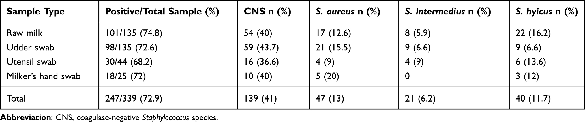 |
Table 1 Prevalence of Staphylococcus Species Isolates from Smallholders and Dairy Cows in Central Ethiopia |
Univariate logistic regression analysis showed that none of the study variables in cows was found to be significantly associated with the isolation of Staphylococcus as indicated in Table 2.
 |
Table 2 Univariate Analysis of the Risk Factors for Isolation of Staphylococcus Species from Raw Milk of Cows in the Study Area |
Antimicrobial Susceptibility Profiles of Staphylococcus Species
From 50 isolates of Staphylococcus species subjected to antimicrobial susceptibility tests, most isolates (90% to 100%) were resistant to the beta-lactam antibiotics, namely ampicillin, penicillin, and amoxicillin (Table 3). From 10 S. intermedius isolates subjected to antimicrobial susceptibility testing, all isolates were resistant to ampicillin, 7 (70%) to nalidixic acid and penicillin, 5 (50%) to vancomycin. S. hyicus isolates exhibited 90% (18/20) resistance to ampicillin, 85% (17/20) to penicillin, 60% (12/20) to amoxicillin. All 50 isolates of Staphylococcus species subjected to antimicrobial susceptibility testing were 100% susceptible to ciprofloxacin. In addition, all 20 tested isolates of S. aureus were susceptible to ciprofloxacin, streptomycin, and sulphamethoxazole–trimethoprim. A total of the 20 tested isolates of S. hyicus were susceptible to doxycycline and ciprofloxacin. All tested S. intermedius isolates were susceptible to ciprofloxacin and sulphamethoxazole–trimethoprim, but all the 10 tested isolates were resistant to ampicillin (Table 3).
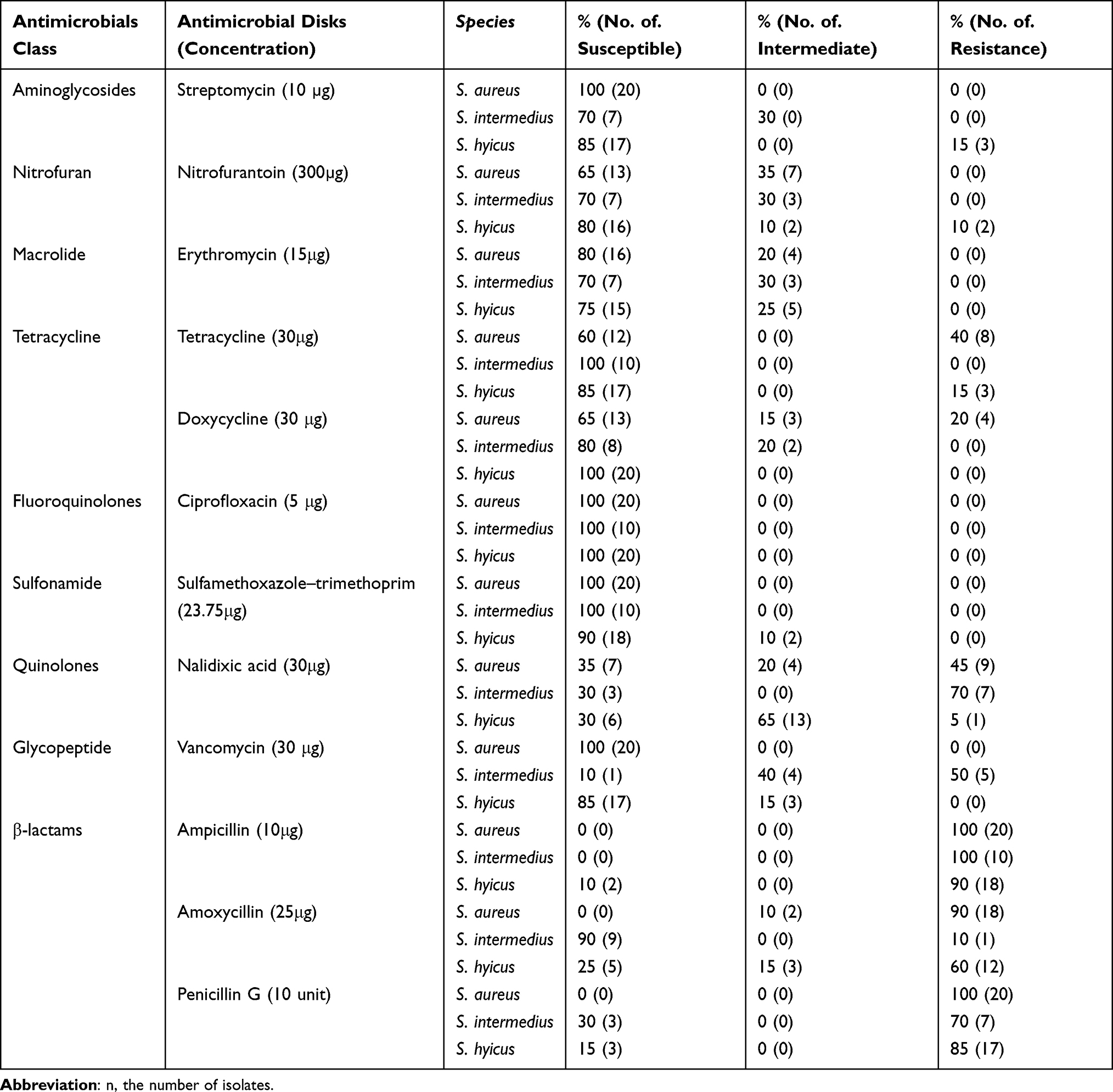 |
Table 3 Percentage of Antimicrobial Susceptibility Profiles of S. aureus (n=20), S. intermedius (n=10), and S. hyicus (n=20) |
Antimicrobial Resistance Pattern of S. aureus, S. intermedius, and S. hyicus
Seven isolates of S. aureus were resistant to three beta-lactam antibiotics (ampicillin, penicillin, and amoxicillin) (Table 4). Likewise, eight isolates of S. hyicus were resistant to these beta-lactam antibiotics.
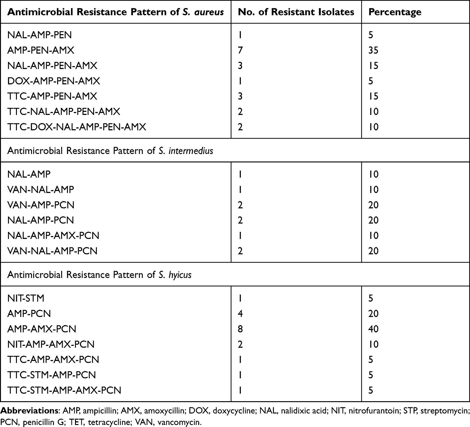 |
Table 4 Antimicrobial Resistance Patterns of S. aureus (n=20), S. intermedius (n=10), and S. hyicus (n=20) |
Multidrug Resistance
In this study, five S. aureus isolates (two from raw milk, two from utensil swabs and one from milker’s hand swabs), three S. intermedius (two from utensil swab and one from raw milk), and two S. hyicus isolates from raw milk were found to be resistant to three antimicrobial classes (Table 5).
 |
Table 5 Multidrug Resistance of Staphylococcus Species Isolates |
Discussion
Cow milk may be contaminated with Staphylococcus and other bacterial species at any stage of the process. At the point of production, milk may be positive if the cow is mastitic and is shedding pathogens, or can be contaminated from unclean udders or teats, from the dust, from the unclean milking machines or milkers’ hands, from dirty milking containers. Milk can also be contaminated at post-harvest stages. In this study, the contamination of milk with Staphylococcus species was investigated at the smallholders and dairy farms from various sources such as the skin of udder, hands of a milking personnel, and surface of milk containers. A total of 247 (72.9%) isolates of staphylococci were found from 339 total samples, which consists of raw milk samples, swabs of milking utensils, udder swabs, and swabs of milkers’ hands. The overall prevalence of Staphylococcus species identified in this study was slightly higher than the report of Gizaw,32 who reported 55.1% of Staphylococcus species isolates from milk in Bishoftu, Ethiopia. The variation in the prevalence of Staphylococcus species reported may be due to the usage of cleaning agents and the sample size.
The prevalence of S. aureus in raw milk in the current study (13.9%) was comparable with the reports of Asrat et al33 who reported 15.5% S. aureus from milk samples of dairy cows and nasal swabs of farmworkers in selected dairy farms around Addis Ababa, Ethiopia. Likewise, Gizaw32 reported 17.2% of S. aureus from cattle, food chains, and humans in Bishoftu, Ethiopia. However, our finding was lower than that of Bendahou et al,34 who reported 40% S. aureus isolates from milk and milk products in North Morocco. The low prevalence of S. aureus in the current study compared to the study in the north of Morocco could be attributed to the collection of the milk samples directly from the cows’ udders before contacting milking utensils in this study, which might have reduced the prevalence of S. aureus.
The prevalence of S. intermedius (6.2%) and S. hyicus (11.7%) recorded in this study agrees with the study conducted by Gizaw,32 who reported 7.4% of S. intermedius and 8.2% of S. hyicus from milk and samples of human origin (udder milk, tank milk, bucket swab, tank swab farm hand swab, nasal swab) and is similar to the study by Addis et al,35 who reported the prevalence of 4% of S. hyicus from raw bovine milk in Bishoftu, Ethiopia. Our findings were also similar to the study of Bendahou et al,34 who reported 2% S. intermedius and 4% S. hyicus in dairy cows in North Morocco. Similarly, the prevalence of CNS (41%) recovered from milk, swab samples from the udder, milking utensils, and milkers’ hands in the current study was closely comparable with the findings of Bendahou et al,34 who reported a 54% CNS from milk and milk products in North Morocco.
Out of 44 swab samples collected from milking utensils in farms in the study area, 4 (9%) were found to be S. aureus. Parmar et al36 reported a high prevalence of 18.8% of S. aureus from swabs of milking equipment of a dairy farm in Anand, Gujarat, India. The dissimilarity in the prevalence may be due to the difference in the hygienic status of the equipment sampled. The prevalence of S. aureus in swab samples collected from milkers’ hands was 20% (5/25). This finding was higher when compared with a study conducted by Parmar et al,36 who reported a 2.55% prevalence of S. aureus from swabs of milkers’ hands. The variation might be due to the small number of samples in this study.
The results of the questionnaire survey to find out potential risk factors associated with the prevalence of Staphylococcal species indicated that none of the variables studied were significantly associated with the prevalence of Staphylococcal species.
The prevalence and degree of antimicrobial resistance in veterinary medicine are increasing worldwide. The dissemination of antimicrobial-resistant staphylococci are presenting a challenge to both human and animal health professionals.37 The antimicrobial susceptibility tests carried out in the current study indicated that S. aureus was 100% resistant to ampicillin and penicillin, and 90% resistant to amoxicillin. This finding was in agreement with a study conducted by Addis et al,35 who reported 100% resistance of S. aureus to both penicillin G and amoxicillin. This finding is also comparable with the result of Sori et al,38 who reported 98% resistance of S. aureus isolates to penicillin G and 67% to ampicillin. A study conducted in Bangladesh by Begum et al39 showed a slightly lower antimicrobial resistance profile, where S. aureus isolates were found to be 82.86% resistant to penicillin G and 37.14%) to amoxicillin.
S. aureus isolates found in this study have also shown 45% resistance to nalidixic acid, 40% to tetracycline, and 20% to doxycycline. This is similar to the findings of Sori et al,38 where S. aureus was reported to have 46% resistance to amoxicillin–clavulanic acid, 87.2% to penicillin, 92% to nalidixic acid, and 46% to amoxicillin. However, these isolates were found to be susceptible to tetracycline. The probable explanation for this could be that S. aureus strains can change their resistance behavior to the exposed antimicrobials.13
Al-Thani and Al-Ali37 reported that 78% of S. aureus isolates showed resistance to tetracycline, which is comparable with the report in this study. This is not surprising because penicillin G and tetracycline are the most used antimicrobials for the treatment of infection or mastitis in veterinary practice in Ethiopia.40 Moreover, penicillin resistance is plasmid based and, it spread out very quickly to several other strains.33,41 Similarly, Daka et al42 reported a 67.9% resistance pattern of strains of S. aureus isolates from milk to penicillin G, 70.9% to ampicillin,30.9% to amoxicillin-clavulanic acid, and 0% to ciprofloxacin. Daka et al42 also reported that S. aureus isolates were resistant to erythromycin (32.1%), trimethoprim-sulfamethoxazole (7.7%) and vancomycin (38.5%). On the contrary, this study indicated that S. aureus isolates were susceptible to erythromycin (80%), sulfamethoxazole–trimethoprim (100%) and vancomycin (100%).
Among the S. intermedius isolates identified in this study, 100% were resistant to ampicillin, 70% to nalidixic acid and penicillin, and 100% susceptible to ciprofloxacin, tetracycline, and sulfamethoxazole-trimethoprim. These findings are correlated with the report of Intorre et al43 where 100% S. intermedius isolates were susceptible to ciprofloxacin.
In this study, multidrug-resistant S. aureus, S. intermedius and S. hyicus, which were resistant to three classes of antimicrobials were found. Five S. aureus isolates were recovered from raw milk, milk utensil swabs and milkers’ hand swabs suggesting that this pathogen is circulating at human animal interface. This signifies a public health risk as S. aureus is known to be one of the most common foodborne pathogens worldwide.44 In addition, the emergence of multidrug-resistant S. intermedius and S. hyicus complicates treatment and poses a serious threat to infection control and public health.
Conclusion
The present study showed a high prevalence of Staphylococcus species in dairy cows in Guder and Ambo towns. The result of swab samples from the udder of the cow, milkers’ hands, and storage containers also showed prevalence of Staphylococcus species. This shows the possibility of milk contamination with Staphylococcus species from milkers’ hands, milking utensils, and udder of the cow after milking. The presence of pathogenic S. aureus poses a public health hazard and concerns about the safety of milk and milk products. The results obtained in this study also showed that the Staphylococcus species isolated from raw milk, swab samples from milking utensils, and milker’s hands were resistant to common antibiotics that could be a threat to the control of mastitis and public health. Therefore, raw milk should be pasteurized and hygienically handled. Further research on the investigation of factors that help the increase of antimicrobial-resistant Staphylococcus species and characterization of CNS at species level are suggested.
Abbreviations
BHI, brain heart infusion; CNS, coagulase-negative Staphylococcus; CPS, coagulase-positive Staphylococcus; MSA, mannitol salt agar; PAB, purple agar base; SFD, staphylococcal foodborne disease.
Data Sharing Statement
The corresponding author and first author will provide datasets that support the results of this article upon reasonable request.
Ethics Approval and Consent to Participate
We confirm that the animals were handled with the best practice of veterinary care. We also confirm that verbal informed consent was acceptable and approved by the College of Veterinary Medicine and Agriculture, Addis Ababa University. The study’s aims and methods were explained to all participating farm owners. The information utilized in this study was acquired from the participants and coded to maintain confidentiality after getting a verbal agreement. Ethical clearance for the study was obtained from the College of Veterinary Medicine and Agriculture, Addis Ababa University (AAU, CVMA), minutes of the Animal Research Ethics and Review Committee (Min NO: VM/ERC/04/10/14, Review Date: 23/10/2014).
Acknowledgments
We would like to thank Addis Ababa University, Thematic Research Fund for its financial support. We would also like to thank the cattle owners, the veterinarians, and the staff of the Ambo University department of Veterinary Laboratory Technology for their help in this study. The study was the first author’s master’s thesis presented to Addis Ababa University, College of Veterinary Medicine, and Agriculture, and it can be found online in the Addis Ababa University Institutional thesis repository. It was part of the thematic research project funded by Addis Ababa University, College of Veterinary Medicine and Agriculture, and all co-authors contributed significantly to the research work. We would like to confirm that the thesis work or part of it has not been submitted nor published in any journal. Mention of trade names or commercial products in this publication is solely for the purpose of providing specific information and does not imply recommendation or endorsement by the US Department of Agriculture (USDA). USDA is an equal opportunity provider and employer.
Funding
This work was supported by Addis Ababa University, Thematic Research Fund. The fund supporter had no role in research design, data collection, and analysis.
Disclosure
The authors declare that there are no conflicts of interest regarding the publication of this paper.
References
1. Quinn P, Carter M, Markey B, Carter G. Clinical Veterinary Microbiology. USA: Mosby; 1999.
2. Foster T. Staphylococcus. Medical Microbiology.
3. Larsen H, Sloth K, Elsberg C, et al. The dynamics of Staphylococcus aureus intramammary infection in nine Danish dairy herds. Vet Microbiol. 2000;71(1–2):89–101. doi:10.1016/S0378-1135(99)00161-3
4. Wakabayashi Y, Umeda K, Yonogi S, et al. Staphylococcal food poisoning caused by Staphylococcus argenteus harboring staphylococcal enterotoxin genes. Int J Food Microbiol. 2018;265:23–29. doi:10.1016/j.ijfoodmicro.2017.10.022
5. Hennekinne J-A. Chapter 7 - Staphylococcus aureus as a leading cause of foodborne outbreaks worldwide. In: Fetsch A, editor. Staphylococcus aureus. Academic Press; 2018:129–146.
6. Fagundes H, Barchesi L, Nader Filho A, Ferreira LM, Oliveira CAF. Occurrence of Staphylococcus aureus in raw milk produced in dairy farms in São Paulo state, Brazil. Braz J Microbiol. 2010;41(2):376–380. doi:10.1590/S1517-83822010000200018
7. Argaw S, Addis M. A review on staphylococcal food poisoning. Food Sci Qual Manag. 2015;40:59–72.
8. Quinn PJ, Markey BK, Leonard FC, Hartigan P, Fanning S, Fitzpatrick EI. Veterinary Microbiology and Microbial Disease. John Wiley & Sons; 2011.
9. Gautam V, Sethuraman N, Kaur R, Sachdev S, Marwaha N, Ray P. Changing epidemiology of coagulase-negative staphylococci in normal flora of skin. Indian J Med Microbiol. 2017;35(2):277. doi:10.4103/ijmm.IJMM_16_282
10. Zecconi A, Hahn G. Staphylococcus aureus in raw milk and human health risk. Int Dairy Fed. 1999;1999:(345):15–18.
11. de Oliveira LP, Silva V, Cirqueira M. Study of Staphylococcus aureus in raw and pasteurized milk consumed in the Reconcavo area of the State of Bahia, Brazil. Int J Food Process Technol. 2011;2(6):128.
12. Zelalem Y. Sanitary Conditions and Microbial Qualities of Dairy Products in Urban and Peri-Urban Dairy Shed of the Central Ethiopia. Lyon, France: DEA; 2003.
13. Chambers HF, Deleo FR. Waves of resistance: staphylococcus aureus in the antibiotic era. Nat Rev Microbiol. 2009;7(9):629–641. doi:10.1038/nrmicro2200
14. Mekonnen H, Workineh S, Bayleyegn M, Moges A, Tadele K. Antimicrobial susceptibility profiles of mastitis isolates from cows in three major Ethiopian dairies. Rev Med Vet. 2005;156(7):391.
15. Getahun K, Kelay B, Bekana M, Lobago F. Bovine mastitis and antibiotic resistance patterns in Selalle smallholder dairy farms, central Ethiopia. Trop Anim Health Prod. 2008;40(4):261–268. doi:10.1007/s11250-007-9090-5
16. Abera M, Demie B, Aragaw K, Regassa F, Regassa A. Isolation and identification of Staphylococcus aureus from bovine mastitic milk and their drug resistance patterns in Adama town, Ethiopia. J Vet Med Anim Health. 2010;2(3):29–34.
17. Girma S, Mammo A, Bogele K, Sori T, Tadesse F, Jibat T. Study on prevalence of bovine mastitis and its major causative agents in West Hararghe zone, Doba district, Ethiopia. J Vet Med Anim Health. 2012;4(8):116–123.
18. Belayneh R, Belihu K, Wubete A. Dairy cows mastitis survey in Adama town, Ethiopia. J Vet Med Anim Health. 2013;5(10):281–287.
19. Tamiru F, Alemu S, Tsega A. Aerobic microorganisms isolated from mastitic bovine milk and their antimicrobial susceptibility profile, Ethiopia. Am-Euras J Agric and Environ Sci. 2013;13(7):920–925.
20. Wodajo H. Hygienic practices during milking and bacteriological quality of the milk in raw bovine bulk milk in the selected milk collection centers: smallholder dairy processing Ethiopia; 2014.
21. Abera B, Lemma D, Iticha I. Study of bovine mastitis in Asella government dairy farm of Oromia Regional state, South Eastern Ethiopia. Int j Curr. 2013;1:134–145.
22. Amenu K, Wieland B, Szonyi B, Grace D. Milk handling practices and consumption behavior among Borana pastoralists in southern Ethiopia. J Health Popul Nutr. 2019;38(1):6. doi:10.1186/s41043-019-0163-7
23. ADLFDO. Ambo district livestock and fishery development office, the annual report. Ambo, Ethiopia; 2014.
24. TKDLFDO. Toke kutaye districts livestock and fishery development office, the annual report. Guder, Ethiopia; 2016.
25. CSA. Cities & Towns; 2015. Available from: https://www.citypopulation.de/Ethiopia.html.
26. York M, Isenberg H. Clinical microbiology procedures handbook; 2007.
27. Forbes BA, Sahm DF, Weissfeld AS. Study guide for Bailey & Scott’s diagnostic microbiology. Mosby USA; 2007.
28. Quinn P, Markey B, Carter M, Donnelly W, Leonard F. Veterinary Microbiology and Microbial Disease. Blackwell Science Ltd. A Blackwell publishing company; 2005.
29. Bennett R, Lancette G. Bacteriological analytical manual: Staphylococcus aureus; 2001. Available from: http://www.fda.gov/Food/FoodScienceResearch/LaboratoryMethods/ucm071429.htm.
30. CLSI. M100-S24 Performance Standards for Antimicrobial Susceptibility Testing: Twenty Fourth Information Supplement. Vol. 342014. CLSI; 2007:68–150.
31. STATACORP T. Statistical Software: Release 11 College Station. College Station, TX: StataCorp LP; 2009.
32. Gizaw F. Staphylococcus: Epidemiology and Its Drug Resistance in Cattle, Food Chains and Humans in Central Ethiopia. Bishoftu, Ethiopia: Addis Ababa University; 2014.
33. Asrat AMD, Woldeamanuel Y, Tefera G. Identification and antimicrobial susceptibility of Staphylococcus aureus isolated from milk samples of dairy cows and nasal swabs of farm workers in selected dairy farms around Addis Ababa, Ethiopia. Afr J Microbiol Res. 2013;7(27):3501–3510.
34. Bendahou A, Lebbadi M, Ennanei L, Essadqui FZ, Abid M. Characterization of Staphylococcus species isolated from raw milk and milk products (lben and jben) in North Morocco. J Infect Dev Ctries. 2008;2(03):218–225. doi:10.3855/jidc.266
35. Addis M, Pal M, Kyule MN. Isolation and identification of Staphylococcus species from raw bovine milk in Debre Zeit, Ethiopia. Vet Res. 2011;4(2):45–49.
36. Parmar B, Brahmbhatt M, Dhami A, Nayak J, Sch J. Isolation, characterization and antibiotics sensitivity pattern of Staphylococcus aureus from man, animal and environment. Sch J Agric Vet Sci. 2014;1(4):173–179.
37. Al-Thani RF, Al-Ali F. Incidences and antimicrobial susceptibility profile of Staphylococcus species isolated from animals in different Qatari farms. Afr J Microbiol Res. 2012;6(48):7454–7458. doi:10.5897/AJMR12.1270
38. Sori T, Hussien J, Bitew M. Prevalence and susceptibility assay of Staphylococcus aureus isolated from bovine mastitis in dairy farms of Jimma town, South West Ethiopia. J Anim Vet Adv. 2011;10(6):745–749. doi:10.3923/javaa.2011.745.749
39. Begum H, Uddin M, Islam M, Nazir KHMNH IM, Rahman M. Detection of biofilm producing coagulase positive Staphylococcus aureus from bovine mastitis, their pigment production, hemolytic activity and antibiotic sensitivity pattern. Bangladesh Soc Agric Sci Techno. 2007;4:97–100.
40. Reta MA, Bereda TW, Alemu AN. Bacterial contaminations of raw cow’s milk consumed at Jigjiga City of Somali Regional State, Eastern Ethiopia. Int J Food Contam. 2016;3(1):4. doi:10.1186/s40550-016-0027-5
41. McCarthy AJ, Lindsay JA. The distribution of plasmids that carry virulence and resistance genes in Staphylococcus aureus is lineage associated. BMC Microbiol. 2012;12(1):104. doi:10.1186/1471-2180-12-104
42. Daka D, G/Silassie S, Yihdego D. Antibiotic-resistance Staphylococcus aureus isolated from cow’s milk in the Hawassa area, South Ethiopia. Ann Clin Microbiol Antimicrob. 2012;11(1):26. doi:10.1186/1476-0711-11-26
43. Intorre L, Vanni M, Di Bello D, et al. Antimicrobial susceptibility and mechanism of resistance to fluoroquinolones in Staphylococcus intermedius and Staphylococcus schleiferi. J Vet Pharmacol Ther. 2007;30(5):464–469. doi:10.1111/j.1365-2885.2007.00896.x
44. Kadariya J, Smith TC, Thapaliya D. Staphylococcus aureus and staphylococcal food-borne disease: an ongoing challenge in public health. Biomed Res Int. 2014;2014:827965. doi:10.1155/2014/827965
 © 2022 The Author(s). This work is published and licensed by Dove Medical Press Limited. The
full terms of this license are available at https://www.dovepress.com/terms
and incorporate the Creative Commons Attribution
- Non Commercial (unported, 3.0) License.
By accessing the work you hereby accept the Terms. Non-commercial uses of the work are permitted
without any further permission from Dove Medical Press Limited, provided the work is properly
attributed. For permission for commercial use of this work, please see paragraphs 4.2 and 5 of our Terms.
© 2022 The Author(s). This work is published and licensed by Dove Medical Press Limited. The
full terms of this license are available at https://www.dovepress.com/terms
and incorporate the Creative Commons Attribution
- Non Commercial (unported, 3.0) License.
By accessing the work you hereby accept the Terms. Non-commercial uses of the work are permitted
without any further permission from Dove Medical Press Limited, provided the work is properly
attributed. For permission for commercial use of this work, please see paragraphs 4.2 and 5 of our Terms.