Back to Journals » Journal of Inflammation Research » Volume 18
Anti-Inflammatory Combinations Targeting Oncostatin M Gene to Remodel Intestinal Flora and Alleviate Inflammation of Perianal Ulcer in Rats
Authors Wu Y, Ge H, Zhao H, Zou K, Wang P, Wang Y, Zhang Y
Received 10 June 2025
Accepted for publication 19 November 2025
Published 17 December 2025 Volume 2025:18 Pages 17769—17788
DOI https://doi.org/10.2147/JIR.S546010
Checked for plagiarism Yes
Review by Single anonymous peer review
Peer reviewer comments 3
Editor who approved publication: Dr Anish R. Maskey
Yanlan Wu,1,* Hao Ge,2,* Haoran Zhao,3 Kaiping Zou,4 Pei Wang,5 Yi Wang,1 Yang Zhang5
1Department of Colon and Rectal Surgery, Nanjing Hospital of Chinese Medicine Affiliated to Nanjing University of Chinese Medicine, Nanjing, Jiangsu, People’s Republic of China; 2School of Clinical Medicine, Jiangsu Health Vocational College, Nanjing, Jiangsu, People’s Republic of China; 3First Clinical Medical College, Nanjing University of Chinese Medicine, Nanjing, Jiangsu, People’s Republic of China; 4Department of Pharmacy, Affiliated Hospital of Nanjing University of Chinese Medicine, Nanjing, Jiangsu, People’s Republic of China; 5Jiangsu Clinical Innovation Center for Anorectal Diseases of Traditional Chinese Medicine (TCM), Nanjing Hospital of Chinese Medicine Affiliated to Nanjing University of Chinese Medicine, Nanjing, Jiangsu, People’s Republic of China
*These authors contributed equally to this work
Correspondence: Yang Zhang, Jiangsu Clinical Innovation Center for Anorectal Diseases of Traditional Chinese Medicine (TCM), Nanjing Hospital of Chinese Medicine Affiliated to Nanjing University of Chinese Medicine, Nanjing, Jiangsu, People’s Republic of China, Email [email protected]
Background: Perianal Abscess is an inflammatory disease caused by infection in the perianal area, characterized by inflammatory cell infiltration and imbalance of intestinal flora. The herbal medicine anti-inflammatory combination (KYHJ) has therapeutic effects in acute and chronic soft tissue infections, but the specific therapeutic mechanism in perianal inflammatory diseases is unclear. There is a literature evidence showing the role of OSM gene in inflammatory conditions, however this was not previously studied in perianal abscess.
Methods: A perianal inflammation model was constructed in SD rats using 75% glacial acetic acid and treated with different doses of KYHJ; genome-wide changes were detected by RNA sequencing. H&E staining observed pathological states, TUNEL kit detected apoptosis, WB measured apoptotic protein levels, ELISA detected inflammatory factors in serum, and 16S rRNA sequencing analyzed intestinal flora abundance. In vitro, an anal epithelial cell inflammation model induced by LPS was treated with 10% KYHJ-containing serum; EDU assay, flow cytometry, WB, and ELISA were used to detect cell proliferation, apoptosis, related protein levels, and inflammatory factor secretion. Oncostatin M gene was knocked down in rats and overexpressed in epithelial cells for mechanism exploration.
Results: Results showed that KYHJ ameliorated perianal tissue inflammatory infection, inhibited apoptosis, and restored intestinal flora abundance. Initial transcriptome analysis, RT-qPCR and WB performed in this study have additionally supported the role of OSM gene. RNA sequencing linked the tested anti-inflammatory effects of KYHJ to reduced Oncostatin M (OSM) gene expression. RNA transcriptome sequencing showed high Oncostatin M expression in inflamed rats and low expression in the KYHJ group; in vivo knockdown improved perianal inflammation and increased flora abundance. In vitro, KYHJ - containing serum inhibited LPS - induced apoptosis, promoted proliferation, and reduced inflammatory factor secretion, which were reversed by Oncostatin M overexpression.
Conclusion: KYHJ ameliorates perianal inflammatory diseases by targeting Oncostatin M, restoring intestinal flora imbalance, promoting cell proliferation, and inhibiting apoptosis.
Plain Language Summary: We investigated the effects of KYHJ in a rat model of perianal inflammation and in cell experiments. In the rat model, the Oncostatin M (OSM) gene was highly expressed during perianal inflammation but showed reduced expression after KYHJ treatment. KYHJ also modulated the gut microbiota, suppressed secretion of inflammatory factors, and alleviated clinical signs of perianal abscess in treated rats, likely via regulation of OSM. In vitro, serum containing KYHJ promoted proliferation and reduced apoptosis of LPS-treated anal epithelial cells, while decreasing their secretion of inflammatory mediators.
Keywords: anti-inflammatory combinations, oncostatin M, intestinal flora, perianal abscess, apoptosis
Introduction
Perianal Abscess (PA) is an inflammatory disorder caused by the infection of cryptoglandular glands,1 predominantly occurring in the posterior area where the anal glands are situated and within the sphincter interspace.2 It presents as erythematous swelling near the anus. Patients who are immunosuppressed, diabetic, or have systemic sepsis require urgent drainage treatment on the day of presentation to mitigate the risk of deep infection, sepsis, and necrotizing soft tissue infections.1
Nevertheless, delayed intervention and inadequate drainage frequently result in the exacerbation of perianal abscesses and the formation of fistulas.
The gut serves as the largest interface between the body and the external environment. The human gut is inhabited by various microbial communities, which include approximately 1013 bacterial cells, commonly referred to as the “gut microbiota”.3 The human gut flora consists of five main bacterial phyla: the Firmicutes; the Bacteroidetes; the Verrucomicrobia; the Proteobacteria; and the Euryarchaeota.4,5 When the ratio of the five bacterial phyla in the intestinal microbiota changes significantly, disrupting the balance of flora, this can overa-ctivate Toll-like receptors (TLRs). This overactivation brings about the overexpression of inflammatory cytokines, causing injury to intestinal epithelial cells and chronic inflammation.6 Perianal inflammatory lesions can be highly affected by gut microbiota changes whether through a systemic inflammatory route or a local route acting as a source of pathogenic bacteria causing inflammation.
From the experimental side, Glacial acetic acid can induce perianal abscess ulcers in rats by stimulating perianal tissues.7 The mechanism involves the chemical binding to tissue proteins to form complete antigens, triggering an immune response in the body. The activation of local immune cells by this response results in the release of inflammatory agents like TNF-α, IL-1β, and IL-6.8 These inflammatory factors contribute to the inflammatory damage of perianal tissues and subsequent ulceration.9 In this study, glacial acetic acid was applied to incite the perianal subcutis of rats, thereby inducing a rat model of perianal inflammation. This model mimics the mechanisms of inflammatory cell infiltration and tissue destruction observed in perianal lesions associated with perianal abscesses and ulcerative diseases. As a member of the IL-6 family, Oncostatin M (OSM) is key in the inflammatory process, autoimmunity, and cancer.10 OSM protein has key biologic functions acting as a potent pro-inflammatory cytokine and it also has an important role in regulating cell growth. Several studies have demonstrated that OSM promotes malignancy by inducing epithelial-mesenchymal transition and facilitating the movement and infiltration of cancer stem cells.11,12 Beyond its role in tumors, OSM is also involved in inflammatory conditions including inflammatory bowel disease. A previous study has demonstrated that high levels of OSM and its receptor, OSMR, were expressed within the intestinal tissues of people affected by enterocolitis.13 Deletion of the OSM gene significantly attenuates colitis in an animal model of intestinal inflammation. Although showing to act as a potential biomarker and therapeutic target in Inflammatory bowel disease14 the role and mechanism of action of OSM in perianal inflammation remains unclear.
In recent years, Chinese medicine has emerged as a key area of research for regulating intestinal health to treat various diseases.15 Chinese medicine anti-inflammatory combination (KYHJ) is an oral preparation developed by Nanjing Hospital of Traditional Chinese Medicine, based on years of clinical experience. It is clinically used for the treatment of acute and chronic soft tissue infections; however, its therapeutic role in perianal inflammatory diseases has not been previously studied. The pinyin transliteration for traditional Chinese medicine (TCM) formulas is Kāng Yì Huà Jū which means a formula to “Benefit Health and Resolve Carbuncles”. The formula mainly consists of herbs with antimicrobial, anti-inflammatory, and immune-modulating properties. The formula is mainly composed of Honeysuckle Flower, Dandelion, Dazhongye, Red Peony Root, Forsythia, Chenpi, and Licorice. Honeysuckle and Dandelion serve as the primary ingredients for clearing heat, removing toxins, promoting dampness, and reducing swelling. Mudanpi (red peony) cools the blood and clears heat, while Dazhongye also helps cool the blood and stops hemorrhage. Forsythia subdues swelling and disperses knots, and Soapwort supports toxin elimination and reduces swelling. Chenpi promotes the circulation of qi and helps to dry dampness. Licorice eases pain and harmonizes the effects of all the ingredients, contributing to the overall functions of clearing heat, removing toxins, cooling the blood, and reducing swelling. This preparation can be used for both acute and chronic soft tissue infections.
The primary aim of this study was to investigate the mechanism of action of KYHJ in alleviating inflammation by providing experimental evidence supporting the use of this medication for handling inflammatory conditions in the perianal region. We also aimed to specifically identify the molecular mechanisms underlying this effect and the possible role of Oncostatin M (OSM) gene in alleviating perianal inflammation.
Material and Methods
KYHJ Composition and Preparation
Anti-inflammatory Combination (KYHJ, in-hospital preparation) is an oral formulation of traditional Chinese medicine developed by the Nanjing Hospital of Traditional Chinese Medicine. This preparation is based on many years of clinical experience and includes 9g of Honeysuckle, 9g of Forsythia, 9g of Mudanpi, 9g of Paeoniae Alba, 6g of Saponaria, 30g of Ziuhua Dictynium, 30g of Dandelion, 12g of Dazhongye, 3g of Pericarpium Citri Reticulatae, and 5g of Glycyrrhiza Glabra, all combined to make a total of 100mL.
Cell Culture and Group Processing
Rat Rectal Mucosal Epithelial Cells (CP-R254) were sourced from PricellaBiotechnology Co., Ltd. The cells were grown in DMEM (Pricella, Wuhan, CM-R254). To enhance cell adhesion and extension, the culture plates were coated with rat tail collagen I (Solarbio, Beijing, C5062) at a concentration of 5 μg/cm2 before inoculation.
For cell treatment: The Control group cells received PBS treatment for 48 hours. The cells in the LPS group were induced with LPS (10 μg/mL) (Yesen, Shanghai, 60747ES08) for 24 hours, after which the supernatant was discarded. The cells in the LPS+KYHJ group were induced with LPS for 24 hours. Following this induction, the supernatant was discarded, and 10% KYHJ drug-containing serum was added for an additional 24 hours of incubation. Cells in the LPS+KYHJ+pc-NC group and the LPS+KYHJ+pc-OSM group were treated with the negative control plasmid and the plasmid carrying the OSM gene for 4–6 hours. After this, Fresh medium replaced the old one, and the cells were treated with LPS for 24 hours. The supernatant was then discarded, and the incubation continued by adding 10% KYHJ-containing serum for an additional 24 hours. The pc-NC and pc-OSM plasmids were acquired from Hanbio and cells were transfected using Lipofectamine 3000 (Invitrogen, L3000015) as instructed by the manufacturer.
Animal Feeding
SD rats (n=90; 8 weeks; 180±20g) with a 1:1 male-to-female ratio were housed under a 12-hour light/dark cycle at 26 ± 2 °C and 55 ± 10% relative humidity. For the rat perianal inflammation model, 0.05 mL of 75% glacial acetic acid (Sigma-Aldrich, A6283, USA) was injected subcutaneously into the periphery of the anus. An ulcer formed locally 24 hours later, accompanied by redness, swelling, and inflammatory exudation. Referring to the four-level scoring standard for local symptoms (Table 1), rats with a score of 3 points or less were excluded from the experiment.
 |
Table 1 Four-Level Scoring Criteria for Local Symptoms |
Experiment 1: rats were weighed and randomly divided into six groups of ten rats each. The control group consisted of healthy SD rats, while the other groups were perianal inflammation model rats. Rats belonging to the model group were gavaged with saline, rats in the antibiotics group received cefuroxime and metronidazole (50 mg/kg), and rats belonging to the model group were given oral doses of an anti-inflammatory combination at low (0.1 g/kg), medium (0.5 g/kg), and high (2.5 g/kg) doses three times a day, with a 4-hour interval between doses, for 14 consecutive days. The body weights of the rats were continuously monitored throughout the treatment period.
Experiment 2: sh-NC and sh-OSM lentiviral vectors were purchased from RiboBio Co (Guangzhou, China). The perianal inflammation model rats were randomly divided into three groups of 10 rats each. Rats in the Model group were injected with an equal amount of saline in the tail vein, while rats in the Model+sh-NC group and Model+sh-OSM group received an injection of 5×10⁵ TU of sh-NC and sh-OSM lentiviral vectors (in a volume of 10 μL) into the tail vein. OSM knockdown was assessed after 5 days. At the experiment’s conclusion, every rat was anesthetized and euthanized with 5% isoflurane (RWD Lifescience, Shenzhen, China). Perianal tissues were collected for histopathological and molecular biological evaluation. Additionally, fecal samples were collected for 16S RNA sequencing.
Preparation of Drug-Containing Serum
The KYHJ formulation was provided by Nanjing Hospital of Traditional Chinese Medicine. Twelve SD rats were acquired from Beijing Vital River Laboratory Animal Technology Co., Ltd., based in Beijing, China. Once the 7-day acclimatization period was completed, the rats were divided into two equal groups. The rats in the KYHJ group received a high dose of KYHJ (2.5 g/kg) administered by gavage twice a day for 7 days. Blank group was gavaged with an equal amount of saline. Two hours after the last administration, anesthesia of rats with sodium pentobarbital and blood was collected. By centrifuging the blood at 3000 rpm for 15 minutes, serum was extracted. The serum was treated in a 55°C water bath for 30 minutes for sterilization, filtered using a 0.22 μm microporous filter, and then placed in a −80°C storage.
Cell Viability Assay
A CCK-8 assay kit (Beyotime Biotech, C0037) was used for a purpose to detect how KYHJ serum at 2.5%, 5%, 10%, and 20% concentrations affected epithelial cell viability. The epithelial cells were seeded into 96-well plates at a density of 1×10⁵ cells per well and incubated for 24 h using different doses of KYHJ drug-containing serum. In another experiment, the cells were exposed to 10 μg/mL of LPS for a duration of 24 h or 48 h. After that, the epithelial cells received the corresponding concentration of the drug-containing serum for 24 h, respectively, and finally, an enzyme marker was used to measure the absorbance at 450 nm.
H&E Staining
Rat perianal ulcer tissue was set in 4% paraformaldehyde (Service, G1101) for the application of hematoxylin and eosin (H&E) stain. A 1 cm piece of the fixed perianal ulcer tissue was then paraffin-embedded and cut into 4 μm thick slices. These tissue slices were dehydrated using graded concentrations of ethanol and subsequently observed under a high magnification microscope (100× and 400×).
TUNEL Apoptosis Assay Kit Assay
The TUNEL assay of rat perianal tissues was performed using the TUNEL Apoptosis Assay Kit (Beyotime, C1091). As instructed by the manufacturer, the perianal ulcerated tissues were first divided into 4 μm coronal sections. The tissue was then digested with 20 mM proteinase K for 5 minutes. Next, the skin tissues were incubated in methanol for 30 minutes, followed by an incubation with 3% skimmed milk. Subsequently, the samples were incubated at 37 °C for 1 hour after adding 50 µL of biotin labeling solution. This was followed by the addition of 0.2 mL of reaction termination solution, which was incubated for 10 minutes at 37 °C. Then, 50 µL of Streptavidin HRP working solution was added, and the samples were incubated for 30 minutes at 37 °C, followed by the addition of 0.3 mL of DAB color development solution for 10 minutes at 37 °C. Finally, images were obtained using an Axio Lab.A1 microscope (Carl Zeiss AG).
Western Blotting
Using RIPA lysate (NCM Biotech, Product No. WB3100), proteins were extracted. The concentration of protein samples was measured with a BCA kit provided by Beyotime Biotech, quantified, heat-denatured, and stored at −80 °C. A total of 30 μg of protein samples was loaded into wells for electrophoresis, separated by 10% SDS-PAGE, and moved onto a PVDF membrane (EMD Millipore, Billerica, MA). After 1 hour of blocking with 5% skim milk powder, the membranes were incubated overnight at 4 °C with primary antibodies against Bax (Abcam, ab32503, 1:1000), Bcl-2 (Abcam, ab182858, 1:1000), PCNA (Abcam, ab92552, 1:1000), OSM (Abcam, ab133748, 1:1000), and GAPDH (Abcam, ab8245, 1:5000). The membranes were then incubated for 60 minutes at 37 °C with Goat Anti-Rabbit IgG H&L (HRP) (Abcam, ab205718, 1:1000). Bands were visualized using enhanced chemiluminescence reagents (Abbkine, Wuhan) in a LAS-3000 imaging system. Data were analyzed using ImageJ.
Bacterial Abundance Assay
High-throughput sequencing was conducted using the 16S rRNA gene from rat fecal samples. Total DNA was extracted from fecal samples from the Control, Model, KYHJ (L, M, H), and Antibiotics groups (n=3 per group) using the EZNA®Soil DNA Kit. The extracted DNA was confirmed on a 1% agarose gel. The highly variable V3-V4 region of the bacterial 16S rRNA gene was amplified using a thermal cycler PCR system (GeneBio 9700, ABI, USA) with the primers 338 F (5′-ACTCCTACGGGGAGGCAGCAG-3′) and 806 R (5′-GGACTACHVGGGTWTCTAAT-3′). The resulting PCR products were purified with the AxyPrep DNA Gel Extraction Kit (Axygen Biosciences, Union City, CA) and quantified using the Quant Fluor™-ST (Promega, USA) based on the manufacturer’s directions.
Transcriptomics Sequencing
The AxyPrep DNA Gel Extraction Kit (Axygen Biosciences, USA) was used to purify the PCR products, and their concentrations were assessed with a Quantus™ Fluorometer (Promega, USA). After purification, the amplicons were combined in equimolar ratios and sequenced on an Illumina MiSeq PE300/NovaSeq PE250 system at Majorbio BioPharm Technology Co., Ltd. in Shanghai, China (Illumina, USA).
RT-qPCR
RNA was isolated from cell or tissue samples using Trizol chemistry (Beyotime Biotech, Shanghai, R0016). cDNA synthesis was achieved through reverse transcription following the instructions provided in the miRNA All-In-One cDNA Synthesis Kit (abm, Shanghai, G898). Final PCR amplification was performed on an ABI 7900 high-throughput real-time PCR system after adding Taq 2X PCR MasterMix (abm, Shanghai, G888). The primers were designed as follows: OSM forward, 5′-ATTCTGGACACGAAGAGGTCAAG-3′, OSM reverse, 5′-TTCCACTGCAAATCACAGCG-3′; GAPDH forward, 5′-CATCCTGCACCACCAACTGCTTAG-3′, GAPDH reverse, 5′-GCCTGCTTCACCACCTTCTTGATG-3′.
EdU Cell Growth Test
To conduct the proliferation assay of epithelial cells, the BeyoClick™ EdU Cell Proliferation Kit with Alexa Fluor 488 (Beyotime, C0071L) was utilized. Each well of the 24-well plates was inoculated with rat rectal mucosal epithelial cells at a density of 1×10⁶ cells, following the manufacturer’s instructions. A pretreatment with 10 μM EdU was applied for 2 hours. Following the drug intervention, incubation continued in a 37°C incubator. At the conclusion of the incubation, the medium was extracted, and paraformaldehyde was added for fixation at room temperature for 30 minutes. Next, after adding a 0.5% Triton X-100 solution in PBS, the cells were kept at room temperature for 15 minutes. After washing, dyes were added and left in darkness for half an hour. Finally, the nuclei were stained, covered, and observed under an inverted fluorescence microscope (Axio Vert A1, Germany) at 400× magnification. Observation and quantification were performed using ImageJ.
Flow Cytometry
The apoptosis rate of epithelial cells was determined using the Annexin V-EGFP/PI Dual Staining Apoptosis Detection Kit (Keygen Biotech, KGA1101-100). According to the manual, cells were collected by digestion with trypsin (without EDTA), washed with PBS, and then centrifuged (800g, 4°C, 5 min) to discard the supernatant. Next, 5 μL of Annexin V-EGFP and 5 μL of Propidium Iodide were mixed in 500 μL of Binding Buffer and incubated, for 20 min. The samples were then observed and analyzed using a flow cytometer within 1 hour. The apoptosis rate was measured as the sum of ANXA5+ PI− and ANXA5+ PI+.
ELISA
IL-1β (Beyotime, PI303), IL-6 (Beyotime, PI328), and TNF-α (Beyotime, PT516) in the rat bloodstream or the supernatant of cell cultures were quantified using ELISA reagent sets according to the guidelines provided by the manufacturer. Absorbance values at 450 nm were measured by a spectrophotometer (Bio-Rad Model 3550).
Statistical Analysis
Data were expressed as mean ± standard deviation (SD). Differences among groups were analyzed using one-way analysis of variance (ANOVA). If ANOVA revealed significant effect, post-hoc comparison was performed using Tukey’s test. A two-way analysis of variance was performed to find the effect of two different factors and their interaction. If a significant interaction or main effect was identified, posthoc comparison was performed using relevant test. All analyses were performed using GraphPadPrism 8.0. A P value <0.05 was considered statistically significant.
Results
KYHJ Reduce Perianal Inflammation in Rats
With the aim of investigating the therapeutic effect of KYHJ on perianal inflammation in rats at different doses, the study was conducted to treat successfully modeled rats with perianal ulcers by oral administration of KYHJ, and the rats’ body weight was monitored for changes in body weight over the duration of the treatment (Figure 1A). The results demonstrated that the rats in the administered groups had higher body weights than those in the Model group, still, they were all under the values seen in the Control group, with the lowest body weights in the KYHJ (L) group (Figure 1B). The pathological state of the perianal tissues was observed using HE staining, and the results showed significant presence of inflammatory cells in the perianal area of rats in the Model group. Relative to the Model group, inflammatory cell infiltration was reduced in the perianal tissues of rats in the low, medium and high dose groups of KYHJ; the perianal tissues of rats in the Antibiotics group exhibited a marked decrease in inflammatory cell infiltration (Figure 1C), indicating that KYHJ, in agreement with antibiotics, has the effect of improving inflammatory cell infiltration in perianal tissues. To further examine the apoptosis level situation in rat perianal cells, TUNEL assay of skin tissues was performed using TUNEL apoptosis kit, dark brown color represents TUNEL positive cells, which refers to cells with apoptotic nuclei. The study revealed a substantial increase in apoptotic cells in the perianal tissues of rats belonging to the Model group as opposed to the Control group. TUNEL-positive nuclei within the lesion area were reduced compared with the Model group, indicating that KYHJ improved apoptosis in perianal tissues (Figure 1D). To verify the results of the TUNEL assay, the study further used WB to detect the levels of apoptosis-related proteins Bax and Bcl2 in rat perianal tissues. Bax protein levels were significantly higher and Bcl2 protein levels were significantly lower in the Model group (Figure 1E, P<0.001, P<0.001, respectively). In comparison to the Model group, Bax protein levels were dose-dependently decreased (Figure 1E, P<0.05, P<0.001, P<0.001, P<0.001, respectively), and Bcl2 protein levels were dose-dependently increased in the KYHJ (L, M, H) groups (Figure 1E, P<0.01, P<0.001, P<0.001, respectively). The WB results indicated that KYHJ dose-dependently improved apoptosis in perianal ulcer tissues of rats. ELISA experiments pointed to higher levels of IL-1β, IL-6, and TNF-α in rats of the model group. There was no change in the levels of inflammatory factors in the KYHJ (L) group. There was a significant decrease in the levels of inflammatory factors in rats of the KYHJ (M) and KYHJ (H) groups (Figure 1F). Serum for ELISA was obtained at the experimental endpoint by cardiac puncture under anesthesia; after clotting and centrifugation (≈3,000 g, 10 min, 4 °C), the serum was stored at −80 °C until assay. For the in vitro assay, culture supernatants were collected after treatment, clarified (≈12,000 g, 10 min, 4 °C), and stored at −80 °C before ELISA.
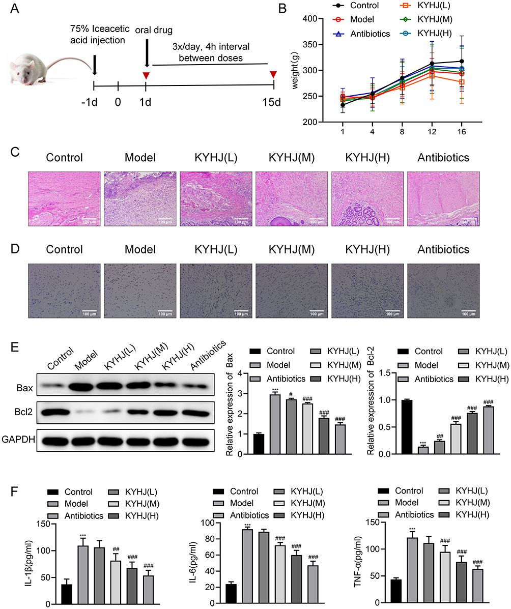 |
Figure 1 Therapeutic effects of KYHJ on rats with perianal inflammation. (A) Schematic diagram of modeling and drug administration in SD rats. (B) Body weight changes in rats with perianal inflammation after treatment with low, medium and high doses of KYHJ and antibiotics, n=10. Body weight was measured at prespecified time points and shown as mean ± SD. Two-way ANOVA (row = group, column = time) indicated a significant main effect of group (F(4,249)=39.48, p<0.0001), with non-significant effects of time (F(5,249)=1.487, p=0.1945) and interaction (F(20,249)=0.4384, p=0.9838). (C) HE staining to observe inflammatory cell infiltration in the perianal tissues of the rats in each group, n=3. (D) TUNEL apoptosis assay kit for detection of apoptosis in rat perianal tissues, n=3. Apoptosis was assessed by TUNEL; the signal indicates DNA fragmentation and is not lineage-specific. Results are expressed as % TUNEL-positive nuclei/DAPI.(E) Western blot analyses of the expression of Bax and Bcl2, n=3. (F) ELISA kits detect the levels of inflammatory factors IL-1β, IL-6 and TNF-α, n=3. The data are expressed as the mean ± standard deviation. Compared with the Control group, *** P<0.001; compared with the Model group, # P<0.05, ## P<0.01, ### P<0.001. |
Effect of KYHJ on the Gut Flora of Rats with Perianal Inflammatory Diseases
In order to analyze the microbial community composition of rat intestinal contents, the study assessed the bacterial diversity and abundance of each group of rat intestinal contents samples by using high-throughput sequencing of the 16S rRNA gene. The results showed that at the Phylum level, Campylobacterota abundance was increased in the Model group, there was a decrease in the relative abundance of Firmicutes. The Model group exhibited a lower relative abundance of Bacilli than the Control group, while this abundance increased after KYHJ treatment. At the Order level, the prevalence of Lactobacillales was lower and the prevalence of Lachnospirales was relatively higher in the Model group, and the abundance of Clostridia_UGG_014 was significantly higher after KYHJ treatment. At the Family level, the quantity of Prevotellaceae was amplified in the Model group, and the quantity of Pepcostreptococcaceae was higher after KYHJ administration (Figure 2A). PCA results showed a significantly larger difference and distance between Control group and the rest of the groups while the difference and distance between the model group and the KYHJ low-dose group were significantly closer (Figure 2B). Using PCoA analysis, overlapping portions were observed in the KYHJ medium-dose and high-dose groups, which indicated that their microbial compositions had a high degree of similarity (Figure 2C). Anosim analysis results indicated that, relative to the Model group, the groups had a significantly higher alpha diversity (Figure 2D).
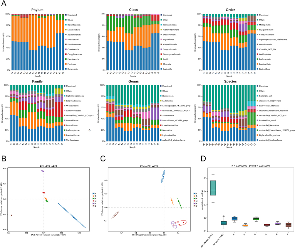 |
Figure 2 Effect of KYHJ on the intestinal flora of rats with perianal infectious diseases. (A) Detection of bacterial abundance in the feces of groups of rats using high-throughput sequencing of the 16S rRNA gene, n=3. From left to right, the levels are phylum, class, order, family, genus, and species level; one color represents one species, and the length of the color block indicates the proportion of relative abundance accounted for by the species; for optimal view, only the ten species with the most abundance were highlighted, and the other species were combined as Others in the figure. (B) Principal Component Analysis (PCA) analyzes compositional differences between groups of samples, n=3. (C) Principal coordinates analysis (PCOA) analyzes compositional differences between groups of samples, n=3. In the PCoA plot, samples in the same group were represented by dots of the same color, and the greater the distance between the sample dots, the greater the difference between the species composition of the samples (D) Anosim (analysis of similarities) analysis tests for significant differences in beta diversity between different subgroups of samples, n=3. (A) Negative control group; (B) Model group; (C) Antibiotics (cefuroxime + metronidazole); (D) Anti-inflammatory combinations (low); E: Anti-inflammatory combinations (medium); F: Anti-inflammatory combinations (high). The data are expressed as the mean ± standard deviation. |
At the phylum level, high anti-inflammatory combination lead to a increase in Bacteroidota and Proteobacteria when compared to negative control and model group. At the class level, Gammaproteobacteria was the most abundant at the high-level anti-inflammatory treatment while bacilli were the least. At the order level, clostridia were enriched in the group exposed to anti-inflammatory medication at medium dose while Enterobacterales showed more abundance in the group exposed to anti-inflammatory combination at high dose. Prevotellaecea and Enterobacteriacae specifically E. coli showed marked enrichment in the group exposed to high levels of anti-inflammatory drugs (Figure 2).
RNA-Seq Reveals Suppression of OSM Signaling and Inflammatory Programs in Perianal Lesions After KYHJ Treatment
RNA sequencing was used to analyze genome-wide changes in perianal inflammatory lesions treated with KYHJ. Differential genes were screened in control/model and KYHJ/model groups. Results showed 1335 up-regulated and 1,076 down-regulated genes between control and model groups, and 1016 up-regulated and 797 down-regulated genes between KYHJ and model groups (Figure 3A). The results of Wayne’s plot showed that there were 1389 genes common to the negative Control vs Model group and Anti-inflammatory Combination vs Model group (Figure 3B). Volcano plot results indicated Control vs Model group and KYHJ group vs Model group with red and blue data points distributed on both sides of the plot indicating genes with significant up-regulation and down-regulation. Gray data points are concentrated in the middle of the plot, indicating insignificant changes in the expression of these genes (Figure 3C). The results of heatmap showed that there were significant differences between Control and Model groups, as well as between KYHJ and Model groups, between genes of different groups, and the functions corresponding to the genes also showed significant differences (Figure 3D). The results of GO annotation classification statistics map showed that there were differences in the expression of genes in terms of biological processes and molecular functions between Control and Model groups, as well as between KYHJ and Model groups. Differences were observed in the expression of genes associated with biological processes, cellular components, and molecular functions between the Control and Model groups, and more genes were differentially expressed and up-regulated in the Control and KYHJ groups (Figure 3E). The annotation results of the differentially expressed genes were categorized according to the type of pathway in KEGG. And according to the bar graph results, the Control group had differentially expressed genes that were mainly associated with human diseases, unlike the Model group. In the KYHJ group, the functions of differentially expressed genes in diseases were mainly in Pathways in cancer and Herpes simplex virus 1 infection (Figure 3F). RNA-seq was performed on perianal lesion tissue collected at endpoint from Control, Model, and KYHJ rats (n=3/group). After euthanasia, a ~5–8 mm biopsy was excised, rinsed in PBS, snap-frozen, and stored at −80 °C; total RNA was extracted with TRIzol, libraries were prepared by the service provider and sequenced on an Illumina platform (paired-end). Together, the RNA-seq data indicate that KYHJ reverses Model-induced transcriptional activation of OSM/OSMR and inflammation-related genes in perianal lesion tissue, providing transcriptomic support for the anti-inflammatory effect.
 |
Figure 3 Role of KYHJ on transcriptomics in rats with perianal inflammatory diseases. RNA sequencing analysis to detect genome-wide changes in KYHJ treatment of perianal infectious diseases, Groups: Control_VS_Model, KYHJ_VS_Model. (A) Bar graph visualizing the number of genes in each differentially expressed gene set, n=3. (B) Wayne plots showing the overlap of differentially expressed genes between different comparison groups, n=3. (C) Volcano map showing distribution of differential genes in two groups, n=3. (D) Heat map showing differences in gene expression in the two groups, n=3. (E) GO annotation system for categorizing differentially expressed genes, n=3. (F) Classification of differentially expressed genes by pathway type in KEGG, n=3. The data are expressed as the mean ± standard deviation. RNA-seq of perianal lesion tissue shows downregulation of OSM signaling and inflammation-related transcripts in KYHJ versus Mo tissue source and sampling are detailed in the Methods. |
Effect of KYHJ on OSM Genes in Perianal Inflammatory Diseases
For a deeper examination of KYHJ’s impact on OSM genes in perianal inflammatory diseases, the study firstly analyzed the differentially expressed genes in Control, Model, and KYHJ groups by using a Wayne diagram. There were 821 genes that were highly expressed in the Model group and less expressed in the Drug group; 568 genes that were less expressed in the Model and are prominently expressed in the Drug group (Figure 4A). The final transcriptome sequencing results revealed that the OSM gene was highly expressed in the Model group of rats compared to the Control group, and then the level of expression of the gene was decreased in the KYHJ group (Figure 4B, P<0.01, P<0.01, respectively). RT-qPCR and WB results showed that OSM levels were higher in the perianal tissues of rats with perianal inflammation than in the control group, whereas OSM levels were lower in rats in the KYHJ group than in rats with perianal inflammation (Figure 4C and D).
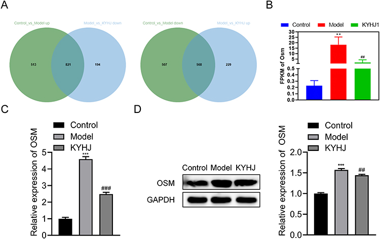 |
Figure 4 Effect of KYHJ on OSM genes in perianal infectious diseases. (A) Wayne diagram analysis of differentially expressed genes in Control, Model, and KYHJ groups, n=3. (B) Transcriptome sequencing analysis revealed that OSM expression was significantly highly expressed in the Model group, and the expression level was significantly reduced after KYHJ treatment. (C) RT-qPCR detection of OSM mRNA expression levels between groups, n=3. (D) Western blot detection of OSM mRNA expression levels between groups, n=3. The data are expressed as the mean ± standard deviation. Compared with the Control group, ** P<0.01, *** P<0.001; compared with the Model group, ## P<0.01, ### P<0.001. |
In vivo Detection of the Effect of Knockdown of OSM Gene on Perianal Inflammation and Intestinal Flora in Rats
To further investigate the effects of OSM genes on perianal inflammation and intestinal flora, the study interfered with OSM gene expression at the perianal site by injecting lentivirus into the rats’ tail vein. OSM knockdown in the perianal region of rats was first detected using RT-qPCR and WB. The results showed no significant change in OSM gene expression in the perianal region of rats in the Model+sh-NC group, whereas the mRNA and protein levels of OSM in the perianal region of rats in the Model+sh-OSM group were significantly reduced (Figure 5A). The next experiment used HE staining to detect disease-related changes in the perianal area, and the outcomes demonstrated that cellular infiltration in perianal tissues was obvious in rats in the Model+sh-NC group, while cellular infiltration in perianal tissues was reduced in rats in the Model+sh-OSM group (Figure 5B), suggesting that OSM genes can promote perianal inflammatory cell infiltration in inflamed tissues. The results of TUNEL Apoptosis Kit exhibited that the total of dark brown cells in the perianal tissues of rats in Model and Model+sh-NC groups was higher, indicating significant apoptosis, while the number of apoptotic cells in the perianal tissues of rats in Model+sh-OSM group was reduced, suggesting that the OSM gene can promote apoptosis in the inflammatory tissues of the perianal area of the rats (Figure 5C). The study further verified the levels of apoptotic proteins Bax and Bcl2 by WB, and the results were consistent with the TUNEL assay, which showed that the levels of Bax protein were significantly decreased and Bax protein levels were significantly increased in rats in the Model+sh-OSM group (Figure 5D, P<0.001, P<0.001, respectively), suggesting that the OSM gene can significantly promote apoptosis in rat perianal inflammatory tissues. The study further examined the levels of IL-1β, IL-6 and TNF-α by ELISA kit, and the results showed that the levels of inflammatory factors IL-1β, IL-6 and TNF-α in the Model+sh-OSM group were significantly reduced (Figure 5E, P<0.001, P<0.001, P<0.001, respectively).
 |
Figure 5 Effect of in vivo knockdown of OSM gene on perianal inflammation in rats. (A) RT-qPCR and Western blot detection of OSM knockdown between groups, n=3. (B) HE staining to observe the inflammatory cell infiltration in the perianal tissues of the rats in each group, n=3. (C) TUNEL apoptosis assay kit for detection of apoptosis in rat perianal tissues, n=3. Apoptosis was assessed by TUNEL; the signal indicates DNA fragmentation and is not lineage-specific. Results are expressed as % TUNEL-positive nuclei/DAPI. (D) Western blot analyses of the expression of Bax and Bcl2, n=3. (E) ELISA kits detect the levels of inflammatory factors IL-1β, IL-6 and TNF-α, n=3. The data are expressed as the mean ± standard deviation. Compared with the Model group, ns: P>0.05; compared with the Model+sh-NC group, ### P<0.001. |
In order to investigate the effect of OSM gene on rat intestinal flora, the study detected the bacterial diversity and abundance of rat intestinal contents after knocking down OSM gene. The intestinal flora abundance was examined by testing three groups of three samples each for a total of nine samples. The data demonstrated that at the Phylum level, the abundance of Cyanobacteria was higher in the feces of rats in the OSM knockdown group relative to the blank knockdown and Model groups. Vampirivibrionia was more abundant at the Class level in rats from the OSM knockdown group. At the Order level, the abundance of Clostridia_UGG_014 was relatively higher in rats of the OSM knockdown group. At the Family level, Lactobacillaceae were found in lower quantities in the OSM knockdown group of rats. (Figure 6A). This may indicate that OSM decreased expression may exert its anti-inflammatory effect by affecting the composition of gut microbiota represented in relatively increased abundance of Cyanobacteria and Vampirivibrionia, and Clostridia while Lactobacillaeceae are reduced. Species diversity differences between samples were further demonstrated by PCA analysis and PCoA analysis, which showed large differences in species diversity between groups (Figure 6B and C) probably indicating a significant role of microbial diversity on the mechanism of inflammatory action of OSM. Anosim analysis showed that alpha diversity was significantly higher in the Model and blank knockdown groups compared to the OSM knockdown group (Figure 6D).
 |
Figure 6 Effect of in vivo knockout of the OSM gene on the intestinal flora of rats. (A) Detection of bacterial abundance in the feces of groups of rats using high-throughput sequencing of the 16S rRNA gene, n=3. (B) Principal Component Analysis (PCA) analyzes compositional differences between groups of samples, n=3. (C) Principal coordinates analysis (PCOA) analyzes compositional differences between groups of samples, n=3. (D) Anosim (analysis of similarities) analysis tests for significant differences in beta diversity between different subgroups of samples, n=3. (A) Mo (B) Model+sh-NC; (C) Model+sh-OSM. The data are expressed as the mean ± standard deviation. |
In vitro Assay of the Effect of KYHJ on LPS-Treated Anal Epithelial Cells via the OSM Gene
The study verified in vitro the effect of KYHJ-containing serum on rectal mucosal epithelial cells of LPS-treated rats via the OSM gene. The effect of 2.5%, 5%, 10%, and 20% KYHJ-containing serum on epithelial cell viability was first examined using the CCK-8 assay, which showed that 20% KYHJ-containing serum significantly reduced epithelial cell viability (Figure 7A, P<0.001), and the 20% dose was removed for the next experiments. The study next induced the production of epithelial cell inflammation with 10 μg/mL of LPS. CCK-8 was continued to detect the effect of KYHJ-containing serum on epithelial cell toxicity in the LPS-induced inflammation model. The results showed that 10 μg/mL of LPS induced epithelial cells for 24 and 48 hours, which significantly reduced the viability of epithelial cells (Figure 7B, P<0.001). At 24 hours of LPS induction, 2.5% KYHJ-containing serum failed to significantly elevate the cell viability of epithelial cells, whereas both 5% and 10% KYHJ-containing serum significantly increased the cell viability of epithelial cells (Figure 7B, P<0.001). At 48 hours of LPS induction, 2.5%, 5%, and 10% of KYHJ were effective in increasing cell viability of epithelial cells (Figure 7B, P<0.05). The study chose to construct a model of epithelial cell inflammation by inducing LPS with 10 μg/mL for 24 hours, and intervened with 10% KYHJ-containing serum for treatment. Next, the epithelial cells were transfected using OSM plasmid, and the study verified the transfection by PCR and WB, which showed that epithelial cells in the pc-OSM group had a markedly higher level of OSM expression (Figure 7C), indicating that the OSM gene was effectively delivered into the epithelial cells.
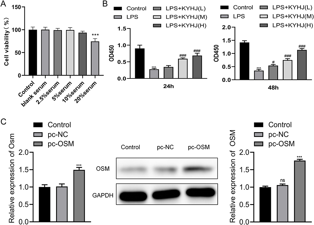 |
Figure 7 In vitro assay of the effect of KYHJ on LPS-treated anal epithelial cells via the OSM gene. (A) Effect of different doses of KYHJ-containing serum on epithelial cell viability as detected by CCK-8 assay, n=3. (B) CCK-8 assay to detect the effect of different doses of KYHJ-containing serum on the viability of LPS-treated epithelial cells, n=3. (C) Validation of OSM manipulation in epithelial cells: left, RT-qPCR of OSM mRNA; right, Western blot of OSM protein. Groups: Control, LPS, LPS+KYHJ, LPS+KYHJ+pc-NC, LPS+KYHJ+pc-OSM (OSM overexpression); n=3. The data are expressed as the mean ± standard deviation. (ns) indicates no statistical significance (P ≥ 0.05); compared with the Control group, *** P<0.001; compared with the LPS group, # P<0.05, ### P<0.001; compared with the pc-NC group, *** P<0.001. |
Next to verify the effect of 10% KYHJ-containing serum on the function of anal epithelial cells via the OSM gene, the study first examined OSM expression in epithelial cells by PCR and WB. The LPS group’s cells exhibited a marked elevation in OSM gene expression according to PCR results (Figure 8A, P<0.001). The expression of OSM gene was decreased in the epithelial cells of the LPS+KYHJ and LPS+KYHJ+pc-NC groups (Figure 8A, P<0.001). While the manifestation of the OSM gene within epithelial cells of LPS+KYHJ+pc-OSM group had a substantial rise (Figure 8A, P<0.001). The WB validation results were consistent with PCR (Figure 8A).
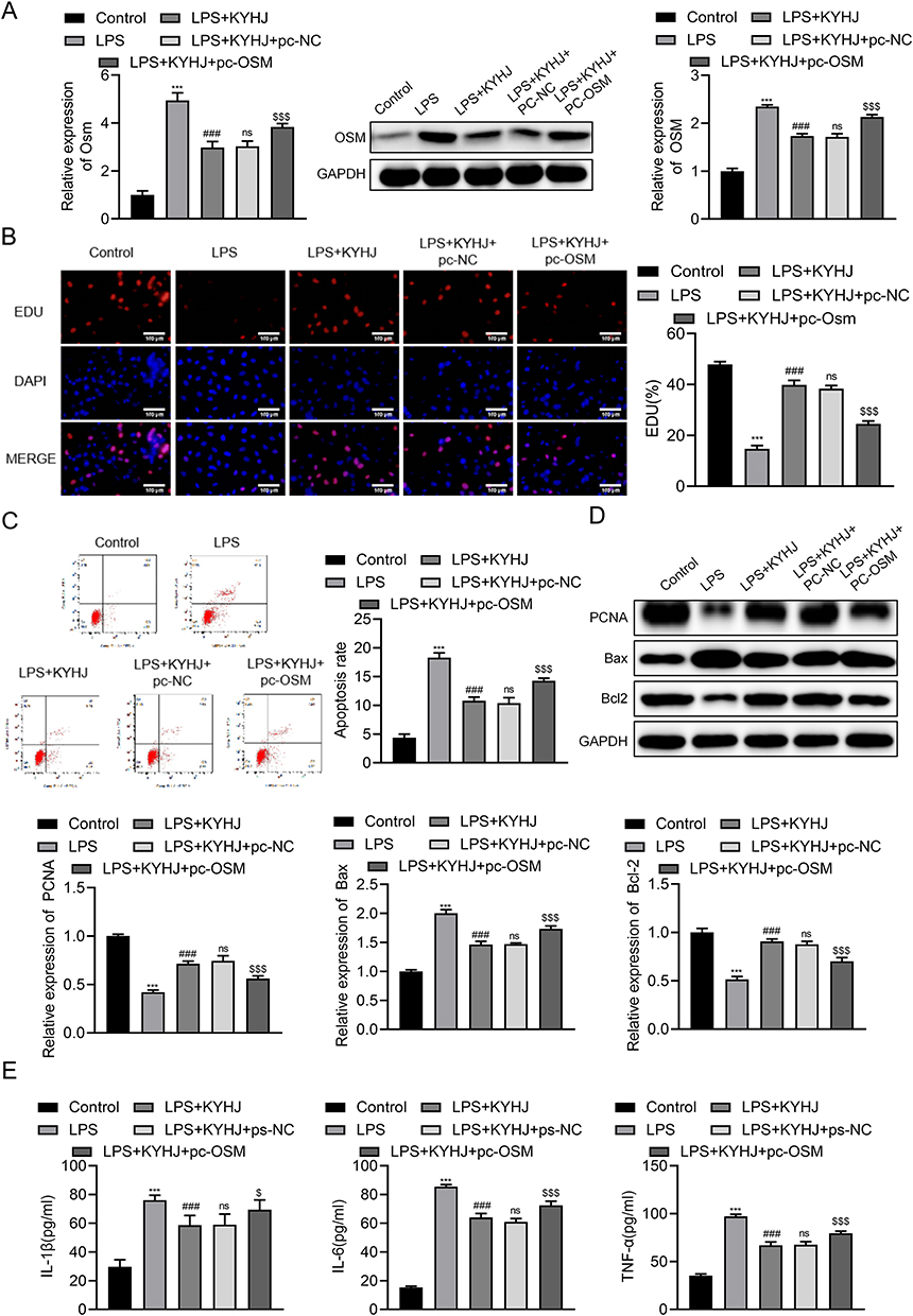 |
Figure 8 In vitro assay of the effect of KYHJ on LPS-treated anal epithelial cells via the OSM gene. (A) RT-qPCR and Western blotting to detect OSM expression levels among different groups, n=3. (B) EdU fluorescence assay to detect cell proliferation between groups, n=3. (C) Flow cytometry assay to detect apoptosis between groups, n=3. (D) Western blot analyses of the expression of PCNA, Bax and Bcl2, n=3. (E) ELISA kits for the detection of inflammatory factors IL-1β, IL-6 and TNF-α in cell supernatants, n=3. The data are expressed as the mean ± standard deviation. Compared with the Control group, *** P<0.001; compared with the LPS group, ### P<0.001; compared with the LPS+KYHJ group, ns: P>0.05; compared with the LPS+KYHJ+pc-NC group, $ P<0.05, $$$ P<0.001. |
The study also used EDU fluorescence assays to detect cell proliferation. The results showed that the proliferation of epithelial cells in the LPS group was inhibited (Figure 8B, P<0.001). The inhibited proliferation of epithelial cells in the LPS+KYHJ and LPS+KYHJ+pc-NC groups was significantly improved (Figure 8B, P<0.001). And the proliferation of epithelial cells in the LPS+KYHJ+pc-OSM group was again significantly inhibited (Figure 8B, P<0.001). The study examined the effect of KYHJ on epithelial cell apoptosis through OSM gene by flow cytometry, results revealed that that LPS promoted apoptosis in epithelial cells, while KYHJ could inhibit apoptosis induced by LPS, while apoptosis rate was significantly increased in the group of LPS+KYHJ+pc-OSM, which indicated that KYHJ inhibited apoptosis through the inhibition of OSM gene (Figure 8C). The study verified KYHJ’s role in modulating the growth and programmed cell death of epithelial cells through OSM gene by detecting the levels of proliferative protein PCNA and apoptotic proteins Bax and Bcl2 by WB assay. It was demonstrated that PCNA levels of cell proliferation proteins were decreased in the LPS group, whereas PCNA protein levels of cells were increased in the LPS+KYHJ group, and decreased in the LPS+KYHJ+pc-OSM group, consistent with the EdU fluorescence assay, which showed that KYHJ promotes cell proliferation by repressing OSM genes. Consistent with the flow-through results, WB apoptotic protein Bax and Bcl2 results showed that KYHJ inhibits epithelial cell apoptosis by repressing the OSM gene (Figure 8D). The study also used ELISA kits to detect the levels of inflammatory factors IL-1β, IL-6 and TNF-α in the supernatants of epithelial cells. As the data revealed that LPS significantly increased the secretion of inflammatory factors from epithelial cells, while KYHJ decreased the secretion of inflammatory factors from epithelial cells, and the levels of inflammatory factors in the supernatants of cells in the LPS+KYHJ+pc-NC group were increased, suggesting that KYHJ may alleviate the secretion of cellular inflammatory factors by inhibiting the OSM gene (Figure 8E).
Discussion
Perianal abscess (PA), an inflammatory disorder of the perianal region, is the most prevalent form of anorectal pathology. This sort of abscess is typically situated on the anal verge and may lead to systemic infections if not treated promptly.16,17 Research revealed that the pathogens cultivated from perianal abscesses consisted of a combination of aerobic and anaerobic bacteria. Among the aerobic microbes, Escherichia coli, Proteus vulgaris, Staphylococcus aureus, and Streptococcus species were most prevalent. In terms of anaerobic microorganisms, Bacteroides and Peptostreptococcus were the most commonly detected types.18,19 Organisms causing perianal abscesses commonly originate from the gastrointestinal tract.20
The normal intestinal flora plays a crucial role in sustaining the functionality of the intestinal mucosal layer, modulating the intestinal immune response, and facilitating nutrient digestion and absorption. When the intestinal flora is imbalanced, the overgrowth of harmful bacteria and the reduction of beneficial bacteria may lead to the impairment of the intestinal mucosal barrier function, so that the harmful bacteria and toxins in the intestinal tract enter the blood circulation, causing systemic inflammatory reactions including perianal abscesses and other perianal inflammatory diseases indicating the strong relation of the gastrointestinal tract flora to these inflammatory conditions. In the present study, the effect of KYHJ in the treatment of perianal inflammation in rats was investigated. In addition, we also investigated its effect on the gastrointestinal flora.
Regarding the flora of the gastrointestinal tract, the results of the study revealed that in the rat model of perianal inflammation, the overall abundance of intestinal flora of the rats especially the number of beneficial bacteria (eg Firmicutes) decreased, and the number of some of the pathogenic bacteria (eg Campylobacterota) increased. When high-level anti-inflammatory treatment was used, Gammaproteobacteria was the most abundant at the class level while bacilli were the least. At the order level, clostridia were enriched in the group exposed to anti-inflammatory medication at medium dose while Enterobacterales showed more abundance in the group exposed to anti-inflammatory combination at high dose. Prevotellaecea and Enterobacteriacae specifically E. coli showed marked enrichment in the group exposed to high levels of anti-inflammatory drugs. After KYHJ treatment, composition of gut microbiota has changed, indicating that KYHJ has the effect of promoting the recovery of beneficial flora or those flora with an effect on regulating the balance of intestinal flora in rats with perianal inflammation which indicates that modulating intestinal flora can be considered a mechanism through which KYHJ may act in reducing inflammation.
In rats model of perianal inflammation, a disruption in the intestinal microbiota could heighten the inflammatory reaction. The body’s defensive response towards injury or infection is manifested as inflammation, but an excessive inflammatory response can cause damage to tissues. The study suggests that an imbalance in the intestinal flora may lead to an overgrowth of harmful bacteria that produce harmful substances, which can trigger an inflammatory response. In addition, the imbalance of the intestinal flora may affect the immune function of the intestine, reducing the body’s resistance to pathogens and further aggravating the inflammatory response.21 It has been found that inflammatory factors such as IL-1β, IL-6 and TNF-α are abundantly expressed in patients with perianal ulcerative diseases.22 Based on that, the same effect was investigated in this study so this study focused on the inflammatory cell infiltration and inflammatory factor expression levels in rats with perianal inflammatory diseases. In addition to looking into the effects of KYHJ on gastrointestinal flora in perianal inflammatory diseases, HE and ELISA were also used to study its effect on inflammatory mediators. HE staining showed that the model group’s rats demonstrated substantial infiltration by inflammatory cells, while KYHJ could alleviate the inflammatory cell infiltration in the perianal tissues. ELISA detected the levels of IL-1β, IL-6 and TNF-α inflammatory factors in the serum of rats, and the conclusions were consistent with HE staining experiments. Rats in the model group exhibited increased serum levels of inflammatory factors. Nevertheless, KYHJ could effectively lower the levels of these inflammatory factors in the serum. And the same conclusion was obtained in the epithelial cells of rectal mucosa of rats showing that KYHJ could reduce LPS - instigated secretion of inflammatory factors in epithelial cells.
Apoptosis participates in the inflammatory response, and during the initiation of apoptotic signaling, inflammatory mediators are produced, activating immune cells to secrete pro-inflammatory cytokines.23–25 B-cell lymphoma-2 (Bcl-2), as an anti-apoptotic protein, inhibits apoptosis mainly by regulating the permeability of the mitochondrial membrane. While Bcl-2-associated X protein (Bax) protein is a member of the Bcl-2 family. When cells receive apoptotic signals, Bax undergoes a conformational change, which increases the permeability of the mitochondrial membrane, thus promoting apoptosis.26,27 We showed that in KYHJ ameliorated apoptosis in a rat model of perianal inflammation. In epithelial cells, KYHJ also ameliorated apoptosis induced by LPS.
In this study, RNA sequencing was used to detect genome-wide changes as a result of KYHJ treatment of perianal inflammatory diseases. OSM was screened by transcriptomics, and ultimately showed to be highly expressed in the model group of rats and expressed at low levels in the KYHJ group. OSM proteins are multifunctional glycoproteins that are members of the IL-6 family and are mainly produced by activated T lymphocytes, monocytes, macrophages, and vascular endothelial cells.28 OSM is also involved in a variety of inflammatory responses, including arthritis, enteritis, and cardiovascular disease.29,30 It has been found that OSM is involved in regulating cardiovascular disease, possibly by promoting JAK2/STAT3 signaling in macrophages to promote atherogenesis.31 Moreover, OSM induces thymic stromal lymphopoietin to promote chronic sinusitis.32 And in this study, we verified the molecular mechanism of KYHJ for the treatment of perianal inflammation and restoration of intestinal flora by knocking down OSM gene in perianal inflammation model rats. Firstly, it was found that after knocking down OSM gene in rats, the inflammatory cell infiltration in perianal tissues of rats was improved, apoptosis was suppressed, and the expression of IL-1β, IL-6, and TNF-α inflammatory factors in the serum of rats were reduced, which indicated that OSM gene could promote apoptosis as well as the infiltration of inflammatory cells, thus aggravating the occurrence of inflammation in perianal area in rats with perianal inflammation. Results of PCA indicate that microbial diversity may have a significant role on the mechanism of inflammatory action of OSM showing that the decreased expression of OSM may exert its anti-inflammatory effect by affecting the composition of gut microbiota represented in relatively increased abundance of Cyanobacteria, Vampirivibrionia, and Clostridia while Lactobacillaeceae are reduced. The study transfected OSM gene in epithelial cells and explored the role of KYHJ. It was found that after transfection of OSM gene, cell proliferation was inhibited, apoptosis level was promoted, and the concentrations of inflammatory factors in the cell supernatants went up. These results indicate that KYHJ inhibits apoptosis, promotes cell proliferation, and reduces the secretion of cellular inflammatory factors by inhibiting the expression of OSM gene, thus alleviating perianal inflammation.
Although a large interest has arisen in finding biologically active compounds isolated from large amounts of natural resources, especially traditional medicinal plants,33 a possible limitation of the current study is the lack of Mass Spectrometry identification (MSI) of different compositions of compounds in the KYHJ herbal medicines. Future studies can include MSI analysis to examine the compositions of specific compounds found in KYJH herbs that would greatly help in identifying how KYHJ herbs interact with and regulate the metabolic pathways and other signaling pathways in the rat models.
In summary, the study investigated the effects of KYHJ on the composition of intestinal flora and inflammation levels showing that the molecular mechanisms underlying its anti-inflammatory action may be related to the regulation of OSM genes. Through establishing a rat perianal inflammation model, our study indicated that KYHJ was able to regulate the composition of intestinal flora and reduce the level of inflammation, and its mechanism of action may be related to the regulation of OSM gene expression. This study provides a theoretical basis for the application of KYHJ in the treatment of perianal abscess, but further studies are needed to verify its clinical efficacy and safety.
Data Sharing Statement
The raw data supporting the conclusions of this manuscript will be made available by the authors, without undue reservation, to any qualified researcher upon reasonable request to the corresponding author.
Ethical Approval
The ethics committee of Animal Ethics Committee of Hangzhou Hebei Technology Co., Ltd approved the animal study. (ZJLL-20230106). All procedures involving animals were performed in accordance with the Guide for the Care and Use of Laboratory Animals (National Institutes of Health, USA) and the Laboratory Animal—Guideline for Ethical Review of Animal Welfare (GB/T 35892-2018, China).
Author Contributions
All authors made a significant contribution to the work reported, whether that is in the conception, study design, execution, acquisition of data, analysis and interpretation, or in all these areas; took part in drafting, revising or critically reviewing the article; gave final approval of the version to be published; have agreed on the journal to which the article has been submitted; and agree to be accountable for all aspects of the work.
Funding
This work was supported by the Jiangsu TCM Science and Technology Development Plan Project (MS2021034); the Jiangsu Province Traditional Chinese Medicine Science and Technology Development Plan Project Project -Clinical application of a new silicone wire integrating unidirectional, quantitative cutting and self-locking in high anal fistula (MS2021034); and 2020 Nanjing Chinese Medicine Young Talent Cultivation Program.
Disclosure
The authors declare that the research was conducted in the absence of any commercial or financial relationships that could be construed as a potential conflict of interest.
References
1. Sahnan K, Adegbola SO, Tozer PJ, Watfah J, Phillips RK. Perianal abscess. BMJ. 2017;356:j475. doi:10.1136/bmj.j475
2. Whiteford MH. Perianal abscess/fistula disease. Clin Colon Rectal Surg. 2007;20(2):102–109. doi:10.1055/s-2007-977488
3. Di Vincenzo F, Del Gaudio A, Petito V, Lopetuso LR, Scaldaferri F. Gut microbiota, intestinal permeability, and systemic inflammation: a narrative review. Intern Emerg Med. 2024;19(2):275–293. doi:10.1007/s11739-023-03374-w
4. Donaldson GP, Lee SM, Mazmanian SK. Gut biogeography of the bacterial microbiota. Nat Rev Microbiol. 2016;14(1):20–32. doi:10.1038/nrmicro3552
5. Dalton A, Mermier C, Zuhl M. Exercise influence on the microbiome-gut-brain axis. Gut Microbes. 2019;10(5):555–568. doi:10.1080/19490976.2018.1562268
6. Karczewski J, Poniedzialek B, Adamski Z, Rzymski P. The effects of the microbiota on the host immune system. Autoimmunity. 2014;47(8):494–504. doi:10.3109/08916934.2014.938322
7. Ferreira-Duarte M, Oliveira LCG, Quintas C, et al. Angiotensin-converting enzymes 1 and 2 in the feces: presence and catalytic activity in the rat 2,4,6-trinitrobenzene sulfonic acid-induced model of colitis. J Gastroenterol Hepatol. 2024;39(9):1885–1894. doi:10.1111/jgh.16541
8. Cheng X, Zhang X, Su J, et al. miR-19b downregulates intestinal SOCS3 to reduce intestinal inflammation in Crohn’s disease. Sci Rep. 2015;5(1):10397. doi:10.1038/srep10397
9. Lucas JH, Wang Q, Meehan-Atrash J, Pang C, Rahman I. Developmental PFOS exposure alters lung inflammation and barrier integrity in juvenile mice. Toxicol Sci. 2024;201(1):48–60. doi:10.1093/toxsci/kfae073
10. Masjedi A, Hajizadeh F, Dargani FB, et al. Oncostatin M: a mysterious cytokine in cancers. Int Immunopharmacol. 2021;90:107158. doi:10.1016/j.intimp.2020.107158
11. Soler MF, Abaurrea A, Azcoaga P, Araujo AM, Caffarel MM. New perspectives in cancer immunotherapy: targeting IL-6 cytokine family. J Immunother Cancer. 2023;11(11):e007530. doi:10.1136/jitc-2023-007530
12. Abaurrea A, Araujo AM, Caffarel MM. The Role of the IL-6 Cytokine Family in Epithelial-Mesenchymal Plasticity in Cancer Progression. Int J Mol Sci. 2021;22(15):8334. doi:10.3390/ijms22158334
13. West NR, Hegazy AN, Owens BMJ, et al. Oncostatin M drives intestinal inflammation and predicts response to tumor necrosis factor-neutralizing therapy in patients with inflammatory bowel disease. Nat Med. 2017;23(5):579–589. doi:10.1038/nm.4307
14. Kim WM, Kaser A, Blumberg RS. A role for oncostatin M in inflammatory bowel disease. Nat Med. 2017;23(5):535–536. doi:10.1038/nm.4338
15. Che Q, Luo T, Shi J, He Y, Xu DL. Mechanisms by Which Traditional Chinese Medicines Influence the Intestinal Flora and Intestinal Barrier. Front Cell Infect Microbiol. 2022;12:863779. doi:10.3389/fcimb.2022.863779
16. Conner JN, Eren S, Tuma F. Perianal Abscess. In: StatPearls. Treasure Island (FL): StatPearls Publishing; 2023.
17. Pogacnik JS, Salgado G. Perianal Crohn’s Disease. Clin Colon Rectal Surg. 2019;32(5):377–385. doi:10.1055/s-0039-1687834
18. Brook I, Frazier EH. Aerobic and anaerobic bacteriology of wounds and cutaneous abscesses. Arch Surg. 1990;125(11):1445–1451. doi:10.1001/archsurg.1990.01410230039007
19. Marcus RH, Stine RJ, Cohen MA. Perirectal abscess. Ann Emerg Med. 1995;25(5):597–603. doi:10.1016/S0196-0644(95)70170-2
20. Liu CK, Liu CP, Leung CH, Sun FJ. Clinical and microbiological analysis of adult perianal abscess. J Microbiol Immunol Infect. 2011;44(3):204–208. doi:10.1016/j.jmii.2011.01.024
21. Zhou M, Liu X, He J, et al. High-fructose corn syrup aggravates colitis via microbiota dysbiosis-mediated Th17/Treg imbalance. Clin Sci. 2023;137(20):1619–1635. doi:10.1042/CS20230788
22. Sugrue J, Nordenstam J, Abcarian H, et al. Pathogenesis and persistence of cryptoglandular anal fistula: a systematic review. Tech Coloproctol. 2017;21(6):425–432. doi:10.1007/s10151-017-1645-5
23. Vringer E, Tait SWG. Mitochondria and cell death-associated inflammation. Cell Death Differ. 2023;30(2):304–312. doi:10.1038/s41418-022-01094-w
24. Wang S, Zhang Y. HMGB1 in inflammation and cancer. J Hematol Oncol. 2020;13(1):116. doi:10.1186/s13045-020-00950-x
25. Liu J, Song K, Lin B, et al. HMGB1 promotes neutrophil PD-L1 expression through TLR2 and mediates T cell apoptosis leading to immunosuppression in sepsis. Int Immunopharmacol. 2024;133:112130. doi:10.1016/j.intimp.2024.112130
26. Spitz AZ, Gavathiotis E. Physiological and pharmacological modulation of BAX. Trends Pharmacol Sci. 2022;43(3):206–220. doi:10.1016/j.tips.2021.11.001
27. King LE, Hohorst L, Garcia-Saez AJ. Expanding roles of BCL-2 proteins in apoptosis execution and beyond. J Cell Sci. 2023;136(22). doi:10.1242/jcs.260790
28. Jones SA, Jenkins BJ. Recent insights into targeting the IL-6 cytokine family in inflammatory diseases and cancer. Nat Rev Immunol. 2018;18(12):773–789. doi:10.1038/s41577-018-0066-7
29. Wolf CL, Pruett C, Lighter D, Jorcyk CL. The clinical relevance of OSM in inflammatory diseases: a comprehensive review. Front Immunol. 2023;14:1239732. doi:10.3389/fimmu.2023.1239732
30. Hermans D, Houben E, Baeten P, et al. Oncostatin M triggers brain inflammation by compromising blood-brain barrier integrity. Acta Neuropathol. 2022;144(2):259–281. doi:10.1007/s00401-022-02445-0
31. Zhang X, Li J, Qin JJ, et al. Oncostatin M receptor beta deficiency attenuates atherogenesis by inhibiting JAK2/STAT3 signaling in macrophages. J Lipid Res. 2017;58(5):895–906. doi:10.1194/jlr.M074112
32. Wang BF, Cao PP, Norton JE, et al. Evidence that oncostatin M synergizes with IL-4 signaling to induce TSLP expression in chronic rhinosinusitis with nasal polyps. J Allergy Clin Immunol. 2023;151(5):1379–1390e11. doi:10.1016/j.jaci.2022.11.029
33. Gu X, Hao D, Xiao P. Research progress of Chinese herbal medicine compounds and their bioactivities: fruitful 2020. Chinese Herbal Medicines. 2022;14(2):171–186. doi:10.1016/j.chmed.2022.03.004
 © 2025 The Author(s). This work is published and licensed by Dove Medical Press Limited. The
full terms of this license are available at https://www.dovepress.com/terms
and incorporate the Creative Commons Attribution
- Non Commercial (unported, 4.0) License.
By accessing the work you hereby accept the Terms. Non-commercial uses of the work are permitted
without any further permission from Dove Medical Press Limited, provided the work is properly
attributed. For permission for commercial use of this work, please see paragraphs 4.2 and 5 of our Terms.
© 2025 The Author(s). This work is published and licensed by Dove Medical Press Limited. The
full terms of this license are available at https://www.dovepress.com/terms
and incorporate the Creative Commons Attribution
- Non Commercial (unported, 4.0) License.
By accessing the work you hereby accept the Terms. Non-commercial uses of the work are permitted
without any further permission from Dove Medical Press Limited, provided the work is properly
attributed. For permission for commercial use of this work, please see paragraphs 4.2 and 5 of our Terms.