
Back to Journals » International Journal of General Medicine » Volume 18
Analysis of the Independent Risk Factors of second-Degree Atrioventricular Block in Patients with Atrial Fibrillation and the Diagnostic Efficacy of Dynamic Electrocardiogram
Authors Lu P, Mo X, Yang X, Lin Y
Received 31 December 2024
Accepted for publication 17 April 2025
Published 8 May 2025 Volume 2025:18 Pages 2487—2495
DOI https://doi.org/10.2147/IJGM.S515113
Checked for plagiarism Yes
Review by Single anonymous peer review
Peer reviewer comments 2
Editor who approved publication: Prof. Dr. Yuriy Sirenko
Ping Lu, Xingchun Mo, Xiaojing Yang, Yaoyao Lin
Department of Cardiology, Linping Campus, Second Affiliated Hospital, Zhejiang University School of Medicine, Zhejiang Chinese Medical University, Hangzhou, Zhejiang, People’s Republic of China
Correspondence: Ping Lu, Department of Cardiology, Linping Campus, Second Affiliated Hospital, Zhejiang University School of Medicine, Zhejiang Chinese Medical University, No. 369 Yingbin Road, Linping District, Hangzhou, 311100, Zhejiang, People’s Republic of China, Email [email protected]
Objective: Exploring the independent risk factors of second-degree atrioventricular block (II AVB) in patients with atrial fibrillation (AF), and to evaluate the clinical value of 24-hour dynamic electrocardiogram (DCG) in its diagnosis.
Methods: A prospective cohort study was conducted on 947 patients with AF diagnosed and treated in our hospital from January 1, 2021 to December 31, 2021. These patients were divided into combined group (98 cases) and uncombined group (849 cases) according to whether they were accompanied by. The clinicopathological data of the patients were collected, and Multivariate logistic regression analysis was used to analyze the independent risk factors. Patients in combined group were further evenly divided into the study group (underwent 24-hour DCG) and the control group (underwent routine ECG) based on the detection methods. The diagnostic value was valued and the positive detection rate was calculated by ROC curve.
Results: The smoking history, left atrial internal diameter (LAD), R-R interval and ventricular rate of patients in two groups had significant differences (P< 0.001). Smoking history (HR=1.531, 95% CI 1.150– 2.038, P< 0.001), LAD> 35.88 mm (HR=1.941, 95% CI 1.301– 2.895, P< 0.001), R-R interval> 2.50 s (HR=2.282, 95% CI 1.231– 4.229, P=0.014) were independent risk factors for AF combined with II AVB, while ventricular rate≤ 70 beats/min (HR=0.506, 95% CI 0.293– 0.873, P=0.014) were independent protective factors for AF combined with II AVB. The mean ventricular rate (70.03± 5.40 beats/min vs 83.11± 8.05 beats/min, P< 0.001) and R-R interval (2.82± 0.26s vs 2.37± 0.14s, P< 0.001) in the study group were longer than the control group. The diagnostic positive rate of DCG (97.96% vs 85.71%, χ²=4.900, P=0.027) was higher than that of conventional ECG.
Conclusion: Smoking history, LAD, R-R interval and ventricular rate were influential factors for AF combined with II AVB. 24-h DCG had potential diagnostic value in the occurrence of AF combined with II AVB.
Keywords: atrial fibrillation, second-degree atrioventricular block, influencing factors, dynamic electrocardiography, diagnostic value
Introduction
Atrial fibrillation (AF) refers to arrhythmia caused by abnormal electrical activity in the upper chamber of the heart, which significantly reduces the efficiency of the heart in delivering blood to the ventricles, thereby increasing the risk of thrombosis and subsequent cerebral infarction.1 According to relevant statistics, the incidence rate of AF among people over 20 years old worldwide is about 3%.2 Patients with AF may experience palpitations, dizziness, shortness of breath, and fatigue. However, AF may be asymptomatic and may only be detected during medical appointments in other situations. Due to the intermittent nature of symptoms, many cases of paroxysmal AF have not yet been diagnosed. Atrioventricular block (AVB) refers to an abnormality in the conduction system of the heart at the atrioventricular node, which limits the pulsation of the lower ventricle of the heart. AVB is mainly caused by abnormal electrical conduction between the ventricles and atria, usually occurring during atrial fibrillation attacks.3 Second-degree AVB (II AVB) is one of the common complications in AF patients, with the electrocardiogram (ECG) commonly shows an R-R interval of ≥ 1.5 s and a high ventricular heart rate. Some II AVB patients also have no obvious clinical symptoms, mainly palpitations, dizziness, fatigue, fatigue, and cardiac arrest, which are difficult to attract individual attention.4 However, in clinical practice, II AVB not only affects the patient’s heart function and reduces their quality of life, but may also leads to complications such as thromboembolism, tachycardia, and cardiomyopathy, endangering the patient’s life.5 Therefore, early diagnosis of AF combined with II AVB plays an important role in reducing the incidence rate of cerebral infarction and other diseases and improving the life quality of patients.
Electrocardiogram (ECG) is one of the most important biological characteristics of the human body, which can capture the electrical activity of the heart, thereby facilitating healthcare professionals in evaluating, diagnosing, and monitoring the patient’s heart condition.6 ECG include dynamic ECG (DCG) and static ECG (SCG), which has different diagnostic efficacy.7 At present, ECG is still the main diagnostic method. However, due to the short recording time of conventional ECG (only 10 seconds), the detection rate of paroxysmal and asymptomatic arrhythmia is low.8 Recent studies have shown9 that 24-hour DCG can capture transient or intermittent arrhythmias that are difficult to be detected by conventional ECG, and its diagnostic sensitivity is higher than that of conventional ECG. However, the diagnostic efficacy of DCG in AF with II AVB is still controversial, especially in the differentiation of first-degree AVB (I AVB) and II AVB needs further verification.
In this study, AF patients admitted in our hospital were included as the subjects to analyze the influencing factors of AF combined with II AVB. Patients with AF combined with II AVB were randomized as two groups, which received routine ECG or 24-hour DCG monitoring respectively, aiming to explore the diagnostic value of DCG in AF combined with II AVB.
Materials and Methods
General Materials
This study has been ratified by the hospital Ethics Committee and complied with Medical Ethics, and was complies with the Declaration of Helsinki. A total of 947 AF patients admitted in our hospital during January 1, 2021 to December 31, 2021 were picked as the subjects based on a randomized, double-blind method. These patients were graded as a combined group (98 cases) and an uncombined group (849 cases) according to whether they were accompanied by II AVB. The inclusion process of the subjects was shown in Figure 1. Patients in combined group were further evenly divided into the study group and the control group based on the detection methods. Inclusion criteria: (1) All subjects met the diagnostic and therapeutic criteria for AF10 and underwent 24-hour DCG monitoring (Unified detection methods were used to identify the disease and ensure the homogeneity and accuracy of the study); (2) The patient’s mental state was normal and they could actively cooperate with treatment; (3) Patients and their families had informed consent to participate in this study (The principle of medical ethics was followed to protect the right of patients to know and choose independently); (4) The clinical pathological data was complete and the mental symptoms were good. Exclusion criteria: (1) Patients with I or II AVB; (2) Patients accompanied with infectious diseases such as AIDS and hepatitis B (Such infectious diseases may affect heart function and metabolism, and then interfere with the progression and diagnosis of AF and AVB); (3) Patients accompanied by a history of pulmonary heart disease or severe pneumonia (These factors may interact with atrial fibrillation combined with II AVB, increasing the complexity of the study results, and it is difficult to accurately determine the relationship between the study factors and the outcome); (4) Patients who withdrew from the study midway due to personal or family reasons; (5) Patients with liver and kidney dysfunction (Liver and kidney dysfunction can affect drug metabolism and the stability of the body’s internal environment, which may affect the condition of AF and AVB).
 |
Figure 1 Inclusion process of study subjects. |
 |
Figure 2 Different methods for detecting ventricular rate and R-R interval in patients with AF combined with II AVB. (A) DCG detection results; (B) ECG detection results. Abbreviations: AF, atrial fibrillation; II AVB, second-degree atrioventricular block. Note: ***P<0.001 compared between two groups. |
Methods
The control group: The control group underwent routine ECG monitoring (Optoelectronic 12 lead automatic analysis ECG machine ECG-2340). Patients should maintain a resting state, relax both physically and mentally, and lie flat. The electrode patches and wires should be connected to the recorder and placed in the corresponding positions to ensure that the patient’s electrocardiogram waveform image was clear.
The study group: The research group underwent 24-hour DCG monitoring (Hangzhou Baihui Medical Equipment Co., Ltd. CT-083S). The electrodes were attached to the corresponding positions on the patient’s chest and fixed. After connected with the recorder, the ECG waveform of the patient in different states within 24 hours was recorded.
Collection of Clinical Pathological Data
The clinical and pathological characteristics of AF patients were collected, including the gender (female, male), age, smoking history (yes, no), drinking history (yes, no), hypertension history (yes, no), coronary heart disease history (yes, no), left atrial diameter (LAD), left ventricular end diameter (systolic, diastolic), R-R interval, and ventricular rate.
Outcome Measures
- The clinical data of patients were collected for Univariate analysis. The factors with statistical differences in Univariate analysis for selected for further logistic multivariate analysis to explore the influencing factors of AF combined with II AVB.
- The effect of DCG on ventricular rate and R-R interval in patients with AF combined with II AVB was analyzed.
- The diagnostic value of DCG for AF combined with II AVB was analyzed, and the positive detection rate was calculated.
Statistical Analysis
The SPSS 23.0 data statistical software was used for data analysis. The measurement data such as the patient’s age, LAD, left ventricular end diameter, R-R interval, and ventricular rate were all tested for normal distribution, and all were in accordance with normal distribution. The measurement data were shown in the form of  . The measurement data between two groups were compared using t-test, and reported the effect size (Cohen’s d). Gender, smoking history, alcohol consumption history, hypertension history, coronary heart disease history, and other enumeration data were expressed in [cases (%)], compared using χ2 inspection, and reported the effect size (Cohen’s d). Logistic multivariate analysis was applied to explore the influencing factors of AF combined with II AVB. The difference was statistically significant with P<0.05. In addition, considering the sample size and study design, the study power was evaluated by calculating the confidence intervals of Odds Ratio (OR) and Hazard Ratio (HR) to ensure that the study was powered to detect the actual effect.
Results
Univariate Analysis of AF Combined with II AVB
The combined group and uncombined group had no obvious difference in gender, age, history of alcohol consumption, history of hypertension, history of coronary heart disease and left ventricular end diameter (P>0.05). The smoking history, LAD, R-R interval and ventricular rate of patients in two groups had significant differences (P<0.001). The history of alcohol consumption, LAD, R-R interval, and ventricular rate were influencing factors for AF combined with II AVB (Table 1).
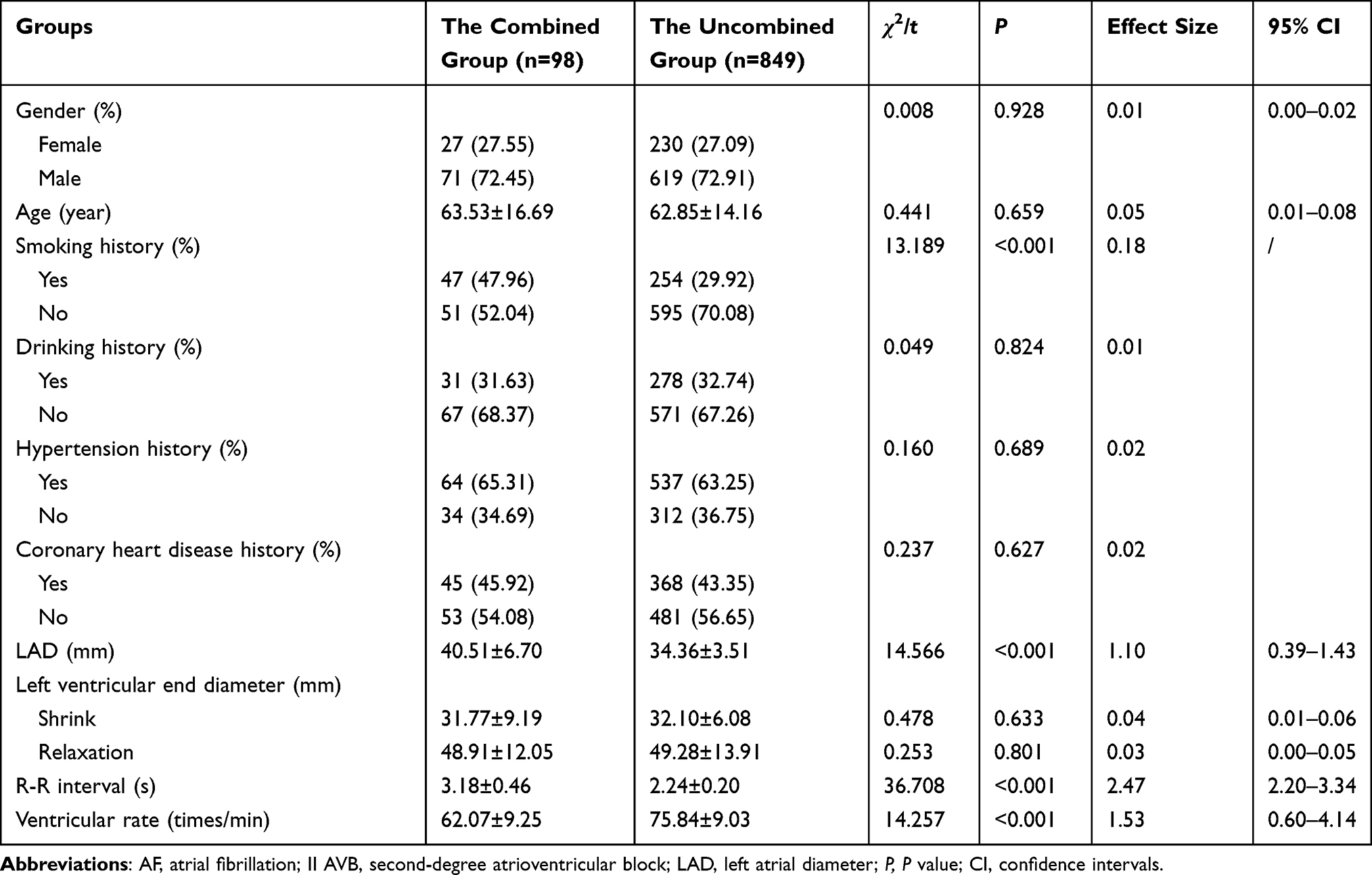 |
Table 1 Univariate Analysis of AF Combined with II AVB [ |

Multivariate Analysis of AF Combined with II AVB
AF patients with II AVB or not were selected as the dependent variable. The statistically significant indicators in Table 1 were also chosen as the dependent variable and analyzed by multivariate logistic analysis. The assignment was shown in Table 2. The result confirmed that smoking history, LAD, R-R interval were independent risk factors (P<0.05) and ventricular rate was protective factor of AF combined with II AVB (P<0.05, Table 3).
 |
Table 2 The Assignment of Each Variable |
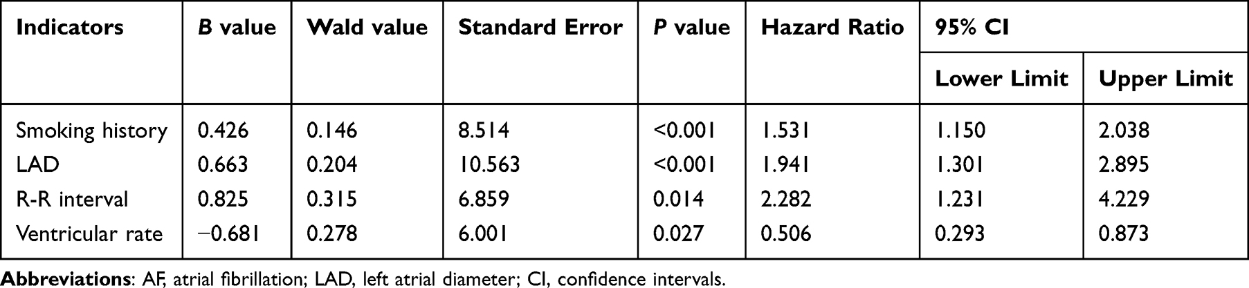 |
Table 3 Multivariate Analysis of AF Combined with II AVB |
DCG Detection of Ventricular Rate and R-R Interval in Patients with AF Combined with II AVB
Compared with the control group, patients in the study group had much lower ventricular rate and longer R-R interval (P<0.001, Figure 2, Table 4).
 |
Table 4 DCG Detection of Ventricular Rate and R-R Interval in Patients with AF Combined with II AVB |

The Diagnostic Value of DCG for AF Combined with II AVB
The positive detection rate of AF combined with II AVB was 97.96% in the study group, which was much higher than 85.71% in the control group (P<0.05, Table 5).
 |
Table 5 The Diagnostic Value of DCG for AF Combined with II AVB [Cases (%)] |
Discussion
AF is an irregular rhythm caused by the disorder of atrial electrical activity and is also the most common persistent arrhythmia in clinical practice. The incidence rate of AF has increased rapidly worldwide, accounting for about one-third of all inpatients.11 The pathogenesis of AF is relatively complex, and increasing evidence suggests12,13 that the occurrence of AF is related to sudden cardiac death, cerebral infarction, and congestive heart failure. In addition, it ultimately increases the risk of thromboembolic events, cardiac and overall mortality.14 AVB is an abnormal conduction along the atrioventricular node or Hipper system, and different etiologies may lead to different outcomes.15 According to the degree of expansion and electrocardiogram characteristics, AVB can be divided into I AVB, II AVB, or third-degree AVB (III AVB). Compared to first-degree and third-degree AVB, the diagnosis of AF combined with II AVB is more difficult. It usually does not show any symptoms and may be atypical or asymptomatic. Especially in the elderly, AF detection still remains challenging.16 Because elderly patients usually have other complications, such as hypertension, congestive heart failure, diabetes, and previous stroke or transient ischemic attack, which further increases the risk of stroke. Even without comorbidities, the average annual risk of stroke in patients over 70 years old is about 3.5%. Besides, the risk may vary due to comorbidities, and the overall risk of stroke or systemic embolism gradually increases over time.17,18 Therefore, finding relevant methods for early and timely diagnosis of diseases is of great significance for effective treatment.
In this study, the clinical data of AF patients with or without concomitant II AVB was analyzed to explore related influencing factors. The results showed that smoking history, LAD, R-R interval, and ventricular rate were all influencing factors of AF combined with II AVB. Upon investigation, smoking remains the main risk factor for cardiovascular disease. Nicotine in tobacco may stimulate the release of catecholamines, block instantaneous outward K+, increase unstable atrial currents, and thus affect cardiac repolarization and membrane transport processes, as well as heart rate variability. The atrium serves as a reservoir, catheter, and active emptying function during the cardiac cycle. A study has found19,20 that LAD can independently predict hospitalization and mortality rates in patients with pulmonary hypertension. The enlargement of the LAD can significantly impair atrial compliance and reduce atrial pumping function, especially during exercise pressure. The R-R interval exceeding 1.5 seconds and the appearance of escape rhythm are important features of atrial fibrillation. In addition, studies have found21,22 that AF patients have a higher ventricular rate compared to normal sinus rhythm, which can lead to a decrease in hemodynamic signals related to heart rate. In this study, potential confounding factors such as hypertension and coronary heart disease were controlled by multivariate Logistic regression to ensure the independence of risk factors. Although no formal power analysis was performed, HR values with 95% confidence intervals were used to quantify the magnitude of the effect of risk factors, enhancing the interpretability of the results. Future studies can combine propensity score matching (PSM) to further reduce selection bias.
With the continuous development of medical technology, the progress of imaging technology has shifted from only displaying the pathological anatomical structure of patient lesions to integrating functions. ECG is a commonly used method for detecting heart diseases in clinical practice, and has important practical value in the diagnosis, evaluation, and prognosis monitoring of various diseases. Some professional association guidelines suggest screening for AF through long-term ECG monitoring. However, due to limited recording time, the detection rate of traditional 12 lead conventional ECG is relatively low.23 DCG is a method which can continuously record and analyze changes in the ECG of the human heart during both active and quiet states for a long time. By recording all abnormal waves, accurate evidence can be provided for the diagnosis of various heart diseases, which has been widely recognized.24 In addition, this study found that 24-hour DCG could more effectively detect the R-R interval and ventricular rate in patients with AF combined with II AVB, and had higher diagnostic value than conventional ECG. In previous studies,25,26 it has been pointed out that long interval time, average ventricular rate, and the proportion of escape rhythm to total heart rate are the main indicators for clinical diagnosis of AF combined with II AVB. When there is a significant difference between the occurrence time of long intervals and the average ventricular rate, it is natural to determine the degree of conduction block in patients.27 In addition, clinical practice can enrich the pathological information of patients based on the data collected from DCG, and draw scatter plots based on this information for clinical diagnosis. Previous studies have shown that the scatter plot distribution of patients with II AVB is relatively uniform, with a wide bar shape, clear upper boundary, indistinct lower boundary, and similar bandwidth. The scatter plot width of patients with I AVB is narrow, with unclear upper and lower boundaries, dense scattering distribution, and no features.28,29 In this case, the difficulty of identification is relatively small, which can help medical staff quickly determine the patient’s condition and develop a reasonable treatment plan. Of course, in practice, some patients do not have typical clinical symptoms and specific DCG, which is related to the patient’s cooperation in examination, disease course, and condition. The study has shown30 that DCG is superior to conventional ECG in the diagnosis of persistent AF with II AVB. However, emerging wearable devices (such as Apple Watch Series8) can achieve real-time AF screening through single-lead monitoring, and their diagnostic accuracy is 91%.31 But, the specificity of wearable devices in the diagnosis of AVB is low (about 75%), and artifacts are prone to occur especially during movement or electrode shedding.32 Therefore, DCG is still the gold standard for the diagnosis of complex arrhythmias, and wearable devices are more suitable for large-scale population screening. A meta-analysis of 12 studies showed33 that elevated vWF levels were significantly associated with thromboembolism risk in patients with AF (OR=1.45, 95% CI 1.12–1.89), and the mechanism may be related to VWF-mediated platelet aggregation and endothelial dysfunction. Although no biomarkers were tested in this study, in the future, a multi-dimensional risk assessment model can be constructed by combining DCG monitoring with vWF and other biomarkers to further optimize patient management strategies.
The clinical value of this study is reflected in two aspects: First, early identification of AF with II AVB by DCG can significantly improve the prognosis of patients. Long R-R interval (>2.5 seconds) and low ventricular rate (≤70 beats/min) are important indications for pacemaker implantation. The high detection rate of Holter monitoring can reduce the risk of syncope, heart failure and even sudden death caused by missed diagnosis. Second, DCG monitoring can guide the optimization of anticoagulation strategies. Studies have shown that the risk of thrombosis in patients with AF combined with AVB is closely related to ventricular rate. The continuous heart rate data provided by DCG can help doctors balance anticoagulation intensity and bleeding risk. In addition, the results of this study support the 2020 ESC guideline recommendations for prolonged ECG monitoring, providing an evidence-based medical basis for clinical practice.
In general, smoking history, LAD, R-R interval and ventricular rate were influential factors for AF combined with II AVB. 24-h DCG had potential diagnostic value in the occurrence of AF combined with II AVB.
Limitations
Although the sample size of 947 patients met the criteria for similar studies, only 98 patients were in the combined group, which may affect the identification of rare risk factors. Considering the disease progression and diagnostic reliability of AF combined with II AVB, a follow-up of 2 years was planned (the start time of follow-up was the time of enrollment) to further observe the development and changes of the disease. Only the data collected at enrollment and during the study period (from January 2021 to December 2021) were analyzed in this study.
The subjects were all from a single center, and patients with severe liver and kidney dysfunction and infectious diseases were excluded, which may lead to a bias of the results to the low-risk population. In the future, multi-center data and a wider population (such as elderly and pregnant patients) are needed to verify the conclusions.
In addition, the subjects of this study were all from a single center and some special patients (patients with severe liver and kidney insufficiency and infectious diseases) were excluded, which may make the results of the study biased to the low-risk population and unable to represent the broader patient population, thus affecting the universality and extrapolation of the study conclusions. Because a formal power analysis was not performed, even the current sample size of 947 patients may have affected the assessment of diagnostic value.
Data Sharing Statement
The datasets used and/or analyzed during the current study are available from the corresponding author on reasonable request.
Ethics Approval and Consent to Participate
This study was approved by The Ethics Committee of Second Affiliated Hospital, Zhejiang University School of Medicine, and was complies with the Declaration of Helsinki. Written informed consent was obtained from participants for the participation in the study and all methods were carried out in accordance with relevant guidelines and regulations.
Funding
There is no funding to report.
Disclosure
The authors declare that they have no competing interests in this work.
References
1. Westerman S, Wenger N. Gender differences in atrial fibrillation: a review of epidemiology, management, and outcomes. Curr Cardiol Rev. 2019;15(2):136–144. doi:10.2174/1573403X15666181205110624
2. Wijesurendra RS, Casadei B. Mechanisms of atrial fibrillation. Heart. 2019;105(24):1860–1867. doi:10.1136/heartjnl-2018-314267
3. Littmann L. Second-degree atrioventricular block. JAMA Intern Med. 2021;181(5):723–724. doi:10.1001/jamainternmed.2020.8601
4. Verma KP, Wong M. Atrial fibrillation. Aust J Gen Pract. 2019;48(10):694–699. doi:10.31128/AJGP-12-18-4787
5. Clark BA, Prystowsky EN. Electrocardiography of atrioventricular block. Card Electrophysiol Clin. 2021;13(4):599–605. doi:10.1016/j.ccep.2021.07.001
6. Shelig M, Ames M, Young GB. Detection of atrial fibrillation in routine EEG recordings. Can J Neurol Sci. 2021;21(1):1–5.
7. Shao M, Zhou Z, Bin G, Bai Y, Wu S. A wearable electrocardiogram telemonitoring system for atrial fibrillation detection. Sensors. 2020;20(3):606. doi:10.3390/s20030606
8. Kim S, Choi Y, Lee K, et al. Comparison of the 11-day adhesive ECG patch monitor and 24-h holter tests to assess the response to antiarrhythmic drug therapy in paroxysmal atrial fibrillation. Diagnostics. 2023;13(19):3078. doi:10.3390/diagnostics13193078
9. Zimetbaum PJ, Josephson ME. The evolving role of ambulatory arrhythmia monitoring in general clinical practice. Ann Intern Med. 1999;130(10):848–856. doi:10.7326/0003-4819-130-10-199905180-00020
10. Sepehri Shamloo A, Dagres N, Hindricks G. ESC-leitlinien 2020 zum vorhofflimmern: zusammenfassung der wichtigsten empfehlungen und neuerungen [2020 ESC guidelines on atrial fibrillation: summary of the most relevant recommendations and innovations]. Herz. 2021;46(1):28–37. German doi:10.1007/s00059-020-05005-y
11. Lau DH, Linz D, Sanders P. New findings in atrial fibrillation mechanisms. Card Electrophysiol Clin. 2019;11(4):563–571. doi:10.1016/j.ccep.2019.08.007
12. Sagris M, Vardas EP, Theofilis P, Antonopoulos AS, Oikonomou E, Tousoulis D. Atrial fibrillation: pathogenesis, predisposing factors, and genetics. Int J Mol Sci. 2021;23(1):6. doi:10.3390/ijms23010006
13. Duarte R, Stainthorpe A, Greenhalgh J, et al. Lead-I ECG for detecting atrial fibrillation in patients with an irregular pulse using single time point testing: a systematic review and economic evaluation. Health Technol Assess. 2020;24(3):1–164. doi:10.3310/hta24030
14. Doi K, Ogawa H, Ishigami K, et al; Fushimi AF Registry Investigators. Impact of valvular heart disease on mortality, thromboembolic and cardiac events in Japanese patients with atrial fibrillation - the fushimi AF registry. Circ J. 2020;84(5):714–722. doi:10.1253/circj.CJ-19-1158
15. Zhao X, Sun C, Cao M, Li H. Atrioventricular block can be used as a risk predictor of clinical atrial fibrillation. Clin Cardiol. 2019;42(4):452–458. doi:10.1002/clc.23167
16. Shan R, Ning Y, Ma Y, et al. Prevalence and risk factors of atrioventricular block among 15 million Chinese health examination participants in 2018: a nation-wide cross-sectional study. BMC Cardiovasc Disord. 2021;21(1):289. doi:10.1186/s12872-021-02105-3
17. Kerola T, Eranti A, Aro AL, et al. Risk factors associated with atrioventricular block. JAMA Network Open. 2019;2(5):e194176. doi:10.1001/jamanetworkopen.2019.4176
18. Rösler A, Schnabel R. Vorhofflimmern und kognitive Störung – bedeutung für die Geriatrie [Atrial fibrillation and impairment of cognition-importance for geriatrics]. Z Gerontol Geriatr. 2021;54(7):704–707. German. doi:10.1007/s00391-020-01754-x
19. Liu YX, Li H, Xia YY, et al. Left atrial diameter and atrial fibrillation, but not elevated NT-proBNP, predict the development of pulmonary hypertension in patients with HFpEF. J Geriatr Cardiol. 2020;17(7):400–409. doi:10.11909/j.issn.1671-5411.2020.07.002
20. Egbe AC, Miranda WR, Connolly HM, Borlaug BA. Coarctation of aorta is associated with left ventricular stiffness, left atrial dysfunction and pulmonary hypertension. Am Heart J. 2021;241:50–58. doi:10.1016/j.ahj.2021.07.005
21. Saglietto A, Scarsoglio S, Ridolfi L, Gaita F, Anselmino M. Higher ventricular rate during atrial fibrillation relates to increased cerebral hypoperfusions and hypertensive events. Sci Rep. 2019;9(1):3779. doi:10.1038/s41598-019-40445-5
22. Westergaard LM, Alhakak A, Rørth R, et al. Ventricular rate in atrial fibrillation and the risk of heart failure and death. Europace. 2023;25(5):euad088. doi:10.1093/europace/euad088
23. Zhang Y, Wang J, Xu Y. Value of heart rate variability on dynamic electrocardiogram in predicting ventricular fibrillation in elderly acute myocardial infarction patients. Ann Palliat Med. 2020;9(5):3488–3494. doi:10.21037/apm-20-1362
24. Chen P, Wu T, Peng Q, et al. Application value between dynamic electrocardiogram and MSCT myocardial perfusion imaging in the diagnosis of myocardial ischemia in coronary heart disease. Ann Palliat Med. 2021;10(10):10720–10725. doi:10.21037/apm-21-2481
25. Spies F, Knecht S, Zeljkovic I, et al. First-degree atrioventricular block in patients with atrial fibrillation and atrial flutter: the prevalence of intra-atrial conduction delay. J Interv Card Electrophysiol. 2021;61(2):421–425. doi:10.1007/s10840-020-00838-3
26. Kambayashi R, Hagiwara-Nagasawa M, Ichikawa T, et al. Analysis of electropharmacological effects of AVE0118 on the atria of chronic atrioventricular block dogs: characterization of anti-atrial fibrillatory action by atrial repolarization-delaying agent. Heart Vessels. 2020;35(9):1316–1322. doi:10.1007/s00380-020-01612-1
27. Chang Q, Liu R. Atrioventricular dissociation with QRS complex change in the atrioventricular block: what are the degree and the site of block? Int J Cardiol. 2016;225:215–217. doi:10.1016/j.ijcard.2016.09.096
28. Barold SS. Definitions and pitfalls in the diagnosis of atrioventricular block. Heart Lung Circ. 2023;32(12):1413–1416. doi:10.1016/j.hlc.2023.09.018
29. Aizawa Y, Nakai T, Ikeya Y, et al. AV timing in pacemaker patients with first-degree AV block: which is preferable, intrinsic AV conduction or pacing? Heart Vessels. 2022;37(8):1411–1417. doi:10.1007/s00380-022-02037-8
30. Zhang L, He J, Lian M, Zhao L, Xie X. Dynamic electrocardiography is useful in the diagnosis of persistent atrial fibrillation accompanied with second-degree atrioventricular block. Acta Cardiol Sin. 2018;34(5):409–416. PMID: 30271091; PMCID: PMC6160510. doi:10.6515/ACS.201809_34(5).20180326E
31. Ford C, Xie CX, Low A, et al. Comparison of 2 smart watch algorithms for detection of atrial fibrillation and the benefit of clinician interpretation: SMART WARS study. JACC Clin Electrophysiol. 2022;8(6):782–791. doi:10.1016/j.jacep.2022.02.013
32. Adasuriya G, Barsky A, Kralj-Hans I, et al. Remote monitoring of atrial fibrillation recurrence using mHealth technology (REMOTE-AF). Eur Heart J Digit Health. 2024;5(3):344–355. doi:10.1093/ehjdh/ztae011
33. Gragnano F, Golia E, Natale F, et al. Von willebrand factor and cardiovascular disease: from a biochemical marker to an attractive therapeutic target. Curr Vasc Pharmacol. 2017;15(5):404–415. doi:10.2174/1570161115666170201114835
 © 2025 The Author(s). This work is published and licensed by Dove Medical Press Limited. The
full terms of this license are available at https://www.dovepress.com/terms
and incorporate the Creative Commons Attribution
- Non Commercial (unported, 4.0) License.
By accessing the work you hereby accept the Terms. Non-commercial uses of the work are permitted
without any further permission from Dove Medical Press Limited, provided the work is properly
attributed. For permission for commercial use of this work, please see paragraphs 4.2 and 5 of our Terms.
© 2025 The Author(s). This work is published and licensed by Dove Medical Press Limited. The
full terms of this license are available at https://www.dovepress.com/terms
and incorporate the Creative Commons Attribution
- Non Commercial (unported, 4.0) License.
By accessing the work you hereby accept the Terms. Non-commercial uses of the work are permitted
without any further permission from Dove Medical Press Limited, provided the work is properly
attributed. For permission for commercial use of this work, please see paragraphs 4.2 and 5 of our Terms.
Recommended articles
Serum microRNA-181a Expression Level in Patients with Acute Liver Failure and Its Correlation with Prognosis
Wang L, Liu P, Han Y
International Journal of General Medicine 2024, 17:4815-4822
Published Date: 18 October 2024
Influencing Factors of New-Onset Atrial Fibrillation in Sepsis Patients and Construction of a Nomogram Prediction Model: Based on LASSO Regression
Tang J, Ou W, Han B, Wen W
International Journal of General Medicine 2026, 19:565434
Published Date: 22 January 2026