
Back to Journals » OncoTargets and Therapy » Volume 18
Analysis and Exploration of the Relationship Between the Status of Serum Tumor Markers and Clinicopathological Features and Curative Effects in Gastric Cancer and Gastroesophageal Junction Tumor
Authors Xiao Y, Xing Z  , Li Z, Jia G, Cheng P, Chen Y, Sun L, Lu C
Received 9 June 2025
Accepted for publication 6 October 2025
Published 11 October 2025 Volume 2025:18 Pages 1149—1157
DOI https://doi.org/10.2147/OTT.S542740
Checked for plagiarism Yes
Review by Single anonymous peer review
Peer reviewer comments 2
Editor who approved publication: Professor Yong Teng
Yifan Xiao,1,* Zhenzhu Xing,2,* Zhaobing Li,3 Gang Jia,4 Peng Cheng,4 Yuming Chen,4 Liang Sun,5 Chuangxin Lu4
1Department of Oncology, Henan University People’s Hospital, Zhengzhou, Henan, 450003, People’s Republic of China; 2Department of Oncology, Zhengzhou University People’s Hospital (Henan Provincial People’s Hospital), Zhengzhou, Henan, 450003, People’s Republic of China; 3Department of Medical Records Management, Henan Provincial People’s Hospital, Zhengzhou, Henan, 450003, People’s Republic of China; 4Department of Oncology, Henan Provincial People’s Hospital, Zhengzhou, Henan, 450003, People’s Republic of China; 5School of Public Health, Zhengzhou University, Zhengzhou, Henan, 450001, People’s Republic of China
*These authors contributed equally to this work
Correspondence: Chuangxin Lu, Department of Oncology, Zhengzhou University People’s Hospital (Henan Provincial People’s Hospital), Zhengzhou, Henan, 450003, People’s Republic of China, Email [email protected]
Background: The detection of tumor markers for predicting the therapeutic efficacy is relatively rare at present. The combined elevation of carcinoembryonic antigen (CEA), CA19-9, CA72-4, and CA125 and others may predict the efficacy of immunotherapy combined with chemotherapy, which is helpful for the precise screening of patients. This study aimed to investigate the correlation between serum tumor marker expression and clinical features, including stage, differentiation, primary site, and metastatic diameter. It also examined the relationship between tumor marker levels and the therapeutic efficacy in advanced gastric cancer patients.
Methods: We analyzed 327 patients with gastric or esophagogastric junction adenocarcinoma at Henan Provincial People’s Hospital. CEA, CA19-9, CA125, and CA72-4 levels were categorized as negative, single-marker elevated, or multiple-marker (≥ 2) elevated. Clinical features and survival outcomes were evaluated.
Results: Elevated marker numbers correlated significantly with advanced clinical stage, lower differentiation, Lauren classification type, and larger metastatic diameter. Patients with stage IV disease exhibited higher marker elevations than those with earlier stages. No significant association was observed between the number of tumor elevated markers and T/N stage, primary/metastatic site, or PD-L1 combined positive score > 5. After first-line chemotherapy, the objective response rate was positively correlated with elevated tumor marker numbers, single- rather than multiple-marker elevation showed better progression-free survival. In immunotherapy combined with chemotherapy, any increase in a tumor marker ≥ 5 times with a total metastasis diameter < 6 cm, indicating better short-term efficacy.
Conclusion: Elevated serum tumor markers are associated with higher tumor burden, advanced stage, and poorer differentiation in gastric cancer, potentially serving as disease severity and treatment response predictors.
Keywords: gastric cancer, serum tumor markers, immunochemotherapy, clinical features, treatment efficacy
Introduction
For intermediate and advanced gastric cancer, drug therapy, including chemotherapy, targeted therapy, and immune checkpoint inhibitors, is the most crucial treatment modality.1 Accurate screening of populations for which treatment would be effective has been a focus of research.
Tumor markers are bioactive substances produced and secreted by malignant tumor cells, primarily entering the bloodstream. These markers—serving as indirect indicators of tumor occurrence, progression, metastasis, and invasion—can provide insights into tumor differentiation. Consequently, they play a valuable role in tumor diagnosis and treatment. Current tumor markers utilized in clinical diagnosis include CA19-9 (associated with pancreatic cancer), alpha-fetoprotein (linked to liver cancer), and prostate-specific antigen (related to prostate cancer).2–4 However, progress in identifying tumor markers specific for gastric cancer has been limited. Non-specific serum markers such as CEA and the carbohydrate antigens, CA50 and CA19-9, are extensively employed in the screening and therapeutic evaluation of malignant tumors in the digestive tract. Research on tumor markers for gastric cancer has primarily focused on (1) early detection and diagnosis, (2) monitoring of postoperative recurrence, and (3) evaluation of drug therapy efficacy in patients with advanced-stage disease.
Clinically, we have observed diverse phenomena among patients with gastric cancer: some do not exhibit abnormal elevation in serum tumor markers, others exhibit elevation in a single marker, and some display elevations in multiple markers. The association of these variations with factors such as clinical stage, prognosis, and therapeutic outcomes remains unclear. In recent years, several studies have explored the relationship between the number of elevated tumor markers and the prognosis of gastric cancer, and found that there are significant differences in survival rates among patients with different numbers of elevated tumor markers, making them independent risk factors for gastric cancer patients. Some even believe that they can provide more accurate prognostic information than the traditional TNM staging system.5–7 However, these studies mainly focused on patients who underwent early surgery. Our research is a correlation analysis of the therapeutic efficacy of patients with advanced chemotherapy. At present, the efficacy prediction of immunotherapy combined with chemotherapy in advanced gastric cancer mainly relies on the detection of MSI and PDL1. However, the predictive value is limited, and the proportion of the population with MSI-H and PDL1 > 5 is relatively small. In clinical practice, it is necessary to find other convenient and feasible markers to make up for this deficiency. In our previous clinical work, we discovered a case of advanced gastric cancer patient who almost achieved CR after PD1 monoclonal antibody combined chemotherapy. We found that the tumor markers of this patient showed significant increases in multiple aspects before treatment. It is speculated that the combined increase of CEA, CA19-9, CA72-4, and CA125, etc. may predict the efficacy of immunotherapy combined with chemotherapy, which is helpful for precise screening of patients. Moreover, as tumor-associated antigens, tumor markers might indicate increased immunogenicity. Recently, combining programmed cell death protein 1 monoclonal antibodies with chemotherapy has become the standard treatment for advanced gastric cancer.8,9 This raises the question of whether patients with significantly elevated tumor markers might derive greater benefit from immunotherapy.
In this study, we analyzed data of patients with advanced gastric cancer to explore the correlation between the serum expression of tumor markers and factors such as clinical stage, degree of differentiation, tumor primary site, and total metastatic diameter. Additionally, we investigated the relationship between varying expressions of tumor markers and the efficacy of immunotherapy combined with chemotherapy.
Materials and Methods
Patients
This study included 327 patients diagnosed with gastric adenocarcinoma or esophagogastric junction adenocarcinoma who were treated at Henan Provincial People’s Hospital between January 1, 2021, and April 1, 2024. The follow-up deadline is December 31, 2024. The cohort comprised 228 men and 99 women, aged 23–88 years, with a median age of 64 years. All diagnoses were pathologically confirmed as adenocarcinoma. Among the total of 225 patients who underwent chemotherapy, 111 received immune checkpoint inhibitors. Chemotherapy regimen: Oxaliplatin combined with fluorouracil (including fluorouracil injection, capecitabine, tegafur-gimeracil-oteracil potassium), immune checkpoint inhibitor: Sintilimab, Pembrolizumab.
Observation Indicators and Evaluation Criteria
All patients’ venous blood samples were taken into serum separator tubes within one week before receiving treatment. Hemato-logical parameters were determined immediately after blood sample collection using an electrochemical luminescence immunoassay analyzer (e602, Roche, Switzerland) in the clinical laboratory. PD-L1 CPS was assessed via 22C3 assay. The cut-off points for these tumor markers were according to clinical standards. Tumor marker levels within the reference range were considered negative, whereas markers above the baseline limit were classified as positive. Specifically, CEA ≤5.0 ng/mL, CA19-9 ≤35 U/mL, CA125 ≤35 U/mL, and CA72-4 ≤6.9 U/mL were considered negative. Patients were categorized into three groups based on their tumor marker levels: negative, single-marker elevated, and multiple-marker elevated (≥2 markers elevated).
Statistical Analysis
Statistical analysis was conducted using the SPSS 22.0 software (IBM Corp, Armonk, NY, USA). Use frequency and rate to describe the clinical medical records of the research subjects. Categorical data were compared using the chi-square test. Survival analysis was conducted using the Kaplan–Meier method and Log rank test to compute survival curves and compare progression-free survival (PFS) among the groups. The statistical correction for multiple comparisons was carried out using the Bonferroni method. A p-value of <0.05 was considered statistically significant.
Results
There were 144 cases of esophageal and gastric junction adenocarcinoma and 183 cases of cancer in gastric body and gastric antrum. Clinical staging revealed 51 cases in stages I–II, 51 in stage III, and 225 in stage IV. The patients included 135 diagnosed as high differentiation, 195 as low differentiation, 149 as the Lauren intestinal type, 157 as the diffuse type, and 21 as the mixed type. Previous research has identified correlations between CA19-9 and N stage, and between CEA and the T stage.10 Consistent with these findings, our results demonstrated a strong correlation between tumor marker levels and later tumor stage, and poorer tumor differentiation, consistent with previous studies.11,12 Moreover, analysis of the total metastasis diameter in advanced unresectable gastric cancer using computed tomography or magnetic resonance imaging with a 6-cm threshold revealed a significant correlation with elevated tumor marker levels (Table 1). This suggests that a higher number of positive markers corresponds to a larger tumor burden and a poorer prognosis. Our analysis revealed that CEA had the highest positivity rate (54.9%), followed by CA19-9 (51.8%), CA72-4 (39.9%), and CA125 (37.8%) (Table 2).
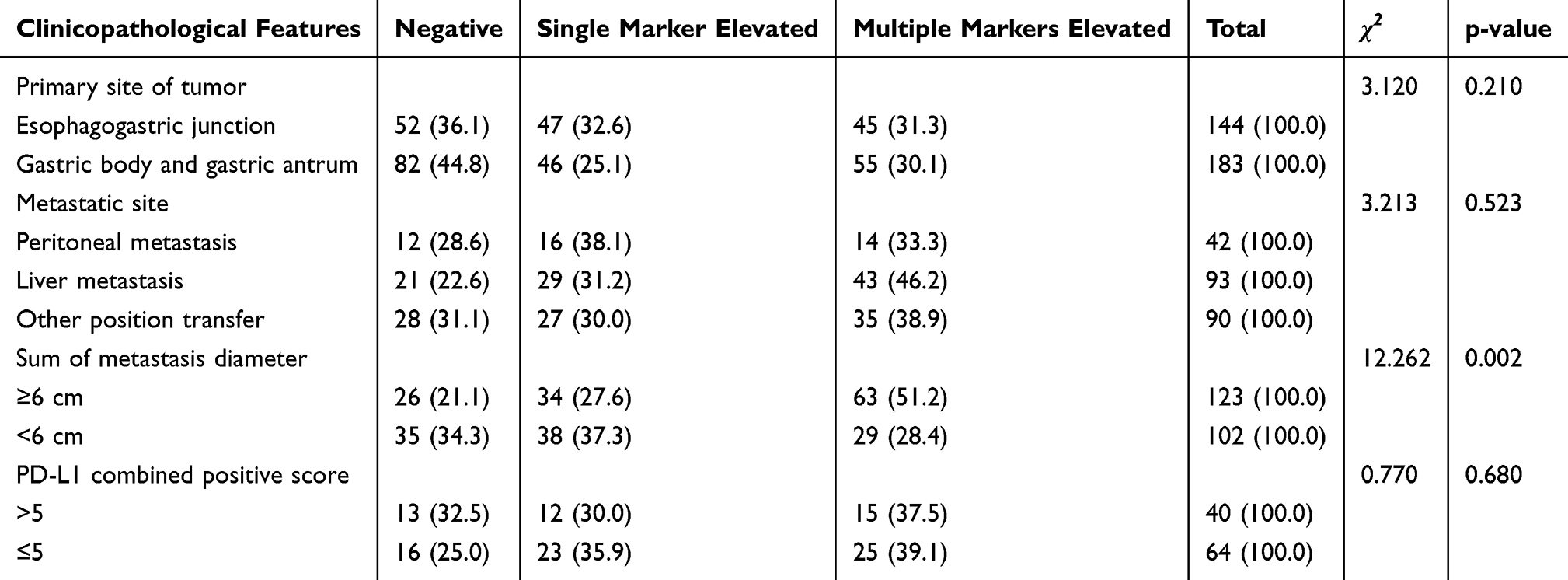 |
Table 1 Relationship Between the Number of Elevated Serum Tumor Markers and Clinicopathological Features |
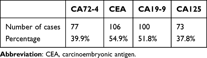 |
Table 2 Proportion of Different Markers in Patients with Positive Markers |
After first-line treatment for advanced gastric cancer, a relationship was observed between treatment efficacy (assessed through objective response rate [ORR], disease control rate [DCR], and PFS) and the number of elevated tumor markers. The ORR was positively correlated with the number of elevated tumor markers, with a higher number of elevated markers correlating with more favorable short-term chemotherapy outcomes. However, a higher number of elevated tumor markers was correlated with shorter PFS, indicating a poorer prognosis. The correlation between the number of elevated tumor markers and DCR was not statistically significant (p = 0.578). Pairwise comparisons revealed significant differences in PFS between patients with a single elevated marker and those with multiple elevated markers (Table 3, Figures 1 and 2).
 |
Table 3 Relationship Between Tumor Markers and the Efficacy of First-Line Drug Therapy in Stage IV Disease |
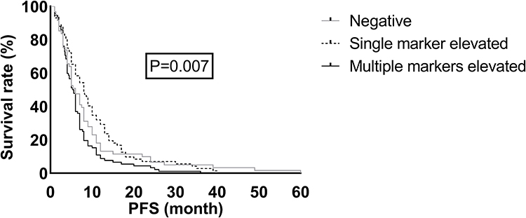 |
Figure 1 Progression-free survival curve of each group after first-line treatment. After first-line treatment for advanced gastric cancer, a significant difference was found between PFS and the number of elevated tumor markers (p = 0.007). Patients with multiple elevated markers had the shortest PFS and poor prognosis. |
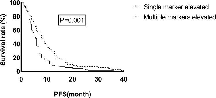 |
Figure 2 Progression-free survival curve of single-marker elevation versus multiple-marker elevation after first-line treatment. After first-line treatment, a significant difference was found in PFS between the single-marker elevated and multiple-marker elevated groups (p = 0.001). The prognosis of the single-marker elevated group was better than that of the multiple-marker elevated group. |
Statistical analysis of the therapeutic effects of immunotherapy combined with chemotherapy across various tumor markers did not reveal significant differences in efficacy (ORR, DCR, and PFS) related to tumor markers (p = 0.509, 0.288, and 0.477, respectively) (Table 4 and Figure 3).
 |
Table 4 Relationship Between Tumor Markers and the Efficacy of Immunotherapy Plus Chemotherapy in Stage IV Disease |
 |
Figure 3 Progression-free survival curve of each group after immunotherapy combined with chemotherapy. No difference was found between PFS and the number of increased tumor markers after immunotherapy combined with chemotherapy (p = 0.477). |
Among patients undergoing combination immunotherapy and chemotherapy, two groups were compared based on tumor marker levels and metrics, such as ORR, DCR, and PFS. The first group had a ≥5-fold increase in tumor markers and a total metastasis diameter <6 cm, whereas the second had negative tumor markers and a total metastasis diameter ≥6 cm (p = 0.045). The first group demonstrated superior short-term efficacy. However, no significant differences were observed in DCR or PFS between the groups (p = 0.516, p = 0.162) (Table 5 and Figure 4).
 |
Table 5 Efficacy Relationship According to Tumor Marker Levels and Total Metastasis Diameter Following Immunochemotherapy Treatment |
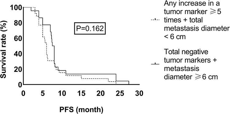 |
Figure 4 Progression-free survival curves of two subgroups after immunotherapy combined with chemotherapy. After immunotherapy combined with chemotherapy, patients with advanced gastric cancer were divided into two groups based on the degree of increase in tumor markers and the total metastatic diameter. The relationship between PFS and the two groups (any increase in a tumor marker ≥5 times + total metastasis diameter <6 cm vs negative tumor markers + metastatic diameter ≥6 cm) was compared, with no significant differences (p = 0.162). |
Discussion
Tumor markers, such as hormones, enzymes, and antigens secreted by tumor cells, are detectable in tumor tissues and body fluids.13 Serological markers such as CEA, CA125, CA19-9, and CA72-4 are frequently elevated in gastrointestinal tumors. Numerous studies have demonstrated their strong association with the development of gastric cancer. Consequently, in the absence of specific serum markers for gastric cancer, these markers play a critical role in early diagnosis. The combined use of three or more markers is particularly effective for early screening and diagnosis of gastric cancer.14 In advanced stages, an increase in serum tumor markers is strongly correlated with disease progression and prognosis. Gastric cancer positive for CA19-9 is associated with clinicopathological characteristics, such as poor histological differentiation, extensive lymphatic and venous invasion, and a high incidence of lymph node metastasis.15 Researchers have reported positivity rates of 21.1%, 27.8%, and 30.0% for CEA, CA19-9, and CA72-4, respectively, in gastric cancer. These markers significantly correlate with the clinical stage and patient survival, with CA72-4 demonstrating the strongest correlation and prognostic value.16 Other domestic studies also highlighted the relevance of CA72-4, linking it to tumor invasion depth, lymph node metastasis, peritoneal metastasis, and distant metastasis. The positivity rate of CA72-4 was higher in patients with gastric cancer experiencing lymph node, peritoneal, and serous involvement.17,18
Currently, numerous clinical treatments are available for gastric cancer, with chemotherapy being a commonly utilized approach. However, its efficacy as a standalone treatment for advanced gastric cancer is limited, with a median survival time typically less than 1 year.19 The addition of immunotherapy into the treatment regimen has shown promise in improving outcomes and extending patient survival. Clinical trials such as ATTRACTION-2, CHECKMATE-649, and ORIENT-16 have demonstrated that incorporating immune checkpoint inhibitors can significantly prolong the median survival time.8,9,20 Despite its potential to achieve long-term survival, the benefits of immunotherapy are not universal, as some patients fail to experience improved outcomes when it is combined with chemotherapy. Thus, a key research focus is identifying biomarkers that can effectively predict which patients will most likely benefit from treatment. Tumor markers, as antigens associated with tumor cells, may indicate heightened immunogenicity in certain patients. Hence, it is crucial to determine whether patients with significantly high tumor marker levels gain greater benefits from immunotherapy.
In this study, we investigated the therapeutic effectiveness and prognosis of patients with gastric cancer with varying levels and degrees of elevated tumor markers. In first-line treatment for advanced gastric cancer, significant differences were observed in remission rates across different tumor marker levels (p=0.039). Notably, the ORR increased with the number of elevated tumor markers, suggesting that patients with a higher number of elevated markers were more likely to achieve remission. Additionally, in first-line chemotherapy for gastric cancer, patients with varying levels of elevated tumor markers exhibited statistically significant differences in PFS (p=0.007). Further analysis revealed disparities in PFS between patients with a single elevated marker and those with multiple elevated markers. However, no significant difference in PFS was found between patients with negative tumor markers and those with single or multiple elevated markers. No correlation was found between tumor markers and ORR, DCR, or PFS in patients treated with a combination of immunotherapy and chemotherapy. However, an increasing trend in the ORR was observed in this treatment group as the number of tumor markers increased.
Patients with elevated tumor markers had a poor prognosis. After first-line treatment, patients with multiple elevated markers had a PFS of 5.3 months, which is significantly lower than a PFS of 8 months observed for those with a single elevated marker. The addition of immune checkpoint inhibitors improved the prognosis for patients with multiple elevated markers, increasing their PFS to 6 months. This finding suggests that patients with multiple elevated markers could benefit from the addition of immune checkpoint inhibitors. To further identify the immunologically advantaged population, we compared the relationship between ORR, DCR, and PFS between the groups: patients with any increase in a tumor marker ≥5 times and a total metastasis diameter <6 cm versus those with negative tumor markers and a metastasis diameter ≥6 cm. A significant difference in the ORR was observed (p=0.045), with the first group demonstrating superior short-term efficacy. This result indicates greater sensitivity to immune checkpoint inhibitors and better therapeutic outcomes when metastasis is smaller and tumor marker elevation is higher. Multi-marker elevation (≥5-fold) with limited metastatic burden (<6cm) predicts superior immunotherapy response, such a result may occur for the following reasons: 1) Immunogenicity perspective: Elevated markers (CEA/CA19-9/CA72-4/CA125) may indicate increased tumor-associated antigen presentation, enhancing T-cell activation potential with checkpoint inhibitors; 2) Tumor biology implications: The ≥5-fold threshold could reflect hypermetabolic/aggressive tumors more vulnerable to immune-mediated cytotoxicity; 3) Microenvironment interactions: The <6cm metastasis constraint suggests limited immunosuppressive stroma, permitting better drug penetration and immune cell infiltration.
Conclusion
The simultaneous expression of multiple tumor markers is significantly correlated with the differentiation degree, clinical stage, Lauren classification, and tumor burden of gastric cancer. Tumor marker levels are closely linked to both short-term efficacy and patient prognosis. In the treatment of advanced gastric cancer, the short-term efficacy of groups with elevated levels of a single or multiple tumor markers is better than that of the groups with negative markers. Especially, the PFS of the group with elevated single marker levels is longer and the prognosis is better. In the subgroup of patients receiving immunotherapy combined with chemotherapy, any increase in a tumor marker ≥5 times and a total metastasis diameter <6 cm showed better short-term efficacy. The incorporation of immunotherapy has shown potential to improve outcomes for patients with elevated tumor markers, providing valuable insights for optimizing treatment strategies.
Of note, our research does have some limitations. First, the retrospective study did not provide comprehensive information on patients’ eating habits, nutrition, physical condition, and the enthusiasm for treatment, resulting in an imbalance in the baseline data. Second, the cut-off value for the degree of marker elevation that indicates therapeutic efficacy is currently uncertain and requires more data to gradually analyze and determine. Third, age differences need to be stratified. Finally, there is a lack of data on OS. Future studies should be designed as prospective studies, and more patients with better compliance need to be included in the analysis to obtain objective results.
Abbreviations
CEA, carcinoembryonic antigen; PFS, progression-free survival; ORR, objective response rate; DCR, disease control rate.
Data Sharing Statement
The data generated in this study are included in the figures or tables of this article.
Ethics Approval and Consent to Participate
The study was approved by the relevant ethical committee of Henan Provincial People’s Hospital (approval number: (2021) Lun Shen No. (31)). Patient data used in this study will only be used for academic research purposes and will not be used for commercial gain to ensure there is no conflict of interest. All data were obtained with the patients’ verbal informed consent. Because this study was a retrospective one, the signing of the consent form was waived with the approval of the ethics committee. This study was conducted in accordance with the declaration of Helsinki.
Consent for Publication
Consent for publication was obtained from the participants.
Funding
This research did not receive any specific grant from funding agencies in the public, commercial, or not-for-profit sectors. The funder had no role in the conceptualization, design, data collection, analysis, decision to publish, or preparation of the manuscript.
Disclosure
The authors declare that they have no competing interests.
References
1. Zhang P, Zhang X, Zhu N, Zhuang F, Zhou D, Wang P. Clinical efficacy, survival, and adverse reaction evaluation of immune checkpoint inhibitor in advanced gastric cancer: a systematic review and meta-analysis. Comput Math Methods Med. 2022;2022:6998090. doi:10.1155/2022/6998090
2. Luo G, Jin K, Deng S, et al. Roles of CA19-9 in pancreatic cancer: biomarker, predictor and promoter. Biochim Biophys Acta Rev Cancer. 2021;1875(2):188409. doi:10.1016/j.bbcan.2020.188409
3. Zhao K, Zhou X, Xiao Y, Wang Y, Wen L. Research progress in alpha-fetoprotein-induced immunosuppression of liver cancer. Mini Rev Med Chem. 2022;22(17):2237–2243. doi:10.2174/1389557522666220218124816
4. Pezaro C, Woo HH, Davis ID. Prostate cancer: measuring PSA. Intern Med J. 2014;44(5):433–440. doi:10.1111/imj.12407
5. Zhang R, Chen X, Chen G, et al. Combined use of tumor markers in gastric cancer: a novel method with promising prognostic accuracy and practicality. Ann Surg Oncol. 2023;30(13):8561–8571. doi:10.1245/s10434-023-14194-9
6. Li L, Ma B, Liu F, et al. The Number of Positive Tumor Markers (NPTM) achieves higher value in the prognosis prediction of gastric cancer. Dis Markers. 2022;2022:5145918. doi:10.1155/2022/5145918
7. Sun AH, Zhang XY, Huang YY, Chen L, Wang Q, Jiang XC. Prognostic value and predictive model of tumor markers in stage I to III gastric cancer patients. World J Clin Oncol. 2024;15(8):1033–1047. doi:10.5306/wjco.v15.i8.1033
8. Xu J, Jiang H, Pan Y, et al. Sintilimab plus chemotherapy for unresectable gastric or gastroesophageal junction cancer: the ORIENT-16 randomized clinical trial. JAMA. 2023;330(21):2064–2074. doi:10.1001/jama.2023.19918
9. Janjigian YY, Shitara K, Moehler M, et al. First-line nivolumab plus chemotherapy versus chemotherapy alone for advanced gastric, gastro-oesophageal junction, and oesophageal adenocarcinoma (CheckMate 649): a randomised, open-label, Phase 3 trial. Lancet. 2021;398(10294):27–40. doi:10.1016/S0140-6736(21)00797-2
10. Huang ZB, Zhou X, Xu J, et al. Prognostic value of preoperative serum tumor markers in gastric cancer. World J Clin Oncol. 2014;5(2):170–176. doi:10.5306/wjco.v5.i2.170
11. Kochi M, Fujii M, Kanamori N, et al. Evaluation of serum CEA and CA19-9 levels as prognostic factors in patients with gastric cancer. Gastric Cancer. 2000;3(4):177–186. doi:10.1007/PL00011715
12. Choi SR, Jang JS, Lee JH, et al. Role of serum tumor markers in monitoring for recurrence of gastric cancer following radical gastrectomy. Dig Dis Sci. 2006;51(11):2081–2086. doi:10.1007/s10620-006-9166-5
13. Jiang T, Mei L, Yang X, Sun T, Wang Z, Ji Y. Biomarkers of gastric cancer: current advancement. Heliyon. 2022;8(10):e10899. doi:10.1016/j.heliyon.2022.e10899
14. Virgilio E, Proietti A, D’Urso R, et al. Measuring intragastric tumor markers in gastric cancer patients: a systematic literature review on significance and reliability. Anticancer Res. 2017;37(6):2817–2821. doi:10.21873/anticanres.11632
15. Kannagi R, Yin J, Miyazaki K, Izawa M. Current relevance of incomplete synthesis and neo-synthesis for cancer-associated alteration of carbohydrate determinants--Hakomori’s concepts revisited. Biochim Biophys Acta. 2008;1780(3):525–531. doi:10.1016/j.bbagen.2007.10.007
16. Shimada H, Noie T, Ohashi M, Oba K, Takahashi Y. Clinical significance of serum tumor markers for gastric cancer: a systematic review of literature by the Task Force of the Japanese Gastric Cancer Association. Gastric Cancer. 2014;17(1):26–33. doi:10.1007/s10120-013-0259-5
17. Xu Y, Zhang P, Zhang K, Huang C. The application of CA72-4 in the diagnosis, prognosis, and treatment of gastric cancer. Biochim Biophys Acta Rev Cancer. 2021;1876(2):188634. doi:10.1016/j.bbcan.2021.188634
18. Huang C, Liu Z, Xiao L, et al. Clinical significance of serum CA125, CA19-9, CA72-4, and fibrinogen-to-lymphocyte ratio in gastric cancer with peritoneal dissemination. Front Oncol. 2019;9(1159). doi:10.3389/fonc.2019.01159
19. Smyth EC, Nilsson M, Grabsch HI, van Grieken NC, Lordick F. Gastric cancer. Lancet. 2020;396(10251):635–648. doi:10.1016/S0140-6736(20)31288-5
20. Kang YK, Boku N, Satoh T, et al. Nivolumab in patients with advanced gastric or gastro-oesophageal junction cancer refractory to, or intolerant of, at least two previous chemotherapy regimens (ONO-4538-12, ATTRACTION-2): a randomised, double-blind, placebo-controlled, phase 3 trial. Lancet. 2017;390(10111):2461–2471. doi:10.1016/S0140-6736(17)31827-5
 © 2025 The Author(s). This work is published and licensed by Dove Medical Press Limited. The
full terms of this license are available at https://www.dovepress.com/terms
and incorporate the Creative Commons Attribution
- Non Commercial (unported, 4.0) License.
By accessing the work you hereby accept the Terms. Non-commercial uses of the work are permitted
without any further permission from Dove Medical Press Limited, provided the work is properly
attributed. For permission for commercial use of this work, please see paragraphs 4.2 and 5 of our Terms.
© 2025 The Author(s). This work is published and licensed by Dove Medical Press Limited. The
full terms of this license are available at https://www.dovepress.com/terms
and incorporate the Creative Commons Attribution
- Non Commercial (unported, 4.0) License.
By accessing the work you hereby accept the Terms. Non-commercial uses of the work are permitted
without any further permission from Dove Medical Press Limited, provided the work is properly
attributed. For permission for commercial use of this work, please see paragraphs 4.2 and 5 of our Terms.