")
Back to Journals » Clinical, Cosmetic and Investigational Dermatology » Volume 16
An Unusual Case of Cutaneous Pili Migrans in a 45-Day-Old Infant
Authors Yang D , Zhu R, Yang X, Huang S, Guo M
Received 3 April 2023
Accepted for publication 10 June 2023
Published 15 June 2023 Volume 2023:16 Pages 1549—1552
DOI https://doi.org/10.2147/CCID.S415566
Checked for plagiarism Yes
Review by Single anonymous peer review
Peer reviewer comments 3
Editor who approved publication: Dr Jeffrey Weinberg
Dingbin Yang,* Renheng Zhu,* Xuejun Yang, Shuqiong Huang, Menglu Guo
Department of Dermatology, People’s Hospital of Leshan, Leshan, 614000, People’s Republic of China
*These authors contributed equally to this work
Correspondence: Dingbin Yang, Department of Dermatology, People’s Hospital of Leshan, No. 238, Baita Street, Leshan, 614000, People’s Republic of China, Tel +86-15681383138, Email [email protected]
Abstract: Cutaneous Pili Migrans (CPM), a rare skin condition, is composed of hair fragments embedded in the skin epidermis and dermis after skin trauma or for unknown reasons. To the best of our knowledge, there are few reports on cases of CPM in which hair is exposed outside of the skin. Herein, we report an unusual and rare case of 45 days old Chinese male infant with CPM.
Keywords: cutaneous pili migrans, creeping hair, burrowing hair
Introduction
Creeping Eruption means the creeping linear damage that occurs following infection by parasitic organisms, such as Hookworms and Nematodes. Rarely, hair shafts also imitate the parasites to form black lines. In 1957 Yaffee reported a case of linear damage caused by a migratory hair shaft in the superficial skin.1 The symptom was called imbedded hair at that time. Thai and Sinclair reported a middle aged man who presented with a 7cm beard hair that grew under the stratum corneum, they named the condition Cutaneous Pili Migrans (CPM) in 2001.2 The clinical presentation of CPM is a meandering, creeping, slightly raised, linear lesion that moves forwards in an irregular pattern. The source can be the shaft and bristle of the hair and pubic hair, especially the hair segments with sharp ends. Making a shallow incision at the end of the lesion and pulling out the hair is the treatment. We report an infant with a 1-day history of curled linear skin disorder in the neck. This case provides an opportunity to remind clinicians that in not all CPM cases is the hair completely embedded in the skin.
Case Report
The patient was a 45-day old male infant. He was brought to the dermatology department of our hospital as a result of his mother finding a black linear lesion in his neck. The black crooked line was partly revealed, and the skin was without redness or swelling on the surface. The infant was in good health, had no fever or other discomforts. His mother denied that there was an obvious history of trauma, and no history of infection diseases.
The infant’s general condition was good, and he was otherwise healthy. On physical examination, a black linear lesion was noted in the neck folds. It included a total of five segments. Three segments (yellow arrows) were embedded in the skin, and the rest (red arrows) of the strand exposed. It looked like a snake, and there was no erythematosus nor papules around it (Figure 1). We clamped the line exposed on the surface with forceps and pulled it out slowly, without bleeding. The extraction was a curly hair and was about 4cm long. There were no hair follicles at either end (Figure 2). The eruption diminished immediately after hair removal (Figure 3), and a final diagnosis of CPM was made. A 4-year follow-up revealed no recurrence.
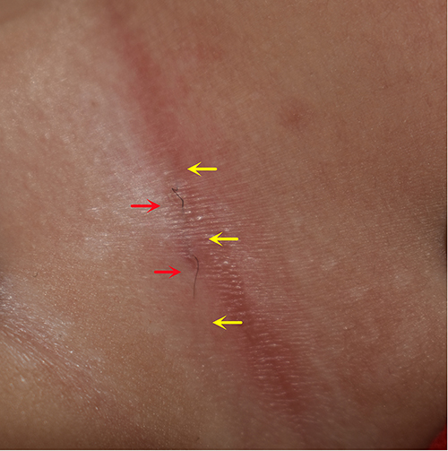 |
Figure 1 A black linear lesion was noted in the neck folds. It included a total of five segments. Three segments (yellow arrows) were embedded in the skin, and the rest (red arrows) of the strand exposed. It looked like a snake, and there was no erythematosus nor papules around it. |
 |
Figure 2 The extraction was a curly hair and was about 4cm long. There were no hair follicles at either end. |
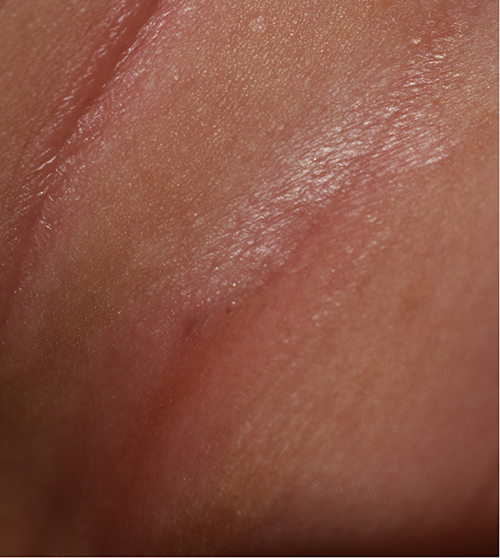 |
Figure 3 The eruption diminished after hair removal. |
Discussion
CPM is a rare disease which is characterized by creeping eruption with linear hair. Since 1957, more than 40 cases, including some with different names such as creeping hair, burrowing hair, moving hair, embedded hair, and mane migration, have been reported.3–5 It was not until 2001 that Thai and Sinclair coined the term CPM.2 Most of the cases were from Asia, including Japan, China, and South Korea because hair has a larger diameter with a higher tensile strength in Asians than in other ethnic groups.4 The site of predilection includes the ankle, plantar, toes, breast, cheeks, neck, and abdomen. The abdomen is the most common site in adults, whereas in children it is more likely to occur on the soles of feet.3,6
The exact etiology of CPM is still unclear. However, it is speculated that skin with wounds and hair with sharp ends may be the two main reasons for the hair entering the skin.3 In our case, the skin lesion was presented in the neck, where skin had been soaked and softened after sweating, or there may have been a tear so that hair could more easily to enter the skin. CPM moves linearly in an irregular pattern in one direction without obvious itching or pain. Because of the migratory feature of CPM, it is often misdiagnosed as Cutaneous Larval Migrans (CLM), despite the fact that CLM is characterized by intensely pruritus and the ability to move in any direction.
Compared with previous classic clinical presentation of single site or single hair, a CPM at multiple sites or with tufted hairs has been reported recently. NAM et al reported a case of hair migration lesions in two regions of the body with a 7-year-old patient having embedded short hairs in the armpit and neck. The authors measured the hair thickness of family members with a scanning electron microscope (SEM) and concluded that the father’s beard was the most likely source.7 Willems et al reported a 34-year-old patient who had grey-black linear hair in the skin above his pubis for 10 years. Dermoscopic examination revealed that the lesion was a hair cluster including about twenty single hairs.8
We report a case of CPM in which two segments of hair were exposed outside the skin. We speculate that this distinctive manifestation may be attributed to the early onset of the disease, as the hair did not completely enter the skin. Interestingly, the infant was only 45 days old and to our knowledge, he is the youngest reported patient with CPM to date.
Conclusion
Our case highlights an unusual occurrence of CPM where the hair migrants like a small snake.
Ethical Concerns
The patient’s mother signed the informed consent. She agreed to publish the details of this case. Institutional approval has been obtained.
Disclosure
The authors report no conflicts of interest in this work.
References
1. Yaffee HS. Imbedded hair resembling larva migrans. AMA Arch Derm. 1957;76(2):254. doi:10.1001/archderm.1957.01550200098027
2. Thai KE, Sinclair RD. Cutaneous pili migrans. Br J Dermatol. 2001;144(1):219. doi:10.1111/j.1365-2133.2001.03998.x
3. Luo DQ, Liu JH, Huang YB, et al. Cutaneous pili migrans: a case report and review of the literature. Int J Dermatol. 2009;48(9):947–950. doi:10.1111/j.1365-4632.2009.04118.x
4. Jang YH, Kim MJ, Kim SL, et al. Creeping Hair in the Beard Area. Ann Dermatol. 2015;27(5):635–636. doi:10.5021/ad.2015.27.5.635
5. Ingkapairoj K, Triwongwaranat D, Jiamton S, et al. Cutaneous Pili Migrans: a case report. Skin Appendage Disord. 2020;6(1):52–54. doi:10.1159/000504234
6. Winkler A, Prindaville B, Wiss K. Curvilinear, erythematous plantar patch in a toddler. Pediatr Dermatol. 2018;35(2):251–252. doi:10.1111/pde.13395
7. Nam KH, Jung ES, Park J, et al. An unusual case of cutaneous pili migrans: pili cuniculati multiplex. Acta Derm Venereol. 2021;101(7):1–3. doi:10.2340/00015555-3861
8. Willems A, Sinclair R. Tufted cutaneous pili migrans. Clin Exp Dermatol. 2021;46(6):1102–1103. doi:10.1111/ced.14622
 © 2023 The Author(s). This work is published and licensed by Dove Medical Press Limited. The full terms of this license are available at https://www.dovepress.com/terms.php and incorporate the Creative Commons Attribution - Non Commercial (unported, v3.0) License.
By accessing the work you hereby accept the Terms. Non-commercial uses of the work are permitted without any further permission from Dove Medical Press Limited, provided the work is properly attributed. For permission for commercial use of this work, please see paragraphs 4.2 and 5 of our Terms.
© 2023 The Author(s). This work is published and licensed by Dove Medical Press Limited. The full terms of this license are available at https://www.dovepress.com/terms.php and incorporate the Creative Commons Attribution - Non Commercial (unported, v3.0) License.
By accessing the work you hereby accept the Terms. Non-commercial uses of the work are permitted without any further permission from Dove Medical Press Limited, provided the work is properly attributed. For permission for commercial use of this work, please see paragraphs 4.2 and 5 of our Terms.