Back to Journals » Pharmacogenomics and Personalized Medicine » Volume 15
An Integrative Analysis Identifying RAB40C as an Oncogenic Immune Protein and Prognostic Marker of Lung Squamous Cell Carcinoma
Authors Wu H, Dong X, Liao L, Huang L
Received 8 February 2022
Accepted for publication 5 May 2022
Published 22 May 2022 Volume 2022:15 Pages 525—537
DOI https://doi.org/10.2147/PGPM.S357166
Checked for plagiarism Yes
Review by Single anonymous peer review
Peer reviewer comments 2
Editor who approved publication: Dr Martin H Bluth
Hong Wu,1 Xuhui Dong,1 Lixian Liao,2 Lihaoyun Huang2
1Department of Pneumology, Yiwu Central Hospital, Yiwu, Zhejiang, People’s Republic of China; 2Department of Oncology, Affiliated Tumor Hospital of Guangxi Medical University, Nanning, Guangxi, People’s Republic of China
Correspondence: Hong Wu, Department of Pneumology, Yiwu Central Hospital, Yiwu, Zhejiang, People’s Republic of China, Email [email protected]
Background: RAB40C, a member of the Ras oncogene family, is a protein with GTPase and GTP-binding activity and is also predicted to be important in immunomodulation. However, the link between RAB40C and lung squamous cell carcinoma (LUSC) has not yet been elucidated. Exploring the relationship between RAB40C and LUSC could help expand the repertoire of immunotherapeutic targets for LUSC and provide more effective therapeutic options for LUSC patients, which behalf of our aim for our study.
Methods: We analyzed the RAB40C expression in different tumor types and stages based on the TCGA database. Subsequently, we explored the differences in RAB40C expression in LUSC versus paracancerous tissues through immunohistochemical analysis. The prognostic value of RAB40C was assessed by Cox regression and Kaplan-Meier analysis. Gene set enrichment analysis-based RAB40C impact pathways and the correlation between RAB40C expression and immune infiltration were obtained using the TIMER2.0 and the CIBERSORT analytical tools. Tumor mutational load and microsatellite instability (MSI) were assessed by the Spearman correlation analysis. Finally, the close association of RAB40C with LUSC was explored by correlating immune cell infiltration with immunomodulator expression, assessing risk scores in combination with other factors, and analyzing prognostic nomogram.
Results: The expression of RAB40C was significantly elevated in LUSC. RAB40C expression was significantly associated with immune factors, immune-related pathways, and MSI. Moreover, RAB40C significantly negatively correlated with LUSC-associated immune infiltrating cells, CD4 memory-activated cells, γδ T cells, M1-like macrophages, and the immune regulator CD28, while it positively associated with the activation of Tregs and natural killer cells. Further, a risk model constructed from RAB40C and its associated immune genes showed that RAB40C might be an independent prognostic factor for LUSC.
Conclusion: RAB40C can be used as an effective prognostic biomarker and a potential immunotherapeutic target for the treatment of LUSC.
Keywords: RAB40C, LUSC, immune regulator, prognostic model, pan-cancer
Introduction
Lung squamous cell carcinoma (LUSC) is a major type of non-small cell lung cancer (NSCLC) that affects the central part of the lungs or the main airways. Due to its malignancy and poor prognosis, LUSC is a global threat. Hence, research on the prevention and treatment of LUSC at its different stages is gaining increasing attention. Age, smoking, and second-hand smoking inhalation are risk factors for LUSC.1 Although surgery and radiotherapy are optimal treatment options for LUSC in its early stages, targeting therapy of genes and molecules such as EGFR, ALK, and MET may be more beneficial for patients with advanced LUSC.2 However, to date, there are many unknown genetic aberrations or unclear oncogenetic drivers of LUSC. Hence, unraveling these unknown factors will provide a promising future for targeting therapies and immunotherapeutic developments.
RAB40C is a member of the Rab GTPases that plays a central role in membrane transport, like transporting proteins among organelles.3 The Rab GTPase family proteins, including RAB40C enriched in oligodendrocytes and located in perinuclear domains, are vital for neurite and glia cell growth and signal transportation of chemical synapses. RAB40C is tightly associated with lipid droplets and regulates their formation.4,5 Further, owing to its function in transporting intracellular proteins, RAB40C can regulate Varp expression, influence the melanogenic enzymes in melanocytes, and affect the growth of melanocytes.6,7 Moreover, RAB40C has been identified as a member of the Ras oncogenic gene family.8 A high level of RAB40C may restrict the survival of osteosarcoma. Besides, methylated RAB40C is considered a potential marker for the assessment of breast cancer.9 Furthermore, in gastric cancer, RAB40C helps oncocyte proliferation. Thus, downregulating RAB40C transcription has been indicated to improve the prognosis of gastric cancer.10
Hence, in this study, we aimed to complement the recent discoveries in RAB40C and tried to explore more therapeutic possibilities for RAB40C in cancers, especially LUSC for more possibility in personal treatments. Initially, we comprehensively analyzed RAB40C expression in pan-cancer, including gene expression, its prognosis at different stages, pathway enrichment analyses, and immune factor analyses. Next, we focused on the correlation between immune cell infiltration and immunomodulator expression, together with evaluating the risk score with other factors. We hope that our findings will help in the advancement of immunotherapies for LUSC, a tumor highly related to RAB40C.
Methods
Clinical Specimens and Immunochemical Analysis
LUSC and paracancerous tissues were obtained from 20 patients treated at the Yiwu Central Hospital from June to December 2019. The patients were pathologically diagnosed with LUSC and did not undergo any radiotherapy, chemotherapy, or immunotherapy before sampling. The tissues were preserved in formalin. We cut the tissues into 5 μm pieces and blocked their endogenous peroxidase activity by de-paraffinization, rehydration, and 5% bovine serum albumin at 37 °C for 30 mins. Subsequently, we incubated the tissues overnight with anti-RAB40C, and then washed them with phosphate-buffered saline (PBS) thrice. Next, the tissue was then incubated with a secondary antibody at 37 °C for 30 mins and washed with PBS three times. Lastly, the tissues were stained with diaminobenzidine and visualized under a microscope. This study was approved by the Ethics and Anthropology Committee of Yiwu Central Hospital and all patients gave informed consent.
Public Databases and Software for Analyzing RAB40C Expression
We downloaded the data of RAB40C RNA expression between normal and tumor tissues in pan-cancer from The Cancer Genome Atlas (TCGA) and Genotype-Tissue Expression (GTEx) database using UCSC Xena (xenabrowser.net) and analyzed the data using the SangerBox software. Additionally, the RAB40C expression in different pathological stages of cancer with reference to the World Health Organization (WHO) criteria acquired from the TCGA database was profiled by the Gene Expression Profiling Interactive Analysis 2 (GEPIA2) webserver (http://gepia2.cancer-pku.cn/).
Enrichment Analysis of Pathways Related to RAB40C Expression
Gene Set Enrichment Analysis (GSEA) selected the pathways with a strong relationship with RAB40C. Interaction pathways with RAB40C integrated by the Kyoto Encyclopedia of Genes and Genomes (KEGG) pathway database and the HALLMARK pathway database were explored by the R package “clusterProfiler.”
Immune Cell Infiltration Analysis Related to RAB40C Expression
The correlation of RAB40C with the immune infiltration level in different tumor types of the TCGA database was analyzed by tools such as the Tumor Immune Estimation Resource (TIMER2) (https://cistrome.shinyapps.io/timer) and Cell-type Identification by Estimating Relative Subsets of RNA Transcripts (CIBERSORT) (http://cibersort.stanford.edu/). The association between RAB40C expression and the abundance of immune cells, including CD4 T memory cells, M1-like macrophages, and Treg cells was analyzed using TIMER2, CIBERSORT, and Pearson’s correlation coefficients. CIBERSORT is a versatile and intelligent approach to precisely calculate immune components of tumor tissue from gene expression profiles of immune infiltrating leukocytes obtained by RNA-sequencing or high-throughput sequencing.11 Further, we employed the Spearman’s test incorporated with the SangerBox tool to comprehensively and accurately evaluate the significance of RAB40C in tumor immunity based on the correlation analysis between RAB40C and the estimated score, stromal score, and immune score acquired from the ESTIMATE algorithm.
Immunoregulators Related to RAB40C Expression
Analysis of the association of RAB40C expression with immune stimulators and suppressors was based on the TISIDB database (http://cis.hku.hk/TISIDB/). Next, we integrated the significant immune regulators and analyzed their correlation with LUSC using Spearman’s test (p<0.05).
Statistical Analysis
We performed the Mann–Whitney U-test for analyzing statistical differences between non-normally distributed variables and the unpaired t-test for normally distributed variables. Furthermore, more than two groups were compared using the Kruskal–Wallis one-way ANOVA analysis. The RAB40C mRNA expression was described in Transcripts Per Million. Based on Spearman’s test, the correlation of RAB40C mRNA expression with both tumor mutation burden (TMB) and microsatellite instability (MSI) was assessed to validate the potential of RAB40C expression as a novel biomarker in cancers. The R version 3.5.0 software was used for all statistical analyses. Results with the p-value < 0.05 were considered statistically significant by the Cox regression analysis.
Prognostic Survival Analysis
Combined with the data from the TCGA database, we used the Kaplan-Meier analysis to assess the overall survival (OS), disease-specific survival (DSS), and progression-free survival interval (PFI) of patients with low or high RAB40C expression. We also performed the Cox regression analysis to evaluate the predictive model for RAB40C in cancer prognosis by showing the correlation of RAB40C expression with the OS, DSS, and PFI of tumors. The R environment was used for both analyses.
Moreover, to generate a prognostic model on RAB40C-related immune regulators of LUSC, we used a univariate Cox regression analysis and showed the relationship between OS and significant immunoregulators using hazard ratios. Multivariate Cox regression analysis with age, gender, and clinical tumor–node–metastasis (TNM) stages was designed to screen for more meaningful immune-related prognostic models. Further, we used Kaplan-Meier curves to establish the survival trend associated with the risk of LUSC (Log rank test, p<0.001). Finally, to validate the accuracy and sensitivity of the risk score combined stage model for the prognosis of LUSC, the receive operator characteristic (ROC) curve dependent on the different LUSC stages was used for assessment based on the survivalROC package.
Prognostic Nomogram in LUSC
We evaluated the prognosis of LUSC by scoring the contribution of each parameter, including risk score, age, sex, and TNM stage, to a patient’s death or the recurrence of LUSC and synthesized each score to construct a nomogram. A higher score indicates that the patient is at higher risk for a poor prognosis. Nomograms for 3-year and 5-year survival developed by the “rms” package in R were incorporated with calibration curves gauged with the consistency index (C-index) to anticipate the individual prognosis of LUSC.
Result
Differential Expression Analysis of RAB40C Between Tumor and Normal Tissues
We obtained the differences in RAB40C expression between cancer and paracancerous tissues in individual tumor samples from the TCGA database. Its expression was significantly higher in many cancers compared to normal tissues, including bladder urothelial carcinoma (BLCA), breast invasive carcinoma (BRCA), cholangiocarcinoma (CHOL), head and neck squamous cell carcinoma (HNSC), kidney renal clear cell carcinoma (KIRC), liver hepatocellular carcinoma (LIHC), lung adenocarcinoma (LUAD), LUSC, prostate adenocarcinoma (PRAD), rectum adenocarcinoma (READ), stomach adenocarcinoma (STAD), uterine corpus endometrial carcinoma (UCEC) (p<0.001), and colon adenocarcinoma (COAD) (p<0.05). In contrast, its expression was significantly lower only in glioblastoma multiforme (GBM) (p<0.001), kidney chromophobe (KICH) (p<0.05), and brain lower-grade glioma (LGG) (p<0.01) (Figure 1). Considering the limited number of normal samples in the TCGA database, we integrated data from normal tissues in the GTEx database with data from the TCGA tumor tissues to analyze the expression differences of RAB40C in 27 tumor-normal tissue pairs. RAB40C was also significantly highly expressed in adrenocortical carcinoma (ACC), cervical squamous cell carcinoma and endocervical adenocarcinoma (CESC), esophageal carcinoma (ESCA), acute myeloid leukemia (LAML), ovarian serous cystadenocarcinoma (OV), pancreatic adenocarcinoma (PAAD), and uterine carcinosarcoma (UCS), in addition to the 13 tumors mentioned above. RAB40C had a significantly low expression only in a few tumors, including skin cutaneous melanoma (SKCM), testicular germ cell tumors (TGCT), and thyroid carcinoma (THCA) (Supplementary Figure 1).
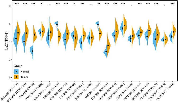 |
Figure 1 Expression of RAB40C in cancer tissues and adjacent normal tissues in the TCGA database. *p<0.05; **p<0.01; ***p<0.001. |
Analysis of Relationship Between RAB40C Expression and Tumor Stage
We analyzed the expression of RAB40C in the different cancer stages according to the WHO and found that RAB40C expression was significantly elevated in ACC and LIHC in the advanced stages and significantly decreased in BLCA, ESCA, KICH, and STAD (Figure 2A–F). RAB40C expression in BRCA, COAD, LUSC, TGCT, CHOL, uveal melanoma (UVM), and HNSC showed an elevated trend, but the differences with the normal tissues were not statistically significant (Supplementary Figure 2).
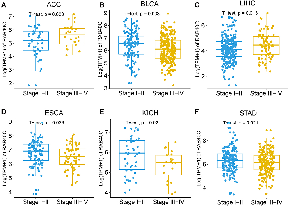 |
Figure 2 RAB40C expression at different stages of pan-cancer according to WHO. (A–F) Pan-cancer differential expression of RAB40C in the tumor types indicated in the TCGA database according to the WHO staging of cancer. |
Prognostic Analysis of RAB40C in TCGA
We evaluated the potential prognostic significance of RAB40C by the univariate Cox regression and Kaplan-Meier analysis. Univariate Cox regression showed that high expression of RAB40C was associated with a high risk of tumorigenesis. OS results showed that high RAB40C expression was a risk factor for patients with BRCA, lymphoid neoplasm diffuse large b-cell lymphoma (DLBC), KIRC, LIHC, LUSC, OV, THCA, and UCS, while being a protective factor for patients with BLCA, HNSC, READ, STAD, and thymoma (THYM) (Figure 3A). DSS analysis showed that RAB40C was a risk factor for patients with BRCA, DLBC, KIRC, LIHC, LUSC, OV, THCA, and UCS and a protective factor for patients with BLCA, HNSC, READ, STAD, and THYM (Figure 3B). Finally, PFI analysis showed that RAB40C was a risk factor for patients with ACC, CESC, KIRC, LIHC, LUSC, PRAD, and UVM and a protective factor for patients with BLCA, COAD, ESCA, GBM, HNSC, KICH, kidney renal papillary cell carcinoma, LGG, OV, PAAD, SKCM, THYM, and UCEC (Figure 3C).
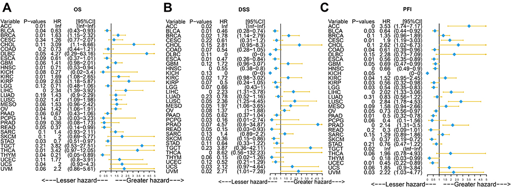 |
Figure 3 Prognostic analysis via Univariate Cox regression of RAB40C. (A) Effect of RAB40C on the overall survival (OS) of pan-cancer. (B) Effect of RAB40C on the disease-specific survival (DSS) of pan-cancer. (C) Effect of RAB40C on the progression-free survival interval (PFI) of pan-cancer. |
Next, we evaluated the OS, DSS, and PFI by the Kaplan-Meier analysis. The OS data showed RAB40C as a risk factor in ACC, BRCA, DLCA, OV, KIRC, LIHC, LUSC, THCA, and UCS (Supplementary Figure 3A). DSS showed RAB40C as a risk factor in ACC, BRCA, LIHC, UCS, and UVM and a protective factor in THCA (Supplementary Figure 3B). Finally, according to the PFI, RAB40C was found to be a risk factor in ACC, CESC, KIRC, LIHC, LUSC, PRAD, UVM, and TGCT (p<0.05) (Supplementary Figure 3C).
GSEA of RAB40C
According to the GSEA, the functional pathways found in the KEGG and HALLWAY databases showed different levels of enrichment between the high and low expression of RAB40C. According to the KEGG analysis, pathways related to glycerophospholipid, taurine, hypotaurine, and fatty acid metabolism were enriched in tissues with a low expression of RAB40C, suggesting that RAB40C might be an inhibitor for these pathways (Figure 4A). On the contrary, pathways related to graft-host disease, primary immunodeficiency, antigen processing and presentation, and natural killer cell-mediated cytotoxicity were enriched when the RAB40C expression was high, suggesting that RAB40C might activate these pathways (Figure 4B). Further, analysis of the HALLMARK database showed that estrogen response early, notch signaling and p53 pathway were enriched in tissues with low expression of RAB40C, while inflammatory response, interferon-γ response, complement pathway, and allograft rejection were enriched in tissues with high expression of RAB40C (Figures 4C and D).
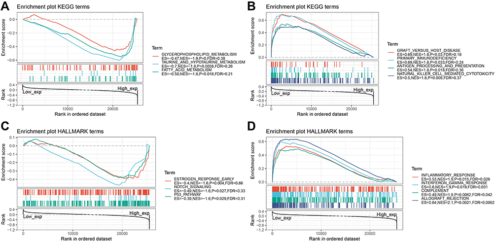 |
Figure 4 Gene Set Enrichment Analysis (GSEA) of RAB40C. (A and B) Enrichment of Kyoto Encyclopedia of Genes and Genomes (KEGG) pathways correlated to RAB40C expression. (C and D) Enrichment of KEGG pathways correlated to RAB40C expression. |
Immune Cell Infiltration Level Associated with RAB40C Expression
According to the association of 22 types of immune cells infiltration level and pan-cancer, we found out that CD4 memory activated cells negatively correlated with RAB40C in BLCA, BRCA, CESC, LUAD, LUSC, STAD, and TGCT in a statistically significant manner (p<0.01), but significantly positively correlated only in KIRC (p<0.001). For BRCA, KIRC, LUAD, LUSC, PAAD, TGCT, and THCA, RAB40C negatively and significantly correlated with γδT cells (p<0.01). Additionally, the negative relationship between M1-like and M2-like macrophages and RAB40C in BLCA, BRCA, LUAD, LUSC, and THCA was also significant (Figure 5).
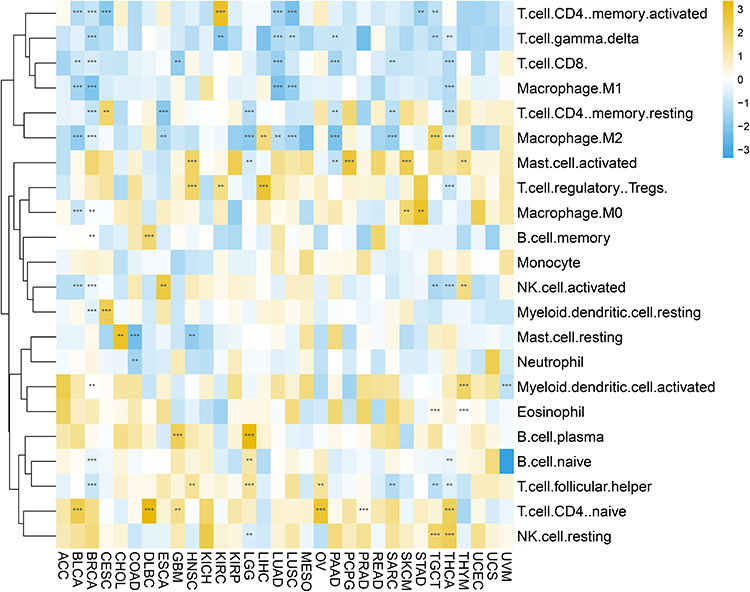 |
Figure 5 Evaluation of the association of RAB40C and immune cell infiltration level in different cancers. **p<0.01; ***p<0.001. |
Further, to investigate the link of immunoregulatory factors with RAB40C in pan-cancer, we measured the ESTIMATE score, stromal score, and immune score based on Spearman correlation test. The association of the ESTIMATE score, which was considered as the synthetic assessment of stromal and immune elements in cancers, and RAB40C was relatively high in GBM (R=−0.499), mesothelioma (MESO) (R=−0.495), PAAD (R=−0.487), BLCA (R=−0.471), and UCS (R=−0.435) (Supplementary Figure 4). The relationship between the stromal score and RAB40C was also significantly high in MESO (R=−0.505), GBM (R=−0.454), and BLCA (R=−0.434) (Supplementary Figure 5). Finally, we observed a positive correlation between the immune score and RAB40C in GBM (R=−0.513), LGG (R=−0.501), ACC (R=−0.488), and BLCA (R=−0.465) (Supplementary Figure 6).
Correlation of Immunoregulatory Factors and RAB40C Expression
Next, we tried to further examine the vital correlations between RAB40C expression and immune factors like regulators and chemokines. The regulators which activated the immune system, including CD48, CD86, CD28, and TNFRSF17, were negatively correlated with RAB40C in BRAC, LUAD, LUSC, PAAD, and THCA in a statistically significant manner (p<0.01). However, TNFRSF2 positively correlated to RAB40C in most cancers with statistical significance (Supplementary Figure 7A). Furthermore, the immune inhibitors HAVCR2, PDCD1LG2, and IL10 negatively correlated to RAB40C in BRCA, LGG, LUAD, LUSC, PAAD, and THCA (p<0.01) (Supplementary Figure 7B). As for chemokine ligands and receptors, CCL5 and CCR5 negatively correlated with RAB40C in BRCA, LUAD, LUSC, PAAD, and THCA (p<0.01) (Supplementary Figures 7C and D).
Correlation of Immune Checkpoints and RAB40C Expression
Currently, immune checkpoints are at the forefront of developing immunotherapies for gene-related tumors. Therefore, we also analyzed the relationship between immune checkpoints and RAB40C in pan-cancer. RAB40C expression in BLCA negatively correlated with 30 out of the 47 immune checkpoints (p<0.001). Even in LUAD and PAAD, the RAB40C expressions negatively correlated with 26 out of 47 immune checkpoints (p<0.01). Further, in LUSC, RAB40C expression negatively correlated with 24 out of 47 immune checkpoints (p<0.05) (Supplementary Figure 8).
Correlation of RAB40C Expression with TMB and MSI
We found that a high TMB negatively correlated in a statistically significant manner with RAB40C in BRCA (p=9.5e-07), COAD (p=0.00088), THCA (p=0.002), and LAML (p=0.035) (Supplementary Figure 9A). Simultaneously, MSI negatively correlated with RAB40C in READ (p=2.3e-07) and COAD (p=0.0038). However, in LUSC (p=7.1e-09), KICH (p=0.0019), LUAD (p=0.0022), and LIHC (p=0.00061), MSI positively correlated with RAB40C (Supplementary Figure 9B).
Thus, all data previously observed suggest that RAB40C is a promising candidate for prognostic prediction and immunotherapy in cancer. In addition, according to the correlations between RAB40C and the tumors mentioned above, LUSC might be treated through cancer immunotherapy targeting RAB40C.
Immunohistochemical Analysis of RAB40C in LUSC
Immunohistochemistry and statistical analysis comparing LUSC with adjacent normal tissues showed that the expression of RAB40C in LUSC was significantly high (p=0.034) (Supplementary Figure 10).
Infiltration of Immune Cells in LUSC Related to RAB40C Expression
While comparing the infiltration level of immune cells in tissues with low and high expression of RAB40C in LUSC, we found that the proportion of T cell CD4 memory activated and M1-like macrophages in LUSC were significantly lower when the RAB40C expression was high than when it was low (p<0.05). Inversely, the level of Tregs in LUSC positively correlated with RAB40C expression (p<0.01) (Figure 6).
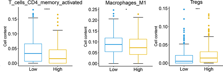 |
Figure 6 Evaluation of the association of RAB40C and CD4 memory-activated cells, M1-like macrophages, and Tregs infiltration level in lung squamous cell carcinoma (LUSC). *p<0.05; ***p<0.001; ****p<0.0001. |
Immunoregulators in LUSC Related to RAB40C
We used the TISIDB database to understand the correlation between immune modulators and RAB40C expression in LUSC. We speculated that RAB40C expression may affect the expression of immunoregulatory genes to some extent, such as CD96, PDCD1LG2, IL6, NT5E, TNFSF13B, HAVCR2, and CD48, since the immune response to LUSC decreased with a high RAB40C expression, with a significant negative relationship between RAB40C expression and the immunomodulators (p<0.001). However, RAB40C also positively modulated some immune regulators, including VTCN1, PVRL2, TNFRSF18, KLRK1, TNFRSF25, and TNFRSF14 (p<0.05) (Supplementary Figure 11).
Analysis of the Prognostic Value of RAB40C-Related Immunomodulators in LUSC
To investigate the prognostic value of RAB40C-associated immunomodulators in LUSC, we performed a single-factor Cox regression analysis of RAB40C with its associated 44 immune genes. The results showed that the abnormal expression of RAB40C, ADORA2A, CTLA4, TGFBR1, CD70, IL6, KLRC1, KLRK1, NT5E, and TNFRSF13C genes were risk factors for LUSC (Figure 7A). Subsequently, we performed a multifactorial Cox regression analysis of the above immune genes and found that RAB40C, ADORA2A, CTLA4, TGFBR1, CD70, IL6, and NT5E were the risk factors for LUSC (Figure 7B). The Kaplan-Meier survival curve analysis of our prognostic model revealed that the low-risk population survived significantly longer than the high-risk population (p<0.001) (Figure 7C).
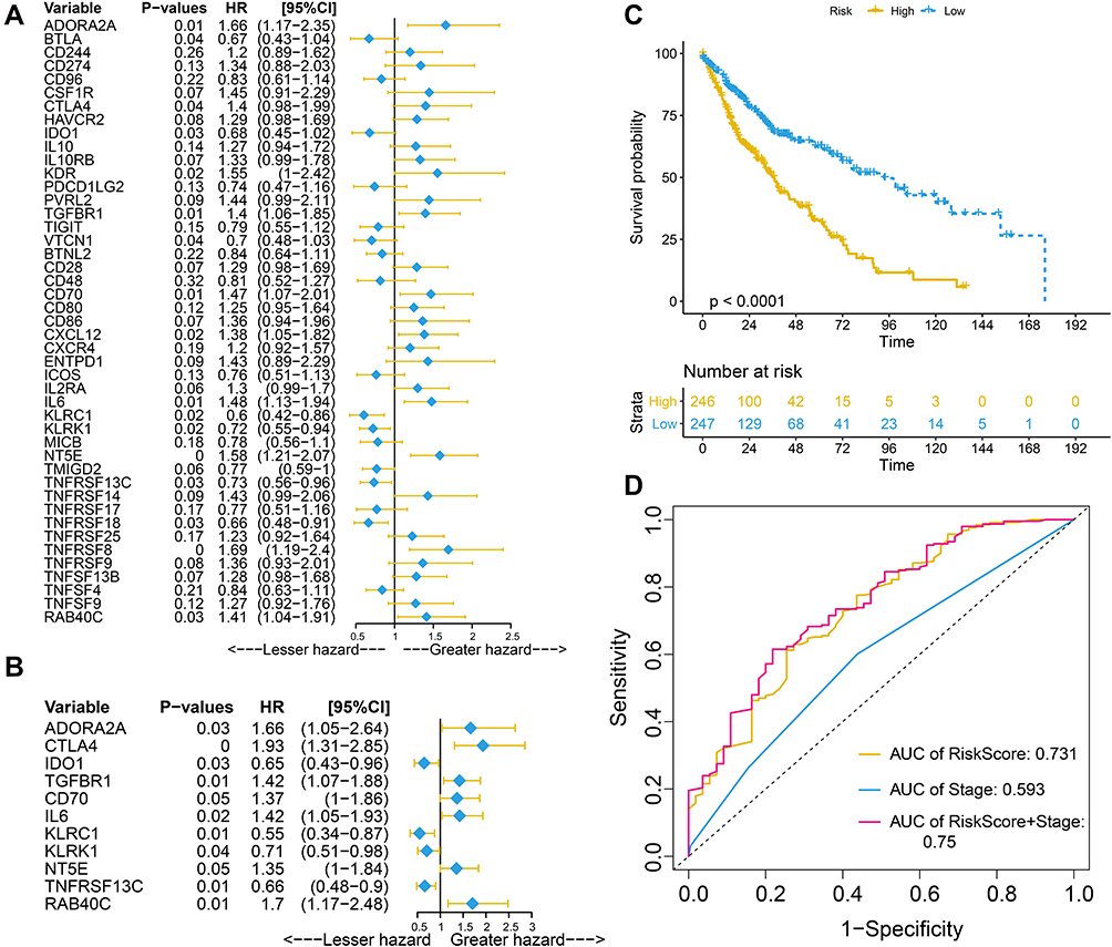 |
Figure 7 Univariate (A) and multivariate (B) Cox regression analysis. (C) Kaplan–Meier curves for LUSC regarding the risk scores. (D) Time-dependent receiver operating characteristic curves at 5 years for LUSC. |
To predict the accuracy of the prognostic model, we generated a time-dependent ROC curve. The results showed that the area under the curve (AUC) values for risk score and staging were 0.731 and 0.593, respectively. When combining stage and risk score, the AUC reached 0.75 (Figure 7D), thus showing that the model predictive efficacy was superior to staging. We investigated the distribution of risk scores, survival status, and characteristic gene expression profiles in LUSC and found that RAB40C, NT5E, IL6, CD70, TGFBR1, and ADORA2A were highly expressed in the high-risk population (Figures 8A–C). In the univariate Cox regression model, the risk model significantly correlated with survival in LUSC [hazard ratio (HR) = 2.72, 95% confidence interval (CI) = 2.17–3.41, p= 0] (Figure 8D). In a multivariate Cox regression analysis, the risk model was found to be an independent prognostic factor for LUSC by adjusting for age, sex, stage, T (size of the tumor and any spread of cancer into nearby tissue), M (metastasis), and N (spread of cancer to nearby lymph nodes) (HR = 2.9, 95% CI = 2.24–3.77, p = 0) (Figure 8E).
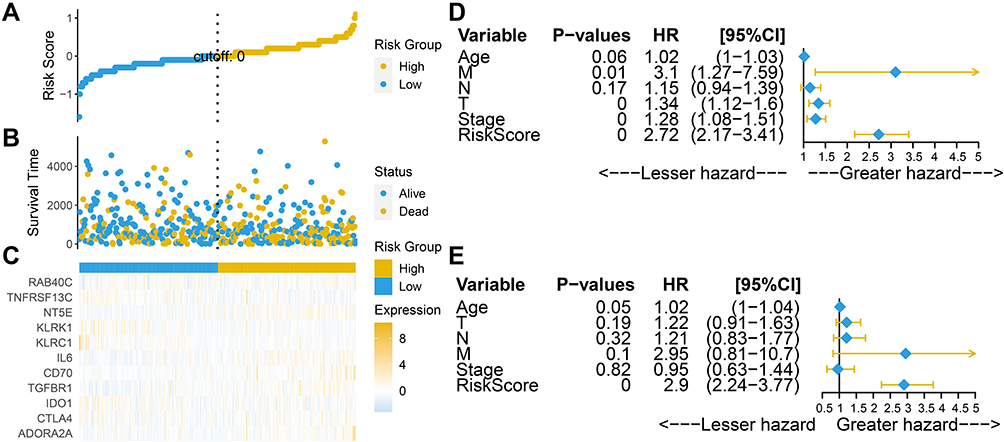 |
Figure 8 Prognostic value of risk scores in TCGA LUSC. (A–C) Distribution of risk scores and survival and gene expression profiles in LUSC. (D and E) Univariate and multivariate Cox regression analyses of risk scores for OS in LUSC. |
Construction of Nomogram
We constructed a prognostic line plot for LUSC to predict the probability of survival of individuals by weighing the risk score, stage, T, N, M, age, and sex. The line plot was calibrated and the calibration curve showed a high agreement between the probabilities predicted by the line plot (solid line) and the ideal reference line (dashed line) for 3- and 5-year survival (Figures 9A–C).
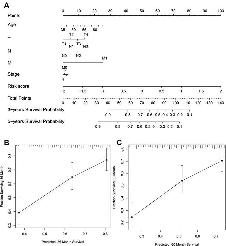 |
Figure 9 Construction of LUSC prognostic nomogram (A) Nomogram predicting the likelihood of survival at 3 and 5 years for individual LUSC patients. (B and C) Calibration curves for 3-year and 5-year survival rates for LUSC patients. Forty-five-degree dashed lines represent the high agreement between predicted and actual likelihood line plots. |
Discussion
LUSC is a major subtype of NSCLC, accounting for approximately 30% of NSCLC with high patient mortality. LUSC is often diagnosed at an advanced stage and is associated with poor clinical prognosis and a lack of clinically appropriate targeted therapies. RAB40C is a member of the Ras oncogene family that recruits ubiquitin ligase complexes. To date, RAB40C expression has been shown to be associated with a few tumors, such as osteosarcoma and breast cancer. Furthermore, only a few studies have assessed the diagnostic and prognostic potential of RAB40C in LUSC. Therefore, it was necessary to investigate the diagnostic and prognostic value of RAB40C in LUSC.
In this study, by looking for pan-cancer RAB40C expression, we found that RAB40C had significantly high expression in multiple cancers. Univariate Cox regression and Kaplan-Meier analysis showed that RAB40C is a risk factor for LUSC, PRAD, and STAD. The miRNA let-7a is a tumor suppressor, and its expression is low in gastric cancer tissues and cell lines.10 let-7a expression significantly negatively correlated with RAB40C protein levels, thus demonstrating that RAB40C has high expression levels in gastric cancer. Aberrant methylation in RAB40C is also associated with the pathogenesis of prostate cancer. Therefore, it is reasonable to consider RAB40C as a risk factor for LUSC.
Interferon-γ is a polymorphic molecule used by cancer cells to mediate the suppression of antitumor immune responses, thus exerting a pro-tumor effect. Inflammatory responses, interferon-γ responses, and other pathways are enriched upon high expression of RAB40C. Cancer immunotherapy is an effective therapeutic strategy for advanced tumors, and studying tumor-infiltrating immune cells will help find prognostic markers against these tumors.12 In this study, immune infiltrating cells, including CD4 memory-activated cells, γδ T cells, and M1-like and M2-like macrophages showed a significant negative correlation with RAB40C in various tumors. Li et al demonstrated that the proportion of γδ T cells was significantly lower in the peripheral blood of patients with advanced lung cancer. CD28 signaling plays a critical role in many T cell processes and is mainly expressed on activated T cells.13 In recent years, CD28 has received increasing attention as a key therapeutic target for several immune diseases.14 Since CD28 is expressed at low levels in peripheral blood T cells of lung cancer patients, and our results showed that CD28 negatively correlated with RAB40C in BRAC, LUAD, and LUSC, we inferred that RAB40C might be highly expressed in LUSC. Further, RAB40C acts as a ubiquitin E3 ligase responsible for RACK1 ubiquitination, and the role of RAB40C in controlling RACK1 levels is essential for cancer cell growth and T cell migration.15 Thus, the above findings imply that RAB40C might be a potential immunotherapeutic target and predictor of LUSC.
RAB40C expression had a significant negative correlation with 24 out of the 47 immune checkpoints associated with LUSC. Further, RAB40C expression positively correlated with MSI in LUSC, KICH, LUAD, and LIHC. The incidence of MSI is significantly higher in lung cancer than in normal lung tissue. Thus, a positive MSI on 3p (Chromosome 3P) is specific for lung cancer and can act as a diagnostic indicator for early lung cancer. Accordingly, as the positive association between RAB40C expression and MSI in LUSC, it stands to deduce RAB40C is inclined to be a negative factor in LUSC.
To verify that RAB40C has a strong correlation with LUSC, we compared immunohistochemistry and statistical analysis of paired lung squamous cell carcinoma and paraneoplastic patients and found a significantly high expression of RAB40C in lung squamous cell carcinoma. We then compared the immune cell infiltration levels between low and high RAB40C expression in LUSC. The abundance of CD4 memory-activated cells, M1-like macrophages, and M2-like macrophages in LUSC were significantly lower but the abundance of Tregs, natural killer cells, and T-cell follicular helpers were significantly higher when the RAB40C expression was high than when it was low. The accumulation of tumor-specific CD4+ effector T cells is essential for an effective antitumor response.16 M1-like macrophages are key effector cells for eliminating pathogens, viral infections, and cancer cells.17 Conversely, Tregs play a negative role in stimulating an effective antitumor immune response.18 Thus, the relationship between RAB40C and the corresponding immune cells shows that RAB40C is a risk factor for LUSC.
The TISIDB database was used to obtain correlations between immunomodulators and RAB40C expression in LUSC. IL-6R and its most related genes are mainly involved in tumor angiogenesis, invasion, and metastasis in LUSC, and correlation analysis showed that IL-6 and IL-6R were positively correlated in LUSC.19 Our results showed that when RAB40C was highly expressed, multiple stimulatory and inhibitory factors, including IL6, showed decreased immune responses to LUSC. Although our results differ from previous studies, they demonstrate that RAB40C is closely associated with immunomodulatory factors in LUSC, which can be explored further. To investigate the prognostic value of RAB40C-associated immunomodulators in LUSC, we performed a one-way Cox regression analysis of 44 immune genes of interest and found that an abnormal expression of RAB40C, CTLA-4, CD70, IL6, and other genes were risk factors for LUSC. CTLA-4 is involved in the negative regulation of immune response and is now considered a promising new immunosuppressive agent that has been incorporated into the standard of care for patients with advanced LUSC.20 The immune system plays a critical role in LUSC development, and the expression of immune-related genes may be an important predictor of LUSC prognosis. Liu et al constructed a risk model using four risk genes (S100P, PLAU, NOD1, and TRAV39) and accurately predicted the prognosis of LUSC patients, thus validating that the risk scores generated by the model can act as independent prognostic indicators.21 The validation of the risk model in LUSC provided us with an idea to explore the use of immune genes such as RAB40C to construct an immune-related prognostic model for LUSC. Upon analyzing the reliability and stability of our model, we found that the risk model can be an independent prognostic factor for LUSC and that the model significantly correlated with survival in LUSC. Finally, by constructing a nomogram for predicting the probability of survival of an individual, the risk score model was consistent with the actual situation and thus showed high performance. Taken together, the success of our risk model provides a stronger indication that RAB40C is a potential prognostic marker for LUSC.
However, our study also has certain limitations. First, this study was analyzed based on a public data set, and the accuracy of the results was subject to some errors. Second, there was a lack of in vivo and in vitro experiments to demonstrate the immune mechanism and prognostic value of tumors in the presence of RAB40C. Hence, without experimental validation, the authenticity of our findings was lost to some extent. Thus, we still need to explore the immunomodulatory mechanism of RAB40C in LUSC to correctly predict the prognosis of LUSC patients.
Conclusion
In conclusion, we comprehensively analyzed the expression profile and prognostic significance of RAB40C in pan-cancer and explored the correlation of RAB40C with the tumor immune microenvironment and immune checkpoints in pan-cancer. We found that immunomodulatory genes associated with RAB40C expression in LUSC independently predicted the OS in LUSC and that RAB40C was an independent prognostic biomarker in LUSC. This study showed the potential of RAB40C as a therapeutic target, laying the foundation for further molecular mechanistic studies, with the ultimate goal of using RAB40C for the clinical management of LUSC.
Data Sharing Statement
The data used to support the findings of this study are deposited in TCGA and GEO databases.
Ethics Approval and Consent to Participate
Written informed consent was acquired from all patients. This study was approved by the Ethics and Human Subject Committee of Yiwu Central Hospital. All experiments and methods were performed in accordance with relevant guidelines and regulations.
Funding
There is no funding to report.
Disclosure
The authors declare no conflicts of interest.
References
1. Li Y, Gu J, Xu F, Zhu Q, Ge D, Lu C. Transcriptomic and functional network features of lung squamous cell carcinoma through integrative analysis of GEO and TCGA data. Sci Rep. 2018;8(1):15834. doi:10.1038/s41598-018-34160-w
2. Hirsch FR, Scagliotti GV, Mulshine JL, et al. Lung cancer: current therapies and new targeted treatments. Lancet. 2017;389(10066):299–311. doi:10.1016/S0140-6736(16)30958-8
3. Lee RH, Iioka H, Ohashi M, Iemura S, Natsume T, Kinoshita N. XRab40 and XCullin5 form a ubiquitin ligase complex essential for the noncanonical Wnt pathway. EMBO J. 2007;26(15):3592–3606. doi:10.1038/sj.emboj.7601781
4. Tan R, Wang W, Wang S, et al. Small GTPase Rab40c associates with lipid droplets and modulates the biogenesis of lipid droplets. PLoS One. 2013;8(4):e63213. doi:10.1371/journal.pone.0063213
5. Tan R, Xu X, Hong W, Wang T. Analysis of biogenesis of lipid droplets by examining Rab40c associating with lipid droplets. Methods Mol Biol. 2015;1270:125–135.
6. Fukuda M. Multiple roles of VARP in endosomal trafficking: rabs, retromer components and R-SNARE VAMP7 meet on VARP. Traffic. 2016;17(7):709–719. doi:10.1111/tra.12406
7. Yatsu A, Shimada H, Ohbayashi N, Fukuda M. Rab40C is a novel Varp-binding protein that promotes proteasomal degradation of Varp in melanocytes. Biol Open. 2015;4(3):267–275. doi:10.1242/bio.201411114
8. Liu K, Huo H, Jia W, et al. RAB40C gene polymorphisms rs62030917 and rs2269556 are associated with an increased risk of lumbar disc herniation development in the Chinese Han population. J Gene Med. 2021;23(4):e3252. doi:10.1002/jgm.3252
9. Khakpour G, Noruzinia M, Izadi P, et al. Methylomics of breast cancer: seeking epimarkers in peripheral blood of young subjects. Tumour Biol. 2017;39(3):1010428317695040. doi:10.1177/1010428317695040
10. Yang Q, Jie Z, Cao H, et al. Low-level expression of let-7a in gastric cancer and its involvement in tumorigenesis by targeting RAB40C. Carcinogenesis. 2011;32(5):713–722. doi:10.1093/carcin/bgr035
11. Chen B, Khodadoust MS, Liu CL, Newman AM, Alizadeh AA. Profiling tumor infiltrating immune cells with CIBERSORT. Methods Mol Biol. 2018;1711:243–259.
12. Krucken J, Schroetel RM, Muller IU, et al. Comparative analysis of the human gimap gene cluster encoding a novel GTPase family. Gene. 2004;341:291–304. doi:10.1016/j.gene.2004.07.005
13. Esensten JH, Helou YA, Chopra G, Weiss A, Bluestone JA. CD28 costimulation: from mechanism to therapy. Immunity. 2016;44(5):973–988. doi:10.1016/j.immuni.2016.04.020
14. Xia S, Chen Q, Niu B. CD28: a new drug target for immune disease. Curr Drug Targets. 2020;21(6):589–598. doi:10.2174/1389450120666191114102830
15. Day JP, Whiteley E, Freeley M, et al. RAB40C regulates RACK1 stability via the ubiquitin-proteasome system. Future Sci OA. 2018;4(7):FSO317. doi:10.4155/fsoa-2018-0022
16. Kortekaas KE, Santegoets SJ, Sturm G, et al. CD39 Identifies the CD4(+) tumor-specific T-cell population in human cancer. Cancer Immunol Res. 2020;8(10):1311–1321. doi:10.1158/2326-6066.CIR-20-0270
17. Italiani P, Boraschi D. From monocytes to M1/M2 macrophages: phenotypical vs. functional differentiation. Front Immunol. 2014;5:514. doi:10.3389/fimmu.2014.00514
18. Yan S, Zhang Y, Sun B. The function and potential drug targets of tumour-associated Tregs for cancer immunotherapy. Sci China Life Sci. 2019;62(2):179–186. doi:10.1007/s11427-018-9428-9
19. Xu B, Chen Q, Yue C, et al. Prognostic value of IL-6R mRNA in lung adenocarcinoma and squamous cell carcinoma. Oncol Lett. 2018;16(3):2935–2948. doi:10.3892/ol.2018.9044
20. Yuan H, Liu J, Zhang J. The current landscape of immune checkpoint blockade in metastatic lung squamous cell carcinoma. Molecules. 2021;26(5):1392. doi:10.3390/molecules26051392
21. Liu Z, Wan Y, Qiu Y, et al. Development and validation of a novel immune-related prognostic model in lung squamous cell carcinoma. Int J Med Sci. 2020;17(10):1393–1405. doi:10.7150/ijms.47301
 © 2022 The Author(s). This work is published and licensed by Dove Medical Press Limited. The
full terms of this license are available at https://www.dovepress.com/terms
and incorporate the Creative Commons Attribution
- Non Commercial (unported, 3.0) License.
By accessing the work you hereby accept the Terms. Non-commercial uses of the work are permitted
without any further permission from Dove Medical Press Limited, provided the work is properly
attributed. For permission for commercial use of this work, please see paragraphs 4.2 and 5 of our Terms.
© 2022 The Author(s). This work is published and licensed by Dove Medical Press Limited. The
full terms of this license are available at https://www.dovepress.com/terms
and incorporate the Creative Commons Attribution
- Non Commercial (unported, 3.0) License.
By accessing the work you hereby accept the Terms. Non-commercial uses of the work are permitted
without any further permission from Dove Medical Press Limited, provided the work is properly
attributed. For permission for commercial use of this work, please see paragraphs 4.2 and 5 of our Terms.