Back to Journals » Clinical, Cosmetic and Investigational Dentistry » Volume 8
Alveolar bone loss in osteoporosis: a loaded and cellular affair?
Authors Jonasson G, Rythén M
Received 9 April 2016
Accepted for publication 14 May 2016
Published 13 July 2016 Volume 2016:8 Pages 95—103
DOI https://doi.org/10.2147/CCIDE.S92774
Checked for plagiarism Yes
Review by Single anonymous peer review
Peer reviewer comments 3
Editor who approved publication: Professor Christopher E. Okunseri
Grethe Jonasson,1,2 Marianne Rythén,2,3
1Department of Behavioral and Community Dentistry, Institute of Odontology, Sahlgrenska Academy, University of Gothenburg, Gothenburg, 2Research and Development Centre, Borås, 3Specialist Clinic for Pediatric Dentistry, Public Dental Service, Mölndal, Sweden
Abstract: Maxillary and mandibular bone mirror skeletal bone conditions. Bone remodeling happens at endosteal surfaces where the osteoclasts and osteoblasts are situated. More surfaces means more cells and remodeling. The bone turnover rate in the mandibular alveolar process is probably the fastest in the body; thus, the first signs of osteoporosis may be revealed here. Hormones, osteoporosis, and aging influence the alveolar process and the skeletal bones similarly, but differences in loading between loaded, half-loaded, and unloaded bones are important to consider. Bone mass is redistributed from one location to another where strength is needed. A sparse trabeculation in the mandibular premolar region (large intertrabecular spaces and thin trabeculae) is a reliable sign of osteopenia and a high skeletal fracture risk. Having dense trabeculation (small intertrabecular spaces and well-mineralized trabeculae) is generally advantageous to the individual because of the low fracture risk, but may imply some problems for the clinician.
Keywords: bone density, bone fracture, human, mandible, radiography, periodontitis
Introduction
A large proportion of the population visit their dentist annually, and dental radiographs are routinely taken then. Dentists are highly experienced in interpreting radiographs, but use them chiefly for the diagnosis of caries, marginal and apical periodontitis, and before implant treatment. However, there is much more information available in periapical radiographs. Therefore, many research teams have tried to develop methods for using the jawbones to predict osteoporosis 1–4 and fracture risk.5–9
Possible links between osteoporosis and the degree of periodontitis have been debated for years. Both diseases are multifactorial and have many risk factors in common. The aims of the present article are to describe parallels between skeletal bone and alveolar bone and to illuminate some aspects of hormone function, loading and aging, which may explain alveolar bone loss caused by osteoporosis and/or periodontitis.
Bone
Bone is a dynamic tissue. During childhood and adolescence, the formation of bone dominates over the resorption of bone, this is the modeling phase. In mature adults, there is a balance between bone formation and resorption, whereas bone resorption dominates after menopause, and in older males. Skeletal loading produces microcracks in bone, which are replaced by remodeling.10 The remodeling process is regulated systematically by hormones and locally by growth factors and cytokines.11,12 It takes place at the endosteal surfaces of cortical and trabecular bone.13 At first, osteoclasts identify a site for renewal and start resorption. After ∼60 days, osteoblasts initiate bone formation. Although it takes 2 weeks to deposit ∼60% of the mineral content of new bone, full mineralization takes several months. Remodeling occurs at ∼1–2 million sites.11 The osteocytes are imbedded in the mineralized bone; their role is to sense bone strains and communicate with neighboring osteoblasts and osteoclasts.
All bones are constructed with an envelope of compact bone (cortical bone), which encloses the trabecular bone and marrow spaces. The ratio between compact and trabecular bone varies with ∼10% compact and 90% trabecular bone in both the spine and maxilla, whereas the ratio in the mandible is close to that of the distal radius and other long bones: 80% compact and 20% trabecular bone.1 Because of its construction as a network of trabeculae, plates and rods, trabecular bone has a total surface ten times larger than compact bone. Consequentially, it has more endosteal surfaces, more cells, and more remodeling.14
Bone turnover speed has been examined in adult dogs, where alveolar, trabecular bone remodeling in the mandible is double that in the maxilla (37%/year versus 19%/year) and six times faster than in the femur (6.4%/year).15 In addition, the rate of alveolar bone turnover at the alveolar crest is twice the rate of bone turnover at the level of the mandibular canal and three to five times the rate of the mandible at the inferior compact border.16 No exact remodeling rate has been established for human beings, but alveolar, trabecular bone remodels very quickly, especially in the mandible. The remodeling rate was estimated by the number of formation and resorption foci per area in autopsy materials.1
Hormones
Delayed puberty decreases bone mineral density (BMD) and spinal growth. Males with late puberty have longer legs and lower bone density than those with early puberty.17 Males retain hormone protection throughout life and suffer half as many fractures as females.18 In females, early menarche and early menopause are considered to have a strong relationship with high and low BMD, respectively.19 Early menarche is associated with a significant decrease in fracture risk, whereas delayed menarche, as seen in amenorrheic athletes, increases fracture risk.20 Age-related (nonsex-hormone dependent) bone loss may begin in the third decade of life in both sexes at the spine and a decade later at the appendicular sites. With aging, compact bone strength is diminished by approximately 15%–20% and trabecular bone strength by ∼50%.10
The estrogen-related bone loss in females takes place predominantly in the trabecular bone with its larger endosteal surfaces, followed by a slower loss of both trabecular and cortical bone. Decreasing levels of estrogen are thought to be responsible for increased bone resorption and decreasing testosterone levels for decreased bone formation.11,21 Testosterone is associated with bone apposition periosteally even in aged males, and, therefore, what is lost endosteally may be compensated periosteally in males, but not in females.22 In Figure 1, the large variations of bone loss and gain in 80 years olds are shown. Figure 1A illustrates dense trabecular bone and extremely thick basal compact bone in an old male. Figure 1B demonstrates the sparser trabeculation, larger intertrabecular spaces, and the thin, eroded basal bone in an old female. In Figure 1C, the extremely resorbed mandible in an edentulous female, also 80 years old, is presented. Age is strongly correlated with BMD in females but not in males, whereas bone size is more correlated with BMD in males than females.23
 | Figure 1 The three panoramic radiographs show the large variation in bone mass, trabeculation, and basal cortex in persons 79- or 80-years-old. Notes: (A) Shows a male with dense trabecular bone and thick basal compacta, (B) a female with sparse trabecular bone and thin eroded compacta, and (C) an old edentulous female with extremely resorbed alveolar process. |
Hormones influence the jaws and the rest of the skeleton to the same extent but with dissimilarities concerning loading, which, besides genetic factors and hormones, is the strongest factor influencing bone density.11,21
Loading
Weight-bearing physical activities increase skeletal bone mass,24 whereas space flight decreases bone mass.25 Increased loading leads to periosteal apposition (on the outer surface of compact bone), thereby increasing the cross-sectional area in the vertebrae26 and long bones.27 The legs and spine are “loaded” bones, the arms and the mandible half-loaded, and the skull relatively unloaded. As an example, weight-lifters have 3% higher BMD for the total body and 12% for the hip, whereas BMD for the upper part of the skull was 10% lower than in controls.28 Furthermore, after hip fracture, BMD decreases in legs and spine, remains the same in the arms, and increases in the skull.29
Exercise by the elderly (>65 years) is associated with improved muscle strength, coordination, balance, and decreased fall frequency, and there is an association between continued physical activity throughout life and lower hip fracture risk.20
Effects of loading are seen in the same way in the jaws, where the alveolar bone mass and the cross-sectional dimension of the alveolar bone in growing rats increase with increasing functional loading.30,31 Bone mass can be redistributed to the most loaded sites to fortify the bone where it is most needed. When vertical trabeculae are resorbed, the horizontal trabeculae may be fortified (Figure 2). Bone around human molars is generally denser than the bone around premolars and canines, which can be explained by the findings that the highest biting force is recorded in the molar area.32
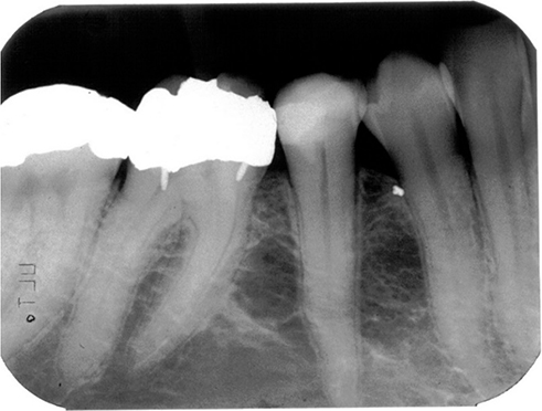 | Figure 2 The radiograph is of a 48-year-old osteoporotic female. The trabecular network is disrupted in two locations around the second premolar. Notes: The vertical trabeculae have disappeared, whereas the horizontal ones have been reinforced. |
After tooth extraction, reduced function leads to local bone loss,33,34 and great interindividual variation in the remodeling pattern of the edentulous areas, with some individuals losing little bone, and others undergoing extensive resorption (Figure 1C).35–37 However, no association, or only a weak one, has been found between skeletal bone mass and residual ridge resorption.37–39
Local factors, such as occluding tooth pairs and the size of the masseter muscles, influence the distal area of the mandible. In patients with heavy occlusion, bruxism, the jaw bone may be denser than the skeletal bones, and therefore symptoms of skeletal osteopenia may become masked in the jaws.40 However, if trabeculation is sparse in areas with occluding teeth (areas under bridge pontics excluded), it is an indication that something is “wrong”, bone formation may be impaired, and fracture risk increased.41,42 Thus, mandibular bone often reflects the condition of the skeleton but differences in loading should be considered.
Bone size and alveolar process width
In females, the mandibular alveolar bone thickness is correlated with BMD.43 The alveolar bone is mostly thicker in the apical part in females with normal BMD compared to the crestal part, whereas it is significantly thinner in osteopenic and osteoporotic females.43
A decreased bone size with time seems to be specific to the alveolar process, as it has not been recorded in any other bone. Not only has it been seen in edentulous regions35,36 but also in dentate areas,44 where the largest decrease after 5 years was found in perimenopausal females. A decreased buccolingual dimension in the dentate alveolar process may be caused by periosteal resorption of this area, and the largest size changes correspond to areas where resorption was most evident in the modeling process during mandibular growth in young individuals.45
Skeletal bone loss/osteoporosis
Osteoporosis occurs when bone mass decreases faster than it is replaced. It is a multifactorial disease characterized by low bone mass and deterioration of bone microarchitecture, leading to bone fragility and a subsequent increase in fracture risk.46 Osteoporosis may be the result of a deficiency of sex hormone, hyperparathyroidism, hyperthyroidism, chronic renal failure, posttransplantation, or medication with glucocorticosteroids.46
Fracture risk increases exponentially with age, due not only to a decrease in BMD but also to the increased rate of falls among the elderly. Relatively, osteoporotic females have more fractures than nonosteoporotic, but up to 70% of all fractures, in absolute numbers, occur in osteopenic females.47 In a 15-year follow-up, the best predictors of future fracture were a previous fracture and glucocorticoid medication followed by alveolar bone texture, rheumatoid arthritis, gastrointestinal disease, and secondary osteoporosis.7 All variables, except alveolar bone texture, are identical with those identified by the large meta-analyses, on which the World Health Organization Fracture Risk Assessment Tool is based.48 Also included in the Fracture Risk Assessment Tool are age, sex, height, weight, smoking, alcohol, and parents with fractured hip.48
Osteoporosis affects ∼75 million people in the Western world, causing >2.3 million fractures a year in Europe and the US.48 Osteoporotic fractures lead to a high morbidity and mortality rate, and BMD predicts survival for subjects over 70 years of age.49 Bone strength depends on the degree of mineralization, bone size, and microstructural features, such as relative trabecular volume, trabecular spacing, and connectivity.50
Measurements of compact bone in the mandible for assessment of osteoporosis
Measurements of bone mass and density in the mandible have been performed since the 1980s1 with different techniques, but most are not useful in the dental clinic, being too complicated, costly, or having an excessively high radiation dose. The dual X-ray absorptiometry method, which is the gold standard for diagnosis of osteoporosis, has low radiation, but for the jawbones, it is only applicable in edentulous individuals, and therefore other methods have been developed.
Maxillary bone consists mostly of trabecular bone, and compact bone is too thin for use as an osteoporosis indicator. Maxillary trabecular bone has been assessed but not as frequently as the mandible due to the difficulty of finding a standard site.51,52
The largest proportion of mandibular compact bone is situated in the inferior cortex, which is well imaged on panoramic radiographs. The mandibular cortical index (MCI) is the most frequently used method in osteoporosis studies.53–56 Compact bone lying distal to the mental foramen is categorized by three groups (Figure 3): normal cortex (MCI-1) having a relatively even endosteal margin; moderately eroded cortex (MCI-2) with semilunar defects, and severely eroded cortex (MCI-3) with heavy endosteal porosities. A severely eroded compacta is associated with osteoporosis,5,55–57 but not consistently with fracture.8,57 Compact bone loss is seen ∼20 years later than trabecular bone loss, which can be seen in females as young as 38 years old.8,9
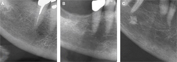 | Figure 3 Visual index for assessment of cortical shape. Notes: Reference images presenting dense trabeculation and a normal mandibular cortex with even and sharp endosteal margin (A), mixed trabeculation, and a moderately eroded cortex with endosteal margin showing semilunar defects (B), sparse trabeculation, and severely eroded cortex, with the cortical layer being clearly porous (C). |
The thickness of the basal compacta increases up to the age of 50 years and decreases significantly thereafter.8 A cortex thickness <3 mm is associated with osteoporosis57–60 but not fracture.8,57 The severely eroded inner cortex in MCI-3 creates difficulties when measuring cortex thickness. Therefore, a computer-based method has been developed.60
Measurements of trabecular bone in the mandible for assessment of osteoporosis
Mandibular trabecular bone becomes denser in the jaws from puberty to middle age,61 thereafter, alveolar trabecular bone becomes sparser in most females,1,8,62 whereas males more often maintain their trabecular pattern (Figure 1A and B).
Trabecular bone structure can be assessed on radiographs by the thickness of the trabeculae, the spacing between the trabeculae, trabecular connectivity,62–64 and by measuring trabecular volume by computed tomography and magnetic resonance. However, the cost and complexity of these methods limit their utility for routine use.63–66 Therefore, a simple three-step visual index has been introduced. It was initially meant for bone evaluation before implant treatment,67 but the index has been proven a valuable indicator for osteoporosis risk4,23,41,42,51 and for fracture risk assessment.6–9
The index classifies the mandibular premolar bone, which is the standard site, as having either sparse, mixed dense plus sparse, or dense trabecular bone (Figure 4).41,67 Sparse trabeculation has large intertrabecular spaces in most of the alveolar processes, especially in the crestal, dentate, and premolar area. Dense trabeculation has small intertrabecular spaces and well-mineralized trabeculae in the entire radiographed area. Mixed dense plus sparse trabeculation is mostly dense crestally and sparse apically. In case of uncertainty, the mixed category is chosen. Most important is identifying individuals with sparse trabeculation because of their high fracture risk. Dense trabeculation is protective. Most individuals have mixed dense and sparse trabeculation in the mandible; BMD varies greatly in this trabeculation group and fracture risk is moderate. The older the participants, the better the fracture prediction.8,9
 | Figure 4 Visual index for assessment of trabecular bone. Notes: Reference images presenting the trabecular pattern as sparse trabeculation in females with: large intertrabecular spaces (A); mixed dense plus sparse trabeculation with small intertrabecular spaces cervically and larger spaces more apically (B); and dense trabeculation with small intertrabecular spaces (C). |
The Kaplan–Meier curve in Figure 5 shows the graded association between trabecular density and fracture risk.9 All participants start free of fracture. The upper curve describing dense trabeculation remains relatively stable, which means ∼90% of those with dense trabeculation stay free of fracture during the period 1980–2006, whereas the lowest curve describing sparse trabeculation dives distinctly; only 25% remained fracture free. The curve in between for mixed dense plus sparse also dives, illustrating ∼62% free of fracture at the end of the 26-year follow-up. The hazard ratio of future fracture for sparse trabeculation compared to mixed trabeculation was 2.9 (95% CI: 2.2–3.8, P<0.0001) and for dense versus mixed trabeculation 0.21 (95% CI: 0.1–0.4, P<0.0001).9
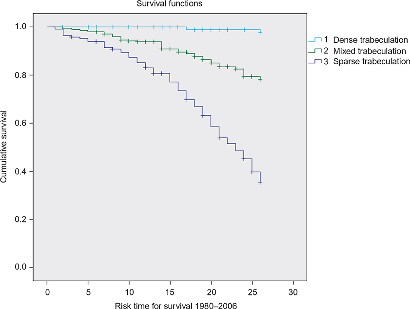 | Figure 5 Kaplan–Meier survival curve showing cumulative “fracture” survival and risk time for fracture in three different trabeculation groups. Notes: 1) Survival of women with dense trabeculation, 2) mixed trabeculation, and 3) sparse trabeculation. Risk time represents the time interval between baseline assessment and fracture event. All participants included (n=518), started “fracture-free” at baseline (1980) and experienced 136 first, incident fractures during the period 1980-2006. |
When the visual trabecular pattern was tested in males, it was not a significant factor for the prediction of osteoporosis,23 but significant for fracture prediction.6
Trabeculation does not change visually after 5 years,42 but radiographs of females performed after 12 and 24 years show a gradual transformation with increased intertrabecular spaces and less mineralized trabeculae.8,9 Similar bone changes to those that could be seen after 12 years in the mandible could be measured in the radius after 1 year using three-dimensional, high-resolution, peripheral quantitative computed tomography.6 In females (mean age 77 years), total density and trabecular number decreased, while trabecular thickness, separation, and heterogeneity increased.68
Both the trabecular visual index and the automated methods work best on intraoral radiographs, which are most widely used in dental practices. With training, the visual trabecular index can be used on panoramic radiographs but the noise level is larger.69
Periodontitis
Periodontitis is induced by specific bacteria from biofilms on tooth surfaces, which triggers an immunoinflammatory response in the adjacent bone tissue. Bacteria are required to initiate the disease process where host proteolytic enzymes mediate direct destruction of the periodontal tissue.70 The progression of periodontitis is influenced by factors such as genetics, general health, smoking, and diet. Furthermore, education and socioeconomic factors are determinants.70 Severe periodontitis is linked to some systemic diseases, for example, diabetes mellitus,71–73 cardiovascular disease,74–76 rheumatoid arthritis,77 and adverse pregnancy outcomes.78
Periodontitis and osteoporosis both progress with advancing age, smoking, estrogen deficiency, and family history.79 Current knowledge regarding the effects of osteoporosis or osteopenia on periodontal disease and alveolar bone loss is inconclusive.12,80–91 Some previous studies have indicated a relationship between periodontal disease and osteoporosis,83–87 while others have not shown any significant relationship.88,89 The lack of consistency in the results may be due to differences in the alveolar bone structure and thickness, which were not considered. This could be supported by the fact that individuals with high mineral levels in the skeleton seem to retain their teeth with deep periodontal pockets more easily than those with osteoporosis.91 Furthermore, individuals with broad, dense jaws have more bone substances to lose than those with thin jaws, and therefore the size of an individual may play an important role for the vertical changes of the alveolar process.91
The multifactorial etiology and confounding factors have limited the understanding of the relationship between osteoporosis and periodontitis. The coarseness of trabeculation may be one such confounding factor. In a new report, the group with sparse trabeculation had more skeletal bone loss, lower BMD, and larger marginal bone level decrease after 5 years than those with not-sparse trabeculation, but surprisingly the little group with dense trabeculation experienced both the greatest 5-year skeletal bone loss and marginal bone level decrease.92 The reason may be that dense trabeculation implies the greatest trabecular endosteal bone surface, and consequently the largest number of bone cells and remodeling sites. Negative events may lead to the most negative responses in dense trabecular bone but research is warranted to clarify whether dense trabeculation leads to the best results after appropriate treatment?
Estrogen deficiency influences BMD,21,93,94 and the remodeling of the alveolar process negatively, whereas treatment with hormone replacement therapy ameliorates the periodontal condition and local bone mass.85,95
Patients with cancer and bisphosphonate-related osteonecrosis of the jaw have significantly fewer teeth, and lower average bone height compared to controls after adjusting for the number of bisphosphonate infusions.96 The antiresorptive effect of bisphosphonates is therapeutic when used in treatment of osteoporosis for a limited period, but the problem is the extremely long elimination time due to large accumulations of bisphosphonate in bone, diminishing bone turnover and preventing bone renewal.16
Clinical relevance
To maintain quality of life for the elderly, targeting individuals with high fracture risk is an important challenge to the dentist. An assessment of the trabecular bone can easily be included in an annual examination.
The trabeculation pattern is of direct interest to the dental profession, since a dense trabeculation indicates a need to exercise prudence when drilling for implants because of increased heating and consequently increased risk of local necrosis.97 A periapical radiograph revealing sparse trabeculation may indicate a need for cortical fixation and long duration of the healing process before the implant can be loaded.98
In adult orthodontics, a different tissue reaction could be expected when teeth are moved with sparse or dense trabeculation. This could be supported by the findings that rats with lower initial bone density have a faster orthodontic tooth movement than rats with significantly higher initial bone density.99 Orthodontic tooth movement is faster in lactating rats on a calcium-deficient diet than in rats on a normal diet.100 Dense trabeculation implies that extraction and surgical extirpation of wisdom teeth are more complicated and probably more difficult to obtain full anesthesia.
Because dentists are in an ideal position to assess oral bone loss on radiographs, they may be the first to discover early signs of osteoporosis. Identifying individuals with high fracture risk before the first fracture occurs is important in order to avoid suffering and minimize high costs to society. If these patients can be easily identified by a dentist, they can improve their bone quality through training and nutrition, and appropriate medication for older people can be prescribed.
Disclosure
The authors report no conflicts of interest in this work.
References
Von Wowern N. Bone mass of mandibles. In vitro and in vivo analyses. Dan Med Bull. 1986;33:23–44. | ||
Kribbs PJ, Chesnut CH, Ott SM, Kilcoyne RF. Relationships between mandibular and skeletal bone in an osteoporotic population. J Prosthet Dent. 1989;62:703–707. | ||
Southard KA, Southard TE, Schlechte JA, Meis PA. The relationship between the density of the alveolar process and that of post-cranial bone. J Dent Res. 2000;79:964–969. | ||
Lindh C, Horner K, Jonasson G, et al. The use of visual assessment of dental radiographs for identifying women at risk of having osteoporosis: the OSTEODENT project. Oral Surg Oral Med Oral Pathol Oral Radiol Endod. 2008;106:285–293. | ||
Bollen AM, Taguchi A, Hujoel PP, Hollender LG. Case-control study on self-reported osteoporotic fractures and mandibular cortical bone. Oral Surg Oral Med Oral Pathol Oral Radiol Endod. 2000;90:518–524. | ||
Hassani-Nejad A, Ahlqwist M, Hakeberg M, Jonasson G. Mandibular trabecular bone as fracture indicator in 80-year-old men and women. Eur J Oral Sci. 2013;121:525–531. | ||
Jonasson G, Billhult A. Mandibular bone structure, bone mineral density, and clinical variables as fracture predictors: a 15-year follow-up of female patients in a dental clinic. Oral Surg Oral Med Oral Pathol Oral Radiol. 2013;116:362–368. | ||
Jonasson G, Sundh V, Hakeberg M, Hassani-Nejad A, Lissner L, Ahlqwist M. Mandibular bone changes in 24 years and skeletal fracture prediction. Clin Oral Investig. 2013;17:565–572. | ||
Jonasson G, Sundh V, Ahlqwist M, Hakeberg M, Björkelund C, Lissner L. A prospective study of mandibular trabecular bone to predict fracture incidence in women: a low-cost screening tool in the dental clinic Bone. 2011;49:873–879. | ||
Martin B. Aging and strength of bone as a structural material. Calcif Tissue Int. 1993;53:S34–S40. | ||
Lerner UH. Bone remodeling in post-menopausal osteoporosis. J Dent Res. 2006;85:584–595. | ||
Reddy MS, Morgan SL. Decreased bone mineral density and periodontal management. Periodontol 2000. 2013;61:195–218. | ||
Seeman E, Delmas PD. Bone quality – the material and structural basis of bone strength and fragility. N Engl J Med. 2006;354:2250–2261. | ||
Kanis JA. Osteoporosis. Oxford: Blackwell Science Ltd; 1994. | ||
Huja SS, Fernandez SA, Hill KJ, Li Y. Remodeling dynamics in the alveolar process in skeletally mature dogs. Anat Rec Discov Mol Cell Evol Biol. 2006;288:1243–1249. | ||
Marx RE, Cillo Jr JE, Ulloa JJ. Oral bisphosphonate-induced osteonecrosis: risk factors, prediction of risk using serum CTX testing, prevention, and treatment. J Oral Maxillofac Surg. 2007;65:2397–2410. | ||
Lorentzon M, Norjavaara E, Kindblom JM. Pubertal timing predicts leg length and childhood body mass index predicts sitting height in young adult men. J Pediatr. 2011;158:452–457. | ||
SBU. Osteoporosis - prevention, diagnosis and treatment. Stockholm: Swedish Council on Health Technology Assessment in Health Care (SBU); SBU report no 165/1; 2003. | ||
Ito M, Yamada M, Hayashi K, Ohki M, Uetani M, Nakamura T. Relation of early menarche to high bone mineral density. Calcif Tissue Int. 1995;57:11–14. | ||
Karlsson M, Bass S, Seeman E. The evidence that exercise during growth or adulthood reduces the risk of fragility fractures is weak. Best Pract Res Clin Rheumatol. 2001;15:429–450. | ||
Riggs BL, Khosla S, Melton III LJ. Sex steroids and the construction and conservation of the adult skeleton. Endocrin Rev. 2002;23:279–302. | ||
Ulm CW, Solar P, Ulm MR, Matejka M. Sex-related changes in the bone mineral content of atrophic mandibles. Calcif Tissue Int. 1994;54:203–207. | ||
Jonasson G, Jonasson L, Kiliaridis S. Skeletal bone mineral density in relation to thickness, bone mass, and structure of the mandibular alveolar process in dentate men and women. Eur J Oral Sci. 2007;115:117–123. | ||
Lanyon LE. Functional strain in bone tissue as an objective, and controlling stimulus for adaptive bone remodelling. J Biomech. 1987;20:1083–1093. | ||
Lang TF. What do we know about fracture risk in long-duration spaceflight? J Musculoskelet Neuronal Interact. 2006;6:319–321. | ||
Mosekilde L. Age-related changes in bone mass, structure, and strength – effects of loading. Z Rheumatol. 2000;59(Suppl 1):1–9. | ||
Ruff CB, Hayes WC. Sex differences in age-related remodeling of the femur and tibia. J Orthop Res. 1988;6:886–896. | ||
Karlsson MK, Hasserius R, Obrant KJ. Bone mineral density in athletes during and after career: a comparison between loaded and unloaded skeletal regions. Calcif Tissue Int. 1996;59:245–248. | ||
Magnusson H, Obrant K, Johnell O, Karlsson KM. Bone mass changes in weight- loaded and unloaded skeletal regions following a fracture of the hip. Osteoporos Int. 2001;69:78–83. | ||
Bresin A, Johansson CB, Kiliaridis S. Effects of occlusal strain on the development of the dentoalveolar process in the growing rat. Eur J Exp Musculoskel Res. 1994;3:112–122. | ||
Kiliaridis S, Bresin A, Holm J, Strid KG. Effects of masticatory muscle function on bone mass in the mandible of the growing rat. Acta Anat. 1996;155:200–205. | ||
Erhardson S, Sheikholeslam A, Forsberg C-M, Lockowandt P. Vertical forces developed by the jaw elevator muscles during unilateral maximal clenching and their distribution on teeth and condyles. Swed Dent J. 1993;17:23–34. | ||
Elovic RP, Hipp JA, Hayes WC. Maxillary molar extraction causes increased bone loss in the mandible of ovariectomized rats. J Bone Miner Res. 1995;10:1087–1093. | ||
Von Wowern N, Hjorting-Hansen E, Stoltze K. Changes in bone mass in rat mandibles after tooth extraction. Int J Oral Surg. 1979;8:229–233. | ||
Carlsson GE, Persson G. Morphologic changes of the mandible after extraction and wearing of dentures; a longitudinal, clinical, and X-ray cephalometric study covering 5 years. Odontol Revy. 1967;18:27–54. | ||
Tallgren A. The continuing reduction of the residual alveolar ridges in complete denture wearers: a mixed-longitudinal study covering 25 years. J Prosthet Dent. 1972;27:120–132. | ||
Von Wowern N, Kollerup G. Symptomatic osteoporosis: a risk factor for residual ridge reduction of the jaws. J. Prosthet Dent. 1992;67:656–660. | ||
Klemetti E. Review of residual ridge resorption and bone density. J Prosthet Dent. 1996;75:512–514. | ||
Klemetti E, Kröger H, Lassila V. Relationships between body mass index and the remaining alveolar ridge. J Oral Rehab. 1997;24:808–812. | ||
Jonasson G, Kiliaridis S. The association between the masseter muscle, the mandibular alveolar bone mass and thickness in dentate women. Arch Oral Biol. 2004;49:1001–1006. | ||
Jonasson G, Bankvall G, Kiliaridis S. Estimation of skeletal bone mineral density by means of the trabecular pattern of the alveolar bone, its interdental thickness, and the bone mass of the mandible. Oral Surg Oral Med Oral Pathol Oral Radiol Endod. 2001;92:346–352. | ||
Jonasson G, Jonasson L, Kiliaridis S. Changes in the radiographic characteristics of the mandibular alveolar process in dentate women with varying bone mineral density: a 5-year prospective study. Bone. 2006;38:714–721. | ||
Jonasson G, Kiliaridis S, Gunnarsson R. Cervical thickness of the mandibular alveolar process and skeletal bone mineral density. Acta Odontol Scand. 1999;57:155–161. | ||
Jonasson G, Kiliaridis S. Changes in the bucco-lingual thickness of the mandibular alveolar process and skeletal bone mineral density in dentate women: a 5-yr prospective study. Eur J Oral Sci. 2005;113(2):114–120. | ||
Enlow DH, Harris DB. A study of the postnatal growth of the human mandible. Am J Orthodont. 1964;50:25–50. | ||
WHO Scientific Group. Prevention and management of osteoporosis. WHO Technical Report Series No 921, Geneva: WHO Press; 2003. | ||
Pasco JA, Seeman E, Henry MJ, Merriman EN, Nicholson GC, Kotowicz MA. The population burden of fractures originates in women with osteopenia, not osteoporosis. Osteoporos Int. 2006;17:1404–1409. | ||
Kanis JA, Johnell O, Oden A, Johansson H, McCloskey E. FRAXTM and the assessment of fracture probability in men and women from the UK. Osteoporos Int. 2008;19:385–397. | ||
Johansson C, Black D, Johnell O, Odén A, Mellström D. Bone mineral density is a predictor of survival. Calcif Tissue Int. 1998;63:190–196. | ||
Genant HK, Jiang Y. Advanced imaging assessment of bone quality. Ann N Y Acad Sci. 2006;1068:410–428. | ||
Lindh C, Obrant K, Petersson A. Maxillary bone mineral density and its relationship to the bone mineral density of the lumbar spine and hip. Oral Surg Oral Med Oral Pathol Oral Radiol Endod. 2004;98:102–109. | ||
Southard KA, Southard TE. Detection of simulated osteoporosis in human anterior maxillary alveolar bone with digital subtraction. Oral Surg Oral Med Oral Pathol. 1994;78:655–661. | ||
Klemetti E, Kolmakov S, Kröger H. Pantomography in assessment of the osteoporosis risk group. Scand J Dent Res. 1994;102:68–72. | ||
Taguchi A, Asano A, Ohtsuka M, et al. Observer performance in diagnosing osteoporosis by dental panoramic radiographs: results from the osteoporosis screening project in dentistry (OSPD). Bone. 2008;43:209–213. | ||
Taguchi A, Tsuda M, Ohtsuka M, et al. Use of dental panoramic radiographs in identifying younger postmenopausal women with osteoporosis. Osteoporos Int. 2006;17:387–394. | ||
White SC, Taguchi A, Kao D, et al. Clinical and panoramic predictors of femur bone mineral density. Osteoporos Int. 2005;16:339–346. | ||
Okabe S, Morimoto Y, Ansai T, et al. Assessment of the relationship between the mandibular cortex on panoramic radiographs and the risk of bone fracture and vascular disease in 80-year-olds. Oral Surg Oral Med Oral Pathol Oral Radiol Endod. 2008;106:433–442. | ||
Devlin H, Karayianni K, Mitsea A, et al. Diagnosing osteoporosis by using dental panoramic radiographs: the OSTEODENT project. Oral Surg Oral Med Oral Pathol Oral Radiol Endod. 2007;104:821–828. | ||
Horner K, Devlin H, Harvey L. Detecting patients with low skeletal bone mass. J Dent. 2002;30:171–175. | ||
Devlin H, Allen PD, Graham J, et al. Automated osteoporosis risk assessment by dentists: a new pathway to diagnosis. Bone. 2007;40:835–842. | ||
Pham D, Kiliaridis S. Evaluation of changes in trabecular alveolar bone during growth using conventional panoramic radiographs. Acta Odontol Scand. 2012;70:127–132. | ||
White SC, Rudolph DJ. Alterations of the trabecular pattern of the jaws in patients with osteoporosis. Oral Surg Oral Med Oral Pathol Oral Radiol Endod. 1999;88:628–635. | ||
White SC, Atchison KA, Gornbein JA, et al. Change in mandibular trabecular pattern and hip fracture rate in elderly women. Dentomaxillofac Radiol. 2005;34:168–174. | ||
Verheij JG, Geraets WG, van der Stelt PF, et al. Prediction of osteoporosis with dental radiographs and age. Dentomaxillofac Radiol. 2009;38:431–437. | ||
Chiba K, Burghardt AJ, Osaki M, Majumdar S. Heterogeneity of bone microstructure in the femoral head in patients with osteoporosis: an ex vivo HR-pQCT study. Bone. 2013;56:139–146. | ||
Geraets WG, Verheij JG, van der Stelt PF, et al. Prediction of bone mineral density with dental radiographs. Bone. 2007;40:1217–1221. | ||
Lindh C, Petersson A, Rohlin M. Assessment of the trabecular pattern before endosseous implant treatment: diagnostic outcome of periapical radiography in the mandible. Oral Surg Oral Med Oral Pathol Oral Radiol Oral Endod. 1996;82:335–343. | ||
Kawalilak CE, Johnston JD, Olszynski WP, Kontulainen SA. Characterizing microarchitectural changes at the distal radius and tibia in postmenopausal women using HR-pQCT. Osteoporos Int. 2014;25:2057–2066. | ||
Pham D, Jonasson G, Kiliaridis S. Assessment of trabecular pattern on periapical and panoramic radiographs: a pilot study. Acta Odontol Scand. 2010;68:91–97. | ||
Bartold PM, Van Dyke TE. Periodontitis: a host-mediated disruption of microbial homeostasis. Unlearning learned concepts. Periodontol 2000. 2013;62:203–217. | ||
Santos Tunes R, Foss-Freitas MC, Nogueira-Filho GR. Impact of periodontitis on the diabetes-related inflammatory status. J Can Dent Assoc. 2010;76:a35. | ||
Koromantzos PA, Makrilakis K, Dereka X, Katsilambros N, Vrotsos IA, Madianos PN. A randomized, controlled trial on the effect of non-surgical periodontal therapy in patients with type 2 diabetes. J Clin Periodontol. 2011;38:142–147. | ||
Chapple IL, Genco R, working group 2 of the joint EFP/AAP workshop. Diabetes and periodontal diseases: consensus report of the Joint EFP/AAP Workshop on Periodontitis and Systemic Diseases. J Periodontol. 2013;84:S106–S112. | ||
Buhlin K, Gustafsson A, Pockley AG, Frostegård J, Klinge B. Risk factors for cardiovascular disease in patients with periodontitis. Eur Heart J. 2003;24:2099–2107. | ||
Belstrøm D, Damgaard C, Nielsen CH, Holmstrup P. Does a causal relation between cardiovascular disease and periodontitis exist? Microbes Infect. 2012;14:411–418. | ||
Tonetti MS, Van Dyke TE. Periodontitis and atherosclerotic cardiovascular disease: consensus report of the joint EFP/AAP workshop on periodontitis and systemic diseases. J Periodontol. 2013;84:24–29. | ||
Kaur S, White S, Bartold PM. Periodontal disease and rheumatoid arthritis: a systematic review. J Dent Res. 2013;92:399–408. | ||
Sanz M, Kornman K. Working group 3 of the joint EFP/AAP workshop. periodontitis and adverse pregnancy outcomes: consensus report of the joint EFP/AAP workshop on periodontitis and systemic diseases. J Periodontol. 2013;84:S164–S169. | ||
Lerner UH. Inflammation-induced bone remodeling in periodontal disease and the influence of post-menopausal osteoporosis. J Dent Res. 2006;85:596–607. | ||
Chesnut CH 3rd. The relationship between skeletal and oral bone mineral density: an overview. Ann Periodontol. 2001;6:193–196. | ||
Jeffcoat M. The Association between osteoporosis and oral bone loss. J Periodontol. 2005;76:2125–2132. | ||
Otomo-Corgel J. Osteoporosis and osteopenia: implications for periodontal and implant therapy. Periodontol. 2000. 2012;59:111–139. | ||
Hildebolt CF, Pilgram TK, Yokoyama-Crothers N, et al. Alveolar bone height and postcranial bone mineral density: negative effects of cigarette smoking and parity. J Periodontol. 2000:71:683–689. | ||
Payne JB, Reinhardt RA, Nummikoski PV, Patil KD. Longitudinal alveolar bone loss in postmenopausal osteoporotic/osteopenic women. Osteoporos Int. 1999:10:34–40. | ||
Tezal M, Wactawski-Wende J, Grossi SG, Ho AW, Dunford R, Genco RJ. The relationship between bone mineral density and periodontitis in postmenopausal women. J Periodontol. 2000:71:1492–1498. | ||
Yoshihara A, Seida Y, Hanada N, Miyazaki H. A longitudinal study of the relationship between periodontal disease and bone mineral density in community-dwelling older adults. J Clin Periodontol. 2004;31:680–684. | ||
Wactawski-Wende J, Hausmann E, Hovey K, Trevisan M, Grossi S, Genco RJ. The association between osteoporosis and alveolar crestal height in postmenopausal women. J Periodontol. 2005:77:2116–2124. | ||
Elders PJ, Habets LL, Netelenbos JC, van der Linden LW, van der Stelt PF. The relation between periodontitis and systemic bone mass in women between 46 and 55 years of age. J Clin Periodontol. 1992:19:492–496. | ||
Kinane DF, Marshall GJ. Periodontal manifestations of systemic disease. Aust Dent J. 2001;46:2–12. | ||
Civitelli R1, Pilgram TK, Dotson M, et al. Alveolar and postcranial bone density in postmenopausal women receiving hormone/estrogen replacement therapy: a randomized, double-blind, placebo-controlled trial. Arch Intern Med. 2002;24;162:1409–1415. | ||
Klemetti E, Collin HL, Forss H, Markkanen H, Lassila V. Mineral status of skeleton and advanced periodontal disease. J Clin Periodontol. 1994;21:184–188. | ||
Jonasson G. Five-year alveolar bone level changes in women of varying skeletal bone mineral density and bone trabeculation. Oral Surg Oral Med Oral Pathol Oral Radiol. 2015;120:86–93. | ||
Rizzoli R, Bonjour J-P. Hormones and bones. Lancet. 1997;349:120–123. | ||
Riggs BL, Khosla S, Atkinson EJ, Dunstan CR, Melton III LJ. Evidence that type I osteoporosis results from enhanced responsiveness of bone to estrogen deficiency. Osteoporos Int. 2003;14:728–733. | ||
Jacobs R, Ghyselen J, Koninckx P, van Steenberghe D. Long-term bone mass evaluation of mandible and lumbar spine in a group of women receiving hormone replacement therapy. Eur J Oral Sci. 1996;104:10–16. | ||
Thumbigere-Math V, Michalowicz BS, Hodges JS, et al. Periodontal disease as a risk factor for bisphosphonate-related osteonecrosis of the jaw. J Periodontol. 2014;85:226–233. | ||
Friberg B, Jemt T, Lekholm U. Early failures in 4,641 consecutively placed Brånemark dental implants: a study from stage 1 surgery to the connection of completed prostheses. Int J Oral Maxillofac Implants. 1991;6:142–146. | ||
Friberg B, Ekestubbe A, Mellstrom D, Sennerby L. Branemark implants and osteoporosis: a clinical exploratory study. Clin Implant Dent Relat Re. 2001;3:50–56. | ||
Bridges T, King G, Mohammad A. The effect of age on tooth movement and mineral density in the alveolar tissue of the rat. Am J Orthod Dentofacial Orthop. 1988;93:245–250. | ||
Goldie RS, King GJ. Root resorption and tooth movement in orthodontically treated, calcium-deficient, and lactating rats. Am J Orthod. 1984;85:424–430. |
 © 2016 The Author(s). This work is published and licensed by Dove Medical Press Limited. The
full terms of this license are available at https://www.dovepress.com/terms
and incorporate the Creative Commons Attribution
- Non Commercial (unported, 3.0) License.
By accessing the work you hereby accept the Terms. Non-commercial uses of the work are permitted
without any further permission from Dove Medical Press Limited, provided the work is properly
attributed. For permission for commercial use of this work, please see paragraphs 4.2 and 5 of our Terms.
© 2016 The Author(s). This work is published and licensed by Dove Medical Press Limited. The
full terms of this license are available at https://www.dovepress.com/terms
and incorporate the Creative Commons Attribution
- Non Commercial (unported, 3.0) License.
By accessing the work you hereby accept the Terms. Non-commercial uses of the work are permitted
without any further permission from Dove Medical Press Limited, provided the work is properly
attributed. For permission for commercial use of this work, please see paragraphs 4.2 and 5 of our Terms.