
Back to Journals » Journal of Asthma and Allergy » Volume 18
Age-Dependent Clinical Manifestations of Eosinophilic Gastrointestinal Disorders Beyond Eosinophilic Esophagitis
Authors Du Z, Li J, Zhou W  , Zhi Y
Received 15 December 2024
Accepted for publication 7 April 2025
Published 15 May 2025 Volume 2025:18 Pages 757—768
DOI https://doi.org/10.2147/JAA.S510375
Checked for plagiarism Yes
Review by Single anonymous peer review
Peer reviewer comments 2
Editor who approved publication: Dr Luis Garcia-Marcos
Zhirong Du,1 Ji Li,2 Weixun Zhou,3 Yuxiang Zhi1
1Department of Allergy, Peking Union Medical College Hospital, Chinese Academy of Medical Sciences and Peking Union Medical College, Beijing Key Laboratory of Precision Medicine for Diagnosis and Treatment of Allergic Disease, National Clinical Research Center for Dermatologic and Immunologic Diseases, Beijing, 100730, People’s Republic of China; 2Department of Gastroenterology, Peking Union Medical College Hospital, Chinese Academy of Medical Sciences, Beijing, 100730, People’s Republic of China; 3Department of Pathology, Peking Union Medical College Hospital, Chinese Academy of Medical Sciences, Beijing, 100730, People’s Republic of China
Correspondence: Yuxiang Zhi, Department of Allergy, Peking Union Medical College Hospital, No. 1 Shuaifuyuan Street, Wangfujing, Beijing, 100730, People’s Republic of China, Email [email protected]
Background: Eosinophilic gastrointestinal disorders (EGIDs) are chronic immune-mediated conditions characterized by pathological eosinophilic infiltration. The clinical features and therapies for eosinophilic esophagitis (EoE) vary between children and adults. However, there is limited comparison of non-EoE EGIDs across different ages of disease onset.
Methods: We retrospectively analyzed data that included 94 patients (12 juveniles, 57 young adults, and 23 older adults) with a history of non-EoE EGIDs managed in a tertiary hospital.
Results: The study included patients with a mean onset age of 36.6 years, 52.1% of whom had allergic comorbidities. Diarrhea was more common in young adults (18– 49 years), while nausea and vomiting were predominant in juveniles (0– 17 years) compared to older adults (≥ 50 years). Reduced flatulence and/or defecation were less common in young adults. Ascites were less frequent in older adults. Notably, patients with the same affected gastrointestinal site also exhibited varying manifestations across age groups: young adults with gastric or ileal involvement had higher diarrhea rates (64% and 68.4%, respectively, p < 0.05), while juveniles with gastric/duodenal involvement exhibited higher nausea, vomiting, and ascites, and those with ileal involvement showed higher ascites (p =0.031). Young adults with colonic involvement had reduced flatulence/defecation less frequently (p = 0.005). Juveniles had significantly higher peripheral eosinophil counts (p = 0.040), and higher total serum IgE levels (p =0.002) compared to older adults. Serum albumin levels were significantly higher, and erythrocyte sedimentation rate (ESR) was significantly lower in the juvenile group compared to the young adult group (p = 0.004, p = 0.045) and the older adult group (p = 0.002, p = 0.002).
Conclusion: Clinical phenotypes of patients with non-EoE EGIDs vary by age of onset. A comprehensive understanding of the features in symptoms and therapies across different age groups can help accelerate diagnosis and enhance patient care.
Keywords: eosinophilic gastrointestinal disorders, eosinophilic esophagitis, glucocorticoid
Introduction
Eosinophilic gastrointestinal disorders (EGID) are chronic, immune-mediated conditions, characterized by gastrointestinal symptoms and pathological eosinophilic infiltration.1–3 EGIDs are classified into eosinophilic esophagitis (EoE) and non-EoE EGIDs. Based on the location of eosinophilic infiltration, non-EoE EGIDs are further subclassified into eosinophilic gastritis (EoG), eosinophilic enteritis (EoN) [including eosinophilic duodenitis (EoD), eosinophilic jejunitis, eosinophilic ileitis], and eosinophilic colitis (EoC).2–5 EoG and EoN were once collectively referred to as eosinophilic gastroenteritis (EoGE),6 but this terminology is no longer preferred.3 EoE is the most well-studied EGID; However, reports on non-EoE EGID are limited.
The prevalence of non-EoE EGIDs is estimated to be 3–8 per 100000 individuals or approximately 50000 cases in the US.7 These disorders are likely underdiagnosed8 but are becoming more frequently recognized and are receiving increased attention. The etiology and pathogenesis of non-EoE EGIDs are complex and not fully understood. A key histopathological characteristic is abnormally increased eosinophil infiltration of the gastrointestinal tract.9
EGIDs are commonly associated with atopy and, to a lesser extent, autoimmunity.10 Food allergens drive eosinophil infiltration in EoE and may play a role in non-EoE EGIDs.11 Th2-type immune responses appear to be involved in EoG.12 Elevated T2 cytokines have been identified in gastric biopsy samples of individuals with EoG.13,14 A recent study further demonstrated that EoG is a type 2 immunity-driven disorder featuring increased gastric type 2 cytokine-producing CD3+CD4+GATA3+TH2 cells strongly correlating with various pathologies.15
EGIDs can occur across all ages, from infancy to adulthood.9,16 Recent studies16,17 have demonstrated different clinical presentations and endoscopic findings between children and adults in patients with EoE. However, comparative studies of the clinical features of patients with non-EoE EGID across various age groups are lacking. Unlike EoE, which has established guidelines, non-EoE EGIDs are less well-studied and lack FDA-approved treatments.18–20 Glucocorticoids are the most widely used clinical treatments for non-EoE EGIDs.21 However, Kobayashi et al22 demonstrated that in pediatric EoE EGIDs, the most common treatment was eliminating suspected pathogenic foods combined with anti-allergic agents.
Whether treatment differs among patients with non-EoE EGIDs across various ages remains unknown. There is also a lack of comparisons of the clinical characteristics of patients receiving different treatment options. This study aims to compare the clinical features across various age groups and therapeutic intervention groups, which might assist diagnosis and patient care improvement.18
Materials and Methods
Patients and Study Design
We retrospectively enrolled 94 patients diagnosed with non-EoE EGIDs at a tertiary hospital in China between 1993 and 2022.
Diagnostic Criteria
Non-EoE EGIDs were diagnosed based on the following criteria:23,24 (1) the presence of GI symptoms such as abdominal pain, nausea, and vomiting. (2) pathological eosinophilic infiltration in the GI tract and/or eosinophilic ascites fluid. (3) exclusion of other causes of gastrointestinal eosinophilia, such as parasite infections, inflammatory bowel diseases, high eosinophilia syndrome, connective tissue disease, malignant tumors, drug allergies, and other diseases that could cause elevated eosinophil levels.
There are no consensus guidelines for diagnosing non-EoE EGIDs; therefore, pathology reports were reviewed based on the pathological cut-offs proposed in the literature.18,23,25–27 1) stomach and/or duodenum ≥30 eosinophils/HPF, 2) small intestine ≥50 eosinophils/HPF, 3) right colon ≥100 eosinophils/HPF, 4) transverse and left colon ≥80 eosinophils/HPF, 5) rectosigmoid colon ≥60 eosinophils/HPF, 6) or an eosinophil count > 10% of the total white blood cells in ascites).
An experienced gastroenterologist and one experienced pathologist reviewed the data and verified the non-EoE EGID diagnoses.
Data Collection
Patient data were extracted from medical records and included demographics (age, sex, body mass index (BMI), duration from symptom onset to diagnosis, symptoms at diagnosis, laboratory findings (serum total immunoglobulin E (IgE) levels, peripheral blood eosinophil counts, and endoscopic and histological findings), and medical history of atopic diseases (asthma, allergic rhinitis, other food allergies, or atopic dermatitis). Treatment data were also collected, including food elimination diets, medications (ie, steroids, proton pump inhibitors, anti-histamines), or combined therapies.
Patients were divided into three groups based on the age at onset: juveniles (0–17 years), young adults (18–49 years), and older adults (≥50 years) (Figure 1A).28 Additionally, patients were categorized according to endoscopic and histopathological findings into groups with specific gastrointestinal involvement.
 |
Figure 1 Characterization of the 94 patients with non-EoE EGIDs. (A) Age distribution of patients at onset. (B) Patient volume trends in newly diagnosed cases. (C) Prevalence of atopic complications. (D) Clinical symptoms in patients. Abbreviations: EGIDs, eosinophilic gastrointestinal disorders; EoE, eosinophilic esophagitis; EGIDs, eosinophilic gastrointestinal disorders. |
Statistical Analysis
Descriptive statistics were used to characterize the study population. Continuous variables were expressed as mean ± standard deviation and compared using Student’s t-test or expressed as medians with their relevant range. They were also compared using the Wilcoxon–Mann–Whitney or Kruskal–Wallis test. Categorical variables were reported as numbers and percentages and compared using the chi-square or Fisher’s exact tests. A two-tailed P < 0.05 was considered significant. Statistical analyses were performed using SPSS Statistics for Windows, version 25.0 (IBM Corp., Armonk, NY, USA).
Results
Baseline Patient Characteristics
This study included 94 patients diagnosed with non-EoE EGID. Over the study period, the number of patients diagnosed with non-EoE EGID showed an increasing trend (Figure 1B). The mean onset age of the patients was 36.63±15.64 years. Of these patients, 52 (55.3%) were male. The median disease course was 4 months (range 0.4–255).
Patients had a high frequency of atopy, with 52.1% reporting a history of allergic comorbidities. The most common self-reported atopic disorder was drug allergy (31.9%), followed by allergic rhinitis (16.0%), asthma (14.9%), food allergy (12.8%), and atopic dermatitis (4.3%) (Figure 1C).
The incidence of food allergies was higher in the juvenile group, but the difference was not statistically significant, possibly due to the small sample size. There were no differences in sex, disease course, allergic comorbidities, or family history of allergies between the different age groups (Table 1).
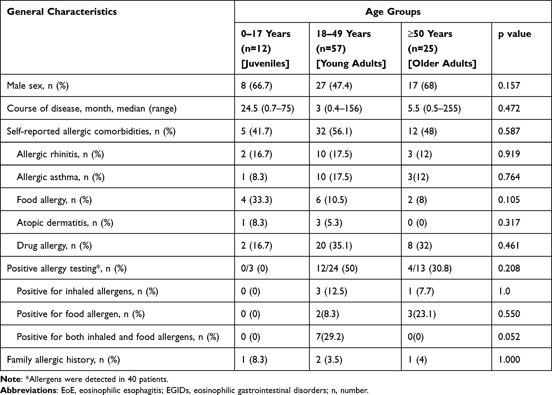 |
Table 1 General Characteristics of the 94 Patients with Non-EoE EGIDs Across Various Age Groups |
Clinical Manifestations
Abdominal pain was the most common manifestation (87/94, 92.6%), followed by weight loss (55/94, 58.5%), vomiting (47/94, 50%), diarrhea (44/94, 46.8%), and nausea (44/94, 46.8%) (Figure 1D and Supplementary Table 1). Young adults had higher diarrhea rates (24 vs 59.6%, p = 0.003), and juveniles had higher rates of nausea (28 vs 83.3%, p = 0.002) compared to older adults. The frequency of vomiting differed significantly among the three age groups (juveniles, 91.7%; young adults, 52.6%; older adults, 24%; p < 0.001). Reduced flatulence and/or defecation were less common in young adults (3.5%, 2/57) compared to juveniles (25%, 3/12, p =0.034) and older adults (20%, 5/25, p = 0.025). Ascites was less frequent in older adults (4%, 1/25) than in juveniles (75%, 9/12; p < 0.001), and young adult (43%, 25/57; p < 0.001) (Figure 2 and Supplementary Table 1).
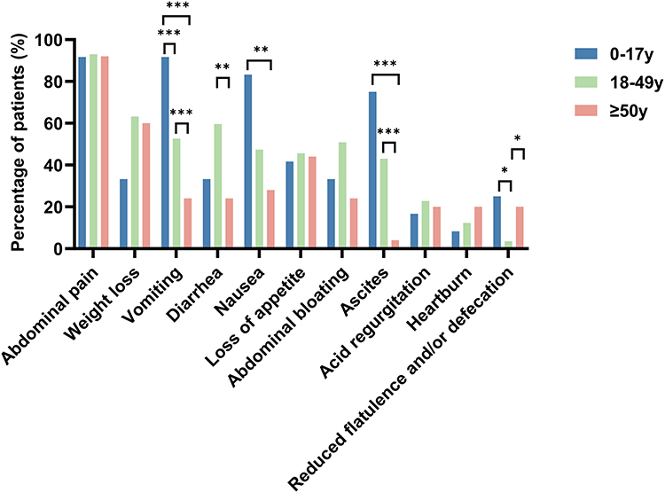 |
Figure 2 Clinical manifestations of the 94 patients with non-EoE EGIDs across various age groups. *P < 0.05, **P < 0.01, ***P < 0.001. Abbreviations: EoE, eosinophilic esophagitis; EGIDs, eosinophilic gastrointestinal disorders. |
The relationship between age groups and disease presentation was further investigated, with stratification based on the inflammatory lesion location. In patients with esophageal involvement, young adults had a significantly higher incidence of diarrhea compared to older adults (83.3 vs 0%, p = 0.048). Among those with gastric involvement, diarrhea was more prevalent in young adults than in juveniles and older adults (64 vs 20 vs 18.2%, p = 0.019). In patients with gastric or duodenal involvement, adolescents exhibited significantly higher rates of nausea (p = 0.027, 0.009), vomiting (p = 0.021, 0.002), and ascites (p = 0.002, <0.001) compared to the other two groups. In cases of ileal involvement, diarrhea was significantly more common in young adults than in juveniles and older adults (68.4 vs 25 vs 18.2%, p = 0.019), while ascites was more prevalent in juveniles (p = 0.031). For colonic involvement, young adults had a significantly lower incidence of reduced flatulence and/or defecation compared to juveniles and older adults (p = 0.005) (Supplementary Table 2).
Laboratory Test
Overall, 84.9% (79/93) of patients showed an increased eosinophil count, and 88% (81/92) showed increased eosinophil percentage in peripheral blood. Additionally, 17.4% (16/92) had decreased serum albumin, 8.8% (7/80) had increased erythrocyte sedimentation rate (ESR), 41.5 (27/65) had increased high-sensitivity C-reactive protein (hsCRP), and 84% (68/81) showed an increased level of total IgE (Table 2). Compared with older adults, juveniles had significantly higher white blood cell counts (12.23 vs 7.77×10⁹/L, p = 0.033) and eosinophil counts (5.52 vs 1.23×10⁹/L, p = 0.040). The serum albumin level in the juvenile group was significantly higher than in the young adult group (45 vs 40 g/L, p =0.004) and the older adult group (45 vs 38.7g/L, p =0.002). Serum total IgE levels were significantly higher in the juvenile group compared to the older adult group (467.5 vs 118g/L, p =0.002). ESR levels were higher in young adults (4 vs 2 mm/h, p =0.045; and older adults (7 vs 2 mm/h, p =0.002) compared to juveniles (Figure 3).
 |
Table 2 Laboratory Test of the 94 Patients with Non-EoE EGIDs in Various Age Groups |
 |
Figure 3 Laboratory test of the 94 patients with non-EoE EGIDs across various age groups. (A) Peripheral WBC counts by age group. (B) Peripheral Eos counts by age group. (C) Percentage of peripheral Eos by age group. (D) T-IgE levels by age group. (E) ESR levels by age group. (F) Serum alb levels by age group. *P < 0.05, **P < 0.01. Abbreviations: EoE, eosinophilic esophagitis; EGIDs, eosinophilic gastrointestinal disorders; WBC, white blood cell; Eos, eosinophils; T-IgE, total IgE; ESR, erythrocyte sedimentation rate; Alb, albumin. |
Allergen Testing
Allergy assessment using skin prick tests or specific serum IgE levels were conducted for 40 patients (42.6%), with a positivity rate of 40% (16/40). Four (10.0%) tested positive for inhalation allergens, including three for dust and grass pollen, and one tested positive for mold. Among the five patients (12.5%) that tested positive for food allergens, four were positive to seafood, while one was positive to wheat and one to fruits (banana and orange). Seven patients (17.5%) were positive for both inhaled and food allergens, with inhaled allergens including mold (n=1), dust mite (n=6), tree pollen (n=4), grass pollen (n=3), animal dander (n=1), and food allergens including seafood (n=4), wheat (n=2), tree nuts (n=1), peanut (n=2), soybean (n=1), sesame (n=1), egg (n=2), cow’s milk (n=1). Sensitization patterns were similar among the three age groups (p=0.813).
Endoscopic and Histological Finding
All patients received a gastroscopy, and 66.0% (62/94) performed colonoscopy. Results from 91 available gastroscopies showed that 97.8% (89/91) exhibited abnormal findings involving the esophagus (10/91, 11.0%), stomach (63/91, 69.2%), and duodenum (76/91, 83.5%). The abnormalities mainly presented as hyperemia, edema, erythema, and erosion. Among the 51 colonoscopies, 17.6% (9/51) were normal, and 82.4% (42/51) showed abnormalities in the Ileum (25/51, 49.0%), jejunum (2/51,3.9%), colon (19/51,37.3%) and rectum (14/51,27.5%), characterized by hyperemia, erosion, and edema (Supplementary Table 3). There were no differences in gastrointestinal endoscopy findings across the three age groups.
According to histological finding, duodenal involvement was the most common, affecting 85.1% ((80/94) of patients, followed by stomach (41/94, 43.6%), ileum (34/94, 36.2%), colon (22/94, 23.4%), esophagus (9/94, 9.6%), jejunum (1/94, 1.1%), ileocecal valve (4/94, 4.3%), and rectum (4/94, 4.3%). There were no differences in eosinophilic involvement sites in gastrointestinal tract across the three age groups (Table 3).
 |
Table 3 Gastrointestinal Involvement of the 94 Patients with Non-EoE EGIDs in Various Age Groups |
Therapeutic Management
Among the 94 patients with non-EoE EGIDs, 12.8% avoided food that showed sensitization on IgE testing (8/12, 66.7% combined the avoidance measures with glucocorticoids (prednisone or its equivalents), and 1.7% (2/12) combined with anti-allergic agents (antihistamines or leukotriene receptor antagonists). Glucocorticoids were used by 81.9% of patients, acid-inhibiting drugs by 37.2%, gastric motility drugs by 8.5%, leukotriene receptor antagonists by 6.4%, probiotics by 21.3%, antihistamines by 20.21%, cyclophosphamide by 2.1%, mesalazine by 3.2%, and 1.1% underwent surgery. Symptoms improved in 90.4% (85/94) of the patients. There were no significant differences in therapy among the three age groups.
Discussion
Although the exact epidemiology of this disease remains unclear, diagnoses of non-EoE EGIDs have increased over the last decade.29 Non-EoE EGIDs can occur at any age, from infancy to adulthood, but their prevalence varies among age groups.9,30 Non-EoE EGIDs tends to be slightly more common in children (< 18 years of age) than in adults (5.3/100,000 vs 5.1/100,000, p=0.537), although this difference is not statistically significant. In contrast, EoC is more prevalent in adults than in children (1.6/100,000 vs 2.3/100,000, p=0.0013).30 We found significant variability in the time from symptom onset to diagnosis, with a median duration of 4 months and a maximum of 255 months. The diagnostic delay aligns with findings from our center and is consistent with a population-based study in the USA, which reported an average delay of 3.6 years for non-EoE EGID patients between initial symptom presentation and diagnosis.31,32 As a national referral center for complex diseases, the median time to diagnosis in our cohort may be shorter than that in the general population. Non-EoE EGIDs are heterogeneous disorders,4 but reports on their clinical characteristics across different age groups are limited. This single-center retrospective study is the first to demonstrate the differences in clinical manifestations of non-EoE EGIDs across various age groups.
An allergic mechanism has been proposed for non-EoE EGID.11 Compared to control patients, individuals with Non-EoE EGIDs and EoC were more likely to suffer from allergic disorders.30 The coexistence of atopic comorbidities, though present in only a subset of patients, should increase the suspicion for EoG/EoD.5 Between 50% - 70% of patients with non-EoE EGID have elevated serum total IgE levels,4,27 a finding corroborated by our study, which showed an 84% incidence of elevated IgE levels. Consistent with previous studies,14 52.1% of our patients with non-EoE EGID self-reported allergic comorbidities, and 40% (16/40) tested positive for specific IgE (sIgE) either to food allergens or aeroallergens. However, it is challenging to confirm whether it represents a true allergy rather than mere sensitization. Non-EoE EGIDs are suspected to originate from food allergens crossing the intestinal mucosa and inducing mast cell degranulation and eosinophil recruitment.33 Previous studies14,34 indicated that 30–50% of patients with EoG tested positive for food allergies, and our study showed similar results, with 30% of patients showed positive IgE to specific foods.34 In addition to the IgE-dependent pathway in non-EoE EGIDs, the pathogenesis also includes IgE-independent mechanisms and cell-mediated immune responses.35 The role of food allergies in non-EoE EGIDs remains uncertain, and it is difficult to establish a causal relationship between specific foods and disease onset. In pediatric patients with non-EoE EGIDs, approximately 23.3% have been reported to have atopic comorbidities,36 which is lower than our data. Our findings suggest that food allergies may be more common in juveniles than in adults.
Symptoms of EGID vary according to the site and layer of the gastrointestinal tract and include abdominal pain, nausea, vomiting, early satiety, weight loss, and diarrhea, but all are non-specific.27,31 Consistent with published data,22 abdominal pain was the most common symptom in patients with non-EoE EGIDs. Weight loss was another major symptom observed, though our incidence rates for abdominal pain and weight loss were higher than previously reported (62% and 27%, respectively).27 Consistent with our study, diarrhea (47.1%) was also one of the most common symptoms reported.37 Previous reports22,36 indicate that pediatric patients with non-EoE EGIDs commonly present with abdominal pain (73.3–76.1%) and diarrhea (23.9–58.3%). We found higher frequency of abdominal pain in the juvenile group, with vomiting (91.7%), nausea (83.3%), and ascites (75%) being the most frequently reported symptoms. Ascites is an uncommon complication in non-EoE EGIDs.38 This discrepancy may be attributed to several factors, including racial differences, the small sample size, and the tendency for hospitalized patients to present with more advanced or complicated disease compared to the general population of non-EoE EGID patients. This may have resulted in an overrepresentation of severe cases, including rare complications such as ascites. Our study identifies age-related differences in the clinical features of non-EoE EGIDs. Vomiting was more common in juveniles, reduced flatulence/defecation less frequent in young adults, and ascites less prevalent in older adults. Notably, patients with the same affected gastrointestinal site also exhibited varying manifestations across age groups: young adults with gastric or ileal involvement had higher diarrhea rates, while juveniles with gastric/duodenal involvement showed more nausea, vomiting, and ascites. Juveniles with ileal involvement also had higher ascites prevalence, and young adults with colonic involvement had reduced flatulence/defecation less often. These findings suggest that age may influence disease presentation, potentially due to differences in immune responses or gastrointestinal tract maturity.39 While our study provides insights into age-specific patterns, it is important to note that only a limited number of studies have explored non-EoE EGIDs across age groups, and further research is needed to confirm these observations.
Complete blood count is supportive in suspected non-EoE EGID cases.33 Elevated eosinophil counts in peripheral blood are present in approximately 70–80% of non-EoE-EGID cases.40 Consistent with this finding, we observed that 84.9% of our patients had eosinophilia. Decreased albumin concentration was present in 17.4% of our patients with non-EoE-EGID, similar to the 20–30% reported in other studies.4 Brenner et al41 reported that peripheral eosinophilia and hypoalbuminemia are associated with a higher biopsy diagnostic yield for EoG. Among our patients, 41.5% exhibited elevated hsCRP levels, compared to the 14.7–30% reported in previous studies.37 Our data also showed that the serum albumin levels were significantly higher, and ESR was lower in the juvenile group than in the young and older adult groups. Hypoalbuminemia is a hallmark of severe Non-EoE EGIDs, and patients with hypoalbuminemia at presentation might predict a chronic disease course.18,42 Thus, the severity of non-EoE EGIDs in our juvenile group might have been milder than in the other two adult groups.
The duodenum (85.1%) was the most commonly affected site, followed by the stomach (43.6%), ileum (36.2%), colon (23.4%), and esophagus (9.6%). Consistent with our study, the incidence of small intestine and stomach involvement was reported as 76.5% and 41.2%, respectively.37 Reed et al27 found that 28% of patients with non-EoE EGIDs had colonic involvement, while another study reported a higher incidence of 55.9%.37 Esophageal involvement was less common in our study (9.6%) compared to previous studies (26.5–30%).27,37 Choi et al43 reported that 29% of children with Non-EoE EGIDs had colon involvement, and 13% had esophageal involvement, which was 16.7% and 0%, respectively, in our juvenile group. The reported affected sites are influenced by different study population and various biopsy sites.
Currently, there are no standardized guidelines for the optimal treatment of non-EoE EGIDs.9 Therapeutic management is primarily empirical, which typically involves dietary modifications, steroids, immunosuppressive agents, and biological therapies.26,44 Diet therapy is often the first-line treatment, but its efficacy in patients with non-EoE EGIDs remains uncertain. Amino acid-based elemental diets effectively treat Non-EoE EGIDs.11 Gastric and duodenal EGIDs may be related to food allergies.1 Kobayashi et al22 demonstrated that in pediatric eosinophilic gastroenteritis, the most common treatment was eliminating suspected food allergens combined with anti allergy agents, resulting in symptom resolution in 39.4% of patients and symptom improvement in 48.6%. In our study, 12.8% avoided foods that showed sensitization on IgE testing; 66.7% (8/12) combined this approach with glucocorticoids; 1.7% (2/12) combined with anti-allergic agents. IgE sensitization alone does not necessarily confirm causality in driving EGID inflammation. The gold standard for identifying causative foods involves elimination diets followed by endoscopic confirmation of inflammation resolution.39 This study found that previous treatment strategies for hospitalized patients, which relied solely on specific IgE results to guide dietary interventions, had significant limitations. In future clinical practice, it is essential to establish a clear relationship between specific foods and disease activity before providing dietary recommendations. This approach will ensure more accurate and effective management of EGID, ultimately improving patient outcomes. Corticosteroids were the most common therapeutic alternative (81.9%), and therapeutic management was consistent across different age groups.
The strength of this study is the large cohort of 94 Chinese patients with non-EoE EGIDs from China. This is the first study to investigate the clinical profiles of Chinese patients with non-EoE EGIDs at various ages at disease onset. Given the close relationship between EGIDs and allergic disorders, we provide a detailed analysis of the sensitization spectrum in our patients. However, this study has certain limitations. First, due to the availability of more comprehensive clinical documentation, only hospitalized patients were included in the study. As a result, the proportion of patients with severe disease may be higher than the overall population of non-EoE EGID patients. Second, our hospital primarily treats adult patients, potentially underestimating the proportion of juvenile non-EoE EGID cases. The small juvenile subgroup necessitates cautious interpretations of conclusions, and more extensive cohort studies are needed in the future. Third, as a retrospective study, some clinical data were incomplete. Patients with non-EoE EGIDs are more likely to have allergic diseases. Evaluating the proportion of allergic diseases across age groups may help understand possible disease subtypes. However, many patients were not tested for allergens, and data on allergic diseases primarily came from self-reports rather than professional allergists assessments.
Conclusions
This study highlights significant age-related differences in the clinical presentation and laboratory findings of non-EoE EGID patients. Young adults with gastric or ileal involvement had higher diarrhea rates, while juveniles with gastric or duodenal involvement exhibited higher rates of nausea, vomiting, and ascites. Young adults with colonic involvement experienced reduced flatulence/defecation less frequently. Additionally, juveniles had significantly higher peripheral eosinophil counts and total serum IgE levels compared to older adults, along with higher serum albumin levels and lower ESR than both young and older adult groups. These findings emphasize the importance of considering age-related variations in diagnosing and managing non-EoE EGIDs. Future research should focus on developing and validating a comprehensive diagnostic tool or algorithm to improve the identification of this heterogeneous condition.
Abbreviations
EGIDs, eosinophilic gastrointestinal disorders; EoE, eosinophilic esophagitis; EoG, eosinophilic gastritis; EoGE, eosinophilic gastroenteritis; EoN, eosinophilic enteritis; EoD, eosinophilic duodenitis; EoC, eosinophilic colitis; hsCRP, hypersensitive C-reactive protein.
Data Sharing Statement
The datasets used and/or analyzed during the current study are available from the corresponding author on reasonable request.
Ethics Approval and Consent to Participate
This study was conducted in accordance with the declaration of Helsinki and approved by the ethics committee of Peking Union Medical College Hospital (approval number: S-k2101). Informed consent was obtained from all subjects and/or their legal guardian(s) prior to study commencement.
Authors’ Consent for Publication
All authors have approved the final manuscript and approved of the submission to Journal of Asthma and Allergy.
Acknowledgments
We appreciate all the patients and investigators who participated in this study.
Author Contributions
All authors contributed significantly to the work reported in this article, including the conception and design of the study, data acquisition, analysis, and interpretation. Each author participated in drafting, revising, or critically reviewing the manuscript. All authors have approved the final version to be published, agreed on the journal to which the article has been submitted, and are accountable for all aspects of the work.
Funding
This study was supported by National High Level Hospital Clinical Research Funding (2022-PUMCH-B-090).
Disclosure
None of the authors have any competing interests related to this study.
References
1. Rothenberg ME, Hottinger SKB, Gonsalves N, et al. Impressions and aspirations from the FDA GREAT VI workshop on eosinophilic gastrointestinal disorders beyond eosinophilic esophagitis and perspectives for progress in the field. J Allergy Clin Immunol. 2022;149(3):844–853. doi:10.1016/j.jaci.2021.12.768
2. Low EE, Dellon ES. Review article: emerging insights into the epidemiology, pathophysiology, diagnostic and therapeutic aspects of eosinophilic oesophagitis and other eosinophilic gastrointestinal diseases. Aliment Pharmacol Ther. 2024;59(3):322–340. doi:10.1111/apt.17845
3. Dellon ES, Gonsalves N, Abonia JP, et al. International consensus recommendations for eosinophilic gastrointestinal disease nomenclature. Clin Gastroenterol Hepatol. 2022;20(11):2474–2484e2473. doi:10.1016/j.cgh.2022.02.017
4. Kinoshita Y, Sanuki T. Review of non-eosinophilic esophagitis-eosinophilic gastrointestinal disease (Non-EoE-EGID) and a case series of twenty-eight affected patients. Biomolecules. 2023;13(9):1417. doi:10.3390/biom13091417
5. Wright BL, Abonia JP, Abud EM, et al. Advances and ongoing challenges in eosinophilic gastrointestinal disorders presented at the CEGIR/TIGERs symposium at the 2024 American Academy of Allergy, Asthma & Immunology meeting. J Allergy Clin Immunol. 2024;154(4):882–892. doi:10.1016/j.jaci.2024.07.022
6. Marasco G, Visaggi P, Vassallo M, et al. Current and novel therapies for eosinophilic gastrointestinal diseases. Int J Mol Sci. 2023;24(20):15165. doi:10.3390/ijms242015165
7. Jensen ET, Martin CF, Kappelman MD, Dellon ES. Prevalence of eosinophilic gastritis, gastroenteritis, and colitis: estimates from a national administrative database. J Pediatr Gastroenterol Nutr. 2016;62(1):36–42. doi:10.1097/MPG.0000000000000865
8. Licari A, Votto M, Scudeller L, et al. Epidemiology of nonesophageal eosinophilic gastrointestinal diseases in symptomatic patients: a systematic review and meta-analysis. J Allergy Clin Immunol Pract. 2020;8(6):1994–2003.e1992. doi:10.1016/j.jaip.2020.01.060
9. Li K, Ruan G, Liu S, et al. Eosinophilic gastroenteritis: pathogenesis, diagnosis, and treatment. Chin Med J. 2023;136(8):899–909. doi:10.1097/CM9.0000000000002511
10. Cianferoni A, Spergel JM. Eosinophilic esophagitis and gastroenteritis. Curr Allergy Asthma Rep. 2015;15(9):58. doi:10.1007/s11882-015-0558-5
11. Gonsalves N, Doerfler B, Zalewski A, et al. Prospective study of an amino acid-based elemental diet in an eosinophilic gastritis and gastroenteritis nutrition trial. J Allergy Clin Immunol. 2023;152(3):676–688. doi:10.1016/j.jaci.2023.05.024
12. Zhang M, Li Y. Eosinophilic gastroenteritis: a state-of-the-art review. J Gastroenterol Hepatol. 2017;32(1):64–72. doi:10.1111/jgh.13463
13. Shoda T, Wen T, Caldwell JM, et al. Molecular, endoscopic, histologic, and circulating biomarker-based diagnosis of eosinophilic gastritis: multi-site study. J Allergy Clin Immunol. 2020;145(1):255–269. doi:10.1016/j.jaci.2019.11.007
14. Caldwell JM, Collins MH, Stucke EM, et al. Histologic eosinophilic gastritis is a systemic disorder associated with blood and extragastric eosinophilia, TH2 immunity, and a unique gastric transcriptome. J Allergy Clin Immunol. 2014;134(5):1114–1124. doi:10.1016/j.jaci.2014.07.026
15. Ben-Baruch Morgenstern N, Shoda T, Rochman Y, et al. Local type 2 immunity in eosinophilic gastritis. J Allergy Clin Immunol. 2023;152(1):136–144. doi:10.1016/j.jaci.2023.01.021
16. Pesek RD, Greuter T, Lopez-Nunez O, Bernieh A, Straumann A, Collins MH. Clinicopathologic correlations in eosinophilic gastrointestinal disorders. J Allergy Clin Immunol Pract. 2021;9(9):3258–3266. doi:10.1016/j.jaip.2021.06.002
17. Laserna-Mendieta EJ, Navarro P, Casabona-Francés S, et al. Differences between childhood- and adulthood-onset eosinophilic esophagitis: an analysis from the EoE connect registry. Dig Liver Dis. 2023;55(3):350–359. doi:10.1016/j.dld.2022.09.020
18. Licari A, Votto M, D’Auria E, Castagnoli R, Caimmi SME, Marseglia GL. Eosinophilic gastrointestinal diseases in children: a practical review. Curr Pediatr Rev. 2020;16(2):106–114. doi:10.2174/18756336MTAxdNzcpx
19. Liacouras CA, Furuta GT, Hirano I, et al. Eosinophilic esophagitis: updated consensus recommendations for children and adults. J Allergy Clin Immunol. 2011;128(1):3–20.e26. quiz 21–22. doi:10.1016/j.jaci.2011.02.040
20. Shoda T, Taylor RJ, Sakai N, Rothenberg ME. Common and disparate clinical presentations and mechanisms in different eosinophilic gastrointestinal diseases. J Allergy Clin Immunol. 2024;153(6):1472–1484. doi:10.1016/j.jaci.2024.03.013
21. Kinoshita Y, Furuta K, Ishimaura N, et al. Clinical characteristics of Japanese patients with eosinophilic esophagitis and eosinophilic gastroenteritis. J Gastroenterol. 2013;48(3):333–339. doi:10.1007/s00535-012-0640-x
22. Kobayashi S, Tsunoda T, Umetsu S, Inui A, Fujisawa T, Sogo T. Clinical features of pediatric eosinophilic gastroenteritis. Pediatr Int. 2022;64(1):e15322. doi:10.1111/ped.15322
23. Talley NJ, Shorter RG, Phillips SF, Zinsmeister AR. Eosinophilic gastroenteritis: a clinicopathological study of patients with disease of the mucosa, muscle layer, and subserosal tissues. Gut. 1990;31(1):54–58. doi:10.1136/gut.31.1.54
24. Hasan SH, Taylor S, Garg S, et al. Diagnosis of pediatric non-esophageal eosinophilic gastrointestinal disorders by eosinophil peroxidase immunohistochemistry. Pediatr Dev Pathol. 2021;24(6):513–522. doi:10.1177/10935266211024552
25. Collins MH. Histopathology associated with eosinophilic gastrointestinal diseases. Immunol Allergy Clin North Am. 2009;29(1):109–117,x–xi. doi:10.1016/j.iac.2008.10.005
26. Walker MM, Potter M, Talley NJ. Eosinophilic gastroenteritis and other eosinophilic gut diseases distal to the oesophagus. Lancet Gastroenterol Hepatol. 2018;3(4):271–280. doi:10.1016/S2468-1253(18)30005-0
27. Reed C, Woosley JT, Dellon ES. Clinical characteristics, treatment outcomes, and resource utilization in children and adults with eosinophilic gastroenteritis. Dig Liver Dis. 2015;47(3):197–201. doi:10.1016/j.dld.2014.11.009
28. Du Z, Zhi Y. Age-dependent manifestations of eosinophilic gastrointestinal disorders beyond eosinophilic esophagitis. J Allergy Clin Immunol. 2025;155(2):AB36. doi:10.1016/j.jaci.2024.12.116
29. Pesek RD, Reed CC, Muir AB, et al. Increasing rates of diagnosis, substantial co-occurrence, and variable treatment patterns of eosinophilic gastritis, gastroenteritis, and colitis based on 10-year data across a multicenter consortium. Am J Gastroenterol. 2019;114(6):984–994. doi:10.14309/ajg.0000000000000228
30. Mansoor E, Saleh MA, Cooper GS. Prevalence of eosinophilic gastroenteritis and colitis in a population-based study, from 2012 to 2017. Clin Gastroenterol Hepatol. 2017;15(11):1733–1741. doi:10.1016/j.cgh.2017.05.050
31. Chehade M, Kamboj AP, Atkins D, Gehman LT. Diagnostic delay in patients with eosinophilic gastritis and/or duodenitis: a population-based study. J Allergy Clin Immunol Pract. 2021;9(5):2050–2059.e2020. doi:10.1016/j.jaip.2020.12.054
32. Li KW, Ruan GC, Liu S, et al. Long-term prognosis and its associated predictive factors in patients with eosinophilic gastroenteritis. World J Gastroenterol. 2024;30(2):146–157. doi:10.3748/wjg.v30.i2.146
33. Sunkara T, Rawla P, Yarlagadda KS, Gaduputi V. Eosinophilic gastroenteritis: diagnosis and clinical perspectives. Clin Exp Gastroenterol. 2019;12:239–253. doi:10.2147/CEG.S173130
34. Tien FM, Wu JF, Jeng YM, et al. Clinical features and treatment responses of children with eosinophilic gastroenteritis. Pediatr Neonatol. 2011;52(5):272–278. doi:10.1016/j.pedneo.2011.06.006
35. Yu W, Freeland DMH, Nadeau KC. Food allergy: immune mechanisms, diagnosis and immunotherapy. Nat Rev Immunol. 2016;16(12):751–765. doi:10.1038/nri.2016.111
36. Votto M, Raffaele A, De Filippo M, et al. Eosinophilic gastrointestinal disorders in children and adolescents: a single-center experience. Dig Liver Dis. 2022;54(2):214–220. doi:10.1016/j.dld.2021.06.027
37. Okimoto E, Ishimura N, Ishihara S. Clinical characteristics and treatment outcomes of patients with eosinophilic esophagitis and eosinophilic gastroenteritis. Digestion. 2021;102(1):33–40. doi:10.1159/000511588
38. Fenoglio LM, Benedetti V, Rossi C, et al. Eosinophilic gastroenteritis with ascites: a case report and review of the literature. Dig Dis Sci. 2003;48(5):1013–1020. doi:10.1023/A:1023080419660
39. Simon AK, Hollander GA, McMichael A. Evolution of the immune system in humans from infancy to old age. Proc Biol Sci. 2015;282(1821):20143085. doi:10.1098/rspb.2014.3085
40. Yamamoto M, Nagashima S, Yamada Y, et al. Comparison of nonesophageal eosinophilic gastrointestinal disorders with eosinophilic esophagitis: a nationwide survey. J Allergy Clin Immunol Pract. 2021;9(9):3339–3349.e3338. doi:10.1016/j.jaip.2021.06.026
41. Brenner EJ, Greenberg SB, Chang NC, Corder SR, Cowherd EL, Dellon ES. Peripheral eosinophilia and hypoalbuminemia are associated with a higher biopsy diagnostic yield for eosinophilic gastroenteritis. Clin Res Hepatol Gastroenterol. 2021;45(5):101746. doi:10.1016/j.clinre.2021.101746
42. Havlichek D, Choung RS, Murray JA. Eosinophilic gastroenteritis: using presenting findings to predict disease course. Clin Transl Gastroenterol. 2021;12(10):e00394. doi:10.14309/ctg.0000000000000394
43. Choi JS, Choi SJ, Lee KJ, et al. Clinical manifestations and treatment outcomes of eosinophilic gastroenteritis in children. Pediatr Gastroenterol Hepatol Nutr. 2015;18(4):253–260. doi:10.5223/pghn.2015.18.4.253
44. Ko HM, Morotti RA, Yershov O, Chehade M. Eosinophilic gastritis in children: clinicopathological correlation, disease course, and response to therapy. Am J Gastroenterol. 2014;109(8):1277–1285. doi:10.1038/ajg.2014.166
 © 2025 The Author(s). This work is published and licensed by Dove Medical Press Limited. The
full terms of this license are available at https://www.dovepress.com/terms
and incorporate the Creative Commons Attribution
- Non Commercial (unported, 4.0) License.
By accessing the work you hereby accept the Terms. Non-commercial uses of the work are permitted
without any further permission from Dove Medical Press Limited, provided the work is properly
attributed. For permission for commercial use of this work, please see paragraphs 4.2 and 5 of our Terms.
© 2025 The Author(s). This work is published and licensed by Dove Medical Press Limited. The
full terms of this license are available at https://www.dovepress.com/terms
and incorporate the Creative Commons Attribution
- Non Commercial (unported, 4.0) License.
By accessing the work you hereby accept the Terms. Non-commercial uses of the work are permitted
without any further permission from Dove Medical Press Limited, provided the work is properly
attributed. For permission for commercial use of this work, please see paragraphs 4.2 and 5 of our Terms.