Back to Journals » Drug Design, Development and Therapy » Volume 15
Administration of Dexmedetomidine Does Not Produce Long-Term Protective Effect on Testicular Damage Post Testicular Ischemia-Reperfusion Injury
Authors Xiao J, Wan W, Zhang Y, Ma J ![]() , Yan L, Luo Y, Tang J
, Yan L, Luo Y, Tang J
Received 2 December 2020
Accepted for publication 13 January 2021
Published 27 January 2021 Volume 2021:15 Pages 315—321
DOI https://doi.org/10.2147/DDDT.S293926
Checked for plagiarism Yes
Review by Single anonymous peer review
Peer reviewer comments 3
Editor who approved publication: Professor Anastasios Lymperopoulos
Jing Xiao,1,2 Wenbo Wan,2 Ying Zhang,1,2 Jun Ma,2 Lin Yan,2 Yukun Luo,1,2 Jie Tang1,2
1School of Medicine, Nankai University, Tianjin, People’s Republic of China; 2Department of Ultrasound, The First Medical Center of Chinese PLA General Hospital, Beijing, People’s Republic of China
Correspondence: Jie Tang
Department of Ultrasound, The First Medical Center of Chinese PLA General Hospital, Fuxing Road 28#, Beijing 100853, People’s Republic of China
Tel +86-10-5549-9056
Fax +86-10-5549-9255
Email [email protected]
Background: After surgical correction of testicular torsion, up to 68% of ipsilateral testes undergo atrophy due to ischemia-reperfusion injury (IRI). Recent studies have shown that dexmedetomidine (Dex) alleviates IRI in various vital organs. However, those studies evaluated its protective effect on short-term reperfusion.
Purpose: We aimed to investigate whether Dex has a long-term protective effect against testicular injury after IRI.
Materials and Methods: A total of 24 New Zealand white rabbits were randomly divided into three groups (n = 8/group): the control group (saline-infused rabbits without IRI), the IRI group (saline-injected rabbits with IRI), and the Dex group (Dex-injected rabbits with IRI). The spermatic cord of rabbits in IRI and Dex groups was ligated for 4 h, and 1 h before reperfusion, Dex was administered intraperitoneally at a dose of 50 μg/kg body weight in group Dex, whereas saline was administered at the same dose to the IRI and control groups. Rabbits were kept alive for 4 weeks post reperfusion, then the testes were harvested, and the rabbits were euthanized.
Results: Four weeks post reperfusion, testicular volumes of the affected side decreased considerably in the IRI and Dex groups compared to the control group, with no significant difference between the IRI and Dex groups. Compared to the control group, the Johnson score and the mean seminiferous tubular diameters were significantly decreased in the IRI and Dex groups, but no significant differences were observed after administration of Dex. There were no significant differences in malondialdehyde and superoxide dismutase levels between the groups treated with and without Dex.
Conclusion: Dex administration 3 h after ischemia and 1 h before reperfusion did not demonstrate a significant protective effect against testicular injury 4 weeks after IRI in rabbits. Further research is needed to confirm the potential therapeutic effects of Dex by varying the experimental conditions.
Keywords: dexmedetomidine, testis, reperfusion injury, testicular atrophy, testicular torsion
Introduction
Testicular torsion is an urologic emergency occurring frequently in adolescents and young men, requiring timely intervention. However, despite prompt diagnosis and surgical treatment, up to 68% of affected testes undergo atrophy,1 which may lead to male subfertility and even infertility. The testicular volume has a significant correlation with spermatogenesis, and it is a reliable indicator of testicular function.2 Sperm count and motility decrease in patients with testicular atrophy, and the retention of a nonviable or severely damaged testis is harmful to the contralateral testis.1 Therefore, any medication or method that could prevent this testicular atrophy is of significant benefit to patients.
Testicular torsion/detorsion has a pathophysiological course of ischemia-reperfusion injury (IRI). Ischemic testes recovery requires re-establishment of blood flow; however, the resumption of blood flow can further aggravate any testicular injury. IRI is a complex pathophysiological process, involving excessive generation of reactive oxygen species (ROS).3 An excessive production of ROS results in DNA damage, endothelial injury, and oxidative stress, and it also could initiate the intrinsic apoptosis pathway in testicular tissue cells, leading to the apoptosis of germinal cells.4 Numerous efforts have been made to discover novel and effective chemicals or drugs that can treat IRI.5–7
Dexmedetomidine (Dex), a potent selective α2-adrenoceptor agonist, is mainly used for sedation and as an analgesic in post-anesthesia care unit and intensive care units, respectively.8 Accumulating evidence has shown that Dex has a potential protective effect in various vital organs after IRI,9–12 probably by inhibiting inflammation through inactivation of the toll-like receptor 4 (TLR4)/nuclear factor kappa B (NF-κB) pathway,11 suppressing neuronal autophagy through up-regulation of the hypoxia-inducible factor (HIF)-1α,12 and inhibiting the high mobility group protein B1 Group (HMGB1)/TLR 4/NF-κB pathway.13 However, most research focuses on the protective effect of Dex post short-term ischemia and reperfusion,10,14,15 and research on the long-term effects of Dex on organs after IRI is limited. Therefore, we performed this experimental study to investigate whether Dex has a long-term protective effect against testicular injury at 4 weeks after IRI in order to maximize the preservation of testicular function.
Materials and Methods
Animals and Surgical Procedures
Twenty-four male New Zealand white rabbits, weighing of 2.5–3.2 kg, were purchased from the Laboratory Animal Center of the First Medical Center of Chinese PLA General Hospital (Experimental Animal License: SCXK (Beijing) 2015–0005). This study was approved by the Institutional Animal Care and Use Committee of the Chinese PLA General Hospital (2019-X15-53), and it was carried out in accordance with the Code of Practice for the Housing and Care of Animals Used in Scientific Procedures. All rabbits were acclimated for 7 days in temperature- and humidity-controlled cages with free access to food and water. They were made to fast for at least 8 hours before the experiment while retaining their free access to water.
All rabbits were anesthetized with 10% chloral hydrate (2–2.5 mL/kg body weight) through a catheter inserted into the marginal ear vein, and then the perineal fur was shaved. Under sterile conditions, a longitudinal incision was made about 2 cm above the upper pole of the right testis, and the spermatic layers were dissected in turn till the internal spermatic fascia. The right spermatic cord was then ligated with 3/0 sutures. Then, the incision was sutured. If the blood flow was absent in the testis on color Doppler ultrasonography after ligation, it indicated that the model was successfully established. The ligation was released after 4 h, and the incision was re-sutured.
Four weeks after reperfusion, the testicular tissues were harvested, washed, and divided into two parts. One part was fixed in 10% neutral formalin for histological analysis, whereas the other part was stored at –80°C to determine the markers of oxidative stress and antioxidant status. After the testes were harvested, the rabbits were euthanized with an overdose of anesthesia.
Grouping and Drug Treatment
All the rabbits were randomly divided into three groups (n = 8/group).
- Control group (group C): The rabbits were subjected to the inguinal incision and isolation of the spermatic cord without any ligation. Saline was injected intraperitoneally at a dose of 50 μg/kg body weight 3 h after inguinal incision.
- IRI group: The rabbits were subjected to spermatic cord ligation and released as described above, and saline was injected intraperitoneally at a dose of 50 μg/kg body weight 1 h before releasing the spermatic cord ligation.
- Dex group: The rabbits were subjected to spermatic cord ligation and administered Dex (Cisen Pharmaceutical Co., Ltd., Jining, China) intraperitoneally. Based on a previous study,16 Dex was administered at a dose of 50 μg/kg body weight 1 hour before releasing the spermatic cord ligation.
Ultrasonography
Ultrasonography was performed using Mindray Resona 7 scanners (Mindray, Shenzhen, China) with a 5.6–10 MHz linear array transducer. The depth, gain, and scale were kept identical during the entire experiment. Ultrasonography was carried out to measure the testicular size before ligation, after releasing the ligations, and 4 weeks after testicular reperfusion. Both the testes were measured for their volume calculation using the ellipsoid formula: length × width × thickness × 0.524.17
Markers of Oxidative Stress and Antioxidant Status
Approximately 100 mg of ground testicular tissues in each group were homogenized in 0.9 mL of cold normal saline after cutting the tissues into small pieces with scissors. The homogenate was then centrifuged at 3000 rpm for 30 min. The supernatant was used to measure malondialdehyde (MDA, a marker of oxidative stress) and superoxide dismutase (SOD, a marker of antioxidant status). They were analyzed with MDA and SOD assay kits according to the manufacturer’s instructions (Nanjing Jiancheng Bioengineering Institute, Nanjing, China). The absorbance was measured at 532 nm for MDA and 550 nm for SOD using a microplate reader (Thermo Fisher Scientific, Massachusetts, USA).
Histological Evaluation
All testicular specimens were fixed in 10% neutral formalin for 24 hours at 4°C, and then embedded in paraffin wax. 3-μm-thickness sections were cut and stained with hematoxylin and eosin. The slides were analyzed by pathologists who were blinded to the experiment design, and scored from 1 to 10 according to the Johnson scoring system.18 In this system, seminiferous tubules with complete spermatogenesis and regular tubular architecture are given a score of 10, whereas seminiferous tubules without any spermatogenesis and germ cells are given a score of 1. The mean seminiferous tubular diameter (MSTD) was measured in microns using a microscope-adaptable micrometer (Olympus, Tokyo, Japan). At least 10 seminiferous tubules of each section were scored and measured.
Statistical Analyses
All data were analyzed using IBM SPSS Statistics Base software for Windows version 21.0 (IBM Corp., Armonk). The distribution of the variables was analyzed using the Shapiro–Wilk test, and the Levene test was used to assess the homogeneity of variances. Continuous data are expressed as mean ± standard deviation. The paired t test was used to compare the testicular volumes between the ipsilateral and contralateral sides, and one-way analysis of variance (ANOVA) followed by Tukey’s post hoc test was used to compare the differences in the testicular volume, Johnson scores and MSTDs among the experimental groups. A two-tailed P-value less than 0.05 was considered statistically significant.
Results
During the experimental period, two rabbits died (1 in the IRI group and 1 in the Dex group). At the end of the 4-week follow-up, the number of rabbits in the control, IRI and Dex groups was 8, 7, and 7, respectively.
Comparison of Testicular Volume Changes in the Different Groups
Before spermatic cord ligation, there were no significant differences in the testicular volumes between each side and among the different groups (P > 0.05, Tables 1 and 2, Figure 1). Compared to the contralateral testis, the mean volume of the ipsilateral testis in the IRI group increased significantly just after releasing the spermatic cord ligation, but no significant differences were noted in the Dex group (Table 1). There were no marked differences in the mean volumes of the ipsilateral testes among the three groups (P > 0.05, Table 2, Figure 1A) just after releasing the spermatic cord ligations. Four weeks after reperfusion, testicular volumes of the ipsilateral side in the IRI and Dex groups were considerably lesser than those of the contralateral sides (P < 0.05, Table 1), but there was no significant difference between the IRI and Dex groups themselves. (P = 0.937, Table 2, Figure 1A). A total of six ipsilateral testes developed atrophy (3 in the IRI group and 3 in the Dex group) 4 weeks after reperfusion.
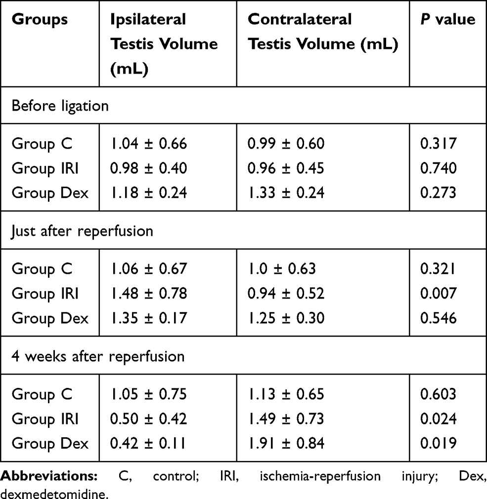 |
Table 1 Comparison of Testicular Volume Between the Ipsilateral and Contralateral Side |
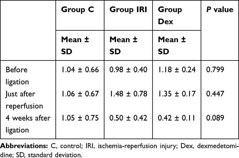 |
Table 2 Comparison of the Ipsilateral Testicular Volume (mL) Between Groups |
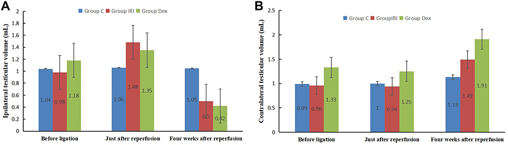 |
Figure 1 Testicular volume changes of both the sides in each group at different time points. (A) the ipsilateral side; (B) the contralateral side. |
Contralateral testicular volumes in the IRI and Dex groups were not considerably different from those of the control group at different time points (P > 0.05, Figure 1B).
Effects of IRI on Markers of Oxidative Stress and Antioxidant Status
MDA concentrations increased in the IRI (4.2 ± 1.7 nmol/mg protein) and Dex (2.8 ± 0.3 nmol/mg protein) groups compared to those of the control group (1.5 ± 0.5 nmol/mg protein), but there were no significant differences in MDA concentrations between the IRI and Dex groups (P > 0.05, Figure 2A). SOD activity reduced in the IRI (2.5 ± 1.0 U/mg protein) and Dex (3.2 ± 0.9 U/mg protein) groups compared with that of the control group (5.6 ± 0.5 U/mg protein), but these differences were not significant between the IRI and Dex groups (P > 0.05, Figure 2B).
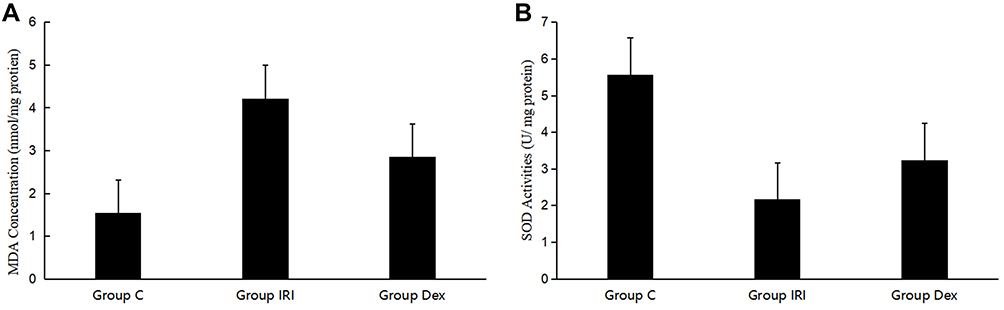 |
Figure 2 Concentrations of MDA (A) and SOD (B) in the affected testicular tissue. Abbreviations: MDA, malondialdehyde; SOD, superoxide dismutase. |
Comparison of Histological Changes in the Different Groups
The histological examination demonstrated that the rabbits’ testes in the control group were normal (Figure 3A), whereas the maturation of the spermatogenic cells in the testes that underwent IRI decreased considerably (Figure 3B and C). The Johnson score and MSTD of the IRI and Dex groups were lower than those of the control group, but no significant differences were observed between the IRI and Dex groups (P >0.05, Table 3).
 |
Table 3 The Johnson Score and MSTD in the Testis of Each Group |
 |
Figure 3 Histological changes in the testis of each group. Microphotographs taken of group C (A, arrowhead, spermatozoa), group IRI (B, arrowhead, spermatids), and group Dex (C, arrowhead, spermatids) testis subjected to HE staining; 400× magnification; scale bars, 50 μm. Abbreviations: C, control; IRI, ischemia-reperfusion injury; Dex, dexmedetomidine; HE, hematoxylin and eosin. |
Discussion
This study investigated whether Dex produced a protective effect on testicular damage 4 weeks after IRI. In the rabbit model of testicular IRI, we found that there were no significant differences in the testicular volumes, MDA concentrations, SOD activity, Johnson scores and MSTDs among the groups that were treated with and without Dex, which indicated that Dex alone could not prevent testicular damage after IRI.
In the process of testicular IRI, the balance between the production of free oxygen radicals and antioxidant capacity is disrupted, generating excessive ROS. The ROS can attack the polyunsaturated fatty acids in the biological membranes, and induce free radical chain reactions, leading to the enhancement of lipid peroxidation.14 There are many chemical agents and drugs, including oxygen radical scavengers, that have been successfully used to reduce IRI in animal models.6,14,15
Dex has been shown to have a protective effect against IRI in several tissues,19–22 but the underlying mechanism of Dex action in preventing IRI is not clear. There are some opinions about its preventive properties during IRI. A previous study demonstrated that Dex shows its effect via presynaptic α-adrenoceptors, reducing the section of noradrenaline induced by ischemia.23 By this mechanism, Dex may prevent potential destructive effects caused by noradrenaline by preventing the increased production of free oxygen radicals production.24 In a study on the anti-inflammatory effects of Dex in different tissues, Ma et al22 suggested that Dex provided renoprotection through anti-inflammatory effects on the parasympathetic nervous system activation. Sun et al10 also reported its anti-inflammatory effect on the spinal cord IRI in rabbits.
Apoptosis is important for regulating the population of germ cells in the testes. Sönmez et al25 demonstrated that testicular IRI caused a significant increase in apoptosis of the ipsilateral testis. One study demonstrated that administration of Dex significantly decreased the germ cell apoptosis indices.15 Another study reported that Dex inhibited the caspase-3 dependent apoptosis in a rabbit spinal cord ischemia-reperfusion model.10
Although many chemicals have been shown to reduce testicular IRI in animal models, most of them have primarily been examined 2–24 hours after testicular IRI.5,6,14,25 Few studies have examined the progress of testicular IRI, and the injury that can occur months and years later. Experimental and clinical case studies that contain long-term follow-up results have suggested that the acute damage that occurs post-torsion might continue and create chronic damage.26 For these reasons, we observed the long-term protective effect of Dex on testicular IRI. We found that there were no significant differences in MDA concentrations, SOD activity, MSTDs, and testicular volumes between the groups treated with and without Dex, suggesting that the long-term protective effect of Dex against testicular IRI is not as significant as that reported of its short-term protective effects against acute damage. Samy et al27 evaluated the protective efficacy of platelet-rich plasma on short-term torsion/detorsion (48 h after detorsion) and long-term torsion/detorsion (30 days after detorsion), and showed that its protective efficacy on short-term torsion/detorsion was superior to that on long-term torsion/detorsion. Their results are similar to ours. This may be associated with the one-time application of Dex. When 0.016 mL/rat zinc was administered with distilled water for 1 to 3 months, its long-term beneficial effects were more evident, and these results were supported histopathologically as well.26
The protective effect of Dex has also been explored by varying the doses in testicular IRI. Tuglu et al16 reported that Dex at a dose of 100 μg/kg significantly increased the tissue antioxidant activity when compared with a dose of Dex 50 μg/kg, indicating that Dex shows an antioxidative function in a dose-dependent manner. A study had revealed that the administration of Dex after ischemia had no protective effects, but was rather proved to be more effective against IRI when Dex was applied before an ischemic insult.9 In a clinical setting, ischemic insults can not be predicted, so the administration of Dex ahead of time is not possible. Therefore, in our study, Dex was injected 1 h before reperfusion. In order to reduce inter-individual variation, we did not twist the spermatic cord directly in this study, as the thickness of the spermatic cord determines the length of the spermatic cord over which twisting occurs,28 which has an influence on the degree of ischemia; however, the spermatic cord was ligated, and if there was no blood flow in the affected testis on color Doppler flow imaging, the model was deemed to be established successfully.
In general, atrophic seminiferous tubules have interstitial fibrosis. This fibrosis is supposed to be the result of inflammation and impairs the transfer of nutrients to the germ cells, which may result in infertility. Fibrosis is inversely correlated to the diameters of seminiferous tubules, length of the lining germ cells, and the number of germ cells.27 In our study, the morphologic analysis demonstrated the marked decrease in the Johnson scores and MSTDs of seminiferous tubules at 4 weeks after IRI. However, there were no significant differences in the histological analysis and testicular volumes between the groups treated with and without Dex, and none of the contralateral testicular volumes among all the groups had any significant differences.
This experimental study has several limitations. Immunohistochemical and electron microscopic findings were not conducted. A study showed that the protective effect of Dex against IRI is dose-dependent,16 but in our study, Dex was only administered once at a dose of 50 μg/kg body weight. Additionally, the ligation period was for only 4 h. Further research is needed to verify the long-term protective effect of repeated administration of Dex with different doses on various degrees of testicular IRI to establish a proper regimen in the post-recovery care after testicular IRI.
Conclusion
The administration of Dex intraperitoneally 3 h after ischemia and 1 h before reperfusion did not induce a significant protective effect against testicular IRI at 4 weeks after reperfusion in rabbits. Additionally, testicular damage did not improve at the pathological level after the administration of Dex. Further research is needed to confirm the potential therapeutic effects of Dex by varying the experimental conditions to establish a preventive treatment for testicular damage after the instances of IRI.
Acknowledgments
This work was financially supported by the National Natural Science Foundation of China (No. 81471682).
Disclosure
The authors report no conflicts of interest in this work.
References
1. Visser AJ, Heyns CF. Testicular function after torsion of the spermatic cord. BJU Int. 2003;3:200–203. doi:10.1046/j.1464-410X.2003.04307.x
2. Bujan L, Mieusset R, Mansat A, et al. Testicular size in infertile men: relationship to semen characteristics and hormonal blood levels. Br J Urol. 1989;6:632–637. doi:10.1111/j.1464-410x.1989.tb05325.x
3. Karaguzel E, Kadihasanoglu M, Kutlu O. Mechanisms of testicular torsion and potential protective agents. Nat Rev Urol. 2014;7:391–399. doi:10.1038/nrurol.2014.135
4. Shokoohi M, Khaki A, Shoorei H, et al. Hesperidin attenuated apoptotic-related genes in testicle of a male rat model of varicocoele. Andrology. 2020;1:249–258. doi:10.1111/andr.12681
5. Ghasemnejad-Berenji M, Ghazi-Khansari M, Yazdani I, et al. Rapamycin protects testes against germ cell apoptosis and oxidative stress induced by testicular ischemia-reperfusion. Iran J Basic Med Sci. 2017;8:905–911. doi:10.22038/ijbms.2017.9112
6. Wei S, Huang Y, Zhou J. Probucol reduces testicular torsion/detorsion-induced ischemia/reperfusion injury in rats. Oxid Med Cell Longev. 2017;2017:5424097. doi:10.1155/2017/5424097
7. Dogan C, Halici Z, Topcu A, et al. Effects of amlodipine on ischaemia/reperfusion injury in the rat testis. Andrologia. 2016;4:441–452. doi:10.1111/and.12464
8. Weerink MAS, Struys MMRF, Hannivoort LN, et al. Clinical pharmacokinetics and pharmacodynamics of dexmedetomidine. Clin Pharmacokinet. 2017;8:893–913. doi:10.1007/s40262-017-0507-7
9. Zhang X, Liu Z, Wen S, et al. Dexmedetomidine administration before, but not after, ischemia attenuates intestinal injury induced by intestinal ischemia-reperfusion in rats. Anesthesiology. 2012;5:1035–1046. doi:10.1097/ALN.0b013e3182503964
10. Sun Z, Zhao T, Lv S, et al. Dexmedetomidine attenuates spinal cord ischemia-reperfusion injury through both anti-inflammation and anti-apoptosis mechanisms in rabbits. J Transl Med. 2018;1:209. doi:10.1186/s12967-018-1583-7
11. Kim E, Kim HC, Lee S, et al. Dexmedetomidine confers neuroprotection against transient global cerebral ischemia/reperfusion injury in rats by inhibiting inflammation through inactivation of the TLR-4/NF-kappaB pathway. Neurosci Lett. 2017;649:20–27. doi:10.1016/j.neulet.2017.04.011
12. Luo C, Ouyang M, Fang Y, et al. Dexmedetomidine protects mouse brain from ischemia-reperfusion injury via inhibiting neuronal autophagy through up-regulating HIF-1α. Front Cell Neurosci. 2017;11:197. doi:10.3389/fncel.2017.00197
13. Liu J, Zhang S, Fan X, et al. Dexmedetomidine preconditioning ameliorates inflammation and blood-spinal cord barrier damage after spinal cord ischemia-reperfusion injury by down-regulation high mobility group box 1-toll-like receptor 4-nuclear factor κB signaling pathway. Spine. 2019;2:E74–E81. doi:10.1097/BRS.0000000000002772
14. Tuglu D, Yuvanc E, Ozan T, et al. Protective effects of udenafil citrate, piracetam and dexmedetomidine treatment on testicular torsion/detorsion-induced ischaemia/reperfusion injury in rats. Andrologia. 2016;6:676–682. doi:10.1111/and.12499
15. Hancı V, Erol B, Bektaş S, et al. Effect of dexmedetomidine on testicular torsion/detorsion damage in rats. Urol Int. 2010;1:105–111. doi:10.1159/000273476
16. Tuglu D, Yuvanc E, Yılmaz E, et al. The antioxidant effect of dexmedetomidine on testicular ischemia-reperfusion injury. Acta Cir Bras. 2015;6:414–421. doi:10.1590/S0102-865020150060000007
17. Mauri G, Pacella CM, Papini E, et al. Image-guided thyroid ablation: proposal for standardization of terminology and reporting criteria. Thyroid. 2019;5:611–618. doi:10.1089/thy.2018.0604
18. Johnsen SG. Testicular biopsy score count—a method for registration of spermatogenesis in human testes: normal values and results in 335 hypogonadal males. Hormones. 1970;1:2–25. doi:10.1159/000178170
19. Wu Z, Davis JRJ, Zhu Y. Dexmedetomidine protects against myocardial ischemia/reperfusion injury by ameliorating oxidative stress and cell apoptosis through the Trx1-dependent Akt pathway. Biomed Res Int. 2020;2020:8979270. doi:10.1155/2020/8979270
20. Kotanoglu MS, Kadioglu E, Emerce E, et al. Antioxidant effects of dexmedetomidine against hydrogen peroxide-induced DNA damage in vitro by alkaline Comet assay. Turk J Med Sci. 2020;5:1393–1398. doi:10.3906/sag-1910-76
21. Zhang Y, Liu M, Yang Y, et al. Dexmedetomidine exerts a protective effect on ischemia-reperfusion injury after hepatectomy: a prospective, randomized, controlled study. J Clin Anesth. 2020;61:109631. doi:10.1016/j.jclinane.2019.109631
22. Ma J, Chen Q, Li J, et al. Dexmedetomidine-mediated prevention of renal ischemia-reperfusion injury depends in part on cholinergic anti-inflammatory mechanisms. Anesth Analg. 2020;130:1054–1062. doi:10.1213/ANE.0000000000003820
23. Jolkkonen J, Puurunen K, Koistinaho J, et al. Neuroprotection by the alpha2-adrenoceptor agonist, dexmedetomidine, in rat focal cerebral ischemia. Eur J Pharmacol. 1999;1:31–36. doi:10.1016/s0014-2999(99)00186-7
24. Hoffman WE, Kochs E, Werner C, et al. Dexmedetomidine improves neurologic outcome from incomplete ischemia in the rat. Reversal by the alpha 2-adrenergic antagonist atipamezole. Anesthesiology. 1991;2:328–332. doi:10.1097/00000542-199108000-00022
25. Sönmez MF, Ozdemir Ş, Guzel M, et al. The ameliorative effects of vinpocetine on apoptosis and HSP-70 expression in testicular torsion in rats. Biotech Histochem. 2017;2:92–99. doi:10.1080/10520295.2016.1259499
26. Oral A, Halici Z, Bayir Y, et al. Effects of oral zinc administration on long-term ipsilateral and contralateral testes damage after experimental testis ischaemia-reperfusion. Andrologia. 2017;6:e12673. doi:10.1111/and.12673
27. Samy A, El-Adl M, Rezk S, et al. The potential protective and therapeutic effects of platelet-rich plasma on ischemia/reperfusion injury following experimental torsion/detorsion of testis in the Albino rat model. Life Sci. 2020;256:117982. doi:10.1016/j.lfs.2020.117982
28. Bentley DF, Ricchiuti DJ, Nasrallah PF, et al. Spermatic cord torsion with preserved testis perfusion: initial anatomical observations. J Urol. 2004;6(Part 1):2373–2376. doi:10.1097/01.ju.0000145527.08591.27
 © 2021 The Author(s). This work is published and licensed by Dove Medical Press Limited. The
full terms of this license are available at https://www.dovepress.com/terms
and incorporate the Creative Commons Attribution
- Non Commercial (unported, 3.0) License.
By accessing the work you hereby accept the Terms. Non-commercial uses of the work are permitted
without any further permission from Dove Medical Press Limited, provided the work is properly
attributed. For permission for commercial use of this work, please see paragraphs 4.2 and 5 of our Terms.
© 2021 The Author(s). This work is published and licensed by Dove Medical Press Limited. The
full terms of this license are available at https://www.dovepress.com/terms
and incorporate the Creative Commons Attribution
- Non Commercial (unported, 3.0) License.
By accessing the work you hereby accept the Terms. Non-commercial uses of the work are permitted
without any further permission from Dove Medical Press Limited, provided the work is properly
attributed. For permission for commercial use of this work, please see paragraphs 4.2 and 5 of our Terms.