
Back to Journals » International Journal of Nanomedicine » Volume 21
Acid-Responsive Nanocarriers for Site-Specific Osteoclast Inhibition and Osteoporosis Therapy: A Review
Authors Shao H  , Chen Y, Feng X, Yang Y, Li X, Ren Y, Wang Z
Received 14 January 2026
Accepted for publication 29 May 2026
Published 8 July 2026 Volume 2026:21 596025
DOI https://doi.org/10.2147/IJN.S596025
Checked for plagiarism Yes
Review by Single anonymous peer review
Peer reviewer comments 2
Editor who approved publication: Professor Jie Huang
Hanrui Shao,1,* Yiran Chen,2,* Xiuzhi Feng,1 Ying Yang,1 Xiaochen Li,1 Yanling Ren,1 Zhimin Wang3
1School of Basic Medical Sciences, Liaoning University of Traditional Chinese Medicine, Shenyang, Liaoning, People’s Republic of China; 2School of Acupuncture-Moxibustion and Tuina, Liaoning University of Traditional Chinese Medicine, Shenyang, Liaoning, People’s Republic of China; 3Department of Endocrinology, Affiliated Hospital of Liaoning University of Traditional Chinese Medicine, Shenyang, Liaoning, People’s Republic of China
*These authors contributed equally to this work
Correspondence: Yanling Ren, School of Basic Medical Sciences, Liaoning University of Traditional Chinese Medicine, Shenyang, Liaoning, People’s Republic of China, Email [email protected] Zhimin Wang, Department of Endocrinology, Affiliated Hospital of Liaoning University of Traditional Chinese Medicine, Shenyang, Liaoning, People’s Republic of China, Email [email protected]
Abstract: The fundamental cause of osteoporosis lies in the imbalance of bone remodeling triggered by the overactivation of osteoclasts. Although existing anti-resorptive medications demonstrate definitive therapeutic efficacy, their lack of lesion specificity often leads to off-target systemic exposure. This, in turn, frequently results in clinical side effects—such as excessive suppression of bone turnover and osteonecrosis of the jaw—which severely compromise the safety and patient compliance of long-term treatment. Consequently, there is an urgent clinical demand for precision delivery strategies with lesion-specific targeting. During the process of bone resorption, osteoclasts actively secrete protons into the sealed zone via proton pumps, establishing a localized, extreme acidic microenvironment. This biological phenomenon provides a natural physicochemical “switch” for achieving site-specific drug delivery. Based on these considerations, this paper introduces the “differential effective site exposure” strategy. This approach aims to leverage the acidic gradient to drive the spatial sequestration and active responsiveness of nanocarriers, thereby maximizing the effective drug exposure gain at bone resorption sites relative to non-target tissues. We systematically review the design principles and drug release kinetic profiles of three categories of acid-responsive nanocarriers based on chemical bond cleavage, charge reversal, and inorganic matrix degradation. Furthermore, this study highlights how biomimetic materials, represented by amorphous calcium carbonate, restore the balance of bone remodeling through a synergistic mechanism of neutralizing the pathological acidic environment and releasing osteogenic active ions. Finally, the paper evaluates the challenges posed by disease heterogeneity and discusses the translational bottlenecks in industrial-scale production and long-term biosafety. This work is intended to provide a theoretical framework and design rationale for the development of highly selective and safe next-generation precision anti-osteoporotic therapeutics.
Keywords: osteoclast-targeted nanocarriers, acid-responsive delivery, resorption lacuna microenvironment, site-effective exposure, osteoporosis therapy
Introduction
The core pathological feature of osteoporosis is the imbalance of bone remodeling coupling; specifically, bone resorption mediated by osteoclasts outpaces bone formation mediated by osteoblasts, leading to bone loss and microstructural damage.1 According to epidemiological data, approximately one-third of women and one-fifth of men over the age of 50 globally will sustain one or more osteoporotic fractures during their remaining lifetime.2 In 1990, the global incidence of hip fractures was approximately 1.26 million. With the accelerating aging of the global population, and assuming age- and sex-specific incidence rates remain constant, this figure is projected to reach approximately 4.5 million by 2050.3 Such high morbidity not only severely compromises patients’ quality of life but also imposes a staggering socioeconomic burden; in China alone, the annual direct medical costs associated with osteoporotic fractures are estimated to exceed 25.4 billion by 2050.4 Consequently, against the backdrop of an increasingly aging population, developing precision therapeutic strategies that can safely and effectively reverse bone loss has emerged as a critical scientific challenge and clinical priority in the fields of orthopedics and biomedicine. Throughout this protracted pathological process, a primary challenge in clinical treatment lies in reconciling the conflict between systemic drug exposure and localized precision inhibition. Although bisphosphonates and denosumab remain the frontline clinical choices for osteoporosis treatment, both exert their therapeutic effects by reducing overall bone turnover, posing a latent risk of excessively suppressing bone remodeling. Owing to their high affinity for hydroxyapatite, bisphosphonates are extensively deposited within the bone matrix, generating a persistent inhibitory effect on bone resorption. However, their extraordinarily long intraosseous half-life results in a lack of reversible pharmacological flexibility. In contrast, denosumab—a monoclonal antibody—achieves systemic RANKL blockade via the circulatory system, offering more potent anti-resorptive efficacy. Yet, due to the absence of intraosseous reservoirs, discontinuation leads to a rapid rebound of bone turnover markers and a significantly increased risk of bone loss, often necessitating sequential bridge therapy with bisphosphonates.5,6 Therefore, achieving steady regulation of the anti-resorptive process—without inducing excessive suppression and associated systemic risks—remains a formidable challenge that urgently needs to be addressed in current long-term clinical management.7
The “resorption lacunae” established by osteoclasts during the bone resorption process exhibit unique acidification characteristics, providing a prerequisite for the specific recognition required in precision drug delivery. Research has confirmed that, mediated by the proton pump mechanism, the local pH within the sealed zone can plummet to 4.0–5.0, creating a significant proton gradient relative to the near-neutral surrounding physiological fluids.8 This extreme microenvironment—which appears only transiently and locally during active bone resorption—constitutes an ideal physicochemical target for distinguishing pathological lesions from healthy tissues.
Against this backdrop, this paper introduces the core concept of “Site-Effective Exposure Difference” (SEED). From the evolutionary logic of nanomedicine, while the SEED strategy draws inspiration from the paradigm of leveraging pathological features for drug enrichment in oncology, its core mechanism extends beyond mere reliance on tissue permeability. Instead, it utilizes a precise response to the localized acidic microenvironment of the resorption lacunae to achieve active drug release and spatiotemporal enrichment at the lesion site.9,10 In terms of design philosophy, this represents a paradigmatic shift from “passive accumulation” to “active intervention”. Unlike the Enhanced Permeability and Retention(EPR) effect, which relies on passive diffusion via vascular leakage, the SEED strategy emphasizes “active responsiveness” and “spatial sequestration” based on chemical kinetics. Addressing the enrichment dilemmas faced by the EPR effect—such as complex interstitial fluid pressure and tissue heterogeneity—SEED aims to realize precision release and effective drug exposure at the lesion through microenvironmental triggering.11 Consequently, the extreme acidic environment established by osteoclasts within the resorption lacunae provides a physicochemical “switch” with a higher energy gradient and more distinct boundaries. By leveraging this pronounced physicochemical disparity, acid-responsive nanocarriers integrated with responsive elements, such as acid-labile bond cleavage or structural disassembly, endow delivery systems with the capacity for intelligent transformation. These systems remain “silent” within the systemic circulation to minimize off-target toxicity, but they are rapidly “activated” to release their therapeutic payload upon reaching the acidic environment of the bone resorption lacunae. This microenvironmentally regulated delivery paradigm is designed to characterize and maximize the effective exposure gain of nanocarriers at bone resorption sites relative to non-target tissues, thereby significantly broadening the therapeutic window of anti-osteoporotic medications.12,13
Based on this premise, the present paper systematically reviews recent advances in nanodelivery systems targeting the acidic microenvironment of osteoclasts. We focus on the carrier design principles and responsive mechanisms within the framework of the SEED strategy, with the aim of providing a theoretical foundation and design rationale for the development of next-generation, highly selective anti-osteoporotic therapeutics.
Osteoclasts Absorb the Microenvironment: The Biological Basis of Acidic Targets
The Biochemical Acidification Mechanism and Transmembrane Proton Gradient Characteristics of Absorption Depression
Localized acidification within the resorption lacunae is considered an indispensable prerequisite for osteoclast-mediated bone resorption. The establishment and maintenance of this acidic microenvironment depend on the metabolic and buffering foundations provided by intracellular enzymatic reactions. This process is coupled with transmembrane ion transport to achieve precise regulation of the pH within the lacunar cavity. Upon adhering to the bone surface, osteoclasts undergo cytoskeletal remodeling to form a sealing zone composed of a dense actin ring. This structure isolates the bone resorption lacuna beneath the cell from the surrounding extracellular fluid, providing the structural support necessary to create a relatively independent local microenvironment.14
Regarding the source of protons, intracellular carbonic anhydrase II acts as the key rate-limiting enzyme, catalyzing the hydration of CO2 to generate H+ and HCO3−, thereby providing a continuous supply of proton substrates for the acidification process.15 Subsequently, the V-ATPase proton pumps, which are heavily clustered on the ruffled border membrane, are activated. These pumps utilize the energy released from ATP hydrolysis to actively transport H⁺ into the enclosed lacunar space against the concentration gradient. Simultaneously, through the synergistic action of the cl−/H+ antiporter and its auxiliary subunit Ostm1, osteoclasts import cl− into the lacuna to neutralize the positive charge. This mechanism maintains the transmembrane potential and prevents the accumulation of an electrochemical gradient from inhibiting proton pump activity.14,16,17
This highly efficient active transport mechanism causes the pH within the resorption lacuna to plummet from physiological neutrality to 4.5 or lower within minutes, while the interstitial bone fluid facing the basolateral membrane of the osteoclast remains at a physiological pH of 7.4. The resulting transmembrane “proton gradient” serves not only as the chemical driving force for hydroxyapatite demineralization but also provides the optimal acidic environment for the enzymatic activity of acid cysteine proteases. This mechanism tightly couples inorganic demineralization with collagen matrix degradation both spatially and temporally.18 This transient, high-concentration acidic microdomain, maintained by specialized organelles, constitutes the unique physicochemical microenvironmental attribute of osteoclasts.
Pathological Specificity of the Acidic Microenvironment and the Basis for Therapeutic Targeting
In addition to serving as a chemical medium for dissolving bone minerals, the acidic microenvironment enhances the bone resorptive activity of osteoclasts through a dual mechanism involving the promotion of osteoclast differentiation and activation, alongside signaling pathways mediated by acid-sensing receptors. This sensitivity to the acidic environment renders it a potential intervention target for the treatment of low-bone-mass diseases.19
The acidification of the local microenvironment is a critical factor in promoting the bone resorptive function of osteoclasts. Existing studies have confirmed that extracellular acidification can activate the Pyk2/Cbl-b/Src signaling cascade via osteopontin-integrin interactions. This activation enhances the survival, adhesion, and migration capabilities of osteoclasts while facilitating the formation of F-actin rings and the generation of resorption lacunae.20,21
Furthermore, extracellular protons act as critical signaling molecules that trigger an elevation in intracellular Ca2+ concentration via a receptor network, thereby exerting multidimensional regulation on osteoclasts through two independent downstream pathways. At the level of terminal differentiation and function, localized acidification and RANKL signaling converge on the Ca2+/calcineurin/NFATc1 axis. The upregulation of cytosolic Ca2+ directly activates calcineurin, driving the dephosphorylation and nuclear translocation of the transcription factor NFATc1, which significantly enhances osteoclast differentiation and resorptive activity. Regarding cell survival, the acidic microenvironment simultaneously initiates a parallel, NFAT-independent anti-apoptotic pathway. Pharmacological interventions have demonstrated that this pro-survival effect is unaffected by NFAT inhibitors but can be significantly blocked by PKC or MEK/ERK inhibitors. This confirms that acidification relies on the activation of the PKC/ERK cascade to maintain osteoclast viability.22,23 Additionally, the activation of acid-sensing ion channel 1a primarily mediates extracellular Ca2+ influx, which subsequently regulates the Integrin/Pyk2/Src signaling pathway. This enhances osteoclast migration and adhesion, thereby promoting their bone resorptive function.24
This multi-pathway positive feedback loop, characterized by the relationship between microenvironmental acidification and enhanced osteolysis, renders the acidic environment not merely a byproduct of bone resorption but a central driver for maintaining the pathological state. Consequently, nanocarriers designed specifically for this microenvironment essentially sever the vicious cycle of osteoclast self-reinforcement. This provides a robust biological rationale for achieving high-selectivity SEED (Figure 1).
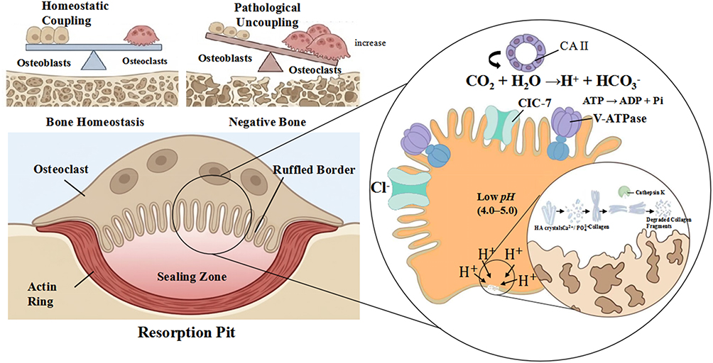 |
Figure 1 Schematic illustration of the mechanism by which the osteoclast-mediated acidic resorption lacuna serves as a specific triggering switch for acid-responsive nanocarriers. |
Construction and Research Advances of Nanocarriers Based on the “Site-Effective Exposure Difference” Strategy
Acid Response and Drug Delivery Kinetic Mechanism Based on Chemical Structure Fracture
The “structural fragmentation” strategy based on acid-labile chemical bond cleavage represents the most direct and classic approach to implementing the SEED system. Such carriers rely on the high sensitivity of specific chemical bonds, including hydrazone, acetal, cis-aconityl, and Schiff base linkages, to low-pH environments (Figure 2). Under physiological conditions, these chemical bonds remain thermodynamically stable, ensuring that the drug is either tightly sequestered or effectively shielded by a protective layer during systemic circulation. This stability satisfies the requirements for the carrier to withstand plasma proteins and esterase-rich environments, thereby significantly reducing background leakage and toxicity toward non-target organs.25
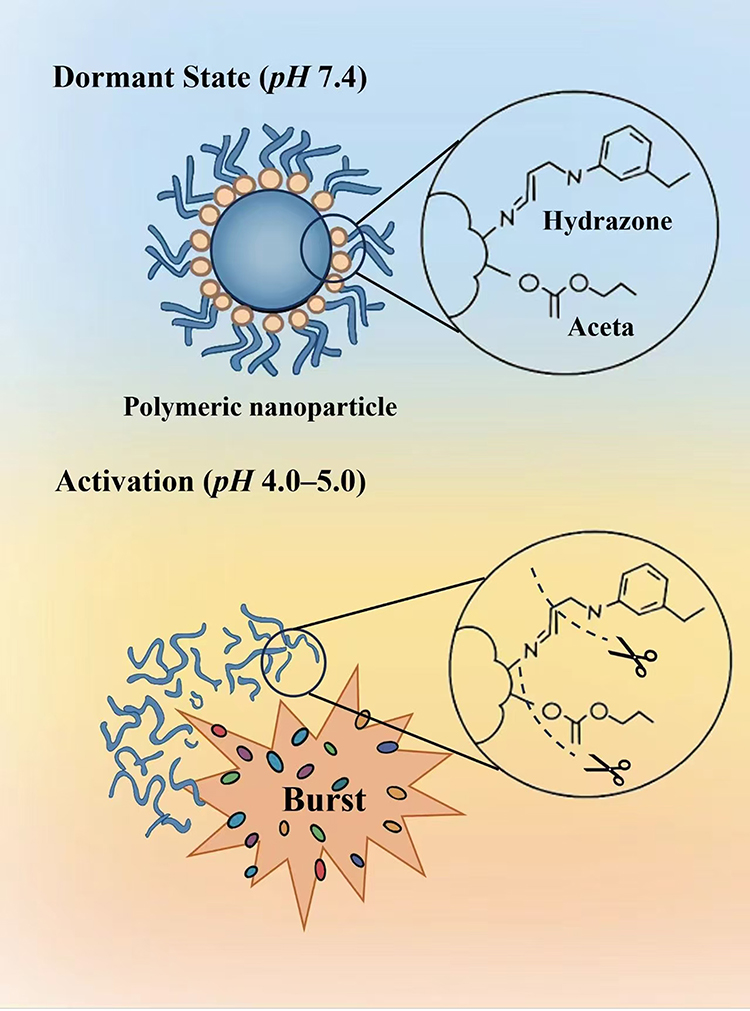 |
Figure 2 Schematic representation of the pH-triggered activation mechanism for acid-responsive polymeric nanocarriers based on the chemical bond cleavage strategy. |
When carriers are enriched via systemic circulation in acidic environments, such as the tumor microenvironment or intracellular lysosomes, the high concentration of protons acts as a catalyst to induce the protonated hydrolysis of acid-labile chemical bonds. Beyond the scenarios primarily discussed in this paper, this mechanism is equally applicable to other physiological acidic microenvironments, including the bone resorption lacunae.
Depending on the distribution of acid-sensitive linkages within the molecular topology of the carrier, this responsive process exhibits distinct drug release kinetics. In prodrug systems where small-molecule drugs are grafted onto polymer side chains via acid-sensitive linkers, the cleavage of these bonds triggered by the acidic environment leads directly to the liberation of the drug from the carrier backbone. Conversely, when acid-sensitive groups are located at the junction points of amphiphilic block copolymers or within the hydrophobic core, protonation disrupts the balance of hydrophilic-hydrophobic affinity. This disruption subsequently induces the total disassembly of the nanostructure, which facilitates the rapid release of encapsulated hydrophobic drugs or biological macromolecules.13
Furthermore, the mechanism of acid-sensitive chemical bond cleavage is extensively applied in the construction of pH-responsive nanocarriers. By incorporating pH-responsive covalent bonds during the synthesis of nanocarriers, this strategy ensures that drug-polymer conjugates remain stable in the neutral pH environment of healthy tissues while undergoing site-specific degradation within the pathological acidic microenvironment of bone. Consequently, this enables the controlled release of the therapeutic payload.
Specifically, pH-responsive charge-transfer polymer nanoparticles can alter their surface charge and/or hydrophilic-hydrophobic balance in response to environmental pH, which subsequently induces structural rearrangements, solubilization, or total disassembly of the nanoparticles. For instance, the hydrophobicity of certain cationic polymers increases as pH decreases, whereas the hydrophilicity of anionic polymers weakens under acidic conditions. Such designs allow nanocarriers to exhibit differential behaviors between systemic circulation and pathological bone microenvironments; they maintain stability under the neutral pH conditions of blood circulation but trigger drug release upon localization within the acidic resorption lacunae created by osteoclast-secreted H+. The core advantage of this strategy lies in its ability to leverage the acidic microenvironmental features common in bone pathologies, thereby providing a versatile platform for the treatment of various bone-related diseases.26
Proton-Driven Surface Charge Reversal and Interface Electrostatic Enhancement Mechanisms
The physicochemical interactions at the interface between nanocarriers and cell membranes are critical factors determining the efficiency of intracellular drug delivery. During this process, the modulation of carrier surface charge presents a significant functional trade-off, the effects of which are highly dependent on the target cell type. Non-phagocytic cells tend to internalize cationic nanocarriers through electrostatic interactions; although this offers higher endocytic efficiency, it is often accompanied by more pronounced cytotoxicity. Conversely, phagocytic cells preferentially uptake anionic nanocarriers.27 This contradiction in charge attributes has established surface dynamic modulation strategies, based on charge reversal, as a primary research focus. This approach has been extensively validated in the field of tumor targeting; for example, carriers based on zwitterionic polymers can transition from a negative to a positive charge as pH decreases, thereby significantly enhancing endocytic efficiency.28 By endowing carriers with responsiveness to pH microenvironments, this strategy effectively reconciles the inherent contradiction between the “systemic circulation stability” and the “target tissue affinity” of nanomedicines as (Figure 3).
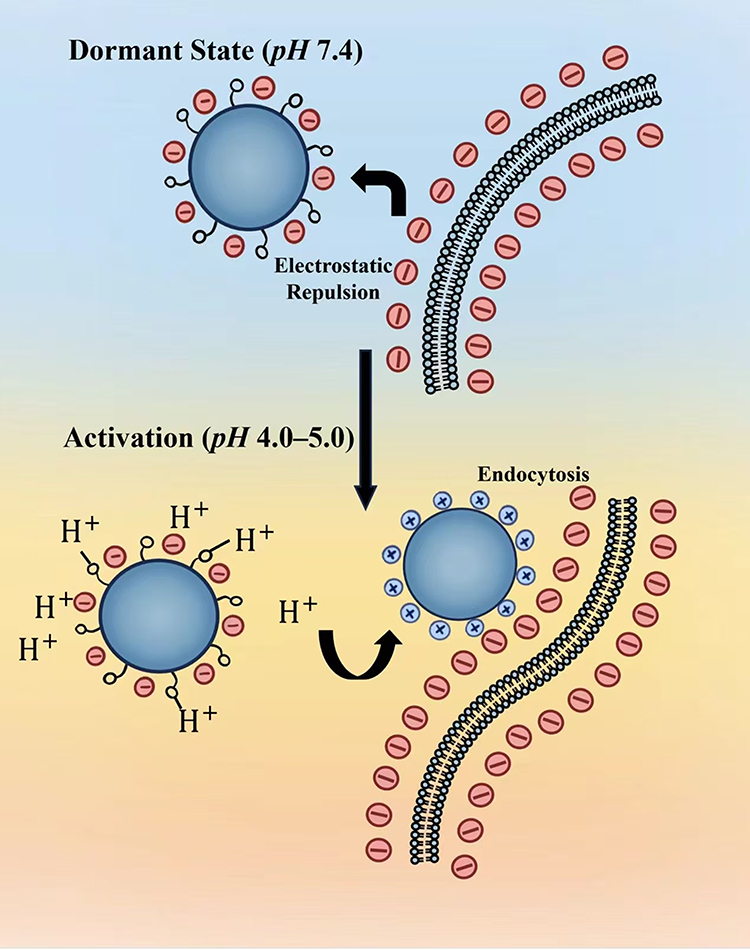 |
Figure 3 Schematic representation of the pH-triggered activation mechanism for acid-responsive polymeric nanocarriers based on the surface charge-reversal strategy. |
This pH-responsive charge-reversal strategy was originally inspired by the precision design required for the acidic tumor microenvironment; however, the underlying interfacial physicochemical principles are equally well-suited for targeted delivery in osteoporosis. Specifically, by introducing acid-sensitive functional moieties such as histidine, tertiary amine groups, sulfonamides, or cis-aconityl derivatives, researchers can construct intelligent interfaces capable of sensing microenvironmental fluctuations. Under physiological pH conditions, these groups maintain a deprotonated state, rendering the carrier slightly negatively charged or electrically neutral. This configuration endows the delivery system with excellent long-circulation characteristics during systemic transit, effectively evading clearance by the reticuloendothelial system.29 Given that the local microenvironment established by osteoclasts during bone resorption possesses a higher proton abundance compared to typical tumor tissues, it provides a more potent biochemical driving force for the charge reversal of nanocarriers. Once the carriers aggregate within the acidic resorption lacunae, the proton-rich microenvironment rapidly drives the protonation of surface groups or induces the cleavage of acid-labile chemical bonds. This process mediates a significant shift in the Zeta potential from negative to positive, thereby enhancing the endocytic efficiency of osteoclasts or the electrostatic adsorption of the drug onto the damaged bone surface. This transmembrane entry into the cell initiates the next stage of the delivery chain, which involves navigating the even more complex intracellular microenvironment.
Upon achieving efficient endocytosis, the focus of the delivery mission shifts toward modulating the intracellular trafficking of the carrier, specifically the circumvention of lysosomal degradation, which is essential for further enhancing drug bioavailability. Cationic polymers with high buffering capacity, represented by polyethylenimine, can trigger the swelling and rupture of organelles by leveraging the “proton sponge effect” upon entering acidic endosomal or lysosomal environments. This mechanism effectively facilitates the escape of the drug from lysosomes into the cytosol, preventing the enzymatic inactivation of biological macromolecules and ensuring effective delivery at the subcellular level.30,31 Such a cascaded charge-responsive mechanism endows the carrier with the ability to adaptively regulate its surface properties within complex physiological environments, thereby establishing an efficient delivery pathway extending from systemic circulation directly to the cytoplasm.
Proton-Consuming Inorganic Matrix Dissociation and in situ Delivery of Bioactive Ions
Utilizing bone mineral components or their analogs as carrier matrices provides a biological foundation for bone-targeted acid-responsive strategies. These inorganic materials are inherently sensitive to acidic environments. Under physiological pH, they maintain a solid state to encapsulate drugs. However, upon exposure to acidic environments ranging from pH 4.0 to 6.0, which encompasses the pH-responsive range of both the extracellular acidified zone of bone resorption lacunae and intracellular lysosomes,32,33, the material bulk undergoes acid-induced dissolution or phase transition12 (Figure 4).
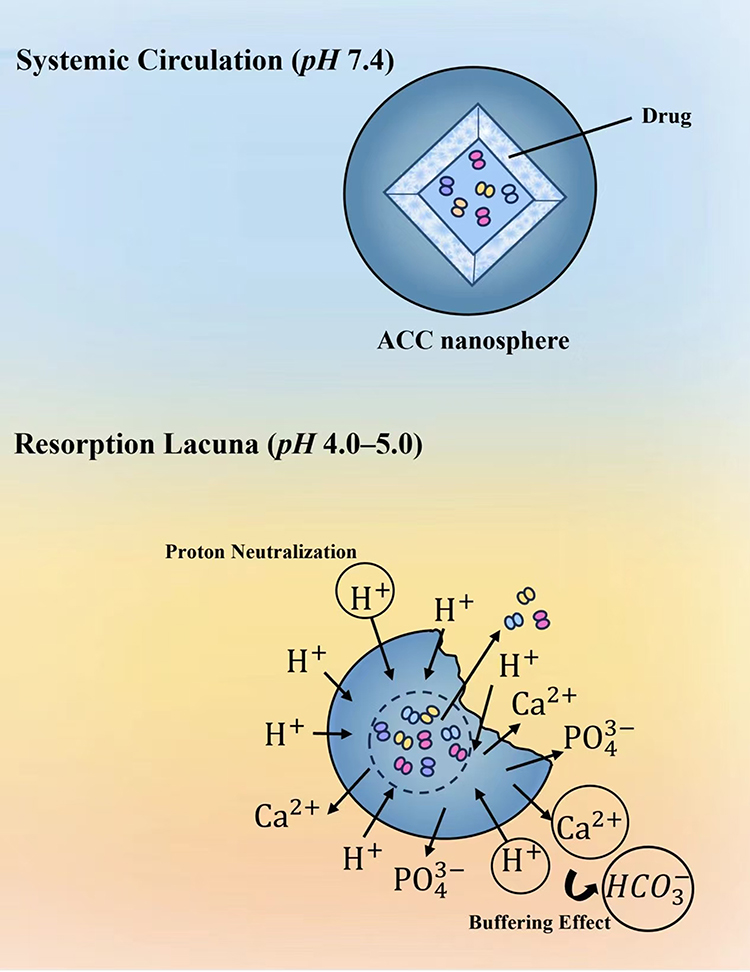 |
Figure 4 Schematic representation of the pH-triggered activation mechanism for acid-responsive nanocarriers based on the inorganic framework dissociation strategy. |
The acid-responsive degradation of inorganic matrices is not merely a physical structural disassembly; rather, it endows the delivery system with a unique therapeutic synergistic effect. From a chemical thermodynamics perspective, the dissolution of calcium carbonate or calcium phosphate is essentially a process accompanied by the stoichiometric consumption of protons. By mimicking the sacrificial dissolution of bone minerals, this delivery system generates a potent “proton buffering effect” that effectively neutralizes the localized acidification within the resorption lacunae. This not only thermodynamically reverses the acidic gradient but also disrupts the specific pH environment required for osteoclast enzyme activation, thereby severing the chemical and biological drivers of pathological bone resorption at their source.34 Furthermore, the high concentrations of calcium and phosphate ions released during the acid-induced degradation of the carrier are not simply metabolic end-products, but are critical bioactive messengers that regulate bone homeostasis. Existing studies have confirmed that a localized surge in extracellular calcium ion concentration can exert a dual inhibitory effect via signaling pathways mediated by calcium-sensing receptors. This not only induces the apoptotic program in mature osteoclasts,35 but also directly suppresses the differentiation and fusion processes of osteoclast precursors.36 Simultaneously, the in situ release of mineral ions provides the necessary nucleation sites and chemical substrates for hydroxyapatite deposition mediated by osteoblasts, thereby endowing the delivery system with additional pro-osteogenic potential.37 This synergistic therapeutic strategy, which integrates the inhibition of bone resorption with the promotion of bone formation, helps to fundamentally correct the imbalanced bone remodeling coupling mechanism characteristic of osteoporosis.
Based on this theoretical foundation, Amorphous Calcium Carbonate (ACC) has emerged as an ideal carrier for constructing microenvironment-responsive systems due to its thermodynamic metastability and superior acid sensitivity. Targeting the osteoclast-dominated acidic microenvironment, Yu et al developed a hexaglutamate-modified, phospholipid-coated ACC nanocarrier loaded with the osteoclast inhibitor oroxylin A. This design utilizes the lipid bilayer to ensure the stability of the carrier during systemic circulation. However, once endocytosed into the acidic resorption lacunae, the internal ACC core undergoes rapid proton-consuming dissolution. This process not only triggers the pH-responsive sustained release of the drug but also effectively reverses the localized acidification within the lacunae by neutralizing protons, thereby blocking the degradation of the bone matrix driven by the acidic environment.12
To further enhance the tissue specificity and regenerative efficacy of delivery systems, Wang et al developed a tetracycline-modified, bone-targeting ACC delivery system. By leveraging the specific affinity of tetracycline for the bone matrix, this system achieves the precise anchoring and enrichment of the carrier on bone surfaces. Research has confirmed that, due to the responsive degradation characteristics of ACC in localized micro-acidic environments, the liberated exogenous calcium ions can activate the BMP-2/Smad signaling pathway. This activation exerts a significant synergistic osteogenic effect with the loaded simvastatin, effectively upregulating osteoblast activity. These studies collectively validate the translational potential of the “microenvironment modulation” strategy, based on acid-responsive inorganic materials, for the precision treatment of osteoporosis.38
These delivery systems, based on the acid-responsive degradation of inorganic matrices, achieve the precise remodeling of the bone resorption lacuna environment at the microscopic scale through a unique “acid erosion–reconstitution” mechanism. This strategy transcends the limitations of conventional single-pathway pharmacological inhibition by leveraging the dual synergistic effects of neutralizing the pathological acidic microenvironment and releasing osteogenic bioactive ions in situ. Consequently, it effectively restores the compromised balance of bone remodeling coupling. This biomimetic mineralization strategy not only significantly enhances the targeting specificity and safety of the treatment but also establishes a robust theoretical and material science foundation for the development of next-generation anti-osteoporotic therapeutics, which are capable of reversing bone loss and possessing the dual efficacy of anti-resorption and pro-regeneration.
In summary, a systematic review of the three predominant acid-responsive strategies—chemical bond cleavage, surface charge reversal, and inorganic framework dissociation—reveals that the core of nanocarrier design lies in the precise modulation of physicochemical parameters. This approach facilitates the effective transformation from systemic circulation stability to site-specific activation at the pathological lesion (Table 1).
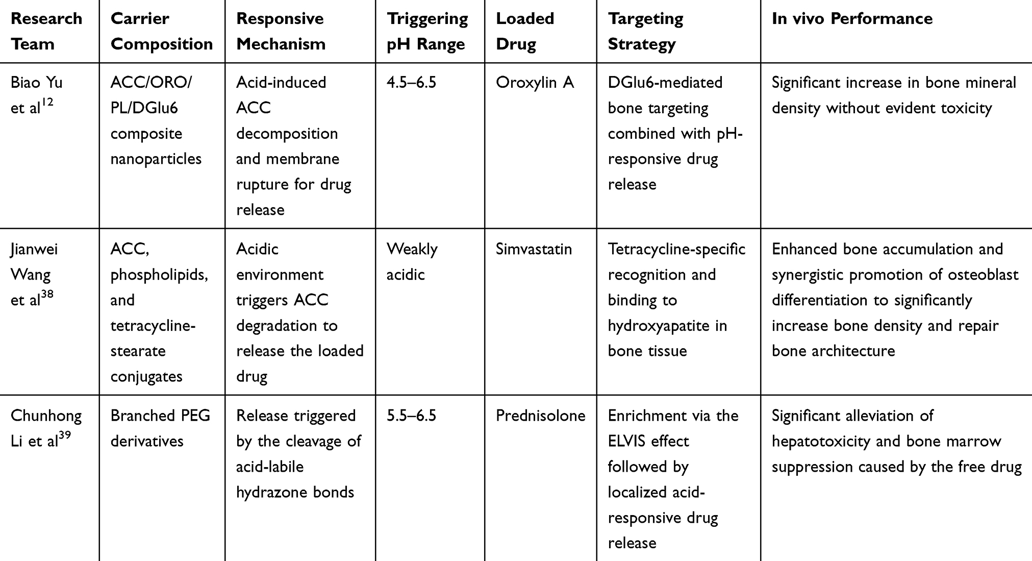 |
Table 1 Comparison of the Performance and Applications of Representative Acid-Responsive Nanocarriers in the Treatment of Bone-Related Diseases |
Benchmarking and Evaluation of Performance for Stimuli-Responsive Nanocarriers
Following a systematic review of the research progress in acid-responsive nanocarriers, it is essential to establish a set of standardized evaluation benchmarks to measure their translational efficacy within complex bone pathological environments. This shift in evaluation paradigm essentially stems from the fundamental differences in pharmacokinetic logic between orthopedic delivery and conventional tumor delivery.
From a mechanistic perspective, tumor delivery has long relied on the EPR, which utilizes the permeability of nascent vasculature to achieve passive accumulation. The average delivery efficiency is only approximately 0.7% ID (Injected Dose), with most cases falling below 5% ID, as a vast majority of the injected dose is cleared by Mononuclear Phagocyte System (MPS) organs such as the liver and spleen.40–42 This passive logic based on vascular leakage faces even more severe challenges in the field of orthopedics, as the bone remodeling microenvironment lacks similar physical pores, which leads to a high susceptibility for drugs to generate non-specific background exposure.43,44 To address this, the present study proposes the SEED Index, which aims to provide a quantitative framework for the orthopedic field that transcends traditional “targeting rates” by evaluating the total ratio of effective exposure at the target site to the systemic circulation background exposure.45 Within this framework, the physical connotation of this ratio is further decomposed into the product of the spatial distribution gain (α) and the kinetic transformation gain (β), expressed as SEED = α×β. Specifically, α is determined based on the increase in bone deposition as listed in the table, measuring the distribution advantage of the carrier at the lesion site relative to the systemic background, thereby addressing the signal-to-noise ratio of spatial occupancy. Meanwhile, β is defined by the inverse relationship between clearance rate and systemic retention, serving to quantify the attenuation effect of systemic circulation on localized effective exposure, which addresses the response efficiency in the temporal dimension.
This algorithm establishes a standardized evaluation benchmark. Retrospective analysis of data from the typical bone-targeting carrier HPMA-ALN reveals that, despite its excellent bone affinity (high α), its SEED index remains suppressed within an inefficient range of 1.1–1.4 due to excessively high systemic retention and low transformation efficiency (extremely low β). This demonstrates that even under conditions of limited α, a system with high β can still achieve an order-of-magnitude leap in target-site exposure relative to background noise, providing a standardized computational foundation for evaluating adaptive nanocarriers.
This “pseudo-responsive” delivery logic faces more severe challenges in the field of orthopedics, as drugs are highly susceptible to excessive non-specific background exposure. If traditional evaluation systems are applied, the typical long-circulating bone-targeting carrier HPMA-ALN is often judged as highly efficient due to its excellent targeting affinity. However, a retrospective estimation integrating the various biodistribution components of this system reveals that its kinetic response efficiency is remarkably low. Specifically, the combination of high systemic retention levels and slow localized clearance rates leads to severe background noise interference, which prevents effective performance coupling with the pathological microenvironment. Consequently, its SEED Index remains suppressed within an inefficient range of 1.1–1.4. Retrospective data analysis indicates that 1.5 constitutes a critical performance demarcation point for measuring delivery efficiency. Systems falling below this value typically exhibit insurmountable off-target effects, whereas exceeding this threshold signifies an effective widening of the therapeutic window. The establishment of this threshold aims to provide a standardized evaluation benchmark for assessing the clinical translational efficacy of nanodelivery systems within complex bone microenvironments by quantitatively weighing the kinetic interplay between site-specific therapeutic gain and systemic toxicity risks.46
In sharp contrast, the polyphosphazene-based bone-targeting delivery system constructed by Zhong et al demonstrates a different paradigm. By leveraging alendronate-mediated ligand-receptor affinity, this system achieves a significant spatial distribution gain (α), with its targeting binding efficiency increasing 2.6-fold compared to the free drug. This improvement fundamentally addresses the signal-to-noise ratio of carrier spatial occupancy within bone tissue. Even more critical is the remarkably high kinetic transformation gain (β) exhibited by its molecular backbone, where the drug release rate in an acidic environment reaches 2.4 times that in a physiological neutral environment. This efficient kinetic profile enables the carrier to sense proton flux instantaneously upon contact with the resorption lacunae and rapidly complete the transition from “locked” to “released” states, thereby resolving the issue of response efficiency in the temporal dimension. Modeling estimations reveal that by virtue of the dual advantages of spatial distribution and kinetic transformation, and through the execution of the SEED algorithmic logic, the SEED Index of this system achieves a leapfrog advancement from the 1.1–1.4 range of traditional carriers to over 6.0. This underscores the index’s utility as a standardized evaluation benchmark for assessing the clinical translational efficacy of nanodelivery systems within complex bone microenvironments.47
The aforementioned comparison profoundly reveals the phenomenon of spatiotemporal dissociation between apparent distribution and target utilization. As a core metric for measuring translational efficacy, the SEED Index is essentially a function constrained by the synergy between spatial-dimensional response thresholds and temporal-dimensional kinetic gains. If a nanocarrier lacks an acid-triggered dynamic response mechanism, it will struggle to overcome the kinetic barriers to functional site transformation, even if high-flux loading is achieved on the bone surface. Based on this, the present study proposes that the research focus for future bone-targeted drugs should shift from a singular “spatial tropism” toward “spatiotemporal precision release” driven by both α and β values. Only when the SEED Index significantly surpasses the critical threshold of 1.5 can a delivery system break through linear distribution constraints and fundamentally suppress non-specific background exposure, thereby possessing substantial potential for clinical translation.
However, although the SEED evaluation framework establishes a theoretical benchmark, its experimental validation within complex physiological environments still faces severe challenges. This difficulty primarily stems from the scarcity of in vivo quantitative monitoring tools; specifically, it remains arduous to perform in situ measurements of proton flux and drug release concentrations within the resorption lacunae without compromising the physiological integrity of the osteoclast sealing zone. Due to the lack of microscopic detection methods, the calculation of β values currently relies heavily on derivations from in vitro static models, which significantly limits the predictive accuracy and practical significance of the SEED framework at the organismal level. Therefore, the development of pathological microenvironment sensing technologies with high spatiotemporal resolution has become a critical requirement to bridge the gap in the transition of the SEED framework from a qualitative concept to a quantitative engineering criterion.
Non-Targeted Accumulation Risks and Safety Considerations of Acid-Responsive Carriers
Upon entering the biological environment, nanocarriers rapidly adsorb plasma proteins to form a biomolecular corona, which confers a new “biological identity” to the nanomaterials. Based on this principle, it can be inferred that this identity transformation may involve the adsorption and activation of complement proteins, subsequently triggering opsonization. This process leads to the recognition of carriers by the MPS and their accumulation in organs such as the liver and spleen, thereby constituting a common bottleneck in bone-targeted delivery.48 However, the bone-targeting effect generated by tetracycline modification enables the drug to be preferentially distributed in bone tissue. In early stages, the concentrations in the liver and kidney are significantly lower than those in the blood, and no significant abnormalities have been observed in short-term blood biochemical or histopathological examinations, initially demonstrating favorable clinical safety.49 However, for nanotherapeutic regimens requiring long-term or repeated administration, short-term evaluations are insufficient to reveal the complex long-term fate of nanoparticles. Bourquin et al pointed out that clearance mechanisms, such as post-uptake release, intracellular degradation, intercellular transfer, and migration across tissue barriers, remain poorly understood. Even in secondary accumulation organs, the long-term retention of nanomaterials may trigger unforeseen biological effects. Of greater concern is that organs rich in the MPS, such as the liver and spleen, are the primary sites of accumulation. For non-degradable inorganic nanomaterials, this entrapment may persist for months or even years. Such chronic accumulation poses risks of pathological alterations including oxidative stress, inflammatory responses, and fibrosis, although the specific molecular mechanisms require further investigation.50
In contrast, while degradable systems such as ACC and polymeric nanoparticles can be metabolized by the physiological environment, the safety of their degradation products or metabolic intermediates still requires comprehensive evaluation, particularly regarding potential cumulative toxicity in organs like the kidneys. For instance, if certain polymer fragments cannot pass through glomerular filtration, they may accumulate within renal tubular epithelial cells and interfere with normal renal function.51,52 Furthermore, although ACC is utilized in bone tissue engineering, controlling its in vivo stability remains a challenge. It is essential to conduct in-depth studies on its degradation kinetics and release behavior in physiological environments to ensure that the material maintains structural integrity before reaching the target tissue, thereby achieving the desired therapeutic effect.53 The in vivo fate of non-degradable materials further substantiates the necessity for such rigor. Taking bovine serum albumin-coated gold nanoparticles as an example, due to the lack of effective degradation pathways, they exhibit long-term retention in MPS organs, whereas the nanoparticle content in the kidneys gradually decreases over time. Such chronic exposure can induce significant inflammatory responses and mild pathological changes in the liver and spleen; although the kidneys harbor a lower particle content, a certain level of oxidative stress response is also observed. This suggests that different organs possess varying sensitivities to nanoparticle exposure, necessitating a comprehensive assessment of their long-term biosafety.54 This comparison strongly demonstrates that in the treatment of osteoporosis, developing bone-targeted delivery systems with precise degradation kinetics is the core prerequisite for evading long-term immuno-inflammatory risks and achieving successful clinical translation.
Challenges in Clinical Translation: From Pathological Heterogeneity to Administration Constraints
Postmenopausal osteoporosis (PMOP) constitutes the most critical clinical application scenario for acid-responsive systems, with its pathological hallmark being the hyperactivation of bone resorption triggered by the precipitous decline in estrogen. According to the description of the bone microenvironment by Lavrador et al, osteoclasts maintain an extreme acidic environment with a pH of approximately 4.0 within the sealed compartment of the bone resorption lacuna, where they release cathepsin K and hydrochloric acid to degrade the organic bone matrix and hydroxyapatite crystals.25 Within the SEED evaluation framework, this significant proton gradient endows the carrier with exceptionally high kinetic transformation gain, ensuring that mechanisms such as acid-sensitive bond cleavage or charge reversal undergo in situ transformation instantaneously upon contact with the resorption sites. This acute responsiveness effectively overcomes the issue of spatiotemporal dissociation of nanocarriers within bone tissue, achieving an order-of-magnitude increase in drug exposure at the effect sites. By suppressing systemic background exposure at non-target sites, this approach establishes a standardized performance-coupling benchmark for the precision treatment of postmenopausal osteoporosis.
Relevant studies indicate that, in significant contrast to the focal acidification characteristics dominated by osteoclasts in PMOP, the pathological microenvironment of inflammation-related secondary osteoporosis exhibits more complex spatiotemporal features. Under these specific pathological conditions, the origin of the acidic environment transcends the spatial limitations of the V-ATPase proton pump on the apical membrane of osteoclasts within the resorption lacunae.55 Driven by pro-inflammatory cytokines such as TNF-α and IL-6, infiltrating macrophages and associated immune cells undergo significant metabolic reprogramming, producing lactic acid accompanied by proton efflux through the enhancement of the aerobic glycolysis pathway.56 This metabolism-derived acidification mechanism creates diffuse zones of background acidification around the bone tissue, which, together with focal functional acidification, constitutes a heterogeneous pathological acidic landscape. This non-specific acidification background presents a severe engineering challenge to the response precision of nanocarriers. If the response threshold of the carrier is set too broadly, it is highly susceptible to premature drug release within peripheral inflammatory infiltration zones before reaching the target resorption sites via the systemic circulation.57 Within the SEED evaluation framework, this non-specific immune recognition induces a dual negative effect. On one hand, once nanoparticles are encapsulated by opsonins such as complement proteins and immunoglobulins, they trigger rapid recognition and clearance by the Mononuclear Phagocyte System, including Kupffer cells. This results in the massive entrapment of particles within RES organs such as the liver and spleen, which significantly increases the risk of off-target accumulation and potential organ toxicity. On the other hand, because particles are prematurely cleared before reaching the target tissue, the effective drug concentration available for uptake in the circulation decreases drastically. This leads to a sharp decline in the targeting delivery gain, defined as the ratio of effective exposure at the target site to systemic exposure.58 This spatiotemporal response distortion fundamentally undermines the overall targeting efficacy and therapeutic index of the delivery system. Therefore, for inflammation-related secondary bone loss, nanocarriers urgently need to establish high-precision acid-responsive mechanisms. The core of this design lies in the accurate discrimination between the high proton concentration gradient of the osteoclast lacunae and the general inflammatory background. By strengthening the specific coupling of materials to extreme acidic environments, carriers can maintain a high SEED Index amidst complex inflammatory noise, ensuring that the therapeutic payload is precisely focused on functional bone resorption sites rather than dissipated across non-specific inflammatory regions.
Unlike primary osteoporosis, glucocorticoid-induced osteoporosis exhibits more complex pathodynamic characteristics, the core of which lies in the profound intervention of glucocorticoids in the osteoblast-osteoclast coupling process. Glucocorticoids promote osteoclastogenesis by upregulating RANKL expression and downregulating OPG in cells of the osteoblast lineage. Simultaneously, they inhibit the differentiation of mesenchymal stem cells into osteoblasts and induce apoptosis in mature osteoblasts, leading to a surge in bone resorption and the suppression of bone formation, which triggers a transient but severe peak of bone loss.59 Within this specific pathological context, the unidirectional response logic that relies solely on acid-triggered mechanisms to inhibit osteoclast activity reveals significant limitations. Although local blockade of the acidic environment in osteoclast lacunae may temporarily alleviate bone resorption, it fails to reverse the substantial deficit in osteoblast function. Furthermore, it may even impair the self-repair capacity of bone tissue due to the excessive suppression of bone turnover.60,61 Consequently, within the SEED evaluation framework, the delivery strategy for glucocorticoid-induced osteoporosis should evolve from singular site-targeting into spatiotemporal functional coupling. The carrier design should utilize the proton flux released from osteoclast lacunae as the triggering signal to synergistically release pro-osteogenic payloads that activate osteogenic signaling pathways or improve the bone marrow microenvironment while precisely inhibiting osteoclast activity. This dual-effect responsive logic driven by the β parameter aims to reconstruct osteoimmune balance within critical pathological time windows. This approach shifts the therapeutic objective from focal blockade to systemic ecological restoration, thereby significantly enhancing the clinical translational potential of nanocarriers in complex pathological models.
The pathological core of senile osteoporosis is not limited to the hyperactivity of bone resorption but resides more significantly in the overall aging phenotype of the bone marrow microenvironment. In elderly patients, the acidification signals within bone remodeling units may not be as intense or concentrated as those observed in postmenopausal osteoporosis due to the depletion of bone marrow mesenchymal stem cells and the decline of osteogenic potential. Conversely, microenvironmental changes driven by chronic systemic low-grade inflammation cause the proton flux in the bone tissue background to exhibit a state of low-amplitude fluctuation.62
Within the pathological context of low-turnover bone remodeling, the SEED evaluation framework reveals potential efficacy traps inherent in traditional delivery strategies. If a carrier continues to employ response thresholds designed for high-turnover models, the kinetic transformation gain β is highly susceptible to dropping to basal levels due to the inability to be activated by weak acidification signals. This results in an efficacy dilemma of targeting without release. Therefore, for senile osteoporosis, the underlying logic of carrier design should undergo a paradigm shift from extreme response to high-sensitivity coupling. The core of this design lies in optimizing the physicochemical properties of the materials to enhance charge reversal efficiency or molecular bond cleavage rates under subtle proton concentration fluctuations. Such optimizations ensure that even in microenvironments with extremely low bone turnover rates, the SEED Index can still surpass the critical efficacy threshold of 1.5, achieving precise coverage and effective intervention at the remaining bone remodeling sites.
From the perspective of systemic administration, the underlying logic of carrier design focuses on the dynamic antagonism between systemic circulation homeostasis and site-specific spatiotemporal responsiveness. Following intravenous administration, carriers first encounter a complex blood environment. Engineering logic dictates that they must possess high physiological inertness to evade opsonization, which requires designers to perform precise modulation of nanocarrier dimensions, typically strictly controlled within the range of tens to hundreds of nanometers to avoid renal clearance and interception by the mononuclear phagocyte system.63
Designers have employed PEGylated lipids and bisphosphonate-lipid conjugates to co-modify liposomal membranes while integrating pH-sensitive lipid components. The PEG layer provides steric hindrance, which significantly enhances the blood circulation stability of the carrier and evades recognition and clearance by the mononuclear phagocyte system. As bone-affinity ligands, bisphosphonates endow the liposomes with active targeting enrichment capabilities toward bone metastatic sites through efficient chelation with hydroxyapatite. Furthermore, the pH-sensitive lipids ensure that the carrier remains relatively stable under physiological pH environments, while undergoing membrane destabilization in the acidic microenvironment of bone metastases or intracellular lysosomes to trigger the release of doxorubicin, thereby achieving a synergy of long circulation, bone targeting, and controlled drug release.64 This sequential strategy of oriented accumulation followed by triggered response not only optimizes the spatial distribution gain α within the SEED framework but also fundamentally safeguards the robustness of the kinetic transformation gain β by inhibiting background noise release within the systemic circulation.
Compared to the rigorous constraints imposed by systemic administration on circulation homeostasis and biosafety, local delivery circumventing the systemic circulation reduces the dependence on long-circulation stability and focuses drug action on the lesion site. By directly bypassing the filtration effect of the mononuclear phagocyte system, this pathway evades the clearance pressure encountered by carriers in the systemic circulation, thus shifting the design logic toward site-specific retention, controlled release, and localized action. Regarding the mechanisms of local retention, carrier design prioritizes matrix interactions for anchoring rather than immune evasion. Designers may employ strong bone-affinity surface modifications or in situ gelation technologies to utilize specific carrier-bone matrix interactions and physical phase-transition retention. These strategies prolong the residence time of drugs within the local microenvironment, establishing a long-residence characteristic that corresponds to the long-circulation feature of systemic administration.65
This logical refinement based on the administration pathway not only alters the distribution kinetics of the carrier in the spatial dimension but also imposes differentiated requirements for the functional modification of materials. Consequently, after defining the dual boundaries of pathological characteristics and administration constraints, the key to enhancing the therapeutic index lies in the targeted optimization of the physicochemical properties of materials to match specific delivery requirements.
Conclusion and Prospects
The precision treatment of osteoporosis has long been constrained by the contradiction between systemic toxicity and local efficacy.66 To address this bottleneck, this paper introduces the SEED evaluation framework, shifting the research focus from simple “spatial tropism” to spatiotemporally precise release driven by spatial distribution gain (α) and kinetic transformation gain (β). This transition from a qualitative concept to a quantitative engineering criterion establishes a critical performance threshold of SEED > 1.5, providing a benchmarking standard for breaking linear distribution constraints and suppressing non-specific background exposure. In particular, systems represented by amorphous calcium carbonate act not only as acid-responsive carriers but also leverage the synergistic effects of “proton buffering” and “in situ release of bioactive ions” to drive the therapeutic paradigm from single-drug anti-resorption toward intelligent microenvironment remodeling. These systems utilize the highly acidic microenvironment of osteoclast resorption lacunae as an intelligent responsive switch, endowing carriers with circulatory inertness at physiological pH and on-demand release characteristics under pathological pH conditions.26 By leveraging deep remodeling of the local microenvironment, inorganic biomimetic nanocarriers achieve the uncoupling synergy of bone resorption inhibition and bone formation preservation. This drives the evolution of osteoporosis therapy from traditional single-drug anti-resorption toward an intelligent intervention paradigm characterized by the synergy of targeted delivery, microenvironmental responsiveness, and acid neutralization.12
Despite the significant advancements of strategies based on the SEED evaluation framework in preclinical research, their clinical translation still faces formidable challenges. A critical factor restricting their application is the substantial discrepancy between in vitro models and the actual in vivo microenvironment. Existing evaluation systems rely heavily on buffers with constant pH values to simulate acidic environments; however, the real bone pathological microenvironment possesses high spatiotemporal dynamism, with acidity levels fluctuating in real-time according to cellular metabolic states and blood perfusion conditions.32 This misalignment between static simulations and dynamic physiological environments may cause nanocarriers to exhibit delayed response or premature release in vivo. Consequently, there is an urgent need to develop dynamic microfluidic models with high biomimetic characteristics to achieve precise assessment of the intelligent responsive behavior of nanocarriers.67
Furthermore, the engineering scale-up and quality control of nanoformulations represent key bottlenecks restricting their industrialization. To pursue optimal targeting efficacy, current carrier designs often integrate complex surface functionalizations, multi-layered core-shell structures, or specific crystalline morphologies. Such sophisticated laboratory-scale processes frequently encounter practical difficulties during transition to industrial-scale production, including poor batch-to-batch consistency, unstable encapsulation efficiency, and high manufacturing costs. Therefore, achieving rational simplification while maintaining acid-responsive sensitivity to construct nanoformulations with well-defined compositions, streamlined structures, and ease of large-scale quality control has become a core imperative for drug developers.68 Furthermore, in view of the distinct acidification profiles associated with postmenopausal, inflammatory, and senile osteoporosis, carrier design must involve the precise regulation of physicochemical parameters to accurately adapt to different response thresholds, thereby mitigating the risk of premature drug release within non-specific inflammatory backgrounds.
The long-term biosafety and in vivo metabolic fate of nanocarriers constitute the mandatory baseline for their clinical translation. Given that osteoporosis is a chronic condition requiring long-term or even lifelong management, patients often necessitate repeated administration. This requirement mandates that carrier materials must not only possess exceptional biocompatibility but also have clearly defined final degradation products and excretion pathways. Although acid-responsive designs effectively circumvent systemic drug distribution, the non-specific accumulation of the nanomaterials themselves within the reticuloendothelial system, or the induction of chronic inflammation by their degradation products, poses potential iatrogenic risks.54 A particularly severe challenge is that the biological fate of clinically relevant nanomaterials after fulfilling their intended functions remains poorly understood. Significant knowledge gaps exist regarding their post-cellular uptake release kinetics, translocation across biological barriers, and ultimate degradation pathways. This lack of transparency in clearance mechanisms presents a formidable obstacle to establishing safety evaluation systems for repeated administration.50 Furthermore, future research perspectives should expand from singular osteoclast targeting to a broader osteoimmunology dimension, investigating how nanocarriers interact with immune components such as macrophages and T cells while modulating the acidic microenvironment. Such insights are essential to ensure that the process of remodeling bone balance does not disrupt the systemic immune homeostasis.
In summary, intelligent nanodelivery strategies responsive to the acidic microenvironment provide a highly promising technical solution for overcoming the barriers of targeted delivery in osteoporosis treatment. Although significant challenges remain in the construction of biomimetic evaluation models, the engineering scale-up of formulations, and the systemic validation of long-term safety, these bottlenecks are expected to be addressed through the profound integration of materials science, pharmaceutics, and osteoimmunology. Future research should focus on developing next-generation carriers that possess both structural simplicity and functional robustness to accelerate their clinical translation from the laboratory. Ultimately, these advancements will provide safe and efficient new clinical strategies for the precision treatment of osteoporosis.
Funding
This study was supported by the National Natural Science Foundation project (NO. 82174260), Liaoning Province Science and Technology Department joint fund doctoral research project (NO: 2023-BSBA-224).
Disclosure
The authors report no conflicts of interest in this work.
References
1. Compston JE, McClung MR, Leslie WD. Osteoporosis. Lancet. 2019;393(10169):364–18. doi:10.1016/S0140-6736(18)32112-3
2. Sözen T, Özışık L, Başaran NÇ. An overview and management of osteoporosis. Eur J Rheumatol. 2017;4(1):46–56. doi:10.5152/eurjrheum.2016.048
3. Gullberg B, Johnell O, Kanis J. World-wide projections for Hip fracture. Osteoporos Int. 1997;7(5):407–413. doi:10.1007/PL00004148
4. Si L, Winzenberg TM, Jiang Q, Chen M, Palmer AJ. Projection of osteoporosis-related fractures and costs in China: 2010–2050. Osteoporos Int. 2015;26(7):1929–1937. doi:10.1007/s00198-015-3093-2
5. Drake MT, Clarke BL, Khosla S. Bisphosphonates: mechanism of action and role in clinical practice. In
6. Baron R, Ferrari S, Russell, RGG. Denosumab and bisphosphonates: different mechanisms of action and effects. Bone. 2011;48(4):677–692.
7. Weinerman S, Usera GL Antiresorptive therapies for osteoporosis. Manag Medication-Relat Osteonecrosis the Jaw. Oral Maxillofac Clin N Am. 2015;27(4):555–560.
8. Qin A, Cheng TS, Pavlos NJ, Lin Z, Dai KR, Zheng MH. V-ATPases in osteoclasts: structure, function and potential inhibitors of bone resorption. Int J Biochem Cell Biol. 2012;44(9):1422–1435. doi:10.1016/j.biocel.2012.05.014
9. Subhan MA, Yalamarty SSK, Filipczak N, Parveen F, Torchilin VP. Recent advances in tumor targeting via EPR effect for cancer treatment. J Pers Med. 2021;11(6):571.
10. Maeda H, Nakamura H, Fang J. The EPR effect for macromolecular drug delivery to solid tumors: improvement of tumor uptake, lowering of systemic toxicity, and distinct tumor imaging in vivo. Adv Drug Deliv Rev. 2013;65(1):71–79. doi:10.1016/j.addr.2012.10.002
11. Khan MS, Alqahtani T, Al Shmrany H, et al. Enhanced permeability and retention (EPR) effect: advances in nanomedicine for improved tumor targeting. Biomater Adv. 2025;181:214636. doi:10.1016/j.bioadv.2025.214636
12. Yu B, Gao Q, Sheng S, et al. Smart osteoclasts targeted nanomedicine based on amorphous CaCO3 for effective osteoporosis reversal. J Nanobiotechnol. 2024;22(1):153. doi:10.1186/s12951-024-02412-9
13. Zhuo S, Zhang F, Yu J, Zhang X, Yang G, Liu X. pH-sensitive biomaterials for drug delivery. Molecules. 2020;25(23):5649.
14. Ribet AB, Ng PY, Pavlos NJ. Membrane transport proteins in osteoclasts: the ins and outs. Front Cell Dev Biol. 2021;9:644986.
15. Rousselle AV, Heymann D. Osteoclastic acidification pathways during bone resorption. Bone. 2002;30(4):533–540. doi:10.1016/S8756-3282(02)00672-5
16. Delaisse JM, Søe K, Andersen TL, Rojek AM, Marcussen N. The mechanism switching the osteoclast from short to long duration bone resorption. Front Cell Dev Biol. 2021;9:644503. doi:10.3389/fcell.2021.644503
17. Stenbeck G. Formation and function of the ruffled border in osteoclasts. Semin Cell Dev Biol. 2002;13(4):285–292. doi:10.1016/S1084952102000587
18. Everts V, Jansen ID, de Vries TJ. Mechanisms of bone resorption. Bone. 2022;163:116499. doi:10.1016/j.bone.2022.116499
19. Yuan FL, Xu MH, Li X, Xinlong H, Fang W, & Dong J The roles of acidosis in osteoclast biology. Front Physiol. 2016;7:222.
20. Ahn H, Kim JM, Lee K, Kim H, Jeong D. Extracellular acidosis accelerates bone resorption by enhancing osteoclast survival, adhesion, and migration. Biochem Biophys Res Commun. 2012;418(1):144–148.
21. Guo DY, Chen ZH, Fu YF, et al. Cilengitide inhibits osteoclast adhesion through blocking the αvβ3-mediated FAK/Src signaling pathway. Heliyon. 2023;9(7).
22. Pereverzev A, Komarova SV, Korčok J, et al. Extracellular acidification enhances osteoclast survival through an NFAT-independent, protein kinase C-dependent pathway. Bone. 2008;42(1):150–161. doi:10.1016/j.bone.2007.08.044
23. Komarova SV, Pereverzev A, Shum JW, Sims SM, Dixon SJ. Convergent signaling by acidosis and receptor activator of NF-κB ligand (RANKL) on the calcium/calcineurin/NFAT pathway in osteoclasts. Proc Natl Acad Sci. 2005;102(7):2643–2648. doi:10.1073/pnas.0406874102
24. Li X, Ye JX, Xu MH, Zhao MD, Yuan FL. Evidence that activation of ASIC1a by acidosis increases osteoclast migration and adhesion by modulating integrin/Pyk2/Src signaling pathway. Osteoporosis Int. 2017;28(7):2221–2231. doi:10.1007/s00198-017-4017-0
25. Lavrador P, Gaspar VM, & Mano JF Stimuli-responsive nanocarriers for delivery of bone therapeutics–barriers and progresses. J Control Release. 2018;273:51–67.
26. Li M, Yu B, Wang S, Zhou F, Cui J, Su J. Microenvironment-responsive nanocarriers for targeted bone disease therapy. Nano Today. 2023;50:101838. doi:10.1016/j.nantod.2023.101838
27. Fröhlich E. The role of surface charge in cellular uptake and cytotoxicity of medical nanoparticles. Int J Nanomed. 2012;5577–5591. doi:10.2147/IJN.S36111
28. Yuan YY, Mao CQ, Du XJ, Du JZ, Wang F, Wang J. Surface charge switchable nanoparticles based on zwitterionic polymer for enhanced drug delivery to tumor. Adv Mater. 2012;24(40):5476–5480. doi:10.1002/adma.201202296
29. Zhang P, Chen D, Li L, Sun K. Charge reversal nano-systems for tumor therapy. J Nanobiotechnol. 2022;20(1):31. doi:10.1186/s12951-021-01221-8
30. Boussif O, Lezoualc’h F, Zanta MA, et al. A versatile vector for gene and oligonucleotide transfer into cells in culture and in vivo: polyethylenimine. Proc Natl Acad Sci U S A. 1995;92(16):7297–7301.
31. Oh YK, Suh D, Kim JM, Choi HG, Shin K, Ko JJ, et al. Polyethylenimine-mediated cellular uptake, nucleus trafficking and expression of cytokine plasmid DNA. Gene Ther. 2002;9(23):1627–1632.
32. Maeda H, Kowada T, Kikuta J, Furuya M, Shirazaki M, Mizukami S, Ishii M, Kikuchi K. Real-time intravital imaging of pH variation associated with osteoclast activity. In: Nat Chem Biol.
33. Jang S, Lee S, Kim H, Ham J, Seo J-H, Mok Y, Noh M, Lee Y. Preparation of pH-sensitive CaP nanoparticles coated with a phosphate-based block copolymer for efficient gene delivery. Polymer. 2012;53(21):4678–4685.
34. Arnett TR. Extracellular pH regulates bone cell function. J Nutr. 2008;138(2):415S–418S. doi:10.1093/jn/138.2.415S
35. Lorget F, Kamel S, Mentaverri R, et al. High extracellular calcium concentrations directly stimulate osteoclast apoptosis. Biochem Biophys Res Commun. 2000;268(3):899–903. doi:10.1006/bbrc.2000.2229
36. Kanatani M, Sugimoto T, Kanzawa M, Yano S, Chihara K. High extracellular calcium inhibits osteoclast-like cell formation by directly acting on the calcium-sensing receptor existing in osteoclast precursor cells. Biochem Biophys Res Commun. 1999;261(1):144–148.
37. Jeong J, Kim JH, Shim JH, Hwang NS, Heo CY. Bioactive calcium phosphate materials and applications in bone regeneration. Biomater Res. 2019;23(1):4. doi:10.1186/s40824-018-0149-3
38. Wang J, Tao S, Jin X, et al. Calcium supplement by tetracycline guided amorphous calcium carbonate potentiates osteoblast promotion for synergetic osteoporosis therapy. Theranostics. 2020;10(19):8591. doi:10.7150/thno.45142
39. Li C, Li H, Wang Q, et al. pH-sensitive polymeric micelles for targeted delivery to inflamed joints. J Control Release. 2017;246:133–141. doi:10.1016/j.jconrel.2016.12.027
40. Hansen AE, Petersen AL, Henriksen JR, et al. Positron emission tomography based elucidation of the enhanced permeability and retention effect in dogs with cancer using copper-64 liposomes. ACS Nano. 2015;9(7):6985–6995. doi:10.1021/acsnano.5b01324
41. Børresen B, Henriksen JR, Clergeaud G, et al. Theranostic imaging may vaccinate against the therapeutic benefit of long circulating PEGylated liposomes and change cargo pharmacokinetics. ACS Nano. 2018;12(11):11386–11398. doi:10.1021/acsnano.8b06266
42. Chen Q, Yuan L, Chou WC, et al. Meta-analysis of nanoparticle distribution in tumors and major organs in tumor-bearing mice. Acs Nano. 2023;17(20):19810–19831. doi:10.1021/acsnano.3c04037
43. Shi J, Kantoff PW, Wooster R, Farokhzad OC. Cancer nanomedicine: progress, challenges and opportunities. Nat Rev Cancer. 2017;17(1):20–37. doi:10.1038/nrc.2016.108
44. Wilhelm S, Tavares AJ, Dai Q, et al. Analysis of nanoparticle delivery to tumours. Nat Rev Mater. 2016;1(5):16014.
45. Klara J, Lewandowska-łańcucka J. How efficient are alendronate-nano/biomaterial combinations for anti-osteoporosis therapy? An evidence-based review of the literature. Int J Nanomed. 2022;17:6065–6094. doi:10.2147/IJN.S388430
46. Pan H, Sima M, Kopeckova P, et al. Biodistribution and pharmacokinetic studies of bone-targeting N-(2-hydroxypropyl) methacrylamide copolymer− alendronate conjugates. Mol Pharmaceut. 2008;5(4):548–558. doi:10.1021/mp800003u
47. Zhong Y, Luo P, Yang X, et al. Bone-targeted acid-responsive polyphosphazene-based resveratrol self-framed delivery system with superior efficacy for osteoporosis treatment in ovariectomized mice. Mater Des. 2026;262:115449. doi:10.1016/j.matdes.2026.115449
48. Monopoli MP, Aberg C, Salvati A, Dawson KA. Biomolecular coronas provide the biological identity of nanosized materials. In: Nano-Enabled Medical Applications. 2020:205–229.
49. Liang Q, Zhang P, Zhang L, et al. Development of tetracycline-modified nanoparticles for bone-targeted delivery of anti-tubercular drug. Front Bioeng Biotechnol. 2023;11:1207520. doi:10.3389/fbioe.2023.1207520
50. Bourquin J, Milosevic A, Hauser D, et al. Biodistribution, clearance, and long-term fate of clinically relevant nanomaterials. Adv Mater. 2018;30(19):1704307. doi:10.1002/adma.201704307
51. Gillings HL, Rojas-Canales DM, Wong SW, et al. Nanoplastic toxicity and uptake in kidney cells: differential effects of concentration, particle size, and polymer type. Cell Biol Toxicol. 2026;42. doi:10.1007/s10565-025-10135-2
52. Shang S, Li X, Wang H, et al. Targeted therapy of kidney disease with nanoparticle drug delivery materials. Bioact Mater. 2024;37:206–221.
53. Liu H, Wen Z, Liu Z, et al. Unlocking the potential of amorphous calcium carbonate: a star ascending in the realm of biomedical application. Acta Pharmaceutica Sinica B. 2024;14(2):602–622. doi:10.1016/j.apsb.2023.08.027
54. Jakic K, Selc M, Razga F, et al. Long-term accumulation, biological effects and toxicity of BSA-coated gold nanoparticles in the mouse liver, spleen, and kidneys. Int J Nanomed. 2024;19:4103–4120. doi:10.2147/IJN.S443168
55. Wu C, Sun Z, He C, Sun R. Ph-dependent osteoimmunomodulation: molecular mechanisms and therapeutic targeting in bone disorders. Pharmacol Res. 2025;218:107838. doi:10.1016/j.phrs.2025.107838
56. Wen J, Liu J, Wan L, Wang F, Li Y. Metabolic reprogramming: the central mechanism driving inflammatory polarization in rheumatoid arthritis and the regulatory role of traditional Chinese medicine. Front Immunol. 2025;16:1659541. doi:10.3389/fimmu.2025.1659541
57. Cheng Y, Long L, Pei H, Yang Y, Wei W, Deng X, Wang L, Tay FR, and Jingzhi M ‘Recent advances in stimulus-responsive nanocarriers for gene therapy.’. Adv Sci. 2021;8(14):2100540):.
58. Peralta-Cuevas E, Degollado-Hernández NY, Martínez-Ortiz IC, Gutierrez-Onofre AJ, Garcia-Atutxa I, Villanueva-Flores F, et al. How do nanoparticle properties shape pharmacokinetics and pharmacodynamics? A mechanistic review. Front Pharmacol. 2025;16:1704814.
59. Peng CH, Lin WY, Yeh KT, Chen H, Wu WT, Lin MD. The molecular etiology and treatment of glucocorticoid-induced osteoporosis. Tzu Chi Med J. 2021;33(3):212–223. doi:10.4103/tcmj.tcmj_233_20
60. Xavier A, Toumi H, Lespessailles E. Animal model for glucocorticoid induced osteoporosis: a systematic review from 2011 to 2021. Int J Mol Sci. 2021;23(1):377. doi:10.3390/ijms23010377
61. Pfeiffenberger M, Damerau A, Gaber T, Buttgereit F. POS0420 generation of a human 3D in vitro bone model that mimics glucocorticoid-induced osteoporosis. 2023.
62. Zhu R, Wan H, Yang H, Song M, Chai Y, Yu B. The role of senescence-associated secretory phenotype in bone loss. Front Cell Dev Biol. 2022;10:841612. doi:10.3389/fcell.2022.841612
63. Guo H, Guo M, Xia Z, Shao Z. Membrane-coated nanoparticles as a biomimetic targeted delivery system for tumour therapy. Biomater Transl. 2024;5(1):33. doi:10.12336/biomatertransl.2024.01.004
64. Ferreira DDS, Faria SD, Lopes SCDA, et al. Development of a bone-targeted pH-sensitive liposomal formulation containing doxorubicin: physicochemical characterization, cytotoxicity, and biodistribution evaluation in a mouse model of bone metastasis. Int J Nanomed. 2016;11:3737–3751. doi:10.2147/IJN.S109966
65. Cheng X, Wei J, Ge Q, et al. The optimized drug delivery systems of treating cancer bone metastatic osteolysis with nanomaterials. Drug Deliv. 2021;28(1):37–53. doi:10.1080/10717544.2020.1856225
66. Farrell KB, Karpeisky A, Thamm DH, Zinnen S. Bisphosphonate conjugation for bone specific drug targeting. Bone Rep. 2018;9:47–60. doi:10.1016/j.bonr.2018.06.007
67. Lipreri MV, Baldini N, Graziani G, Avnet S. Perfused platforms to mimic bone microenvironment at the macro/milli/microscale: pros and cons. Front Cell Dev Biol. 2022;9:760667. doi:10.3389/fcell.2021.760667
68. Joyce P, et al. A translational framework to DELIVER nanomedicines to the clinic. Nat Nanotechnol. 2024;19:1597–1611. doi:10.1038/s41565-024-01754-7
 © 2026 The Author(s). This work is published and licensed by Dove Medical Press Limited. The
full terms of this license are available at https://www.dovepress.com/terms
and incorporate the Creative Commons Attribution
- Non Commercial (unported, 4.0) License.
By accessing the work you hereby accept the Terms. Non-commercial uses of the work are permitted
without any further permission from Dove Medical Press Limited, provided the work is properly
attributed. For permission for commercial use of this work, please see paragraphs 4.2 and 5 of our Terms.
© 2026 The Author(s). This work is published and licensed by Dove Medical Press Limited. The
full terms of this license are available at https://www.dovepress.com/terms
and incorporate the Creative Commons Attribution
- Non Commercial (unported, 4.0) License.
By accessing the work you hereby accept the Terms. Non-commercial uses of the work are permitted
without any further permission from Dove Medical Press Limited, provided the work is properly
attributed. For permission for commercial use of this work, please see paragraphs 4.2 and 5 of our Terms.