
Back to Journals » Infection and Drug Resistance » Volume 19
A Rare Case of Gulosibacter massiliensis Complicating Peritoneal Carcinomatosis

Received 10 March 2026
Accepted for publication 8 June 2026
Published 12 June 2026 Volume 2026:19 606460
DOI https://doi.org/10.2147/IDR.S606460
Checked for plagiarism Yes
Review by Single anonymous peer review
Peer reviewer comments 2
Editor who approved publication: Dr Sandip Patil
Zhineng Xu,1,2 Huiqin Xu,3 Dehua Liu1
1Department of Laboratory, The First Hospital of Kunming, Kunming, Yunnan, 650032, People’s Republic of China; 2School of Medicine, Kunming University, Kunming, Yunnan, 650214, People’s Republic of China; 3School of Medicine, Yunnan University, Kunming, Yunnan, 650500, People’s Republic of China
Correspondence: Dehua Liu, Department of Laboratory, The First Hospital of Kunming, 504 Qingnian Road, Xishan District, Kunming, Yunnan, 650032, People’s Republic of China, Tel +86 158 7799 0175, Email [email protected]
Background: Gulosibacter massiliensis, a Gram-positive, strictly aerobic, and peroxidase-positive bacterium with high motility, was first isolated from a bloodstream infection in 2022. At present, clinical reports on G. massiliensis are limited, with only one case of skin and soft tissue infection reported, and its pathogenicity and infectivity are still unclear.
Case Presentation: A 74-year-old male patient with extensive peritoneal carcinomatosis with ascites infected by G. massiliensis. The patient was admitted to the hospital due to confusion of consciousness. After B-ultrasound, ascites cytology, laboratory testing and multi-department joint diagnosis, it was found that he had malignant ascites, G. massiliensis infection, hepatic encephalopathy, peritoneal secondary malignant tumor and other diseases. During the hospitalization, due to the patient’s critical condition. The patient was deemed unfit for curative treatment and was managed with best supportive care. Ultimately, respecting the family’s wishes, the patient left hospital and went home to receive palliative care. During the laboratory test, we used traditional biochemical reactions and matrix-assisted laser desorption ionization time-of-flight mass spectrometry failed to accurately identify the strain. Finally, the bacteria were finally identified as G. massiliensis by 16S rRNA gene sequencing and NCBI database alignment, and the drug susceptibility results of the bacteria were determined according to the evolutionary relationship and CLSI M45.
Conclusion: This study is the first report that G. massiliensis can cause ascites infection as an opportunistic pathogen in immunocompromised patients. It provides a reference for clinical laboratories to identify this rare pathogen and develops anti-infection programs and also points out the research direction for analyzing its pathogenic mechanism.
Keywords: Gulosibacter massiliensis, clinical report, microbial identification, ascites, Gulosibacter sp.
Introduction
Pathogenic bacteria pose a considerable threat to global public health and patient well-being.1 As of 2021, 1,513 bacterial pathogens affecting humans have been identified, with 197 new species identified between 2011 and 2020, highlighting a notable increase in the diversity of bacterial species responsible for human infections worldwide.2 Gulosibacter massiliensis, a novel bacterium, was identified by Yacouba et al in 2022. This bacterium belongs to the genus Gulosibacter of the family Microbacteriaceae. The species name is derived from the Latin name for Marseille, France, where the type strain was first isolated. G. massiliensis is a high G+C, Gram-positive, motile, non-spore-forming, rod-shaped, aerobic organism that is catalase-positive and oxidase-negative.3,4
Biochemically, the organism exhibits positive reactions for esterase, esterase lipase, leucine arylamidase, and acid phosphatase, and demonstrates the ability to ferment rhamnose, citrate, and D-trehalose. Conversely, it yields negative results for enzymes such as alkaline phosphatase, lipase, and valine arylamidase.3,4 G. massiliensis, a novel bacterium, was first isolated from a blood specimen, suggesting its potential for human pathogenicity.3,4 However, the mechanisms underlying its pathogenicity and transmissibility remain unclear. To date, only one clinical case of infection has been documented, that is, in 2024, Li et al reported its identification in wound exudate from the fourth and fifth toes of a male patient.5 Further clinical investigations were warranted to elucidate the infectious and pathogenic characteristics of this bacterium. In our study, we isolated this strain from the ascitic fluid of a patient with extensive peritoneal carcinomatosis and meticulously documented the entire diagnostic and treatment processes. The comprehensive procedure encompassing culture, isolation, and identification within the clinical laboratory was recorded in detail. In addition, we gathered and systematically organized pertinent data concerning bacteria within the Gulosibacter genus, thereby offering a valuable reference for other clinical laboratories for the detection of G. massiliensis.
Case Report
A 74-year-old man was admitted to the emergency department on December 6, 2024, with a one-day history of confusion and agitation. Subsequently, he was transferred to the neurology department with a diagnosis of “altered consciousness, Etiology Under Investigation”. The patient had a history of diagnoses at another hospital including colon cancer, secondary hepatocellular and intrahepatic bile duct malignancies, secondary malignant tumors of abdominal lymph nodes and peritoneum, severe malnutrition with cachexia, pulmonary embolism, grade III hypertension, and hepatic insufficiency. The patient received chemotherapy with the “mFOLFOX 6” regimen from September to November 2023, followed by “radical resection of right lower quadrant colon cancer + resection of liver metastases at segments S4 and S8 + hilar lymphadenectomy + resection of rectal mesenteric mass” along with associated chemotherapy in December 2023. Upon admission to our hospital, the physical examination revealed that the patient exhibited abdominal distension, dullness on percussion, non-palpable liver and spleen, poor overall condition, fair dietary intake, and absence of bowel movements for two days. Neurologically, the patient was somnolent, uncooperative during conversation and physical examination, and unable to perform the Romberg test successfully. Imaging studies, including abdominal and chest ultrasound and abdominal CT of external hospital, revealed a space-occupying lesion within the liver parenchyma, status post cholecystectomy, a hyperechoic area in the right hepatic lobe (hemangioma may be), splenomegaly and moderate to large volume of ascites (refer to Figure 1A and B).
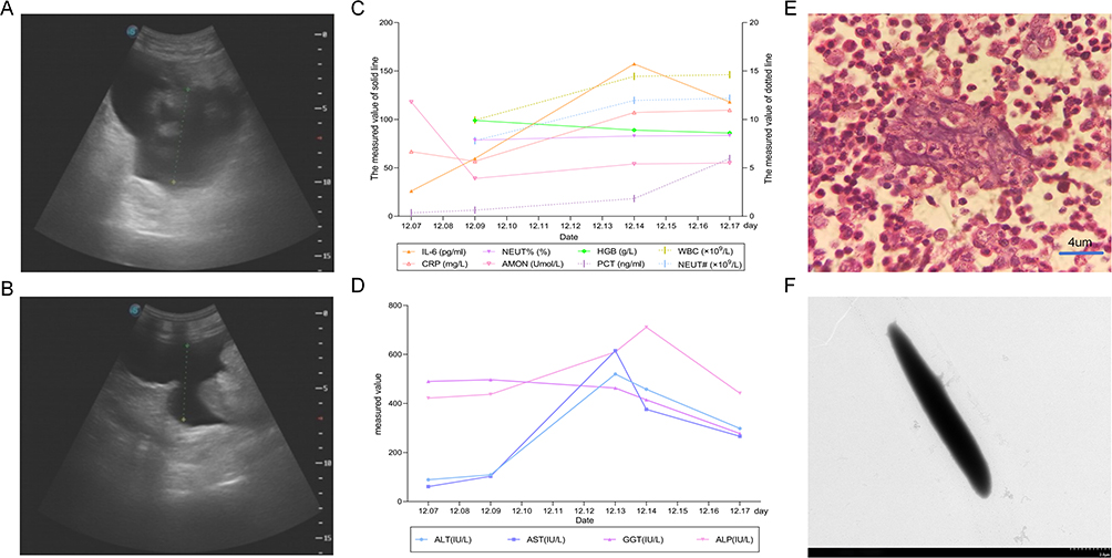 |
Figure 1 Auxiliary test results. (A and B) show abdominal ultrasound images; (C and D) show laboratory test results; (E) shows pathological image of ascitic fluid; (F) shows electron microscopic image of G. massiliensis. Abbreviations: PCT, Procalcitonin; WBC, White Blood Cell Count; NEUT#, Neutrophil Absolute Count; IL-6, Interleukin-6; NEUT%, Neutrophil Percentage; HGB, Hemoglobin; CRP, C-Reactive Protein; AMON, Ammonia; ALT, Alanine Aminotransferase; AST, Aspartate Aminotransferase; GGT, Gamma-Glutamyl Transferase; ALP, Alkaline Phosphatase. |
Laboratory tests showed significant elevations in procalcitonin (PCT, ng/mL), white blood cell count (WBC, ×109/L), absolute neutrophil count (NEUT#, ×109/L), neutrophil percentage (NEUT%), interleukin-6 (IL-6, pg/mL), and C-reactive protein (CRP, mg/L). However, hemoglobin (HGB, g/L) showed a downward trend. (Figure 1C). Stool examination revealed the presence of mucus and blood, with a positive fecal occult blood test confirmed using the monoclonal antibody method. Given the presence of cough, sputum, and ascites, the possibility of pulmonary and abdominal infections was considered in the differential diagnosis. Cranial computed tomography (CT) revealed lacunar lesions in the bilateral basal ganglia and semioval center. These findings, in conjunction with cranial magnetic resonance imaging (MRI) and diffusion-weighted imaging (DWI), which indicated old lacunar cerebral infarctions and ischemic injury foci, as well as significantly elevated blood ammonia levels and transaminases (refer to Figure 1C and D), led to the diagnosis of hepatic encephalopathy as the underlying cause of the patient’s confusion. The other diagnoses were consistent with those made at the external hospital. After cefoperazone/sulbactam (1.5 g, q12h) anti-infection, acidified intestinal tract (Enema with NS 500mL + Acetic Acid 10 mL, bid), L-aspartate ornithine ammonia reduction, nutritional support, maintenance of defecation regularity and symptomatic support treatment, the patient’s consciousness level improved and was transferred to oncology department for treatment.
On the third day of hospitalization, paracentesis was performed at the junction of the lateral and middle thirds of the line connecting the umbilicus to the left anterior superior iliac spine, with the patient in a supine position. A drainage tube was subsequently inserted, allowing gradual removal of ascitic fluid in small, frequent volumes. During the hospital stay, 10.5 liters of ascitic fluid were extracted. During the paracentesis procedure, 10 mL of ascitic fluid was introduced into an aerobic culture bottle (Hapyear, China) and dispatched to the microbiology laboratory for bacterial culture and identification. On the fifth day of hospitalization, Pathological analysis of the ascitic fluid revealed numerous malignant tumor cells with morphological characteristics consistent with adenocarcinoma, confirming the presence of malignant ascites (Figure 1E). On the eighth day of hospitalization, G. massiliensis was isolated from the sample. The morphology of the electron microscope is shown in Figure 1F. Following the results of the antimicrobial susceptibility testing, the antibiotic regimen was adjusted to Combination of meropenem and tigecycline.
In addition to admission diagnosis, hepatic encephalopathy, obstructive jaundice, malignant ascites, right cerebral artery stenosis, multiple old lacunar cerebral infarction, mild anemia, benign prostatic hyperplasia, splenomegaly and extrahepatic bile duct catheterization were also diagnosed. The patient’s condition was complex, the prognosis was poor, and the condition was critical. Following discussions with the patient’s family, who demonstrated understanding and provided consent, palliative symptomatic treatment was initiated. After the ninth day of hospitalization, Esomeprazole was administered for acid suppression and gastric protection to prevent stress-induced ulcer bleeding. Liver-protective therapy with glutathione and polyene phosphatidylcholine resulted in a significant reduction in transaminase levels. L-ornithine L-aspartate therapy was continued to reduce blood ammonia levels, which were stabilized at approximately 55 µmol/L (Figure 1C and D). On the eleventh day of hospitalization, the attending physician observed subcutaneous hemorrhage and extensive ecchymoses on the patient’s soles and left scapula. The coagulation parameters were as follows: prothrombin time (PT), 90.80 S; activated partial thromboplastin time (APTT), 142.30 S; and antithrombin III (AT-III), 39%. Plasma transfusions of 300 mL each were administered on days 11, 12, and 13 to replenish coagulation factors, with no adverse reactions observed post-transfusion. The patient’s condition remained critical, characterized by multiple comorbidities and complications and deteriorating liver function. On the afternoon of December 19, 2024, family members chose to discharge so that patients could receive palliative care at home.
Microbiological Analysis
An aerobic culture bottle containing ascitic fluid provided by the clinical department was incubated in the BacT/Alert 3D 240 blood culture system (bioMérieux, France) at 35°C. A positive signal was detected after 36.72 h of incubation. Subsequently, 1–2 mL of culture broth was inoculated onto Columbia blood agar plates and McConkey agar plates (OXIOD, UK) and incubated at 35°C with 5% CO2. After 24 h of incubation, small white colonies were observed on Columbia blood agar. Gram staining revealed the presence of Gram-positive rods (refer to Figure 2 Ai and Aii). After 48 h, the colonies appeared circular, beige, raised, and smooth, measuring 1–2 mm in diameter, and emitted an odor. Gram staining remained positive (Figure 2 Bi and Bii). Over a period of 5 days, the bacterium grew relatively slowly, with the colony color gradually deepening from beige. Gram staining consistently indicated positive results, and the bacteria tended to cluster (Figure 2 Ai-Eii).
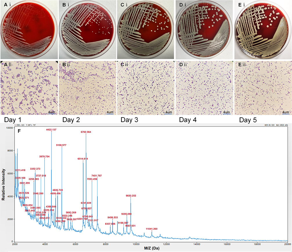 |
Figure 2 Colony morphology and Gram staining of G. massiliensis over five consecutive days of culture. Ai–Ei: Colony morphology on days 1–5; Aii–Eii: Gram stain images on days 1–5; F: Mass spectrometry image of G. massiliensis. |
Using a Hitachi TEM system at an acceleration voltage of 80.0kV and a magnification of 4.0k, the bacterial morphology was observed to be irregular and predominantly coccobacillary, as depicted in Figure 1F. No bacterial growth was detected on MacConkey agar or in anaerobic conditions. The bacterial samples were treated with 70% formic acid and an α-CHCA matrix solution (comprising 50% acetonitrile and 2.5% trifluoroacetic acid) and subsequently analyzed using an Autof ms1000 (AutoBio, China) matrix-assisted laser desorption/ionization time-of-flight mass spectrometer (MALDI-TOF MS). Mass spectrometry analysis identified the bacterium as Pseudoclavibacter faecalis, with a score of 9.025. According to these criteria, confirmation was required for rare species, even with scores ≥6.0, owing to insufficient database coverage, and sequencing was recommended. The mass spectrum, viewed using Auto Acquirer software, revealed characteristic peaks at 3970.754, 4422.137, 5108.997, and 6702.564, among others (Figure 2F).
Subsequent biochemical identification was conducted utilizing the Vitek-2 Compact system, employing specific biochemical cards tailored for Gram-negative bacilli, Gram-positive cocci, and Corynebacterium. Biochemical analysis of the gram-positive cocci and Corynebacterium cards revealed the bacterium’s ability to metabolize D-sorbitol and D-mannose as sugar substrates and ferment mannose, maltose, and xylose. Enzymatic activity assays returned positive results for L-Pyrrolydonyl-Arylamidase, the Amylase, cyclodextrin hydrolysis, alkaline phosphatase, pullulan hydrolysis, phosphatidylinositol-specific phospholipase C, and Arginine dihydrolase. Additionally, the Novobiocin susceptibility test yielded a positive result. The specific biochemical reaction results are shown in Table 1. Despite these biochemical analyses, the precise strain could not be identified in this study. Consequently, 16S rRNA gene sequencing was selected for definitive identification and was performed by RuiBio BioTech. The gene amplification process resulted in a single distinct target band. BLAST analysis of the sequencing results indicated that the highest sequence similarities were observed with G. massiliensis (NR179448.1) at 99.35% and Gulosibacter macacae (NR180318.1) at 97.26%. Based on an integrated assessment of the biochemical characteristics, 16S rRNA sequencing data, and pertinent historical information, the bacterium was conclusively identified as G. massiliensis.
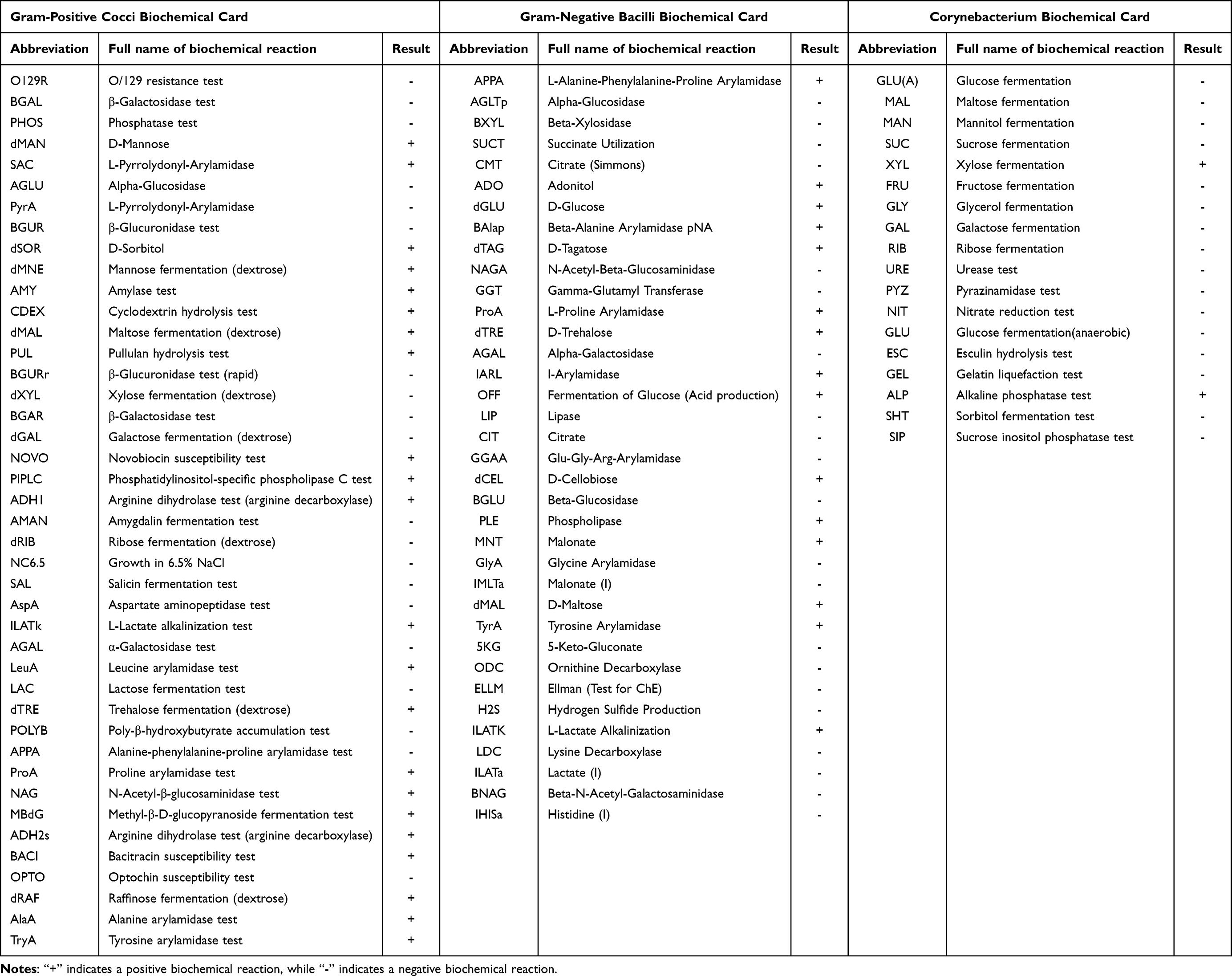 |
Table 1 Biochemical Card Identification Results of Gram-Negative Bacilli, Gram-Positive Cocci, and Corynebacteria |
The 16S rRNA sequencing data were submitted to NCBI-BLAST using the core nucleotide database (core_nt) for Standard Nucleotide BLAST analysis. Appropriate comparison results were selected to construct a phylogenetic tree in MEGA 11, employing the ClustalW algorithm for sequence alignment and the neighbor-joining method for tree construction (Figure 3A and B). The G. massiliensis-3268 strain was classified within the Gulosibacter genus and exhibited the closest relationship to Gulosibacter faecalis strain C2 (PP_930902.1), with a sequence homology of 99.71%. The sequence homology with other species within the same genus exceeded 95%. Genera closely related to Gulosibacter sp., such as Pseudoclavibacter sp. and Zimmermannella sp., are classified within the phylum Actinobacteria, class Actinobacteria, order Micrococcales, and family Microbacteriaceae, suggesting a relatively close phylogenetic relationship, as illustrated in Figure 3A. Furthermore, G. massiliensis-3268 exhibited phylogenetic affinities with various other bacteria, most notably with Zimmermannella faecalis strain IFO15706 (AB 012591.1), demonstrating a sequence homology of 99.06%, indicative of a close relationship. Other species phylogenetically proximate to G. massiliensis-3268 include Klugiella xanthotipulae strain 44C3 (NR_042891.1), as depicted in Figure 3B. The closest relative identified was Pseudoclavibacter sp. Consequently, in accordance with the Clinical and Laboratory Standards Institute (CLSI) M45, 3rd Edition, 2015, the standard for coryneform and other corynebacteria was employed for antimicrobial susceptibility testing. The broth dilution method was used to determine the Minimum Inhibitory Concentration (MIC), while the Kirby-Bauer (K-B) method was used to measure inhibition zones, albeit not for interpretative purposes. According to the Clinical and Laboratory Standards Institute (CLSI) M45 interpretative criteria, G. massiliensis is susceptible to daptomycin, doxycycline, gentamicin, linezolid, vancomycin, meropenem, and tetracycline (Table 2).
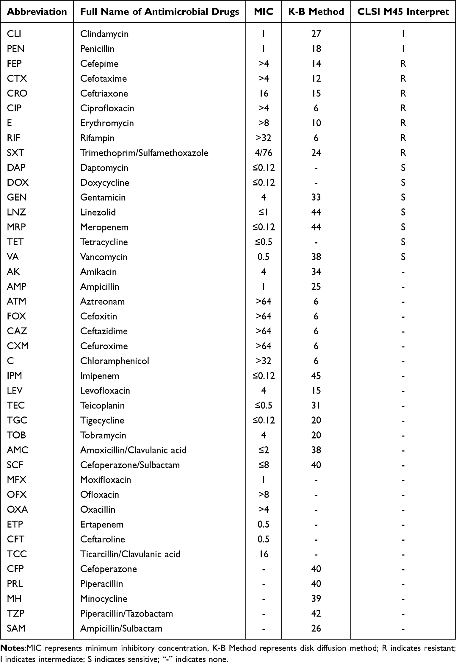 |
Table 2 Drug Susceptibility Test of Gulosibacter massiliensis |
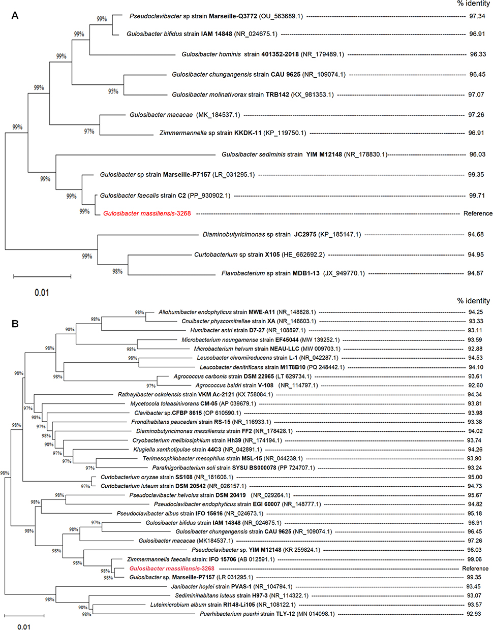 |
Figure 3 Phylogenetic tree based on the 16S rRNA gene sequence of G. massiliensis. (A) Phylogenetic tree relationship of Gulosibacter sp. and its related genera. (B) Phylogenetic relationship of G. massiliensis in Microbacteriaceae. Notes: Reference represents the reference point of the phylogenetic tree; The red font represents the strain detected in this experiment; 3268 represents the strain number; coarsening indicates the specific numbered of bacterial strains; The contents in brackets indicate the accession number in NCBI; % identity represents the similarity ratio. |
Discussion
The genus Gulosibacter is classified within the domain Bacteria, phylum Actinobacteria, class Actinobacteria, order Micrococcales, and family Microbacteriaceae. Members of this genus are gram-positive, strictly aerobic, non-spore-forming, typically presenting as irregular short rods. The predominant fatty acids identified in this genus are 12-methyl tetradecanoic acid (anteiso-C15:0), 14-methyl pentadecanoic acid (iso-C16:0), and 14-methyl hexadecanoic acid (anteiso-C17:0). The polar lipids consisted of diphosphatidylglycerol, phosphatidylglycerol, and an unidentified glycolipid, whereas the major whole-cell sugars were ribose and rhamnose. The primary menaquinone in the respiratory chain is MK-9, and the genomic DNA exhibits a G+C content ranging from approximately 60.0 to 70.0 mol%.6 The optimal growth conditions for this genus are a temperature of approximately 30°C and pH of 8.0. They exhibit positive results for catalase, pyrazinamidase, and various other enzyme activities while testing negative for urease.7 Recognized species within this genus include the type species G. molinativorax and seven additional species: Gulosibacter bifidus, Gulosibacter chungangensis, G. faecalis, Gulosibacter hominis, G. macacae, G. massiliensis, and Gulosibacter sediminis.5 These species have been isolated from diverse samples across different countries and possess distinct characteristics.
G. molinativorax was isolated in 2004 and 2011, respectively, and can mineralize the herbicide Molinate to decompose into ethanethiol and acepan-1-carboxylate. Due to its significant phylogenetic distinctions from the genera Curtobacterium and Brevibacterium helvolum, it was classified as a new genus and species.8 It is the only bacterium known to hydrolyze the sulfate ester bond of molinate.9 This bacteria relies on molinate hydrolase, a cobalt-dependent enzyme with a predicted molecular mass of 50.9 kDa. Structurally, it exists as a homotetramer and belongs to subfamily A of the amidohydrolase superfamily, utilizing cobalt as its sole catalytic center.10,11 In 2017, the microencapsulation of G. molinativorax ON4T using modified chitosan tripolyphosphate crosslinking demonstrated favorable physical properties and effective degradability towards malonate.12 In 2022, a novel composite transposon (Tn6311) harboring the singular catabolic gene molA was identified on a newly discovered low-copy-number plasmid (pARLON1). This gene, located on a chromosome, is predicted to encode an enzyme involved in the cleavage of heterocyclic rings.13
Additionally, in 2004, Lin et al isolated G. faecalis from bovine feces, whereas G. bifidus was isolated from soil, human blood, and wound samples. Initially, these two strains were classified as Zimmermannella species.14 In 2018, Nouioui et al reclassified Actinobacteria into the genus Gulosibacter species. Based on their genomic taxonomy. The transmissibility and pathogenicity of these two strains remain incompletely understood.15 G. chungangensis CAU 9625T, isolated in 2012 from Yellow Sea sediments in South Korea, exhibits 97.8% 16S rRNA gene sequence similarity to model bacteria G. molinativorax ON4T and possesses a comparable fatty acid composition.6 The strain G. chungangensis Gc4EO, isolated in 2021, produces a 4-ethylphenol oxidase. This enzyme shares 42% sequence identity with the vanillyl alcohol oxidase (VAO) from Corynebacterium glutamicum and possesses the same 8α-N3-histidine-linked FAD cofactor. Utilizing oxygen as the sole electron acceptor, it holds promise as a catalyst of choice for the specific dehydrogenation of phenolic compounds.16
Gulosibacter sp. YZ4, isolated from activated sludge in 2012, demonstrated heavy metal tolerance and phenol biodegradation. It can also be preserved via cell immobilization, rendering it suitable for purifying water samples with high phenol concentrations.17 In 2016, Gulosibacter sp. BS4, isolated from the activated sludge of a wastewater treatment facility at the Shchekino-Azot chemical plant in Russia, was confirmed to degrade ε-caprolactam and Nylon-6 oligomers.18 In 2019, Gulosibacter sp. BS38, isolated from soil samples contaminated with industrial waste ε-caprolactam, was capable of growing in a mineral medium with caprolactam as the sole carbon and energy source. It also exhibited the ability to degrade ε-caprolactam and its analogs and possessed 6-aminohexanoate-dimer hydrolase activity.19,20 In 2025, Gulosibacter species. ACHW.36C, isolated from an old cleaning sponge used for applying petroleum-based hoof oil to horses, demonstrated the ability to degrade toluene.21 Although these bacteria have not been formally taxonomically classified, they possess significant bioengineering potential and play important roles in biological processes.
In 2020, G. macacae was isolated from the feces of black langurs, and in 2021, G. sediminis was isolated from the marine sediments of the Indian Ocean. Both strains largely conform to the fundamental characteristics of Gulosibacter species, yet they do not exhibit distinctive features.7,22 Discovered in 2021, G. hominis was isolated from an ear infection and was part of the human skin microbiota and might play a role in opportunistic infections. Notably, it lacks acquired antimicrobial resistance determinants and virulence factors.23 G. massiliensis, isolated from a blood specimen in 2022, is distinguished as the only highly motile strain within the Gulosibacter species and has been associated with wound infections and exudate formation.4,5 Overall, Gulosibacter species are widely distributed in nature and exhibit diverse biological functions, including the degradation of various substances, such as molinate, phenols, caprolactam, toluene, ε-caprolactam, and nylon-6 oligomers. Both G. hominis and G. massiliensis are implicated in opportunistic infections and have the potential to cause bacteremia, thus warranting increased scrutiny. The detailed characteristics of these strains are presented in Table 3.
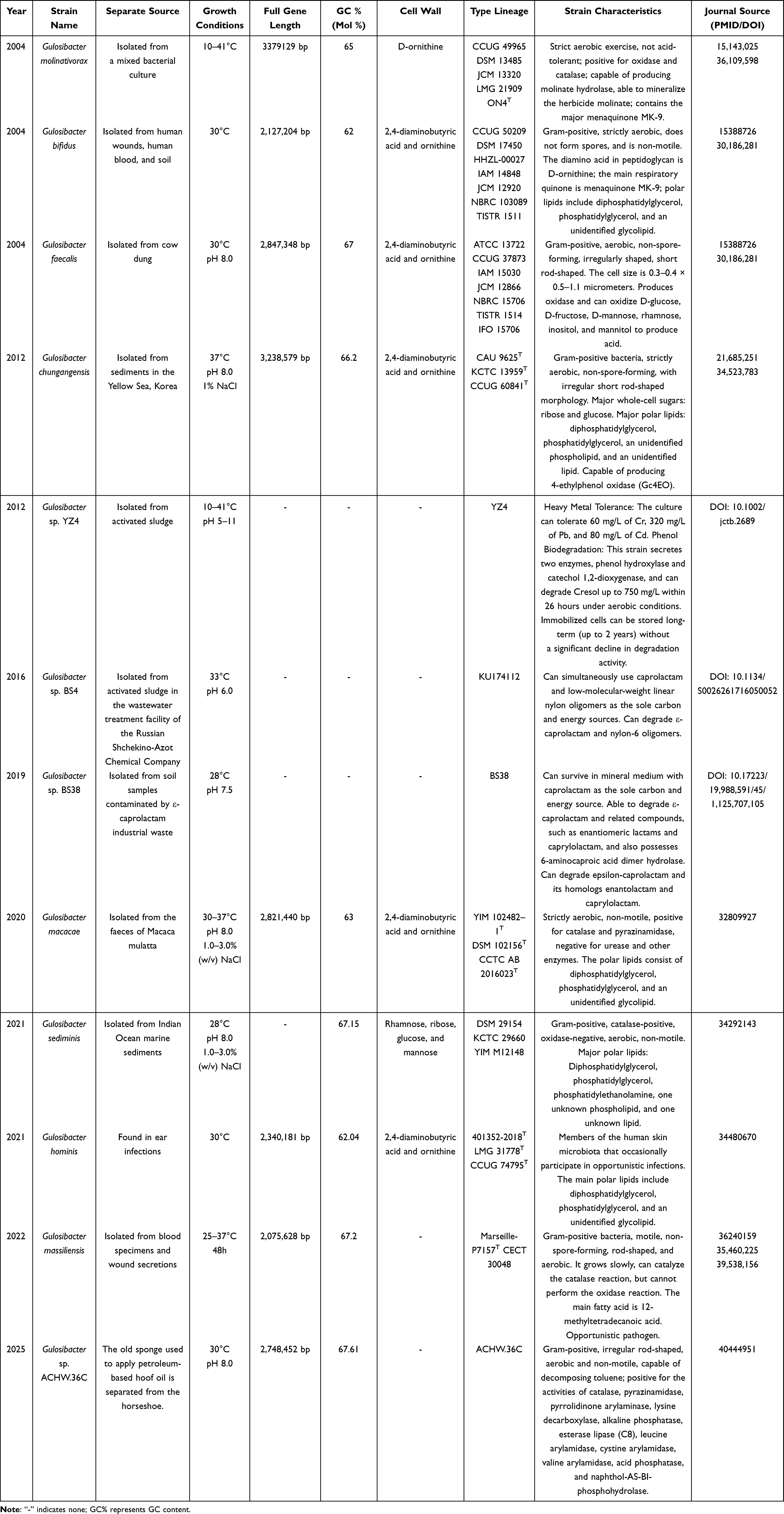 |
Table 3 Basic Information, Characteristics, and Journal Sources of Various Strains in Gulosibacter Sp |
The precise source of the G. massiliensis isolated from the ascitic fluid remains unconfirmed, we hypothesize a translocation from the gastrointestinal tract. This hypothesis is supported by the presence of concurrent gastrointestinal hemorrhage and widespread carcinomatosis peritonei. These conditions likely compromised the intestinal mucosal barrier, facilitating bacterial dissemination. Furthermore, the patient’s immune competence was presumably impaired, either by the systemic immunosuppressive tumor microenvironment24,25 or as a sequela of oncologic therapies.26 Given that G. massiliensisis characterized as the only highly motile strain within its genus, it possesses a distinct mechanistic advantage to invade the peritoneal cavity from the gut lumen. As a typical opportunistic pathogen within the Gulosibacter genus, G. massiliensis may present an elevated infection risk for oncological patients, potentially affecting their in-hospital survival outcomes.27
Upon reviewing the entire clinical course, the patient was admitted with “one-day history of confusion and agitation”, and diagnosed with 18 medical conditions, including extensive peritoneal carcinomatosis, liver failure, moderate-to-large ascites with infection, multiple old lacunar cerebral infarctions, severe malnutrition with emaciation, and grade III hypertension etc. Given the critical condition, poor prognosis, and compromised immune status, only palliative care was feasible for alleviating suffering. Ultimately, respecting the family’s wishes, the patient left hospital and went home to receive palliative care. This case expands the infection spectrum of G. massiliensis reported in the literature. However, this case has limitations such as poor prognosis, no ascites biochemistry, and failure to calculate the SAAG (Serum-Ascites Albumin Gradient) value. Given its atypical clinical manifestations, the exact systemic health effects of G. massiliensis remain unclear, and there is currently a lack of standardized treatment options for antibiotic selection and dosage. These uncertainties highlight the urgent need to further explore its pathogenesis and develop evidence-based treatment strategies. From a diagnostic perspective, clinical laboratories should consider 16S rRNA sequencing when rare coryneform organisms cannot be reliably identified by MALDI-TOF MS, and G. massiliensis should be recognized as a potential opportunistic pathogen in immunocompromised patients.
Ethics Statement
Approval has been obtained from the Ethics Committee of the First Hospital of Kunming, with the ethical approval number: Research Ethics Review (Single) - 2026-030-01. Informed Consent and Publication Consent: Written informed consent and consent for publication in journals have been obtained from the legally authorized representative of the patient (the patient’s daughter) for the detailed case information involved in this study, including clinical data and accompanying images. This study strictly follows all principles in the Declaration of Helsinki and the CARE guidelines and does not involve personal privacy or commercial interests.
Funding
This study was funded by the Kunming Municipal Health Commission Health Research Projects of Yunnan Province of China under Grant 2023-11-01-009.
Disclosure
The authors assert that the study was conducted without any business or financial affiliations that could be construed as a potential conflict of interest.
References
1. Soni J, Sinha S, Pandey R. Understanding bacterial pathogenicity: a closer look at the journey of harmful microbes. Front Microbiol. 2024;15:1370818. doi:10.3389/fmicb.2024.1370818
2. Bartlett A, Padfield D, Lear L, Bendall R, Vos M. A comprehensive list of bacterial pathogens infecting humans. Microbiology. 2022;168(12). doi:10.1099/mic.0.001269
3. Yacouba A, Sissoko S, Tchoupou Saha OF, et al. Description of Acinetobacter ihumii sp. nov. Microbacterium ihumii sp. nov. and Gulosibacter massiliensis sp. nov. three new bacteria isolated from human blood. FEMS Microbiology Letters. 2022;369(1). doi:10.1093/femsle/fnac038
4. Yacouba A, Sissoko S, Tchoupou Saha OL, et al. Correction to: description of Acinetobacter ihumii sp. nov. Microbacterium ihumii sp. nov. and Gulosibacter massiliensis sp. nov. three new bacteria isolated from human blood. J FEMS Microbiol Lett. 2022;369(1). doi:10.1093/femsle/fnac058
5. Li W, Zhang R, Liu L, et al. Emergency wound site infection caused by Gulosibacter massiliensis: a case report. BMC Infectious Diseases. 2024;24(1):1291. doi:10.1186/s12879-024-10187-5
6. Park MH, Traiwan J, Jung MY, Kim W. Gulosibacter chungangensis sp. nov. an actinomycete isolated from a marine sediment, and emended description of the genus Gulosibacter. Int J Syst Evol Microbiol. 2012;62(Pt 5):1055–14. doi:10.1099/ijs.0.032268-0
7. Li G, Li Q, Chen X, et al. Gulosibacter macacae sp. nov. a novel actinobacterium isolated from Macaca mulatta faeces. Int J Syst Evol Microbiol. 2020;70(9):5115–5122. doi:10.1099/ijsem.0.004389
8. Manaia CM, Nogales B, Weiss N, Nunes OC. Gulosibacter molinativorax gen. nov. sp. nov. a molinate-degrading bacterium, and classification of ‘Brevibacterium helvolum’ DSM 20419 as Pseudoclavibacter helvolus gen. nov. sp. nov. Int J Syst Evol Microbiol. 2004;54(Pt 3):783–789. doi:10.1099/ijs.0.02851-0
9. Nunes OC, Lopes AR, Manaia CM. Microbial degradation of the herbicide molinate by defined cultures and in the environment. Appl Microbiol Biotechnol. 2013;97(24):10275–10291. doi:10.1007/s00253-013-5316-9
10. Duarte M, Ferreira-da-Silva F, Lünsdorf H, et al. Gulosibacter molinativorax ON4T molinate hydrolase, a novel cobalt-dependent amidohydrolase. J Bacteriol. 2011;193(20):5810–5816. doi:10.1128/JB.05054-11
11. Leite JP, Duarte M, Paiva AM, et al. Structure-guided engineering of molinate hydrolase for the degradation of thiocarbamate pesticides. PLoS One. 2015;10(4):e0123430. doi:10.1371/journal.pone.0123430
12. Estevinho BN, Lopes AR, Sousa V, Rocha F, Nunes OC. Microencapsulation of Gulosibacter molinativorax ON4(T) cells by a spray-drying process using different biopolymers. J Hazardous Mat. 2017;338:85–92. doi:10.1016/j.jhazmat.2017.05.018
13. Lopes AR, Bunin E, Viana AT, et al. In silico prediction of the enzymes involved in the degradation of the herbicide molinate by Gulosibacter molinativorax ON4(T). Scientific Rep. 2022;12(1):15502. doi:10.1038/s41598-022-18732-5
14. Lin YC, Uemori K, de Briel DA, Arunpairojana V, Yokota A. Zimmermannella helvola gen. nov. sp. nov. Zimmermannella alba sp. nov. Zimmermannella bifida sp. nov. Zimmermannella faecalis sp. nov. and Leucobacter albus sp. nov. novel members of the family Microbacteriaceae. Int J Syst Evol Microbiol. 2004;54(Pt 5):1669–1676. doi:10.1099/ijs.0.02741-0
15. Nouioui I, Carro L, García-López M, et al. Genome-based taxonomic classification of the phylum actinobacteria. Front Microbiol. 2018;9:2007.
16. Alvigini L, Gran-Scheuch A, Guo Y, et al. Discovery, biocatalytic exploration and structural analysis of a 4-Ethylphenol oxidase from Gulosibacter chungangensis. Chembiochem. 2021;22(22):3225–3233. doi:10.1002/cbic.202100457
17. Zhai Z, Wang H, Yan S, Yao J. Biodegradation of phenol at high concentration by a novel bacterium: Gulosibacter sp. YZ4. J Chem Technol Biotechnol. 2012;87(1):105–111. doi:10.1002/jctb.2689
18. Esikova TZ, Taran SA. A novel strain Gulosibacter sp. BS4 degrading epsilon -caprolactam and Nylon-6 oligomers. Microbiology. 2016;85(5):642–645. doi:10.1134/S0026261716050052
19. E TZ. Determination of optimal growth conditions for gram-positive bacterium Gulosibacter sp. BS38, destructor of toxic xenobiotic epsilon-caprolactam. Vestnik Tomskogo Gosudarstvennogo Universiteta, Biologiya. 2019;2019(45):210–219.
20. Esikova TZ, Akatova EV, Taran SA. Bacteria that degrade low-molecular linear epsilon-caprolactam olygomers. Prikladnaia Biokhimiia I Mikrobiologiia. 2014;50(5):481–489. doi:10.7868/S0555109914050043
21. Wai A, Lai G, Leung F, Griffin S. Complete genome sequence of Gulosibacter sp. ACHW.36C, a toluene-degrading strain isolated in Hong Kong. Microbiol Res Announce. 2025;14(7):e0030425. doi:10.1128/mra.00304-25
22. Jiang Z, Zhang WH, Song D, et al. Gulosibacter sediminis sp. nov. isolated from Indian Ocean marine sediment. Int J Syst Evol Microbiol. 2021;71(7). doi:10.1099/ijsem.0.004906
23. Vandamme P, Peeters C, Seth-Smith HMB, et al. Gulosibacter hominis sp. nov.: a novel human microbiome bacterium that may cause opportunistic infections. Antonie Van Leeuwenhoek. 2021;114(11):1841–1854. doi:10.1007/s10482-021-01644-1
24. Demuytere J, Ernst S, van Ovost J, Cosyns S, Ceelen W. The tumor immune microenvironment in peritoneal carcinomatosis. Int Rev Cell Mol Biol. 2022;371:63–95.
25. Ornella MSC, Badrinath N, Kim KA, et al. Immunotherapy for peritoneal carcinomatosis: challenges and prospective outcomes. Cancers. 2023;15(8):2383. doi:10.3390/cancers15082383
26. Charshafian S, Liang SY. Rapid fire: Infectious disease emergencies in patients with cancer. Emerg Med Clin North Am. 2018;36(3):493–516. doi:10.1016/j.emc.2018.04.001
27. Thai V, Lau F, Wolch G, Yang J, Quan H, Fassbender K. Impact of infections on the survival of hospitalized advanced cancer patients. J Pain Sympt Manage. 2012;43(3):549–557. doi:10.1016/j.jpainsymman.2011.04.010
 © 2026 The Author(s). This work is published and licensed by Dove Medical Press Limited. The
full terms of this license are available at https://www.dovepress.com/terms
and incorporate the Creative Commons Attribution
- Non Commercial (unported, 4.0) License.
By accessing the work you hereby accept the Terms. Non-commercial uses of the work are permitted
without any further permission from Dove Medical Press Limited, provided the work is properly
attributed. For permission for commercial use of this work, please see paragraphs 4.2 and 5 of our Terms.
© 2026 The Author(s). This work is published and licensed by Dove Medical Press Limited. The
full terms of this license are available at https://www.dovepress.com/terms
and incorporate the Creative Commons Attribution
- Non Commercial (unported, 4.0) License.
By accessing the work you hereby accept the Terms. Non-commercial uses of the work are permitted
without any further permission from Dove Medical Press Limited, provided the work is properly
attributed. For permission for commercial use of this work, please see paragraphs 4.2 and 5 of our Terms.