Back to Journals » Drug Design, Development and Therapy » Volume 20
A Dual-Function Gastroretentive System: Enhancing Ofloxacin Bioavailability via Floating-Bioadhesive Pellets Based on a Hollow Structure Prepared with a Hybrid Polymer Film
Authors Zhang C ![]() , Yu C, Zhang L
, Yu C, Zhang L
Received 13 January 2026
Accepted for publication 26 May 2026
Published 8 June 2026 Volume 2026:20 589908
DOI https://doi.org/10.2147/DDDT.S589908
Checked for plagiarism Yes
Review by Single anonymous peer review
Peer reviewer comments 2
Editor who approved publication: Dr Leonidas D. Panos
Chungang Zhang,1,* Chenchen Yu,2,* Lin Zhang3
1College of Pharmacy, Liaoning University of Traditional Chinese Medicine, Dalian, People’s Republic of China; 2School of Life and Health, Dalian University, Dalian, People’s Republic of China; 3College of Basic Medical Sciences, Liaoning University of Traditional Chinese Medicine, Shenyang, People’s Republic of China
*These authors contributed equally to this work
Chungang Zhang
College of Pharmacy, Liaoning University of Traditional Chinese Medicine, No. 77, Life One Road, DD Port, Dalian, 116600, People’s Republic of China
, Email [email protected]
Lin Zhang
College of Basic Medical Sciences, Liaoning University of Traditional Chinese Medicine, No. 79, Chongshan East Road, Shenyang, 110847, People’s Republic of China
, Email [email protected]
Purpose: This study aimed to design and characterize a novel dual-function floating-bioadhesive drug delivery system based on a hollow structure to extend gastroretentive time and enhance the bioavailability of ofloxacin.
Methods: Hollow spherical shells were fabricated via fluidized-bed coating using a blended polymer film composed of (SuE (Surelease® E-7-19040) and EuN (Eudragit® NE 30D)). The system integrated a hollow spherical shell, a waterproof layer, a drug layer, a release-retarding ethylcellulose (EC) film, and an outer bioadhesive layer. The mechanical properties, in vitro floating and bioadhesive behavior, drug release profile, and in vivo pharmacokinetics were comprehensively evaluated.
Results: The blended film at a SuE/EuN ratio of 4:4 exhibited optimal mechanical rigidity and strength for the hollow shell. Analysis of the release profile showed a zero-order release for the first 4 h, which was in accordance with the predicted value. The optimized formulation demonstrated excellent buoyancy with a floating rate of 96.3± 0.5% and 100% bioadhesion on gastric mucosa. In vivo pharmacokinetic studies in New Zealand rabbits revealed that the test formulation had a prolonged elimination half-life (6.65± 0.94 h) and a relative bioavailability of 112.2± 14.9% compared to the reference tablet.
Conclusion: The developed floating-bioadhesive system successfully combined hollow-structure buoyancy with mucoadhesion to achieve prolonged gastric retention and sustained drug release, offering a feasible approach for gastroretentive drug delivery systems. The infographic illustrates a dual-function gastroretentive system: floating-bioadhesive. It includes a diagram of the gastrointestinal tract and a chemical structure with the text ’Narrow absorption window in upper GI tract, pH-dependent solubility.’ A cross-section of the system shows layers: bioadhesive layer, release retarding film, drug layer, waterproof layer and the hollowspherical shell. Below, a diagram indicates ’ 8h Floating rate: 96.3± 0.5 percent.’ A graph shows zero-order release (0-4h, R squared = 0.9982). Another diagram highlights ’Prolonged gastric retention, sustained drug release, enhanced bioavailability.’.Infographic on dual-function gastroretentive system: floating and bioadhesive pellets based on a hollow structure.
Keywords: Hollow pellets, mechanical properties, floating, bioadhesive ability, bioavailability
Introduction
The Background of the Research
Developing oral dosage forms that simultaneously ensure sustained drug release and extended gastric residence time remains a primary challenge in clinical pharmacy. This is particularly critical for drugs like Ofloxacin, a broad-spectrum fluoroquinolone antibiotic, which exhibits a narrow absorption window in the upper gastrointestinal tract and pH-dependent solubility (readily soluble in acidic conditions but poorly soluble in alkaline media).1–3 For such compounds, gastroretentive dosage forms offer distinct clinical advantages by prolonging gastric residence time, thereby localizing the drug at its absorption site and preventing dose dumping.4
Despite these advantages, current clinical applications face significant limitations. Single-unit floating systems are prone to “all-or-nothing” gastric emptying, resulting in high variability in gastrointestinal transit time.5,6 Although multiple-unit systems can reduce inter- and intra-subject variability,2 the industrial manufacturing of hollow microspheres, which often relies on organic solvents such as dichloromethane, has been hindered by technical limitations that prevent large-scale production.7 Conversely, the multiple-unit floating carriers (based on a Hollow Structure) documented in extant literature are compatible only with specific drugs. There is a paucity of carriers with general applicability for the floating drug delivery systems that can be used to prepare gastroretentive formulations for a variety of drugs. Meanwhile, from a material science perspective, an ideal membrane for fabricating hollow spherical shells must possess sufficient rigidity and strength to withstand processing stresses. Thus, it is evident that further in-depth research is required on such carrier systems.
Bioadhesive drug delivery systems represented a strategy to extend the gastric residence time of formulations.8 Unlike floating systems, which do not interact with the gastric mucosa, bioadhesive systems utilize mucoadhesive polymers that bind nonspecifically to the mucus layer.9 Consequently, a synergistic multiple-unit system integrating both floating and mucoadhesive properties may offer a solution to these limitations.
The Theory of the Pellets Release Mechanism
The release kinetics from pellets coated with a semi-permeable membrane were founded on an established theoretical model.5 The primary processes governing drug release from the pellets were: Drug liberation is governed by four sequential core processes: (i) penetration of the dissolution medium into the core, (ii) subsequent dissolution of the active pharmaceutical ingredient, (iii) build-up of combined osmotic and hydrostatic pressure, and (iv) convective transport of the dissolved drug through pores in the membrane into the external medium.5 To simplify the mechanistic analysis, the following assumptions were made:
- The drug core was spherical and contains a homogeneous distribution of the drug substance.
- The coating acted as a non-deformable, rigid film.
- The pores within the membrane were of suitable dimensions.
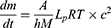
A representative diagram of the pellet release process is provided in Figure 1. The release profile for this semi-permeable membrane system could be expressed by Equation (1):
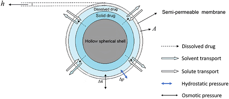 |
Figure 1 Schematic illustration of the release process of the pellets (h: the membrane thickness; (A) the surface area; Δp: the difference in the hydrostatic pressure across the membrane; Δπ: the osmotic pressure difference across the membrane). |
Where  denotes the surface area,
  is the membrane thickness,
  is the mechanical permeability,
  is the concentration of the drug in the dissolved phase with in the pellets.
During the release process,  controls the release rate of the pellets. Beyond a critical point,
  diminishes, leading to a deceleration in release kinetics. The critical point can be determined by the following expression:10–12

Here,  is the amount of drug obtained from at zero-order release,
  is the total drug load, and
  is the density of the solid drug. This model provided a means to estimate the critical value marking the transition from zero-order release kinetics for this type of coated pellet system.
Research Proposal
Based on a hollow structural design, this study focused on developing and characterizing a floating-bioadhesive drug delivery system. A novel blended film, composed of SuE (Surelease® E-7-19040) and EuN (Eudragit® NE 30D), was formulated and evaluated as the primary hollow spherical shell. The thermal and mechanical properties of free films were investigated using Differential Scanning Calorimetry (DSC), tensile, and puncture tests to select the optimal polymer blend. Hollow pellets were fabricated via fluidized-bed coating. Successive layers—a waterproofing stearic acid layer, an ofloxacin drug layer, a release-retarding ethylcellulose (EC) film, and a bioadhesive Carbomer 934P layer—were sprayed onto the sugar spheres, which were subsequently removed to create the hollow core. The entrapped air within this core provides buoyancy, enabling prolonged gastric retention. The resulting system was comprehensively characterized. Key investigations included the coating weight, in vitro buoyancy and gastroretentive behavior, pellet morphology, drug release profile, and pharmacokinetic studies. A water uptake test and mechanical property assessment confirmed that the novel polymer blend met the critical requirements for a robust hollow spherical shell. The present research aims to bridge this gap by designing an innovative floating-bioadhesive drug delivery system. To the best of our knowledge, the above research has not been previously reported.
Materials and Method
Materials
Ofloxacin was sourced from Zhejiang Kangyu Pharmaceutical Co. Ltd. (Zhejiang, China). Suglets®(Sugar Spheres), EC 10cp and Surelease® E-7-19040 were gifts from Colorcon. PVPK30, HPMC E5, PEG 6000, and stearic acid, all of analytical grade, were supplied by Boai Xinkaiyuan (Henan, China), Huzhou Zhanwang (Zhejiang, China), and Bodi (Tianjin, China), respectively. Eudragit® NE 30D was supplied by Evonik (Essen, Germany).
Preparation of Floating Sustained-Release Pellets
The multilayer coating process was performed sequentially, applying the spherical shell polymer, a waterproof layer, the drug (ofloxacin), a release-retarding film, and finally a bioadhesive layer.
A fluidized-bed coater (FD-MP-01, Powrex, Japan) was employed for all coating steps. Sugar spheres of varying sizes served as the inert core. The primary polymeric coating consisted of a blend of SuE and EuN. Core pellets with target weight gains of 15%, 20%, and 25% (w/w) were produced, subsequently cured at 40°Cfor 24 h, washed with water for 8 h to remove the sugar core, and finally freeze-dried to obtain hollow pellets.
A waterproof layer was then applied by coating the freeze-dried hollow pellets with an alcoholic solution of stearic acid. The drug layer was deposited by spraying an aqueous solution of ofloxacin containing 0.5% HPMC E5 onto the pellet surface.
The coating solution was prepared by dissolving EC and PVPK30 in a 5:1 (w/w) ratio in 95% alcohol to achieve an EC concentration of 1.5% (w/v); PVPK30 functioned as both a pore-forming and plasticizing agent. The final bioadhesive layer consisted of a 0.8% w/v alcoholic solution of Carbomer 934P.
The conditions for the coating of pellets were given as follows: spray gun pressure: 1.2 bar; spray rate: 2.5~5 mL/min; inlet air temperature: 30~40 °C.
The optimised formulation was constituted by sugar spheres (840~710 μm), a hollow spherical shell (SuE: EuN = 4:4) with a weight gain of 15% (w/w), a waterproof layer with a weight gain of 20% stearic acid, a drug layer with a weight gain of 60% ofloxacin, a release-retarding film with a weight gain of 6% EC, and a bioadhesive layer with a weight gain of 4% Carbomer 934P.
Preparation and Characterization of Free Films
Composite free films of SuE and EuN were fabricated using a spraying technique. Dispersions of the two polymers were combined at varying mass ratios (0:4, 1:4, 2:4, 4:4, 4:2, 4:1, and 4:0, w/w, SuE to EuN), with the total polymer solid content maintained at 4 grams for each formulation, and stirred for 30 min. The resulting mixtures were sprayed onto Teflon plates (10 cm × 10 cm) and dried at 40 °C for 24 h to obtain the free films. Subsequently, these films were immersed in purified water for 8 h and then freeze-dried. The thickness of all prepared films ranged from approximately 20 to 40 μm.
The puncture strength and percentages of elongation at breaking of the films were determined using a texture analyzer (CT3 Brookfield, USA), A 2 mm diameter stainless-steel puncturing probe was used. The film was cut into samples 20 mm×20 mm in size. The film was fixed at the load cell with a cylindrical hole (r=1.0 cm). The samples were tested with an elongation rate set at 0.1 mm/s and the force displacement curves were recorded by computer. The Puncture strength, Young’s Modulus, Yield strength and elongation were calculated, respectively. All measurements were performed in six independent replicates.
The tensile strength and elongation at breaking were obtained using a tensile strength tester (AI 7000M, GOTECH, TAIWAN). In addition, Young’s modulus was calculated as the slope of the linear region of the stress–distance curve. Films were cut into samples 30 mm×10 mm in size. The tensile properties were tested with an elongation rate set at 0.1 mm/s. The Tensile strength, Young’s modulus, Yield strength and elongation were then calculated, respectively. All measurements were performed in six independent replicates.
Hygroscopicity Testing
Moisture sorption was determined gravimetrically using humidity chambers in conjunction with an analytical balance (FA1104, Shanghai Minqiao Medical Appliance Ltd., Shanghai, China). This method employed saturated salt solutions within the chambers to maintain a precisely controlled relative humidity for the measurements. At predetermined times, samples were withdrawn and accurately weighted. KNO3 was chosen to provide a relative humidity of 92.5% and the chamber was kept at room temperature (25°C). The samples were weighed and all experiments were conducted in six independent replicates (n=6).
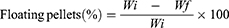
The amount of moisture sorption was calculated as follows:13
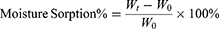
where  and
and  represent the total weight of the samples at time
  and
and  , respectively.
Water Uptake Studies
Water uptake studies were performed in 900 mL of 0.1 N HCl using a USP dissolution apparatus 1 (basket method) at 37 ± 0.5 °C and a rotation speed of 50 rpm. The pellets were placed in the medium and at predetermined intervals, samples were withdrawn and the water on the surface of the pellets was removed using filter paper. The samples were weighed and all experiments were conducted in six independent replicates (n = 6). The amount of moisture sorption was calculated as follows:
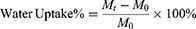
where  represent the total weight of the samples at
  , and
, and  represent the total weight of the samples at
  .
.
Mechanical Strength Investigation
The hollow core pellets should be strong enough to withstand the mechanical collision during coating in the fluidized bed. One hundred hollow spherical shell pellets were tested in an organic glass cylinder and for 10 min under relatively extreme conditions. Then the broken and deformed pellets were removed, and the remaining intact hollow spherical shell pellets were collected. The atomization pressure = 1.5 bar.
The mechanical strength was indicated by the strength index. The higher the strength index was, the greater the mechanical strength was. The index was calculated using the following formula:

where  is the weight of the hollow core pellets remaining undamaged and
  is the initial weight of the hollow core pellets. All experiments were conducted in six independent replicates (n=6).
Differential Scanning Calorimetry
To investigate the effect of new blended materials-different involving combinations of SuE and EuN on the thermal properties, the glass transition temperature (Tg) was examined by differential scanning calorimetry DSC (Mettler-Toledo, Switzerland). Prior to analysis, the samples (about 10 mg) were weighed and sealed into aluminum pans and heated at a rate of 20 °C/min over a temperature range of −30~150 °C under an ultra-high purge of nitrogen at a rate of 80 mL/min. The samples were cycled twice and the Tg was calculated as the midpoint of the transition.
Scanning Electron Microscopy (SEM) Studies
The surface and internal morphology of the pellets were examined using scanning electron microscopy (SEM; model SSX-550, Shimadzu, Japan). Prior to imaging, the samples were mounted on aluminum stubs, sputter-coated with gold, and, for internal visualization, sectioned into hemispheres.
In vitro Release
The in vitro release of ofloxacin from the pellets was evaluated with a USP dissolution apparatus 2 (paddle method) (n=6). The test was conducted in 900 mL of 0.1 N HCl maintained at 37 ± 0.5 °C with a paddle rotation speed of 50 rpm. A pellet sample equivalent to approximately 200 mg of ofloxacin was used for each test. Aliquot samples were withdrawn at predetermined time points, and the drug concentration was quantified by UV-Vis spectroscopy at a wavelength of 293 nm.
Floating Study
The floating capacity of the pellets was evaluated in vitro using a USP dissolution apparatus II (paddle method) under sink conditions (900 mL of 0.1 N HCl, 37 ± 0.5 °C, 50 rpm). For each test, 100 pellets were gently spread onto the medium surface, and their buoyancy was visually monitored at 8 h. The above measurements were performed in six independent replicates. The floating percentage was calculated according to the following equation:4
Where  is the total number of pellets and
  is the number of pellets remaining floating at the end of the test.
Adhesion Behavior Testing
The mucoadhesive performance of the pellets was assessed using the method described by Ranga Rao & Buri.14 Gastric mucosa, freshly excised from male Sprague Dawley rats, was rinsed with a simulated gastric fluid (pH 1.2) containing NaCl and HCl. A section of the mucosa was secured on a glass slide, and 30 pellets were evenly distributed onto its surface. The assembly was then incubated in a humidity chamber maintained at 37 ± 0.5 °C and 92.5% relative humidity for 20 min. Subsequently, the slide was inclined at a 45° angle, and the mucosa was rinsed with the same medium at a controlled flow rate of 22 mL/min for 5 min. The bioadhesive rate was calculated using the following formula:15
Where  is the total number of pellets applied, and
  is the number of pellets retained on the mucosa after rinsing. All measurements were performed in six independent replicates.
In vivo Pharmacokinetic Studies
Establishment of the in Vivo Analysis Method
A SHIMADZU LC-2010AHT HPLC system equipped with a quaternary pump and a UV-Vis detector was used for quantification. Chromatographic separation was achieved on an RX-C18 column (150 mm × 4.6 mm, 5 μm) using an isocratic mobile phase of acetonitrile and 0.05 mol/L citric acid (adjusted to pH 3.5 with triethylamine) (10:90, v/v) at a flow rate of 1.0 mL/min. Detection was performed at 293 nm.
Plasma Sample Processing
Plasma samples (100 μL) were spiked with 10 μL of ciprofloxacin internal standard solution (209 μg/mL), vortexed for 1 min, and then protein-precipitated with 500 μL of acetonitrile. After vortexing and centrifugation (12,000 rpm, 10 min), 500 μL of the supernatant were evaporated to dryness. The residue was reconstituted in 100 μL of mobile phase, vortexed for 2 min, and centrifuged again (12,000 rpm, 10 min). A 10 μL aliquot of the final supernatant was injected into the HPLC system.
Method Validation
Method specificity was confirmed by analyzing blank plasma, blank plasma spiked with standards, and post-dose study samples. A linear calibration curve was constructed by plotting the peak area ratio of ofloxacin to ciprofloxacin (Y) against the nominal concentration (X), using weighted linear least-squares regression. Intra-day and inter-day precision and accuracy were determined with six replicates at three quality control (QC) concentrations (0.2, 5.0, and 25 μg/mL) over three days. Extraction recovery was evaluated at the same QC levels by comparing the analyte response from processed samples to that from unextracted standards. Stability was assessed under various conditions: room temperature for 2 h, post-preparation at 4 °C for 24 h, after one and three freeze-thaw cycles, and following storage at −20 °C for 30 days.
Animal Experiments and Sample Collection
The experiments received approval from the Animal Ethics Committee of Liaoning University of Traditional Chinese Medicine (IACUC Issue No. 210000620250241). All procedures, including euthanasia by pentobarbital sodium overdose (≥100 mg/kg), were performed in accordance with the guidelines for the care and use of laboratory animals. A two-period, randomized crossover study with a one-week washout was conducted in six healthy New Zealand rabbits (1.5~2 kg). The sample size of six rabbits was determined based on preliminary variability data from similar pharmacokinetic studies in rabbits, providing adequate statistical power (>80%) to detect a 20% difference in AUC with a significance level of 0.05. The study was conducted as an open-label trial due to the visible differences in formulation between the test pellets and the reference tablet. Six male New Zealand rabbits were randomly divided into 2 groups. After overnight fasting, each subject were given either the test formulation (ofloxacin floating-bioadhesive sustained-release pellets, 100 mg) or the reference commercial ofloxacin tablet (100 mg) in each period, separated by a 14-day washout period. Water (15 mL) was provided hourly for 8 h post-dose. Blood samples (0.5 mL) were collected from the ear vein into heparinized tubes at scheduled times (test group: 1, 2, 3, 4, 6, 8, 10, 12, 14, 16, 24 h; reference group: 0.5, 1, 1.5, 2, 3, 4, 6, 8, 10, 12, 14, 16, 24 h). Plasma was separated by centrifugation (4000 rpm, 10 min) and stored at −20 °C until analysis.
Pharmacokinetic Data Analysis
Pharmacokinetic parameters (AUC, Cmax, Tmax, t1/2) were calculated using DAS 3.0 software. The relative bioavailability (F) of the test formulation was calculated as: F (%) = (AUCT/AUCR) × 100%. Statistical comparisons of pharmacokinetic parameters between the test and reference formulations were performed using two one side t-test after confirming normality, with a significance level set at p < 0.05.
Results and Discussion
Mechanical Properties
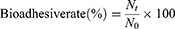
An ideal coating film for the hollow spherical shell should be flexible and rigid enough to withstand any impact during the fluid bed coating process. The hollow spherical shell should be intact and not deformed during the whole process. The stress–strain curve was recorded, and the corresponding mechanical properties were calculated and are listed in Table 1 and Table 2. The tensile strength and puncture strength represent the transverse stress and longitudinal stress, respectively. Pure SuE film had a maximum tensile strength of 2.42±0.36 MPa and a minimum puncture strength of 0.84±0.28 Mpa. Its maximum tensile yield strength was 1.69±0.32 Mpa and the lowest yield strength in puncture strength test was 0.78±0.35 MPa. Both mechanical tests showed a very low elongation and a high Young’s Modulus. Thus, the poor mechanical performance of SuE, characterized by its brittleness and low strength, is attributed to restricted chain mobility caused by intermolecular hydrogen bonding and a rigid, sugar-based molecular architecture.
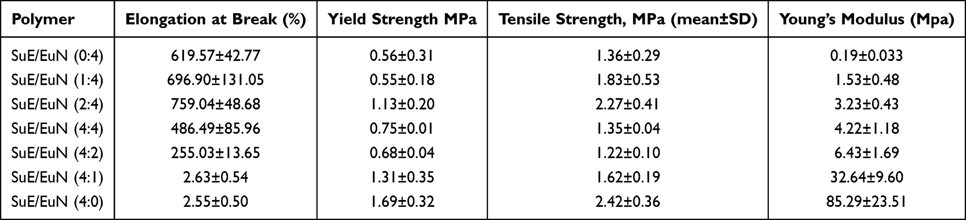 |
Table 1 Mechanical Properties of the Blended Polymer Film in Tensile Test |
 |
Table 2 Mechanical Properties of the Blended Polymer Film in Puncture Test |
Pure EuN had a very high elongation, low Yield strength and a low Young’s modulus in both two mechanical tests. This was because EuN was a neutral ester polymer with the low glass transition temperature of about 6 °C; The molecular chain of the polymer resin exhibited plastic flow during the test process. Therefore, it was flexible, non-rigid and hard to break. These results were in agreement with those reported by Sungthongjeen et al.16 Thus, it was a flexible and soft film and could be easily deformed as a hollow spherical shell.
The mechanical properties varied depending on the blended ratio of SuE and EuN. The percentage elongation values at breaking were found to increase with increasing EuN loading in both mechanical tests. Conversely, Young’s modulus decreased on increasing the amount of EuN. Thus, the mechanical behavior of the blended polymer films changed from brittle to flexible with increasing ratio of EuN. The rigidity also increased on increasing the ratio of SuE. In summary, the addition of EuN to the SuE was a feasible solution to improve the toughness of the blended polymer films in order to ensure an excellent performance during the preparation process. Two findings were noted for all formulations in terms of Yield strength in both the tensile and puncture test. Firstly, the Yield strength decreased on increasing EuN loading in the tensile test. And secondly, on increasing the ratio of EuN, the Yield strength initially increased and then decreased in the puncture test. When the ratio of SuE/EuN was 4:4, the film showed a maximum Yield strength (2.70±0.54 MPa) in the puncture strength test and the values of the elongation and Young’s modulus (28.03±8.01 Kpa) were also satisfactory. The longitudinal stress mainly resisted the impact of the hollow spherical shell moving during the fluid bed coating process. According to these results, the blended polymer film (in a ratio of 4:4) had good rigidity and strength and was suitable candidate for use as the hollow spherical shell.
Thermal Properties
The effect of EuN on the SuE polymer was investigated by measuring the changes in Tg after mixing using different ratios. The Tg of pure SuE polymer was about 35 °C. And the Tg of blended polymer decreased from 35 °C to 6 °C with an increase in the EuN content (Figure 2). This decrease in Tg could be attributed to the weakening effect of EuN which increased segmental motion of the polymer chain. When the Tg of the blended polymer increased, the rigidity increased and the flexibility decreased. This also mean that the blended polymer films would be brittle at a high Tg and soft at a low Tg.
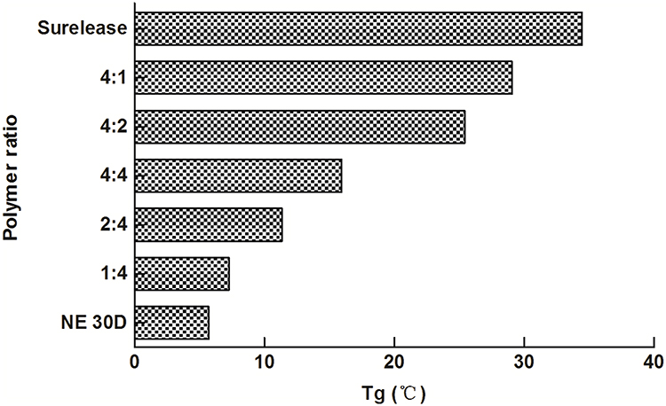 |
Figure 2 Glass transition temperature of the blended polymer film (SuE/EuN) of different ratio. |
Design and Preparation of the Hollow Pellets
This study aimed to develop the floating-bioadhesive pellets with a hollow core structure. As illustrated schematically in Figure 3, the system was fabricated via a fluidized-bed multilayer coating process, sequentially comprising five functional layers: a hollow spherical shell, a stearic acid waterproofing layer, an ofloxacin drug layer, an EC release-retarding film, and an outermost bioadhesive layer of Carbomer 934P.
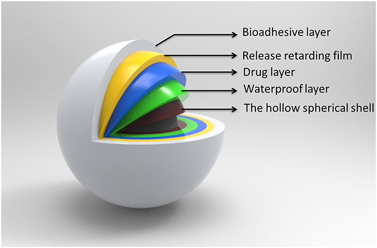 |
Figure 3 Design of the novel the gastric-retentive pellets. |
The surface and cross-sectional morphologies of the pellets were examined by scanning electron microscopy (SEM), as presented in Figure 4. Figure 4A–E depicted the surface of the hollow core coated with blends of EuN and SuE at varying ratios. The surfaces of the hollow core pellets became smoother with an increasing amount of EuN in the blended coating materials. The cross-section of the hollow shell is shown in Figure 4F and G. Since the sugar sphere core was composed of water-soluble sugar and insoluble starch, leaching of the sugar left behind residual starch granules within the hollow interior. The fully coated pellet, comprising all five functional layers, was displayed in Figure 4H–J. The images clearly revealed a central hollow cavity sequentially surrounded by the five distinct coatings: the primary polymeric shell, stearic acid, ofloxacin, ethylcellulose (EC), and Carbomer 934P.
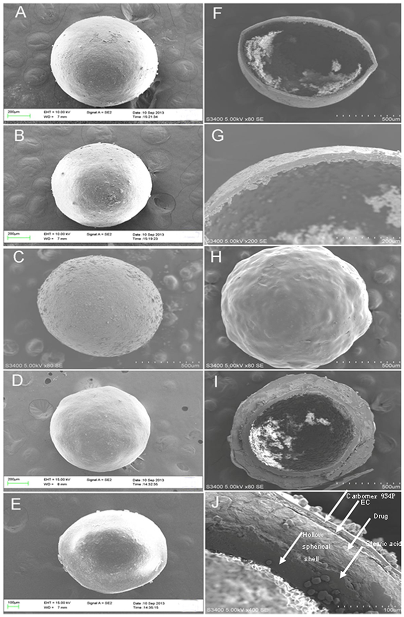 |
Figure 4 SEM photomicrographs of: (A-E) hollow spherical shell (polymer ratio of 4:1, 4:2, 4:4, 2:4 and 1:4, respectively) (F and G) the cross-section morphology of hollow spherical shell (polymer ratio of 4:4), (H) pellets coated with Carbomer, (I and J) the cross-section morphology of the optimized formulation. |
The hollow spherical shell was prepared with the new blended coating materials. The compatibility and stability of coating solution consisted of SuE and EuN in different proportions were good. The two kinds of aqueous dispersion were miscible. When the hollow pellets were coated with SuE, it became too fragile. However, when the hollow pellets were coated with EuN, it was easy to be flexible. The membrane coating used as the hollow spherical shell should be rigid and strong enough to withstand the powerful mechanical collisions in the fluid bed coater and maintain the integrity of the shell.
Sugar spheres (840~710 μm) prepared by the sieving method were collected for optimizing the ratio of SuE to EuN and the level of blended polymer. The final weight and the mechanical strength index were used as evaluation indexes. As shown in Figure 5A, the mechanical strength index increased on increasing the level of blended polymer. When the coating level of blended polymer reached 25%, the mechanical strength index attained its maximum. This enhancement was primarily attributed to the greater shell thickness, which contributed to higher mechanical strength. Under identical polymer coating level, the mechanical strength index initially increased and subsequently decreased with increasing proportion of EuN. The maximum mechanical strength index was achieved when the ratio of SuE to EuN was 4:4. This improvement was likely attributable to the shell prepared at this ratio possessing adequate toughness and strength.
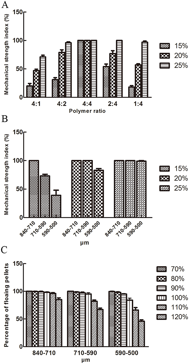 |
Figure 5 (A) Effect of the ratio of blended polymer film on the mechanical strength of the hollow spherical shell. (B) Effect of the particle size on the mechanical strength of the hollow spherical shell. (C) The effect of the different amount of drug loaded of different particle size on the floating ability in water. |
Sugar spheres of different sizes were investigated to obtain the optimal cores. The optimized ratio of the blended polymer was fixed at 4:4. Therefore, different levels of blended polymer coating were investigated. As shown in Figure 5B, the mechanical strength index decreased on decreasing the size of the sugar spheres. This trend could be attributed to the thinning of the shells formed by the same total coating weight gain when the sphere diameter was smaller, resulting in reduced mechanical strength. It also increased on increasing the coating level of the blended polymer. To obtain the greater buoyancy, three groups (840~710 μm, 15% coating level, 710~590 μm, 20% coating level and 590~500 mesh, 25% coating level) were chosen for further investigation.
Three groups of the above hollow spherical shells (840~710 μm, 15% coating level, 710~590 μm, 20% coating level and 590–500 mesh, 25% coating level) were investigated to obtain the highest amount of drug loaded. When the percentage of floating pellets was more than 95%, the formulation could be used for further study. It can be seen from Figure 5C that the percentage of floating pellets decreased on the increasing the amount of drug layer loaded. The highest amount of drug loaded for the three groups was 110%, 100% and 90%, respectively. Thus, the optimal hollow spherical shell consisted of sugar spheres (840~710 μm), SuE: EuN = 4:4, with a weight gain of 15% (w/w).
Waterproof Testing of the Hollow Pellets
The moisture sorption profile of the pellets was determined by measuring the percentage weight change after 48 h of storage at 92.5% relative humidity. As shown in Figure 6A, the uncoated hollow pellets exhibited significant hygroscopicity, rapidly absorbing moisture. In contrast, pellets coated with stearic acid (10% and 20%) demonstrated markedly slower moisture uptake rates. The moisture resistance improved substantially with the application of the waterproof layer, and the equilibrium moisture content decreased progressively as the coating level of stearic acid increased.
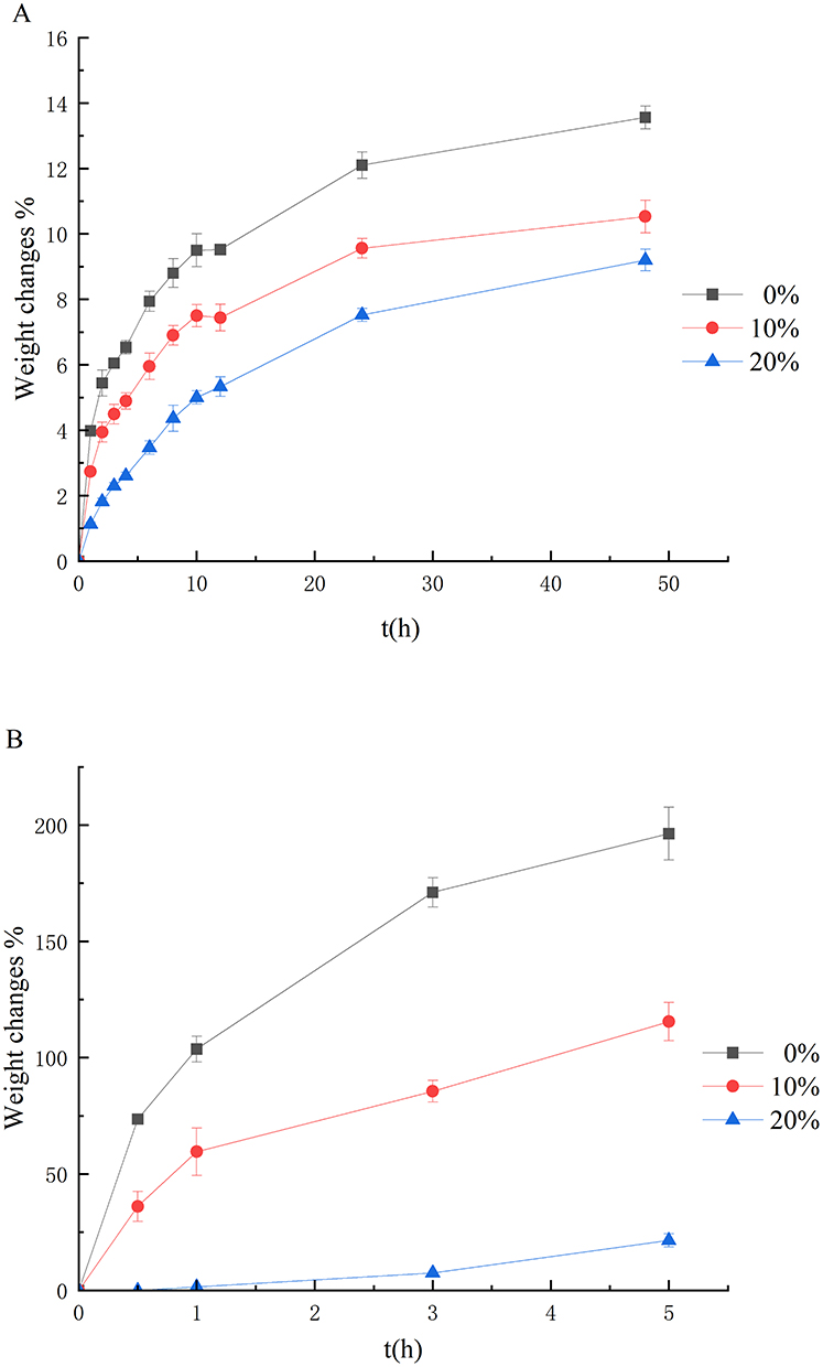 |
Figure 6 (A) Effect of Stearic acid coated on the hollow spherical shell with a 0%, 10% and 20% coating level on water vapor sorption. (B) Effect of Stearic acid coated on the hollow spherical shell with a 0%, 10% and 20% coating level on water uptake. |
Proofing the permeation of liquid through the polymeric film into the hollow core may play a major role in the floating ability. The water uptake profile of the hollow pellets with 0%, 10% and 20% stearic acid coatings is shown in Figure 6B. Water uptake significantly decreased after the addition of stearic acid. When the coating level of stearic acid was 20%, the water uptake was very low until 5 h. This indicates that the pellets coated with 20% stearic acid exhibited a good waterproofing effect and could meet the floating requirement of the hollow pellets.
The weight changes were reduced with an increasing amount of stearic acid in both the moisture sorption test and the water uptake test. However, the moisture sorption increased rapidly after 48 h in comparison with the water uptake test. The results reported above indicated that there was a difference between the moisture sorption behavior and the water uptake behavior of the hollow core pellets. The hollow core pellets were coated with stearic acid to seal the pores on the spherical shell. However, it still has many tiny pores. Because the molecules of water vapor are very small, they could pass through the pores into the inside. The inside of the hollow spherical shell was full of air. The permeation of the aqueous phase into the hollow pellets was hindered by the water–pellet interfacial tension and the internal pressure within the pellets.
Drug Release Study
Floating-bioadhesive pellets not only have the ability to float and exhibit bioadhesive behavior, but also need to have a sustained-release effect. The drug loading of the floating-bioadhesive pellets was fixed at 60% for further coating with good buoyancy. The optimized amount of the bioadhesive layer was fixed at 4%. The drug release profiles from pellets with varying ethylcellulose (EC) coating levels (2% to 6%) are presented in Figure 7. Coatings of 2% and 3% failed to impart a sustained-release effect. Higher EC levels increased film thickness and reduced pore density, thereby limiting water ingress and slowing drug release. All formulations achieved nearly complete release by 24 h. For comparison, the release profile of a commercially available sustained-release ofloxacin tablet is included in Figure 7.17 The pellets with a 6% EC coating exhibited a similar release rate to the tablet for the first 4 h, after which they released the drug more rapidly. This accelerated release phase suggested the potential for enhanced bioavailability compared to the reference tablet.
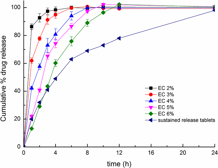 |
Figure 7 Release profiles of the ofloxacin pellets with different EC coating levels in 0.1. |
Characterization of the Release Mechanism
Drug release from the floating-bioadhesive pellets was primarily driven by osmotic pressure through the ethylcellulose (EC) membrane. It was found that the release profile followed zero-order kinetics (R2 = 0.9982) for the initial 4 h, after which the rate declined. To elucidate this mechanism, the process was modeled using Eq. (1). The subsequent decrease in release rate after 4 hours was attributed to a reduction in the osmotic pressure gradient. This time point corresponded to a critical transition and was observed at a cumulative drug release of 60.21% (Figure 8). The theoretical critical value was calculated using Eq. (2). As summarized in Table 3, the calculated range (54.18%~72.60%) encompassed the experimentally observed value of 60.21%. This close agreement between theoretical and experimental resulted validates the proposed release mechanism and the underlying assumptions of the model.
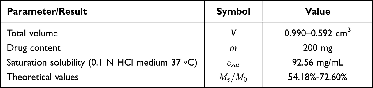 |
Table 3 The Reference Parameters and Results of Theoretical Calculated Value |
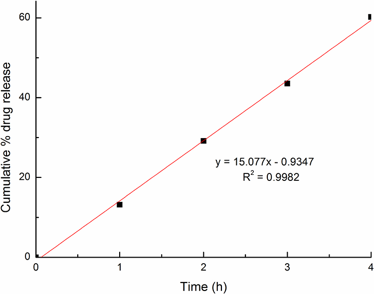 |
Figure 8 The release of optimal formulation over the first 4 h. |
In vitro Floating Ability Study
For the optimized formulation, the floating rate reached 96.3 ± 0.5%, providing a reliable basis for gastric retention through buoyancy. As shown by the white circles in Figure 9, the pellets still floated stably on the dissolution medium surface after 8 hours. These resulted demonstrate the effective barrier function of the waterproof layer in preventing water ingress into the hollow pellets.
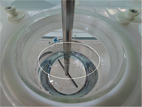 |
Figure 9 Floating pellets in 0.1 N HCl of the optimal formulation. |
Bioadhesive Properties of the Pellets
The retention of pellets on stomach tissues was studied in a test, where the percentage of pellets remaining on the tissues after rinsing for 5 min with medium was observed. On increasing the polymer (Carbomer 934P) coating level, the bioadhesion of the pellets also increased. The percentage of the floating-bioadhesive of pellets with a 4% coating level that remained on the gastric mucosa of the rats was 100%, much higher than that of the non-bioadhesive pellets. These results suggested that the floating-bioadhesive pellets could interact with the mucosubstrate on the surface of the stomach, and adhesion to the mucosa more strongly and the pellets could stay in stomach for prolong period. The mucoadhesion of Carbomer was directly related to the pH of the medium as shown in the case of polycarbophil, polyacrylic acid loosely crosslinked with divinylglycol.18 The mucoadhesion phenomenon occurring between a tissue membrane and Carbomer 934P consisted of a two-step process. There was an interfacial phenomenon influenced by the surface energy effects and spreading of the mucus and the Carbomer 934P hydrogels. Then, interdiffusion or interpenetration of the polymer chains of both phases occurred. This last one, which required hydration of the polymer, was affected by the molecular weight, molecular mobility, viscosity of the adhesive and swelling, properties of both the adhesive and the mucus. A higher binding ability at a pH lower than the pKa of polyacrylic acid (4.75) was reported. This strong bioadhesion occured only when the carboxylic groups were in their acid forms. Thus, the floating- bioadhesive pellets could be expected to exhibit good bioadhesive behavior in vivo.
In vivo Pharmacokinetics Study
Methodological Investigation
Specificity investigation: The results are shown in Figure 10A–C. The peak shapes and retention times of ofloxacin and ciprofloxacin were reproducible and not interfered with by impurities. The retention times were 11.413 min and 13.268 min.
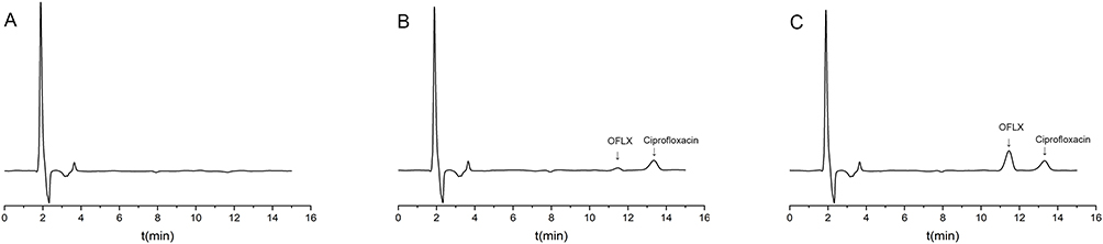 |
Figure 10 HPLC of blank plasma (A), blank plasma + OFLX+ ciprofloxacin (B) and plasma sample + ciprofloxacin (C). |
Calibration curve and limit of quantitation: The calibration curve for the analyte demonstrated good linearity over the range of 0.1~30 μg/mL. The regression equation and correlation metrics were: y = 0.0957x − 0.0292 with R2 = 0.9929, and the correlation coefficient r = 0.9964. The lower limit of quantitation (LLOQ) was 0.1 μg/mL.
Accuracy, precision, and recovery: For ofloxacin, the intra-day and inter-day accuracy and precision were evaluated at three QC levels (0.2, 5.0, and 25 μg/mL). The RSD values were: Intra-day: 2.44%, 2.04%, and 2.19%; Inter-day: 2.29%, 1.79%, and 2.31%. Extraction recovery at the same three concentrations was 99.1%, 99.3%, and 98.9%, with SDs < 1.9%.
Stability: Plasma stability was assessed under multiple storage conditions. The observed relative errors (RE) were within ± 3%, indicating good stability of the samples for the analyzed conditions. Detailed data are presented in Table 4.
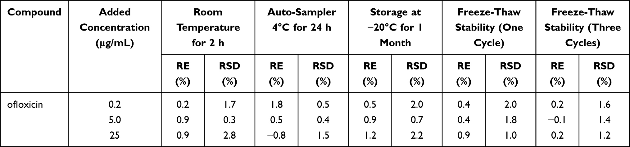 |
Table 4 Stability Results Under Different Storage Conditions |
Pharmacokinetic Study Results
The mean plasma concentrations-time of the test and reference preparations in six New Zealand rabbits were obtained from six New Zealand rabbits (Figure 11) with pharmacokinetic parameters shown in Table 4. Key pharmacokinetic findings: AUC(0-t): Test 138,759.15 ± 14,342.90 ng/mL*h vs. reference 124,702.67 ± 14,361.64 ng/mL*h, indicating a modestly higher overall exposure for the test formulation; Tmax: Test 5.67 ± 0.82 h vs. reference 1.92 ± 0.20 h, indicating a significantly prolonged; Cmax: Test 15142.12 ± 1815.58 ng/mL vs. reference 23584.33± 3514.83 ng/mL indicating marginally lower for the test formulation; t1/2: Test t1/2 = 6.65 ± 0.94 h vs. reference t1/2 = 2.33 ± 0.42 h, reflecting markedly slower elimination with the floating-bioadhesive sustained release system. The relative bioavailability of the floating-bioadhesive sustained release pellets was 112.2 ± 14.9% relative to the tablet. The data were analyzed by a two one-sided t-test and the (1–2α) confidence interval method, and the 90% confidence interval of the AUC(0-t) of the test preparation was 0.993~1.249% of reference preparation, which met the bioequivalence criterion. However, for Cmax the lower bound fell below 80%, demonstrating that the two formulations are not bioequivalent. The floating-bioadhesive sustained release pellets were designed to prolong gastric residence and provide sustained release. While the tablet dissolved rapidly in gastric acid, portions of drug could precipitate in the small intestine. In contrast, the floating-bioadhesive system maintained drug release in the gastric lumen, minimizing precipitation and enabling more consistent absorption, contributing to higher AUC and longer t1/2. The delayed Tmax, lower Cmax and extended t1/2 suggested a more sustained release profile for ofloxacin from the floating-bioadhesive sustained release pellets, supporting continuous plasma drug exposure with reduced peak-trough fluctuations. Given that ofloxacin exhibited concentration-dependent antibacterial activity, the floating-bioadhesive sustained release from the test formulation may enhance therapeutic efficacy relative to the reference tablet. The observed pharmacokinetic improvements indicated superior gastric retention and prolonged gastroretention time with the ofloxacin floating-bioadhesive sustained release pellets, supporting their potential to offer improved oral bioavailability. It was acknowledged that direct evidence of prolonged gastric retention in vivo (eg, via imaging) was not obtained in this study, which was a limitation. Nevertheless, prior investigations utilizing X-ray imaging in rabbit models had confirmed that the hollow-structured floating pellets remained in the stomach for a minimum of 6 h.15
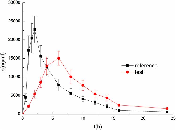 |
Figure 11 Plasma concentration - time profile of ofloxacin floating-bioadhesive sustained release pellets and reference ofloxacin tablets. Results are expressed as the mean ± SD. |
Notes: Statistical significances were indicated as P < 0.05 for AUC(0-t), AUC(0-∞), t1/2, Tmax, Cmax differences between formulations (as reported in Table 5).
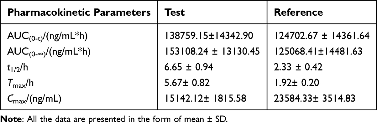 |
Table 5 Pharmacokinetic Parameters of the Test and Reference Preparations |
Gastroretentive drug delivery systems (GRDDS) were extensively explored to prolong gastric residence and enhance oral absorption of drugs with narrow absorption windows, pH-dependent solubility, or local gastric activity.3,4 Various strategies, including floating, swelling/expandable, high-density, raft-forming, mucoadhesive, and recently 3D-printed systems, were developed.3,4,6 Each approach offers advantages, but gastric retention was highly variable and none was universally effective. In this study, a hollow multiparticulate pellet platform was designed to combine floating and bioadhesion, aiming to improve retention reliability while sustaining drug release.
Floating systems were widely studied due to their simplicity and low apparent density.3,4,8 However, single-unit forms may be affected by “all-or-nothing” gastric emptying, causing variable residence and absorption.3,4 Multiparticulate forms disperse more uniformly and were less prone to abrupt emptying.2,3,5 Here, buoyancy was achieved via hollow internal structure rather than solely gas generation, promoting more stable floating under physiological conditions.
Expandable systems relied on post-ingestion swelling to resist pyloric transit, but in vivo performance depends on reproducible swelling, mechanical integrity, and safe size reduction.3,4 The present pellets, in contrast, achieved prolonged residence through combined buoyancy and mucosal interaction without large dimensional changes, offering potentially safer retention.
Mucoadhesive formulations prolong residence via interaction with gastric mucus, yet efficacy was limited by mucus turnover and shear forces.3,4,9 The dual-function pellets demonstrated both high floating (96.3±0.5%) and complete bioadhesion, suggesting synergistic retention: floating maintains gastric localization, while bioadhesion reinforces contact with the mucosa.
Compared with prior hollow microsphere or hollow particulate systems, emphasis on shell integrity and mechanical robustness was critical.5,7,9 The SuE/EuN polymer ratio of 4:4 produced pellets with optimal rigidity, preserving cavity structure for buoyancy and ensuring reproducible release. This highlights that hollow systems’ performance depended on stable structural maintenance, not merely cavity formation.
From a manufacturing perspective, while advanced techniques such as 3D printing offered precise control over geometry and release,6 their scalability remained limited. The current pellets, based on conventional pharmaceutical processing, balanced manufacturability with multifunctionality, including floating, bioadhesion, and sustained release.
Overall, the dual-function hollow pellets combined advantages of multiple GRDDS strategies while mitigating individual limitations. They represented a promising platform for improving oral delivery of ofloxacin and potentially other drugs requiring extended gastric residence.
Conclusion
The multiparticulate floating-bioadhesive system, based on a hollow structure, was designed and developed to achieve gastroretention through two complementary mechanisms: (1) robust buoyancy and (2) strong mucoadhesion. Mechanical characterization of the blended polymeric membrane revealed that increasing the proportion of EuN initially enhanced then reduced tensile yield strength in puncture testing, while Young’s modulus decreased with higher EuN content. A formulation containing 50% EuN and 50% SuE exhibited favorable rigidity and strength. The glass transition temperature (Tg) followed the same trend as Young’s modulus. A 20% coating with stearic acid provided effective waterproofing, and a coating level of EC 6% (w/w) yielded optimal drug release. Release kinetics analysis indicated an initial zero-order phase for the first 4 h. The pellets remained floating for up to 8 h. The in vivo pharmacokinetic study in New Zealand rabbits confirmed the system’s effectiveness, showing a significantly prolonged elimination half-life and enhanced relative bioavailability of  with the AUC meeting bioequivalence criteria. A prolonged Tmax (5.67 ± 0.82), a lower Cmax and a longer t1/2 reflect the intended slow-release and long-term in vivo gastric retention. These findings established a framework for future delivery studies involving drugs that can benefit from dual functionality: a hollow carrier with mucoadhesive properties. Collectively, the results indicated that the hollow, gastroretentive, floating-bioadhesive system could potentially extend gastric residence time and support sustained drug release, as suggested by the pharmacokinetic profile. Consequently, the hollow pellet based on floating-bioadhesive formulation represented a feasible approach for gastric-retentive drug delivery systems. To further substantiate the claimed clinical advantages, future studies should include direct visualization of gastric retention and long-term pharmacokinetic/pharmacodynamic evaluations.
Author Contributions
All authors made a significant contribution to the work reported, whether that is in the conception, study design, execution, acquisition of data, analysis and interpretation, or in all these areas; took part in drafting, revising or critically reviewing the article; gave final approval of the version to be published; have agreed on the journal to which the article has been submitted; and agree to be accountable for all aspects of the work.
Funding
This study was supported by the National Natural Science Foundation of China (No.81503257); Inner Mongolia Major science and technology project (No.2021ZD0017); Liaoning Provincial Science and Technology Programme Joint Programme (Applied Basic Research Project) (2023JH2/101700206); Liaoning University of Traditional Chinese Medicine-Natural Science University Key Project (2021LZY047); Liaoning Provincial Department of Education university basic scientific research project reserve project (LJ212410162055); National Key Research and Development Program (No.2018YFC1706903). Liaoning Province Science and Technology Plan Joint Plan (Natural Science Foundation-Doctoral Research Start-up Project) (2024-BSLH-006).
Disclosure
The authors declare that they have no known competing financial interests or personal relationships that could have appeared to influence the work reported in this paper.
References
1. Hussen NHA, Qadir SH, Rahman HS, et al. Long-term toxicity of fluoroquinolones: a comprehensive review. Drug Chem Toxicol. 2024;47(5):795–20. doi:10.1080/01480545.2023.2240036
2. Baldi A. Recent technological advancements in multiparticulate formulations: the smart drug delivery systems. Asian J Pharm. 2015;9(4):S13–S25. doi:10.22377/ajp.v9i4.481
3. Das S, Kaur S, Rai VK. Gastro-retentive drug delivery systems: a recent update on clinical pertinence and drug delivery. Drug Deliv Transl Re. 2021;11(5):1849–1877. doi:10.1007/s13346-020-00875-5
4. Dubey A, Ovais M, Bisen AC, et al. Advancements and challenges in gastroretentive drug delivery systems: a comprehensive review of research innovation, technologies, and clinical applications. Recent Adv Drug Deliv Formul. 2025;20(1):41–65. doi:10.2174/0126673878342430250414114531
5. Zhang C, Xu M, Tao X, et al. A floating multiparticulate system for ofloxacin based on a multilayer structure: in vitro and in vivo evaluation. Int J Pharm. 2012;430(1–2):141–150. doi:10.1016/j.ijpharm.2012.04.013
6. Turac I-R, Porfire A, Iurian S, et al. Expanding the manufacturing approaches for gastroretentive drug delivery systems with 3D printing technology. Pharmaceutics. 2024;16(6):790. doi:10.3390/pharmaceutics16060790
7. Zhao L, Wei Y-M, Yu Y, et al. Polymer blends used to prepare nifedipine loaded hollow microspheres for a floating-type oral drug delivery system: in vitro evaluation. Arch Pharm Res. 2010;33(3):443–450. doi:10.1007/s12272-010-0314-2
8. More S, Gavali K, Doke O, et al. Gastroretentive drug delivery system. J Drug Delivery Ther. 2018;8(4):24–35. doi:10.22270/jddt.v8i4.1788
9. Liu Y, Zhang J, Gao Y, et al. Preparation and evaluation of glyceryl monooleate-coated hollow-bioadhesive microspheres for gastroretentive drug delivery. Int J Pharm. 2011;413(1–2):103–109. doi:10.1016/j.ijpharm.2011.04.030
10. McClelland GA, Sutton SC, Engle K, et al. The solubility-modulated osmotic pump: in vitro/in vivo release of diltiazem hydrochloride. Pharm Res. 1991;8(1):88–92. doi:10.1023/A:1015890525495
11. Zentner GM, McClelland GA, Sutton SC. Controlled porosity solubility-and resin-modulated osmotic drug delivery systems for release of diltiazem hydrochloride. J Control Release. 1991;16(1–2):237–243. doi:10.1016/0168-3659(91)90047-H
12. Verma RK, Krishna DM, Garg S. Formulation aspects in the development of osmotically controlled oral drug delivery systems. J Control Release. 2002;79(1–3):7–27. doi:10.1016/S0168-3659(01)00550-8
13. Chen H, Shi S, Liu A, et al. Combined application of extrusion-spheronization and hot-melt coating technologies for improving moisture-proofing of herbal extracts. J Pharm Sci. 2010;99(5):2444–2454. doi:10.1002/jps.21990
14. Rao KR, Buri P. A novel in situ method to test polymers and coated microparticles for bioadhesion. Int J Pharm. 1989;52(3):265–270. doi:10.1016/0378-5173(89)90229-9
15. Zhang C, Tang J, Liu D, et al. Design and evaluation of an innovative floating and bioadhesive multiparticulate drug delivery system based on hollow structure. Int J Pharm. 2016;503(1–2):41–55. doi:10.1016/j.ijpharm.2016.02.045
16. Sungthongjeen S, Sriamornsak P, Puttipipatkhachorn S. Design and evaluation of floating multi-layer coated tablets based on gas formation. Eur J Pharm Biopharm. 2008;69(1):255–263. doi:10.1016/j.ejpb.2007.09.013
17. Sungthongjeen S, Paeratakul O, Limmatvapirat S, et al. Preparation and in vitro evaluation of a multiple-unit floating drug delivery system based on gas formation technique. Int J Pharm. 2006;324(2):136–143. doi:10.1016/j.ijpharm.2006.06.002
18. Chede LS, Donovan MD. Evaluation of bioadhesive gels for local action in the esophagus. Int J Pharm. 2023;642:123115. doi:10.1016/j.ijpharm.2023.123115
 © 2026 The Author(s). This work is published and licensed by Dove Medical Press Limited. The
full terms of this license are available at https://www.dovepress.com/terms
and incorporate the Creative Commons Attribution
- Non Commercial (unported, 4.0) License.
By accessing the work you hereby accept the Terms. Non-commercial uses of the work are permitted
without any further permission from Dove Medical Press Limited, provided the work is properly
attributed. For permission for commercial use of this work, please see paragraphs 4.2 and 5 of our Terms.
© 2026 The Author(s). This work is published and licensed by Dove Medical Press Limited. The
full terms of this license are available at https://www.dovepress.com/terms
and incorporate the Creative Commons Attribution
- Non Commercial (unported, 4.0) License.
By accessing the work you hereby accept the Terms. Non-commercial uses of the work are permitted
without any further permission from Dove Medical Press Limited, provided the work is properly
attributed. For permission for commercial use of this work, please see paragraphs 4.2 and 5 of our Terms.