
Back to Journals » Clinical, Cosmetic and Investigational Dermatology » Volume 19
A Case Report of Electrocautery-Assisted Photodynamic Therapy for Subungual Wart with Nail Defects
Authors Zhao X, Zhu L, Li G, Tu X, Cai L
Received 23 March 2026
Accepted for publication 7 May 2026
Published 16 May 2026 Volume 2026:19 606962
DOI https://doi.org/10.2147/CCID.S606962
Checked for plagiarism Yes
Review by Single anonymous peer review
Peer reviewer comments 2
Editor who approved publication: Dr Michela Starace
Xiaoyan Zhao,* Lanyu Zhu,* Guiqiong Li, Xiugu Tu, Liangqi Cai
Department of Dermatology, The First Affiliated Hospital of Xiamen University, Xiamen, Fujian, People’s Republic of China
*These authors contributed equally to this work
Correspondence: Liangqi Cai, Email [email protected]
Abstract: Subungual wart with nail defects presents a therapeutic challenge in dermatology. Conventional treatments such as topical medications, cryotherapy, and laser therapy often yield suboptimal outcomes and are associated with high recurrence rates. This report describes the successful management of a refractory case of subungual wart with nail defects using electrocautery-assisted photodynamic therapy (PDT).
Keywords: subungual wart with nail defects, photodynamic therapy, electrocautery-assisted, combination therapy, refractory wart
Introduction
Subungual wart, a particular localization of verruca vulgaris, is a human papillomavirus (HPV) infection1 that poses significant clinical difficulties. These lesions, located beneath the nail plate, can destroy the nail bed, leading to functional and cosmetic impairment, and considerable inconvenience to patients. Current standard therapies, including topical agents, liquid nitrogen cryotherapy, and CO2 laser, are widely used but have notable limitations. The overlying nail plate often impedes the penetration of topical medications, limiting their efficacy.2 Cryotherapy is exceedingly painful in this location and requires a prolonged course with high recurrence rates.3 Destructive CO2 lasers may cause delayed wound healing, secondary infections, and permanent nail matrix defects or scarring.3 Given the limitations of traditional destructive physical and chemical therapies, recent dermatological literature has increasingly highlighted alternative non-destructive interventions, such as hypertonic salt solution and saturated saline immersion therapies.4 These emerging strategies aim to alter the local osmotic microenvironment, inducing dehydration and natural shedding of virus-infected tissues. In this paper, we report a refractory case of subungual wart successfully treated with electrocautery-assisted PDT.
Case Report
A healthy 19-year-old male presented with a dark brown, verrucous growth under and around the right thumbnail for two years, accompanied by mild pain, but no itching or bleeding. He was diagnosed with a subungual wart via histopathological biopsy at another hospital. He had received multiple sessions of cryotherapy (every two weeks for 10 sessions) and CO2 laser (2W, once a week for 3 weeks), as well as various topical medications, but the lesion progressively enlarged, causing severe nail destruction and deformity (Figure 1).
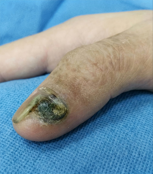 |
Figure 1 Clinical presentation of the refractory subungual wart prior to treatment, showing severe hyperkeratosis and concomitant nail plate destruction. |
We initiated an electrocautery-assisted PDT approach. The procedure was as follows: after routine disinfection with povidone-iodine and surface anesthesia with lidocaine cream, an electrocautery unit operating in the radiofrequency spectrum was used in a blended mode (100VA) to treat the lesion. The operation extended 1 mm beyond the visible border of the wart as a safety margin, vaporizing the tissue down to the superficial dermis. The primary goal was to significantly debulk the hyperkeratotic tissue to clear the physical barrier for the subsequent penetration of the photosensitizer (Figure 2). Immediately after the electrocautery debulking, PDT was performed. A 20% 5-aminolevulinic acid (ALA) solution (354 mg) was applied locally to the exposed lesion and incubated under occlusion for 1 hour. Subsequently, the area was irradiated with a red light source (635 nm, 80 mW/cm2) for 20 minutes. The treatment was administered once a week for a total of 6 sessions. During the first session, electrocautery was employed for deep debulking of the hyperkeratotic verrucous tissue. In subsequent sessions, the electrocautery device was solely utilized for superficial debridement to clear necrotic tissue, debris, and crusts. A cold air device was used concurrently to relieve the burning sensation; the patient’s visual analog scale (VAS) score was 3–4, and no oral analgesics were required. After 4 sessions, significant epithelialization of the nail bed was observed (Figure 3). By the end of 6 sessions, the wart achieved 100% clinical clearance. During the 6-month follow-up, the nail morphology returned to near-normal, with no signs of recurrence (Figure 4).
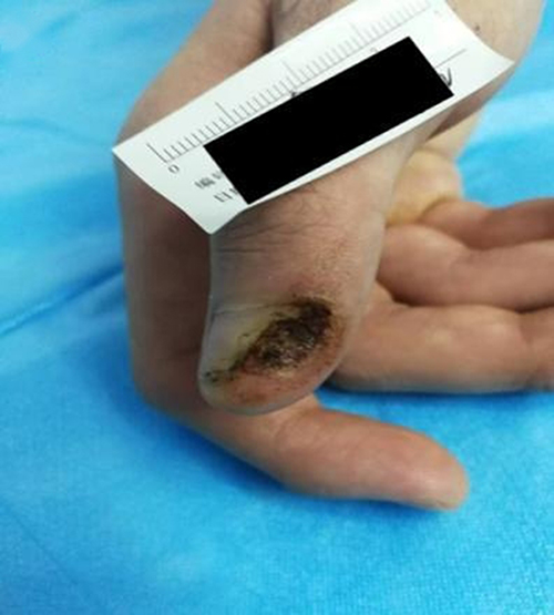 |
Figure 2 The state of the nail bed immediately following electrocautery debulking. |
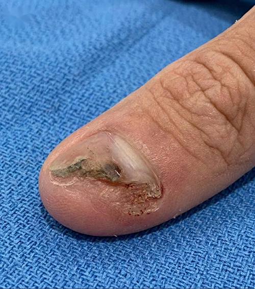 |
Figure 3 Following four sessions of the combined intervention, the volume of the verrucous growth is drastically reduced. |
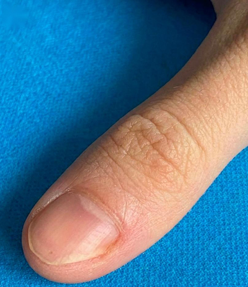 |
Figure 4 Long-term follow-up image at 6 months post-treatment. |
Discussion
Due to its special anatomical location, the subungual wart has always represented a major therapeutic dilemma. The dense hyperkeratosis and the overlying nail plate form a formidable barrier that limits the penetration of topical agents. Even potent keratolytic agents like salicylic acid plasters are often ineffective for deep-seated subungual lesions without aggressive physical debulking, as their purely chemical exfoliation cannot penetrate the dense nail barrier to reach the viral host cells in the basal layer.5 Cryotherapy often provides incomplete clearance for deep lesions and is associated with unbearable pain and high recurrence.3 Destructive CO2 lasers, if not used carefully, can cause irreversible thermal coagulation damage to the fragile nail matrix.5
Intralesional therapies such as bleomycin have shown high clearance rates but are limited by extreme pain and risks of tissue necrosis.6,7 Cidofovir and Vitamin D3 injections have also been reported with varying success.8–10 Antigen-based immunotherapies (eg, Candida antigen or MMR vaccine) aim to stimulate cellular immunity but can cause systemic adverse effects.5
In this context, the role of PDT is increasingly prominent. The core photochemical cascade begins with the highly selective absorption of the prodrug (5-ALA) by hypermetabolic, HPV-infected epidermal cells. When irradiated with red light, the accumulated protoporphyrin IX transfers energy to oxygen, producing highly toxic reactive oxygen species (ROS). These ROS induce cytotoxic necrosis and apoptosis of the targeted HPV-infected basal keratinocytes, rather than directly killing the non-enveloped virus itself. The ultimate clearance of the virus is a secondary process mediated by the host immune system recognizing and phagocytosing the released viral antigens.11
Although the mechanism of PDT is ideal, the thick keratinized tissue still acts as a physical insulator. The key to the success of this case lies in the seamless combination of electrocautery and PDT. While the concept of debulking or lesion preparation using scalpel paring, curettage, or laser ablation is not entirely new, the novelty of our strategy relies largely on the microscopic histological precision of the electrocautery device. The high-frequency alternating current induces explosive intracellular vaporization, allowing for precise and controlled ablation of the hyperkeratotic barrier. This not only mechanically removes the superficial viral load but, crucially, creates numerous microchannels within the dense matrix. These microchannels completely eliminate the physical barrier to the downward penetration of ALA molecules.
Furthermore, recent advances highlight the importance of modifying the local microenvironment for HPV eradication. As discussed by Heymann,12 emerging approaches such as salt-based therapy for recalcitrant warts reflect ongoing innovation in non-destructive and adjunctive treatments. Recent clinical work by Xu et al4 on saturated saline immersion therapy for refractory plantar warts also highlights alternative mechanisms aimed at improving outcomes in difficult cases through cellular dehydration and keratolysis. Additionally, Kumawat and Mukhtar et al13 proposed a simple thermo-sclero-keratolytic therapy for recalcitrant periungual warts, emphasizing the modification of the lesional environment to enhance therapeutic efficacy. Integrating these principles of environmental modification and non-destructive clearance with our physical microchannel-facilitated PDT provides a multifaceted and highly effective perspective against refractory HPV infections.
This clinical case strongly demonstrates that the strategy of electrocautery-assisted PDT is an excellent breakthrough for refractory subungual warts. This clinical victory is not solely due to the electrocautery itself, but stems from the deep synergistic effect: precise debulking and microchannel construction optimize the drug delivery microenvironment, while the subsequent PDT serves as the core of eradication, utilizing targeted photochemical cytotoxicity to destroy the viral safe haven.
However, this report has certain limitations. The 6-month follow-up period is relatively short, considering that the incubation period of common viral warts can range from a few months to up to 1.5 years. Furthermore, molecular pathology has shown that subclinical HPV DNA can be found up to 15 mm around the visible border of the wart. Therefore, future research should focus on long-term cohort studies with extended follow-up periods (eg, over 24 months) and molecular evaluations to further confirm the definitive eradication rate of the virus at the DNA level.
Ethical Approval and Consent to Publish
The publication of this case report was approved by the Ethics Committee of The First Affiliated Hospital of Xiamen University. Written informed consent was obtained from the patient for the publication of this case report and any accompanying images.
Disclosure
The authors report no conflicts of interest in this work.
References
1. Evans BG, Newman-Plotnick H, Prsic A. Fingertip ischemia following intralesional injection of Candida albicans antigen for treatment of a subungual wart. Dermatol Rep. 2023;16(1):9748. doi:10.4081/dr.2023.9748
2. Nofal A, Alakad R, Fouda I, Fawzy MM. Intralesional antigen immunotherapy in the treatment of periungual warts. J Cutan Med Surg. 2021;25(3):286–5. doi:10.1177/1203475420988859
3. Xie K, Cheng M, He B, Li W, Zhong W. Photodynamic therapy combined with liquid nitrogen cryotherapy and curettage for the treatment of recalcitrant periungual and subungual warts: clinical experience and literature review. Indian J Dermatol. 2024;69(1):57–62. doi:10.4103/ijd.ijd_524_22
4. Xu Y, Li T, Liang X, et al. Saturated saline immersion for the treatment of refractory plantar warts: an open-label, one-arm, single-center trial. J Am Acad Dermatol. 2026;94(2):525–529. doi:10.1016/j.jaad.2025.10.014
5. Curtis KL, Davis JC, Di Chiacchio N, et al. Diagnosis and management of subungual and periungual verruca: a clinical review. J Am Acad Dermatol. 2025;92(4):861–871. doi:10.1016/j.jaad.2024.10.081
6. Singal A, Grover C. Efficacy and safety of intralesional bleomycin in the management of ungual warts. Skin Appendage Disord. 2020;6(6):346–350. doi:10.1159/000510989
7. Al-Naggar MR, Al-Adl AS, Rabie AR, Abdelkhalk MR, Elsaie ML. Intralesional bleomycin injection vs microneedling-assisted topical bleomycin spraying in treatment of plantar warts. J Cosmet Dermatol. 2019;18(1):124–128. doi:10.1111/jocd.12537
8. Padilla Espana L, Del Boz J, Fernández Morano T, Arenas‐Villafranca J, de Troya M. Successful treatment of periungual warts with topical cidofovir. Dermatol Ther. 2014;27(6):337–342. doi:10.1111/dth.12161
9. Poppens M, Davis J. Intralesional cidofovir for the treatment of recalcitrant periungual warts. J Dermatolog Treat. 2023;34(1):2154569. doi:10.1080/09546634.2022.2154569
10. Prathibha JP, Varghese N, Aithal VV. Intralesional Vitamin D3 versus bleomycin for difficult-to-heal palmoplantar warts: a comparative study. J Cutan Aesthet Surg. 2023;16(2):114–120. doi:10.4103/JCAS.JCAS_128_21
11. Bartosinska J, Kowalczuk D, Szczepanik-Kulak P, Kwasny M, Krasowska D. A review of photodynamic therapy for the treatment of viral skin diseases. Antivir Ther. 2025;30(2):13596535251331728. doi:10.1177/13596535251331728
12. Heymann WR. Seeking a treatment for recalcitrant plantar warts worth its weight in salt. J Am Acad Dermatol. 2026;94(2):456–457. doi:10.1016/j.jaad.2025.11.048
13. Mukhtar M, Kumawat K, Mukhtar N. Therapeutic pearl: a simple Thermo-sclero-keratolytic therapy for recalcitrant periungual viral warts. J Cutan Aesthet Surg. 2022:10–4103.
 © 2026 The Author(s). This work is published and licensed by Dove Medical Press Limited. The
full terms of this license are available at https://www.dovepress.com/terms
and incorporate the Creative Commons Attribution
- Non Commercial (unported, 4.0) License.
By accessing the work you hereby accept the Terms. Non-commercial uses of the work are permitted
without any further permission from Dove Medical Press Limited, provided the work is properly
attributed. For permission for commercial use of this work, please see paragraphs 4.2 and 5 of our Terms.
© 2026 The Author(s). This work is published and licensed by Dove Medical Press Limited. The
full terms of this license are available at https://www.dovepress.com/terms
and incorporate the Creative Commons Attribution
- Non Commercial (unported, 4.0) License.
By accessing the work you hereby accept the Terms. Non-commercial uses of the work are permitted
without any further permission from Dove Medical Press Limited, provided the work is properly
attributed. For permission for commercial use of this work, please see paragraphs 4.2 and 5 of our Terms.
Recommended articles
The Effect of Myc Inhibition Combined with the Synergistic Effect of Photothermal, Photodynamic and Radiotherapy in Tumor Treatment

Cui Y, Yang W, Liu W, Li Y, Yu C, Yu C, Liu Y, Xu D, Zhu X
International Journal of Nanomedicine 2025, 20:12413-12430
Published Date: 11 October 2025
Recent Advances in Near-Infrared Cyanine Dye-Based Fluorescent Nanoprobes for Tumor Imaging and Therapy
Gao Y, Zhu L, Du Z, Xiong J, Ma R, Aili M, Alifu N, Dong B
International Journal of Nanomedicine 2025, 20:13911-13937
Published Date: 19 November 2025
An Experimental Study into the Photodynamic/Sonodynamic Therapy of Triple-Negative Breast Cancer Mediated by the Novel Nano-Photonic Sensitizer NBDPBr Nanoparticles
Huang Q, Wang L, Zhang Y, Zhao L, Zhang T
International Journal of Nanomedicine 2026, 21:576927
Published Date: 28 March 2026

Nanomaterials for Photodynamic Therapy in Cancer: Classifications, Recent Advances, and Combination Applications
Zhang L, Song L, Pei Y, Zhao M, Shi J, Li J, Qin Y, Liu Y
International Journal of Nanomedicine 2026, 21:591447
Published Date: 21 July 2026