")
Back to Journals » International Medical Case Reports Journal » Volume 15
A Case Report: Clinical Efficacy of Combination Treatment of Dexamethasone and Azathioprine in Recurrent Erythema Multiforme
Authors Suwarsa O , Dewi IP, Sutedja E , Dharmadji HP, Gunawan H , Pangastuti M
Received 8 March 2022
Accepted for publication 25 May 2022
Published 6 July 2022 Volume 2022:15 Pages 355—359
DOI https://doi.org/10.2147/IMCRJ.S364608
Checked for plagiarism Yes
Review by Single anonymous peer review
Peer reviewer comments 2
Editor who approved publication: Professor Ronald Prineas
Oki Suwarsa, Isabella Puspa Dewi, Endang Sutedja, Hartati Purbo Dharmadji, Hendra Gunawan, Miranti Pangastuti
Department of Dermatology and Venereology, Faculty of Medicine, Universitas Padjadjaran - Dr. Hasan Sadikin Hospital, Bandung, Indonesia
Correspondence: Oki Suwarsa, Department of Dermatology and Venereology, Faculty of Medicine, Universitas Padjadjaran - Dr. Hasan Sadikin Hospital, Jl. Pasteur 38, Bandung, West Java, 40161, Indonesia, Tel +62222032426 ext. 3449, Fax +62222032426, Email [email protected]
Abstract: Recurrent erythema multiforme (REM) may have frequent episodes over a period of several years and is considered to be a hypersensitivity reaction associated with infection or medication. REM is a mucocutaneous disorder which is characterized by targetoid lesions. Most of the cases are caused by herpes simplex virus infection. Systemic corticosteroid is frequently used to treat REM due to its effects in suppressing the disease. When REM is unresponsive to systemic corticosteroid, steroid-sparing treatment needs to be instituted. We reported a case of REM in a 49-year-old male. There were complaints of burning sensations on the skin lesions, along with swelling on both hands. On physical examination, erythematous macules and targetoid lesions were found on both palms, arms, and legs. During hospitalization, dexamethasone 20 mg was administered in a tapering dose but new skin lesions still appeared. Two days after azathioprine 50 mg twice daily was added to the treatment, skin lesions and swelling on the patient’s hands were diminished and the burning sensation disappeared. No side effects of azathioprine were found in this patient and no recurrence until two weeks after hospitalization. This case report demonstrated the efficacy of combined treatment of dexamethasone and azathioprine for REM cases unresponsive to systemic corticosteroid.
Keywords: recurrent erythema multiforme, targetoid lesions, azathioprine, dexamethasone
Introduction
Erythema multiforme (EM) is a rare immune-mediated mucocutaneous disorder with acute onset, sometimes recurrent, and sometimes self-limited.1,2 The two main forms of EM are EM major with the involvement of two or more mucosal sites and EM minor with the involvement of one or none mucosal site.3 The etiology of EM remains unknown, but infections, certain drugs, immune conditions, and malignancies can trigger the development of this condition.2,4 Frequent episodes occurring over several years characterized recurrent erythema multiforme (REM).5 Approximately 70% of REM cases are caused by the herpes simplex virus (HSV).1,6 In a study involving 37 patients with REM, HSV deoxyribonucleic acid (DNA) was detected in 57% of patients and the rest was idiopathic.7 Prodromal symptoms are absent in most cases.8 Patients may complain about itching and burning skin sensation, swelling of hands and feet, and pain caused by mucosal erosions.5,9 There is no specific treatment available for this disease.9 Treatment of EM depends on the severity of disease manifestation and the underlying or coexisting causes.3 Systemic corticosteroid therapy is frequently used to treat REM. If the REM is severe, other systemic immunosuppressive treatment needs to be initialized. Azathioprine is one among many immunosuppressive agents reported to be effective for REM cases unresponsive to other treatments.7,10,11
Case Presentation
A 49-year-old male was referred to our department with erythematous macules on the skin and ulceration in the mouth since three days prior to his visit. Four days prior to admission, there were prodromal symptoms such as cough, flu, and headache. The patient went to the general practitioner and was given methampyrone and amoxicillin. In an attempt to alleviate the headache, the patient also took paracetamol. Three days prior to admission, erythematous macules and targetoid lesions started to appear on both palms, arms, and legs along with erosion on eyelid, lips, buccal mucosa, and genital; and bullae on the buttocks (Figure 1A–J). Two days prior to admission, the patient experienced painful urination; with swelling and limited movements on the affected joints of the hands and feet. The patient also complained of sores in his mouth. There were also complaints of burning sensations on the skin lesions. The patient had a history of recurrent targetoid lesions on his both arms and erosions on his lips and buccal mucosa thrice in 2018 and twice in 2019, but the patient forgot the medicine he was given.
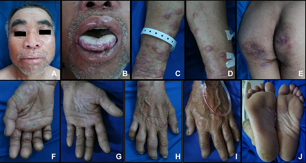 |
Figure 1 Clinical manifestation of recurrent erythema multiforme before treatment. Erosion on both of eyelids (A), lips and tongue (B). Target lesions on both arms (C and D). Bullae on buttocks (E). Target lesions on both palms (F and G). Swelling on both hands (H and I). Target lesions on plantar (J). |
On general physical examination, all vital signs were within their respective normal ranges, except for body temperature (37.6°C) and pain scale (5 out of 10). From laboratory examination, segmented neutrophils were increased, leukocytosis, slightly increased quantitative C-reactive protein (CRP), reactive anti-HSV-1 immunoglobulin G (IgG), non-reactive anti-HSV-1 immunoglobulin M (IgM), non-reactive anti-HSV-II IgG, non-reactive anti-HSV-II IgM, and non-reactive venereal disease research laboratory (VDRL) quantitative. Urinalysis revealed the presence of cocci bacteria. On the erythematous macules and targetoid lesions, 0.25% desoximetasone cream was applied, and 2% mupirocin cream was used on the erosions. For systemic treatment, the patient was given 20 mg of dexamethasone daily for five days and the dose was subsequently reduced to 10 mg on day seven until day thirteen. Upon admission, there were still new skin lesions until day eight hospitalization. On day seven of hospitalization, twice-daily 50 mg of azathioprine was added to the treatment due to the fact that new skin lesions continued appearing. Cefixime 200 mg was also administered twice daily for five days for the urinary tract infection. Ibuprofen 400 mg was given three times daily to reduce the pain. After 13 days of hospitalization, most of the skin lesions were diminished and the swelling on the hands was reduced (Figure 2A–J). After receiving azathioprine, the patient had no more fever, nausea, myalgia, or fatigue. Twice-daily 50 mg azathioprine was continued for one month following hospitalization and no recurrence was observed within one year of follow-up.
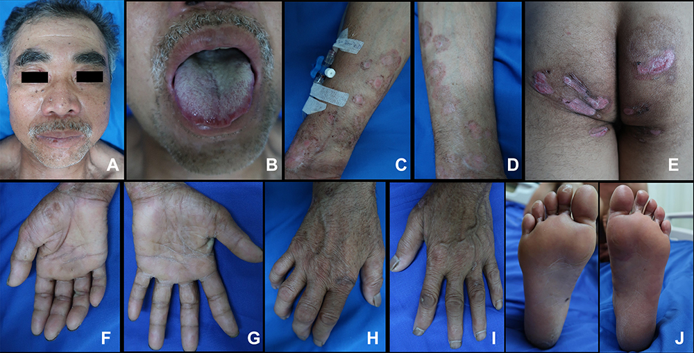 |
Figure 2 After 13 days of hospitalization. Erosion on both eyelids, lips, and tongue diminished (A and B). Target lesions on both arms improved (C and D). No bullae on buttocks (E). Target lesions on both palms improved (F and G). Swelling on both hands diminished (H and I). Target lesion on plantar improved (J). |
Discussion
Erythema multiforme occurs in patients of all ages but is most prevalent in adolescents and young adults (the peak age of 20–40 years),1,7,8 more often in male (male-to-female ratio of 2 to 3:1).4,8 There are several precipitating factors associated with the development of EM, including infections (nearly 90% cases) and medications (10% cases).4 Viral infection, especially HSV is believed to be one of the most common etiological agents for EM.4,7,9 The pathogenesis of herpes-associated EM begins with the transports of DNA fragments to distant skin sites by peripheral blood mononuclear cells. HSV genes within DNA fragments are expressed on keratinocytes, leading to the recruitment of HSV-specific CD4+ Th1 (helper T cells involved in cell-mediated immunity). The CD4 cells respond to viral antigens with the production of interferon-γ, initiating an inflammatory cascade.7 EM is thought to result from cell-mediated immune reaction against viral antigen-positive cells that contain the HSV DNA polymerase.12 Ng et al12 detected HSV-DNA in patients with HSV-related EM and patients with idiopathic EM. In some conditions, frequent episodes of EM over many years lead to REM.13 REM occurs in about 10% of patients with EM major and up to 30% in EM minor.8 Previous studies have reported that HSV was as the most common precipitating factor for REM.5,6,11,13
Prodromal symptoms are rare but may occur one week prior to the onset of EM.4 These include malaise, headache, fever, rhinorrhoea, sore throat, and cough. The clinical manifestation of EM is characterized by targetoid lesions.1,5 These lesions manifest as a central darker red area, a pink zone, and a peripheral red ring,1,4 typically distributed symmetrically along the extensor surfaces of the acral extremities.8,9 The dorsum of the hands and feet, palms and soles are frequently affected.4,8 Lesions may also manifest in the mucous membrane of the ocular, oral, and genital mucosa. Mucosal lesions occur in up to 70% of cases on mucocutaneous areas of the lips, non-attached gingivae, and the ventral side of the tongue.8 Swelling of the nail folds may be seen.4 The lesions are commonly asymptomatic, but itching or burning sensation at the site of the lesions may also occur.9 Our patient reported erythematous macules and targetoid lesions on both palms, arms, and legs; erosion on eyelid, lips, buccal mucosa, and genital; and bullae on the buttocks. Swelling and limited movements on the affected joints of hands and feet were also present, along with sores on the mouth and burning sensations on the skin lesions. There had been instances of recurring targetoid lesions on both arms and erosions on the lips and buccal mucosa, three times in 2018 and twice in 2019.
Erythema multiforme is usually diagnosed based on the patient’s history and clinical manifestation.7–9 There are no diagnostic or laboratory test to confirm the diagnosis.4,8 Laboratory examination can be done to identify a suspected history of HSV infection.7 In a study involving 48 patients with REM, 18 patients had positive serological test for HSV. Positive serological tests indicated the presence of past infection in 13 patients (positive for IgG and negative for IgM).13 The result of serologic test for HSV in our patient, demonstrating past infection (positive for IgG). A biopsy is not necessary when the clinical presentation is clear, but it can be useful to differentiate EM from other diseases that may have similar clinical presentation.5,7 We did not perform a biopsy in this case because the clinical presentation was clear and supported the diagnosis of REM.
There are no specific treatments for the management EM, but treating suspected etiology remains its primary goal.9 Severe cases of EM may require hospitalization, analgesia, corticosteroids, and immunosuppressants therapies.2 Treatment of REM is challenging and prolonged. Systemic corticosteroid (prednisolone 0.5–1.0 mg/ kg/ day tapered over 7–10 days)9 is needed to treat REM and to control the disease, but tapering and discontinuation of corticosteroid also pose risks for flare-ups.3 Immunomodulatory drugs such as azathioprine, cyclophosphamide, dapsone, cyclosporine, levamisole, thalidomide, or interferon-α are tried alone or in combination with steroids.9 Azathioprine is a purine antagonist and acts as an immunosuppressive agent.10 Azathioprine exhibits its cytotoxic and immunosuppressive activities through the disruption of nucleic acids and interference with the activation of T cells. Inflammatory responses may have been suppressed by the effects of azathioprine. Some side effects of this drug are nausea, fever, fatigue, myalgia, hepatotoxicity, and bone marrow depression.14 Azathioprine (100 to 150 mg per day) has been reported successfully in EM patients for whom other treatment have failed.7,11 Wetter et al13 reported that azathioprine was successfully treated five patients with REM. Sen et al10 described one case of REM that was not successfully treated with prednisone 40 mg daily only. The patient required additional treatment with twice-daily 400 mg acyclovir and daily 50 mg azathioprine, and responded very well to this treatment. Patient in this case report did not respond well to dexamethasone. On the seventh day of hospitalization, azathioprine was added to the treatment. One day after the combination therapy was given, no new skin lesions were formed. On day 12, the burning sensation on both palms and soles disappeared, and on day 13, the pre-existing skin lesions had diminished and the swelling on hands was reduced. This patient experienced no azathioprine side effects. At follow-up one year after hospitalization, no recurrence was observed.
Conclusion
This case report demonstrated the successful combination treatment of dexamethasone and azathioprine after no significant improvement with dexamethasone only. This combination therapy successfully reduced the pre-existing skin lesions and swelling on the hands, and eliminated the burning sensation on both palms and soles.
Ethic Statement
This study was conducted in compliance with the Declaration of Helsinki, Good Clinical Practices, local regulatory requirements, and was approved by the Medical Ethics Committee of Hasan Sadikin General Hospital Bandung (approval number: LB.02.01/X.6.5/322/2020).
Consent Statement
The patient signed informed consent forms. He also signed forms giving consent for the use of case details and images for publication and for scientific purposes.
Acknowledgments
Authors would like to thank all staff of the Dermatology and Venereology Department, Faculty of Medicine Universitas Padjadjaran–Hasan Sadikin General Hospital Bandung.
Disclosure
The authors report no conflicts of interest in this work.
References
1. Yavuz IH, Yavuz GO, Bilgili SG, et al. Erythema multiforme; sixty six case series with review of literature. East J Med. 2018;34(4):308–312. doi:10.5505/ejm.2018.70893
2. Hasan S, Jangra J, Choudhary P, et al. Erythema multiforme: a recent update. Biomed Pharmacol J. 2018;11(1):167–170. doi:10.13005/bpj/1358
3. Lerch M, Mainetti C, Beretta-Piccoli BT, et al. Current perspectives on erythema multiforme. Clin Rev Allergy Immunol. 2018;54(1):177–184. doi:10.1007/s12016-017-8667-7
4. Burbulys D, Young KD. Erythema multiforme. In: Rose E, editor. Life-Threatening Rashes. Los Angeles: Springer; 2018:55–77.
5. Sokumbi O, Wetter DA. Clinical features, diagnosis, and treatment of erythema multiforme: a review for the practicing dermatologist. Int J Dermatol. 2012;51(8):889–902. doi:10.1111/j.1365-4632.2011.05348.x
6. Tatnall FM, Schofield JK, Leigh IM. A double-blind, placebo-controlled trial of continuous Acyclovir therapy in recurrent erythema multiforme. Br J Dermatol. 1995;132(2):267–270. doi:10.1111/j.1365-2133.1995.tb05024.x
7. Lamoreux MR, Sternbach MR, Hsu WT. Erythema multiforme. Am Fam Physician. 2006;74(11):1883–1888.
8. Roujeau JC, Mockenhaupt M. Erythema multiforme. In: Kang S, Amagai M, Bruckner AL, editors. Fitzpatrick’s Dermatology.
9. Nair KK, Chaudhuri K, Ashok L. Erythema multiforme: a case series and review of literature. J Trans Med Res. 2018;2(4):124–130.
10. Sen P, Chua SH. A case of recurrent erythema multiforme and its therapeutic complications. Ann Acad Med Singap. 2004;33(6):793–796.
11. Schofield JK, Tatnall FM, Leigh IM. Recurrent erythema multiforme: clinical features and treatment in a large series of patients. Br J Dermatol. 1993;128(5):542–545. doi:10.1111/j.1365-2133.1993.tb00232.x
12. Ng PP, Sun YJ, Tan HH, et al. Detection of herpes simplex virus genomic DNA in various subsets of erythema multiforme by polymerase chain reaction. Dermatology. 2003;207(4):349–353. doi:10.1159/000074112
13. Wetter DA, Davis MD. Recurrent erythema multiforme: clinical characteristics, etiologic associations, and treatment in a series of 48 patients at Mayo Clinic, 2000 to 2007. J Am Acad Dermatol. 2010;62(1):45–53. doi:10.1016/j.jaad.2009.06.046
14. Bardek I, Puretic VM, Lipozencic J. Azathioprine in dermatology. Acta Dermatovenerol Croat. 2007;15(4):264–268.
 © 2022 The Author(s). This work is published and licensed by Dove Medical Press Limited. The full terms of this license are available at https://www.dovepress.com/terms.php and incorporate the Creative Commons Attribution - Non Commercial (unported, v3.0) License.
By accessing the work you hereby accept the Terms. Non-commercial uses of the work are permitted without any further permission from Dove Medical Press Limited, provided the work is properly attributed. For permission for commercial use of this work, please see paragraphs 4.2 and 5 of our Terms.
© 2022 The Author(s). This work is published and licensed by Dove Medical Press Limited. The full terms of this license are available at https://www.dovepress.com/terms.php and incorporate the Creative Commons Attribution - Non Commercial (unported, v3.0) License.
By accessing the work you hereby accept the Terms. Non-commercial uses of the work are permitted without any further permission from Dove Medical Press Limited, provided the work is properly attributed. For permission for commercial use of this work, please see paragraphs 4.2 and 5 of our Terms.