Back to Journals » Clinical, Cosmetic and Investigational Dermatology » Volume 16
A Case of Reactive Eccrine Syringofibroadenoma Associated with Dyshidrotic Eczema
Received 29 August 2023
Accepted for publication 1 November 2023
Published 27 November 2023 Volume 2023:16 Pages 3407—3411
DOI https://doi.org/10.2147/CCID.S437595
Checked for plagiarism Yes
Review by Single anonymous peer review
Peer reviewer comments 2
Editor who approved publication: Dr Jeffrey Weinberg
Yue Chen, Xin Tian
Guangzhou Institute of Dermatology, Guangzhou, Guangdong, People’s Republic of China
Correspondence: Xin Tian, Dermatology, Associate Director of Department of Dermatology, Guangzhou Institute of Dermatology, Guangzhou, Guangdong, People’s Republic of China, Tel/Fax +86-20-83593472, Email [email protected]
Abstract: Eccrine Syringofibroadenoma (ESFA) is an infrequent benign adnexal cutaneous lesion distinguished by hyperplastic epithelium and differentiation into eccrine ducts, displaying diverse clinical presentations. In the present case, a 48-year-old Chinese female patient exhibited a decade-long history of palmoplantar erythema, which clinically resembled Dyshidrotic Eczema. Histological examination suggested the presence of ESFA. Ultimately, considering both the clinical manifestations and histological outcomes, the patients received a diagnosis of reactive ESFA.
Keywords: reactive eccrine syringofibroadenoma, dyshidrotic eczema, histopathology
Introduction
Eccrine Syringofibroadenoma (ESFA) is a rare adnexal tumor characterized by eccrine ductal differentiation and a wide spectrum of clinical manifestations.1 Histologically, ESFA is distinguished by the presence of interconnected strands of epithelial cells embedded within a fibrovascular stroma.2 Despite its distinctive histological features, ESFA presents clinically in a nonspecific and variable manner. In this case report, we present the clinical presentation of a female patient exhibiting persistent erythema, desquamation, and pin-sized blisters on both hands and feet, resembling the clinical features typically associated with dyshidrotic eczema. These symptoms persisted over a prolonged period of several years, leading to the diagnosis of reactive ESFA based on comprehensive clinical and histopathological examination.
Case Report
A 48-year-old Chinese woman presented with a 10-year history of erythematous, desquamating, slowly expanding erythema on her hands and feet (Figure 1a–d). She reported subjective itching and occasional pain, which she noted worsened with prolonged walking. Topical treatment with glucocorticoid ointment and salicylic acid ointment failed to improve her symptoms. The patient denied any history of local trauma prior to the onset of the disease, and there were no family members with similar conditions. Additionally, she had previously tested positive for the hepatitis B antigen and had not undergone any hepatitis B virus-related treatment.
 |
Figure 1 Clinical, dermoscopy and histological image taken at the patient’s initial visit. (a–d) The patient presented with large, symmetrical erythema accompanied by scaling on the palms, soles, and lateral margins of the skin. The patient’s feet appeared dry and chapped, and the Auspitz sign was negative. Some of the nails and toenails exhibited deformities and visible signs of damage. No similar skin lesions were found elsewhere on the body. (e) Dermoscopy of the patient’s hand revealed a reddish background of the hand skin with red areas and surrounding white scales. (f) Dermoscopy of the patient’s foot revealed a light red heterogeneous background. The erosions presented as milky red areas, characterized by a central black scab-like region, surrounded by concentric white circle-like layered scales. (g) Narrow epithelial cords connect to the skin, forming a grid-like structure (H and E × 50). (h) Locally, there was evident typical ductal differentiation (H and E × 200). |
Fungal smear and culture tests of the toenails and skin lesions yielded negative results. Dermoscopy of the plantar lesions revealed clustered or non-specific punctate blood vessels on a dark red background, accompanied by yellow-white scales (Figure 1e–f). Subsequently, biopsy sampling was performed on the left plantar endogenous side, and histopathological examination revealed hyperkeratosis and interconnected chains of fine cuboidal cells extending from the epidermis to the upper dermis. Within these interconnected cell chains, small ductal structures and cystic changes were observed. No mitotic or developmentally abnormal cells were observed (Figure 1g and h). Based on these findings, a diagnosis of reactive ESFA was established.
The patient received treatment with topical mometasone furoate cream and mupirocin ointment, in combination with zinc oxide and 20% cod liver oil ointment. The treatment involved applying a plastic film and securing it with a bandage for 30 minutes once a day. After 3 weeks, a significant improvement was observed in the patient’s skin lesions, characterized by reduced desquamation and erythema compared to the initial presentation (Figure 2). The patient is currently under follow-up, and no recurrences of the skin lesion have been observed.
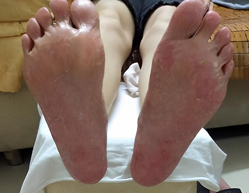 |
Figure 2 Clinical manifestations of the patient after treatment. |
Discussion
ESFA is a rare, benign, cutaneous adnexal neoplasm with eccrine acrosyringeal differentiation.3 The age of the patients ranged from 16 to 80 years, with the majority being 61–80 years old.4 The lesions primarily occurred in the acral region but could also be found on the back, abdomen, buttocks, and other areas. The skin manifestations varied, including papules, plaques, and nodules, which could be isolated or multiple, with symmetrical or linear distribution.2 Based on Starink’s classifications modified by French, there are five subtypes of eccrine fibroadenoma: Solitary ESFA, Multiple ESFA associated with hidrotic ectodermal dysplasia (Clouston and Schopf’s syndrome), Multiple ESFA without associated cutaneous findings (eccrine syringofibroadenomatosis), Non-familial unilateral linear ESFA (naevoid ES) and Reactive ESFA associated with inflammatory or neoplastic processes.5,6 ESFA can occur independently or be accompanied by skin ulcers, chronic lymphedema, amyloidosis, toenail trauma, burns, bruises, bullous pemphigoid, epidermolysis bullosa, and other lesions. It can even be associated with or secondary to squamous cell carcinoma.7 The nature of ESFA, whether reactive or neoplastic, is still controversial, and it is considered a heterogeneous group of lesions. In this case, the patient denied the presence of hypodontia, hypotrichosis, nail dystrophy, palmoplantar keratoderma, as well as periocular and eyelid margin apocrine hidrocystoma. Furthermore, there was no history of a similar case within the patient’s family. Based on the extended duration, clinical features, and histological appearance of the lesions, the patient received a diagnosis of reactive eccrine syringofistulosis associated with dyshidrotic eczema. The occurrence of reactive ESFA may be related to chronic friction of the hands and feet, sweat irritation, and recurrent episodes of sweat blisters.
The treatment approach for ESFA depends on the number, location, and resectability of the lesions. Surgical resection is the preferred option for single small skin lesions, while multiple ESFA requires individualized treatment plans, such as cryotherapy, CO2 laser, curettage, electrocautery, photodynamic therapy, local radiotherapy, topical 5-fluorouracil, imiquimod ointment, and the topical or systemic use of retinoids.8,9 However, reactive ESFA typically does not undergo malignant transformation. Some scholars suggest that symptomatic treatment and regular follow-up can be sufficient for managing reactive ESFA. Therefore, surgical resection may not be necessary for the treatment of reactive ESFA.10,11 In this case, the patient presented with eccrine hidrotic duct fibroadenoma along with sweat herpes, indicating reactive ESFA. The lesions were extensive and difficult to resect. The patient received treatment with topical glucocorticoid ointment, mupirocin ointment, and cod-liver oil and zinc oxide oil for the dry and chapped areas of palmoplantar erythema. The patient experienced relief from clinical symptoms, and the skin lesions did not progress. Our case supports the notion that symptomatic treatment of primary lesions can be effective in managing diffuse reactive ESFA. However, due to the diverse manifestations of reactive ESFA lesions, clinicians often face challenges in diagnosing the lesions during the initial visit. Therefore, a comprehensive pathological examination (such as dermoscopy, fungal smear and culture, and relevant serological tests) is necessary.
Data Sharing Statement
All data used in this work are publicly available.
Ethical Approval
The ethical committee of the hospital gave the agreement to report this case.
Consent Statement
A formal written consent was obtained for publication the case details and associated images from the patient.
Acknowledgments
We thank Dr Xin Zhou for valuable suggestions and discussions.
Author Contributions
All authors made a significant contribution to the work reported, whether that is in the conception, study design, execution, acquisition of data, analysis and interpretation, or in all these areas; took part in drafting, revising or critically reviewing the article; gave final approval of the version to be published; have agreed on the journal to which the article has been submitted; and agree to be accountable for all aspects of the work.
Funding
This study was supported by the Science and Technology Program of Guangzhou (Grant No. 2023A03J0468).
Disclosure
The authors declare no conflicts of interest in this work.
References
1. Takeda H, Mitsuhashi Y, Hayashi M, Kondo S. Eccrine syringofibroadenoma: case report and review of the literature. J Eur Acad Dermatol Venereol. 2001;15(2):147–149. doi:10.1046/j.1468-3083.2001.00213.x
2. Temnithikul B, Jerasutus S, Sudtikoonaseth P, Voravutinon N, Kootiratrakarn T, Kattipathananpong P. Eccrine syringofibroadenoma (ESFA): a report of two cases. Dermatol Pract Concept. 2016;6(1):5–8. doi:10.5826/dpc.0601a03
3. Tiwary AK, Firdous J, Mishra DK, Chaudhary SS. A case report of reactive solitary eccrine syringofibroadenoma. Indian Dermatol Online J. 2017;8(1):35–38. doi:10.4103/2229-5178.198766
4. Bottino CB, Guimarães TF, Gomes FR, Am D, Lima RB, Martins CJ. Solitary eccrine syringofibroadenoma--Case Report. An Bras Dermatol. 2015;90(3 Suppl 1):235–238. doi:10.1590/abd1806-4841.20153802
5. Khan HA, Kumarasinghe P, Wood B. Late-onset eccrine syringofibroadenoma of the feet in a patient with hypohidrotic ectodermal dysplasia. Australas J Dermatol. 2021;62(3):383–385. doi:10.1111/ajd.13604
6. Chukwuma O, Walker A, Motaparthi K, Montanez-Wiscovich M. Recalcitrant erosive plaques on the palms and soles: a rare manifestation of eccrine syringofibroadenoma. JAAD Case Rep. 2020;6(7):590–592. doi:10.1016/j.jdcr.2020.05.004
7. Lee JS, Park H, Yoon HS, Cho S. Eccrine syringofibroadenoma associated with bowen’s disease: a case report and review of the literature. Ann Dermatol. 2020;32(1):57–63. doi:10.5021/ad.2020.32.1.57
8. Sirikham T, Rojhirunsakool S, Vachiramon V. Reactive eccrine syringofibroadenoma associated with neuropathy, venous stasis, and diabetic foot ulcer. Case Rep Dermatol. 2016;8(2):124–129. doi:10.1159/000446469
9. Morganti AG, Martone FR, Macchia G, et al. Eccrine syringofibroadenoma radiation treatment of an unusual presentation. Dermatol Ther. 2010;23(Suppl 1):S20–3. doi:10.1111/j.1529-8019.2009.01282.x
10. Zhou P, Li F, Liu H, Wang L. Solitary eccrine syringofibroadenoma of the dorsum of hand. Indian J Dermatol. 2020;65(6):560–561. doi:10.4103/ijd.IJD_331_18
11. Sugita Y, Makino T, Matsui K, Shimizu T. Reactive eccrine syringofibroadenoma on the heel, clinically mimicking squamous cell carcinoma. Case Rep Dermatol Med. 2019;2019:4735739. doi:10.1155/2019/4735739
 © 2023 The Author(s). This work is published and licensed by Dove Medical Press Limited. The
full terms of this license are available at https://www.dovepress.com/terms
and incorporate the Creative Commons Attribution
- Non Commercial (unported, 3.0) License.
By accessing the work you hereby accept the Terms. Non-commercial uses of the work are permitted
without any further permission from Dove Medical Press Limited, provided the work is properly
attributed. For permission for commercial use of this work, please see paragraphs 4.2 and 5 of our Terms.
© 2023 The Author(s). This work is published and licensed by Dove Medical Press Limited. The
full terms of this license are available at https://www.dovepress.com/terms
and incorporate the Creative Commons Attribution
- Non Commercial (unported, 3.0) License.
By accessing the work you hereby accept the Terms. Non-commercial uses of the work are permitted
without any further permission from Dove Medical Press Limited, provided the work is properly
attributed. For permission for commercial use of this work, please see paragraphs 4.2 and 5 of our Terms.