Back to Journals » Clinical Ophthalmology » Volume 14
532-nm Subthreshold Micropulse Laser for the Treatment of Chronic Central Serous Retinopathy
Authors Sousa K ![]() , Calvão-Santos G, João M
, Calvão-Santos G, João M ![]() , Gomes N, Falcão M
, Gomes N, Falcão M ![]()
Received 23 September 2019
Accepted for publication 8 January 2020
Published 25 February 2020 Volume 2020:14 Pages 525—531
DOI https://doi.org/10.2147/OPTH.S232202
Checked for plagiarism Yes
Review by Single anonymous peer review
Peer reviewer comments 2
Editor who approved publication: Dr Scott Fraser
Keissy Sousa,1 Gil Calvão-Santos,1 Marina João,2 Nuno Gomes,1 Manuel Falcão3,4
1Retina Department of Hospital de Braga, Braga, Portugal; 2Ophthalmology Department of Hospital de Braga, Braga, Portugal; 3Retina Department of Centro Hospitalar Universitário S. João, Porto, Portugal; 4Department of Surgery and Physiology of Faculty of Medicine of the University of Porto, Porto, Portugal
Correspondence: Manuel Falcão
Oftalmologia, Centro Hospitalar S. João, Alameda Prof. Hernâni Monteiro, Porto 4200-319, Portugal
Tel +351 22 551 2100
Email [email protected]
Introduction: Subthreshold micropulse laser treatment with a 532 nm (532-SML) wavelength has been suggested as a treatment option for the treatment of chronic central serous retinopathy (cCSR). The objective is to present its effects and complications.
Methods: We present a retrospective cohort study of cCSR patients submitted to 532-SML. Best-corrected visual acuity (BCVA) and spectral-domain optical coherence tomography (SD-OCT) parameters – central macular thickness, subfoveal outer nuclear layer, external limiting membrane, ellipsoid band, interdigitation band, subretinal fluid and choroidal thickness – were evaluated before and 12 weeks after treatment. A power of 50%, a duty cycle of 5%, exposure time of 200 ms and a spot size of 160 μm were the applied laser parameters.
Results: We included 26 eyes. Overall there were no significant changes in visual acuity (median 0.20 (IQR 0) logMAR before and after treatment) or SD-OCT parameters. However, visual benefits occurred in 42.3% (n=11) of the patients and in half of the cases, subretinal fluid was completely reabsorbed. There were no complications.
Conclusion: In this study, 532-SML was overall ineffective on cCSR as it did not lead to significant changes in the overall median visual acuity and SD-OCT parameters. However, some patients may have benefited functionally and anatomically from the treatment; further investigation is necessary to understand the potential of 532-SML.
Keywords: central serous retinopathy, SD-OCT, subthreshold micropulse laser, 532 nm wavelength, outer retina bands
Introduction
Central Serous Retinopathy (CSR) is characterized by choroidal hyperpermeability, pigment epithelial detachment and retinal neurosensorial detachment with subretinal fluid (SRF); when the fovea is involved central vision loss occurs. In acute CSR, sudden-onset loss of central vision occurs because of a single point of leakage in the retinal pigment epithelium (RPE); such cases tend to resolve spontaneously within several months. However, at least 15% of the patients have chronic SRF accumulation, with associated persistent vision loss and more extensive pathologic features of the retina and choroid.1,2
Photodynamic Therapy (PDT) has been shown to be an effective treatment for chronic CSR (cCSR), both visually and anatomically.2,3 However, PDT is expensive and not always readily available in many centers. Potential side effects as secondary chorioretinal atrophy and reduction of contrast sensitivity have been described.4–6 Other available treatment options include mineralocorticoid antagonist (ie spironolactone and eplerenone), focal laser or anti-vascular endothelial growth factor (VEGF) therapy.7–9 There is overall poor evidence for the use of systemic and intravitreal medications; mineralocorticoid receptor antagonists may have the greatest potential from this class of treatments. Conventional thermal photocoagulation may be used in selected cases.8 Recently, subthreshold micropulse laser treatment (SML) has been used for cCSR with variable results.2,7,10-13 SML is a laser delivery mode that involves a series of repetitive ultrashort laser pulses that more broadly treat RPE. Improved RPE function is proposed to result from the targeted cells’ response to therapy.9 SML diminishes the risk of iatrogenic thermal damage of regular laser, does not induce ophthalmoscopically visible laser burns and can therefore be used to treat subfoveal or juxtafoveal focal and diffuse leaks.
Anatomic success rates were reported to be between 41% and 100% with 577 or 810 nm wavelength.2 The average visual acuity improvement are around nine-letter ETDRS13 or remained within two lines of baseline14 Potential advantages of SML over PDT include cost reduction and the elimination of the adverse effects associated with verteporfin and PDT.14 The use of 5% duty cycle has shown very mild or no visible RPE alterations secondary to local heating in fundus observation, infrared and autofluorescence imaging.10,12 For macular disorders, both green (495–570 nm) and yellow (570–590 nm) wavelengths are suitable as they are well absorbed by melanin and hemoglobin and only minimally by macular xanthophylls.15 There is scarce data using the 532 nm wavelength for cCSR. However, the theoretical principles of the other wavelengths used – 577 or 810 nm2,8,9,14 – could feasibly be applied to the 532 nm wavelength. Due to paucity of data relating to the effectiveness of 532-SML for CSR treatment, the main objective of the study was to present the effects of this laser wavelength (532-SML) using 5% duty cycle.
Methods
We analyzed medical records of patients who had the diagnosis of cCSR that were submitted to 532-SML between June 2017 and April 2019. All investigations were performed in accordance with the principles of the Declaration of Helsinki and approved by the Ethics Committee of Hospital de Braga. Inclusion criteria were patients ≥18 years with cCSR with at least 4 months of persistent subretinal fluid observed by spectral-domain optical coherence tomography (SD-OCT). It was determined a subretinal fluid height until 100 µm. The diagnosis of cCSR was confirmed by fluorescein angiography (FA) (TRC-50DX, Topcon Medical Systems, Inc., Tokyo, Japan) and/or Indocyanine green angiography (ICG) (TRC-50DX, Topcon Medical Systems, Inc., Tokyo, Japan). Data were collected at just before and at 12 weeks after treatment. During these 12 weeks, no other treatment for cCSR was performed including mineralocorticoid receptor antagonists or laser or PDT. We excluded patients with myopia ≥6.0 diopters and macular disorders such as choroidal neovascularization, polypoidal choroidal vasculopathy, age-related macular degeneration, history of vitreomacular disease, cataract or optical media opacity that restricted the examination of ocular fundus. Patients with a history of treatment for their cCSR in the 6 months before the 532 nm MPL treatment (intravitreal anti-VEGF) treatment, laser photocoagulation or prior PDT) were also excluded. Visual acuity was not an inclusion/exclusion criteria.
All included participants underwent a comprehensive ophthalmologic examination, SD-OCT and FA and/or ICG evaluation. Demographic data included sex, age, time of diagnosis, laterality and previous treatments. Ophthalmologic examination included best-corrected visual acuity (BCVA) and slit-lamp biomicroscopy. BCVA was assessed using the decimal scale chart and converted to logarithm of the minimum angle of resolution (logMAR).
All patients performed a Spectralis SD-OCT (Heidelberg Engineering, Heidelberg, Germany) to measure retinal thickness and evaluate the outer retinal layers. The OCT imaging technique consisted in obtaining a macular square (20 x20°) composed of 25 horizontal B-scans, spaced at 240 µm. Each B-scan was averaged 9 times (ART 9). Additionally, for each case, a single horizontal and a single vertical B-scan using the Enhanced Depth Imaging mode, averaged 100 times (ART 100), and centered on the fovea was obtained. The SD-OCTs and BCVA measurements were performed immediately before treatment and 12 weeks later. The presumed foveal center was determined as the area lacking the inner retinal layers in the macular region. Data collected were central macular thickness (CMT) and the following foveal parameters – outer nuclear layer (ONL) thickness; continuity or disruption of external limiting membrane (ELM), ellipsoid band (EZ) and interdigitation band (IZ); subretinal fluid height (SRF) and choroidal thickness (CT). ONL was defined as the distance between the inner limiting membrane and the ELM at the central fovea. SRF was measured as the hyporreflexive space from the IZ to the RPE. Choroidal thickness was manually measured subfoveally with enhanced depth imaging, from the outer portion of the retinal pigment epithelium (RPE) to the inner surface of the sclera. A disruption of the foveal ELM, EZ and IZ bands was classified as absent; its preservation was classified as present. Two independent investigators (K.S. and M.J.) made all measurements and evaluations on the horizontal high-quality scans centered on the fovea. Any prominent difference between the two investigators was discussed with the senior author (M.F.) and the reconciled measurement was recorded.
SML was performed with a Quantel Medical Supra® 532 nm. A pattern of multiple confluent with no space laser spots were applied, covering the fluid seen on SD-OCT and/or the main leakage point in FA. The laser treatment was also applied on even if these areas did not coincide with visible leaking spots on the FA/ICG. All treatments were performed by the same surgeon (K.S.). The power was initially increased upward to the minimum threshold value to cause a barely visible burn on micropulse mode outside vascular area at the posterior pole and then it was reduced to 50%. A duty cycle of 5% was used, exposure time of 200 ms and a spot size of 160 µm. A resorption of ≥50% of SRF was considered a positive anatomical response.16
All statistical analysis was performed using SPSS software version 25.0 (SPSS Inc., Chicago, IL) and p values <0.05 were considered statistically significant. Non-parametric tests were applied after non-normality of the sample was confirmed by Shapiro–Wilk test. All values were presented as mean (± standard-deviation (SD)) or median (interquartile range (IQR)) according to the normality test result. To compare the baseline and post-treatment BCVA, Wilcoxon signed-rank test was used.
Results
Twenty-six eyes of 22 patients were included. The mean age was 51.5±12.1 years, in which 20 (90.9%) were male and 2 (9.1%) were female. Median time from diagnosis until treatment was 19 (20) months. Included previous treatments (≥6 months) for cCSR were 532-SML in 15.4% (n=4), aldosterone receptor antagonist (spironolactone) in 57.7% (n=15), half-dose PDT in 30.8% (n=8) and anti-VEGF (aflibercept) in 26.9% (n=7). Seven eyes (26.9%) were treatment naïve. Table 1 shows demographic features.
 |
Table 1 Demographic Data |
Concerning best-corrected visual acuity, the median BCVA remained at 0.20 (IQR 0) logMAR (z(26)=−1.18, p=0.24) – Figure 1. An average increase of 0.16±0.15 (minimum of −0.1 and a maximum of −0.6) logMAR BCVA was reported in 42.3% (n=11). An improvement of ≥2 lines occurred in 19.2% (n=5) and a decrease of ≥2 lines occurred in 15.4% (n=4). Nevertheless, 46.2% (n=12) had a baseline BCVA superior to +0.2 logMAR.
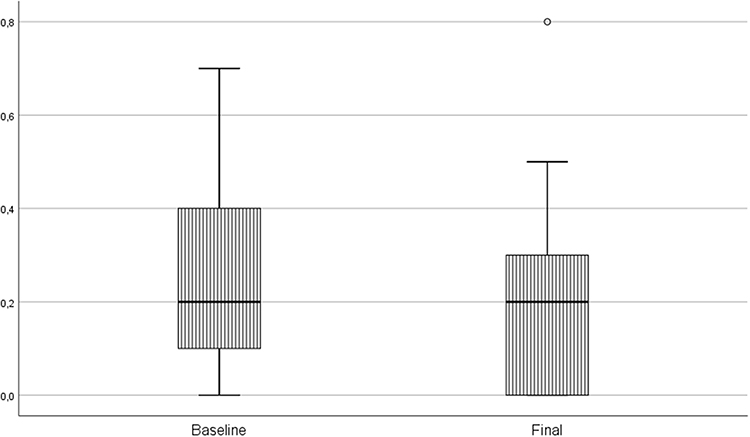 |
Figure 1 Baseline and 12 weeks after 532-SML treatment best-corrected visual acuity (logMAR). There was no significant difference between the two periods of evaluation. 532-SML: Subthreshold micropulse laser treatment with a 532 nm wavelength. |
Overall, the SD-OCT parameters evaluated did not suffer any changes from baseline to 12 weeks after treatment (Table 2). Using SD-OCT for analysis of the macular layers, there was a decrease of CMT and SFR. There was an increase of ONL thickness, CT and a gain of IZ presence. None of these changes were statistically significant. Other external retinal bands (ELM and EZ) did not present any variation. However, 50% of the patients showed an SRF resorption. There was an increase of SRF in 23.1% (n=6). In the group of patients with visual acuity increase, 81.8% (n=9) had a complete anatomic response. In the group of patients treated in the first 12 months (n=8), 50% had a complete anatomical response, the same as the group treated later (n=18) (Spearman’s rho, p=1.00).
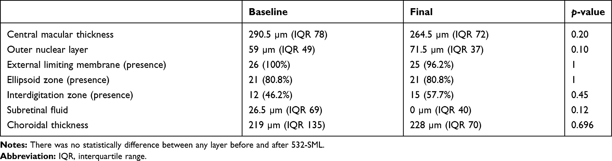 |
Table 2 SD-OCT Parameters at Baseline and After 532-SML Treatment |
We did not document any damage in any external retinal bands or other complications such as laser-induced macular neovascularization or scars during the study period.
Discussion
The main objective of our study was to analyze the efficacy of 532-SML in cCSR. Overall, the results were disappointing with no median change in visual acuity and SD-OCT parameters. Other wavelengths2,13,14 applied demonstrated slightly better results. SML has been used as an optional treatment for cCSR.8,9,13 Advantages over PDT are the availability, less invasive treatment, fewer side effects and economic issues. For example, in our department, SML is always available for treatment whilst PDT requires special approval due to its cost. We have evaluated the results of 532-SML in some cCSR patients concerning efficacy and complications. Overall, we had a 50% of anatomic response with no BCVA variation. Nevertheless, 42.3% (n=9) showed a BCVA increase in which 81.8% (n=9) had a complete anatomic response. No damage were collected in any external retinal bands and no complications or side effects were registered.
Some studies are well recognized and a review was performed by Wood et al13 in 2017. In this review, there was a decrease in mean central macular thickness (80 µm at 3 months). However, this decrease was not statistically significant. Mean best-corrected visual acuity increased about nine letters at 3 months and no study reported a decrease in visual acuity. No major retinal complications were observed. In our study, 42.3% of the patients had an increase in visual acuity (mean of 0.16±0.15 (−0.1 to −0.6) logMAR) which is about nine ETDRS letters. On the other hand, our sample had a greater baseline BCVA: > +0.2 logMAR in 46.2% (n=12) of patients which could explain the non-improvement. There was no statistical difference in CMT which is in line with other papers with no clinically significant difference. In the review by Wood et al, the single study that used a 532nm laser was Behnia et al.17 However, the power titration was 20% instead of 50% used in our study and it was implemented in acute CSR (<1 month). At the end of the follow-up period, BCVA was about the same and it was not reproducible for cCSR patients. Our study has a median time until treatment of 20 months which is a long waiting for treatment. However, anatomic response was not correlated with time until treatment as observed previously.3,16,18
Most studies used a 577 nm or 810 nm wavelength and a 10–15% duty cycle. Five percent duty cycle were used by the PACORES group14 and Malik et al.10 The PACORES group presented 92 eyes treated with 577 nm SML and a 12-month follow-up. Malik et al used an 810 nm wavelength and included 11 eyes with an anatomic efficacy of 72.7% and varied follow-up duration. This wavelength was also used in the PLACE2 trial which compared PDT with micropulse laser treatment. They kept a distance of 500 µm from the foveal center and repeated treatments were possible after 6 to 8 weeks. No significant differences between the two groups (SML versus PDT) were observed based on BCVA, retinal sensitivity and vision-related quality of life. OCT parameters were not evaluated. A greater visual acuity improvement and CMT reduction were observed during the first months after treatment in most of the studies.4,14,19,20
For reference, 12 weeks seems to be enough to test the efficacy of this new wavelength in cCSR; this time frame has been used in other studies to understand the response and to weight other treatment options.2,4,8,9,14,20 In our study, there were no significant changes in BCVA which seem inferior than the other studies that evaluated micropulse laser for cCSR. Also, our patients did not have a significant change in mean CMT. Nevertheless, baseline CMT was better than other studies (290.5 µm vs 402 µm vs 326.5 µm vs 369 µm) which can explain the lower decrease in CMT.7,12,14 Ntomoka et al7 was the only study that evaluated macular layers besides CMT. In our study, SRF was also less severe than in other studies (26.5 µm vs 160.5 µm).
A cut-off of for the amount of SRF to be used to treat cCSR with SML has not been defined. As a first published work with 532 nm, we opted not to treat large volumes of SRF and limited our treatments to patients with an SRF height less than 100 µm. When compared to other studies, the presence of ELM and IS/OS (inner/outer segment) (which corresponds to our EZ) was better in our sample, which may be an indication of less severe disease. CSR as an RPE disease8 and IZ as the representation of the region of the distal portion of the cone's outer segment which indicated contact cylinder with the apposed RPE cell body is expected that the regulation of the disease can be translated in more IZ presence by SML treatment.21 ONL has been described as a biomarker of better prognosis in CSR22–25 and 532-SML marginally increased the ONL thickness in our study, without statistically significant variation. Treatment was applied subfoveally if necessary.
The main limitation of our study is the size and heterogeneity of the sample (patients with a wide variety of previous treatments). A retrospective study has also well-known limitations.
Still, this is the first time that this wavelength is reported in cCSR. As a start for a bigger pilot study, these results could be explored. Even though we did not find statistically significant changes in overall visual acuity, 42% of the patients had visual improvements, in which 81.8% showed a complete anatomic response to treatment. This data suggests that there might be a subgroup of patients that may benefit from this treatment. However, our short sample does not allow a proper subgroup analysis. A prospective study with longer follow-up and more patients should be done to confirm the results and conclude which patients could have better outcomes. It is important to evaluate if this can be an option to PDT, especially because this may not be available in many centers. In our study, 532-SML was also used in less severe patients that were considered for treatment before great loss of visual acuity and irreversible damage. 532-SML, as an available treatment for cCSR, needs to be better evaluated before it can be recommended.
Conclusions
532-SML was ineffective on cCSR as it did not lead to significant changes in the overall median visual acuity and SD-OCT parameters. However, it might have a place in a subgroup of patients with cCSR. In our study, 42% of the patients improved anatomically and visually. Apparently, 532-SML is not harmful so it may explored as an alternative therapy. It should be considered in further prospective studies to better evaluate its efficacy.
Summary
Photodynamic Therapy (PDT) remains the gold-standard of treatment for chronic central serous retinopathy but other treatment options have become available as 577 and 810 nm sub-threshold laser (SML), mineralocorticoid receptor antagonists and anti-VEGF intravitreal injections. Potential advantages of SML over PDT include cost reduction and avoidance of the adverse effects associated with verteporfin and PDT. There is scarce data about subthreshold micropulse laser treatment with a 532 nm wavelength for the treatment of chronic Central Serous Retinopathy. Spectral-domain Optical Coherence Tomography is a non-invasive and useful tool to diagnose and follow-up these patients. The objective of our work is to show the results and adverse effects of 532 nm wavelength SML. The treatment was anatomically successful in half of the patients.
Abbreviations
532-SML, Subthreshold Micropulse Laser using wavelength 532 nm; BCVA, Best-corrected Visual Acuity; CSR, Central Serous Retinopathy; cCSR, Chronic Central Serous Retinopathy; CMT, Central Macular Thickness; CT, Choroidal Thickness; ELM, External Limiting Membrane; ETDRS, Early Treatment Diabetic Retinopathy Study chart; EZ, Ellipsoid Zone; IS/OS, Inner Segment/Outer Segment Junction Layer; IZ, Interdigitation Zone; FA, Fluorescein Angiography; ICG, Indocyanine Green Angiography; IQR, Interquartile Range; logMAR, Logarithm of the Minimum Angle of Resolution; OCT, Optical Coherence Tomography; ONL, Outer Nuclear Layer; PDT, Photodynamic Therapy; RPE, Retinal Pigment Epithelium; SD, Standard-Deviation; SD-OCT: Spectral-domain Optical Coherence Tomography; SML, Subthreshold Micropulse Laser; SRF, Subretinal Fluid; VEGF, Vascular Endothelial Growth Factor.
Ethics Approval and Informed Consent
All procedures were in accordance with the World Medical Association Declaration of Helsinki. The ethical standards of the institutional, document number 132/2017, and national research committee and with the 1964 Helsinki declaration and its later amendments or comparable ethical standards were accomplished. All data were performed based on anonymized data and none of the presented results can identify any patient.
Data Sharing Statement
The data used to support the findings of this study are restricted by the Ethics Committee of Hospital de Braga in order to protect patient privacy. Data are available for researchers who meet the criteria for access to confidential data.
Acknowledgment
Andreia Magalhães (Technician), Joana Pires (Collected Data); Carla Ferreira (Collected Data), Luís Mendonça (Collected Data), Rita Gentil (Collected Data), Petra Gouveia (Collected Data), Fernando Vaz (Ophthalmology Head Department).
Author Contributions
All authors contributed to data analysis, drafting and revising the article, gave final approval of the version to be published, and agree to be accountable for all aspects of the work.
Disclosure
The authors report no conflicts of interest in this work.
References
1. Daruich A, Matet A, Dirani A, et al. Central serous chorioretinopathy: recent findings and new physiopathology hypothesis. Prog Retin Eye Res. 2015;48:82–118. doi:10.1016/j.preteyeres.2015.05.003
2. EHCv D, Fauser S, Breukink MB, et al. Half-dose photodynamic therapy versus high-density subthreshold micropulse laser treatment in patients with chronic central serous chorioretinopathy: the place trial. Ophthalmology. 2018;125(10):1547–1555.
3. Chung CY, Chan YY, Li KKW. Angiographic and tomographic prognostic factors of chronic central serous chorioretinopathy treated with half-dose photodynamic therapy. Ophthalmologica. 2018;240(1):37–44. doi:10.1159/000484100
4. Scholz P, Ersoy L, Boon CJF, Fauser S. Subthreshold micropulse laser (577 nm) treatment in chronic central serous chorioretinopathy. Ophthalmologica. 2015;234:189–194. doi:10.1159/000439600
5. Schatz H, Yannuzzi LA, Gitter KA. Subretinal neovascularization following argon laser photocoagulation treatment for central serous chorioretinopathy: complications or misdiagnosis? Retina. 2012;32(suppl 1):OP893–OP906. doi:10.1097/IAE.0b013e318242fa2d
6. Khosla P, Rana S, Tewari H, Azad R, Talwar D. Evaluation of visual function following argon laser photocoagulation in central serous retinopathy. Ophthalmic Surg Lasers. 1997;28:693–697.
7. Ntomoka CG, Rajesh B, Muriithi GM, Goud A, Chhablani J. Comparison of photodynamic therapy and navigated microsecond laser for chronic central serous chorioretinopathy. R Coll Ophthalmol. 2017;32:1079–1086.
8. Goldhagen BE, Goldhardt R. Diagnosed a patient with central serous chorioretinopathy? Now what? Management of central serous chorioretinopathy. Curr Ophthalmol Rep. 2017;5(2):141–148. doi:10.1007/s40135-017-0133-4
9. Salehi M, Wenick AS, Law HA, Evans JR, Gehlbach P. Interventions for central serous chorioretinopathy: a network meta-analysis. Cochrane Database Syst Rev. 2016;12.
10. Malik KJ, Sampat KM, Mansouri A, Steiner JN, Glaser BM. Low-intensity/high-density subthreshold micropulse diode laser for chronic central serous chorioretinopathy. Retina. 2015;35:532–536. doi:10.1097/IAE.0000000000000285
11. Ozmert E, Demirel S, Yanik O, Batioglu F. Low-fluence photodynamic therapy versus subthreshold micropulse yellow wavelength laser in the treatment of chronic central serous chorioretinopathy. J Ophthalmol. 2016;2016:1–8.
12. Arsan A, Kanar HS, Sonmez A. Visual outcomes and anatomic changes after sub-threshold micropulse yellow laser (577 nm) treatment for chronic central serous chorioretinopathy: long-term follow-up. Eye (Lond). 2018;32:726–733. doi:10.1038/eye.2017.293
13. Wood EH, Karth PA, Sanislo SR, Moshfeghi DM, Palanker DV. Nondamaging retinal laser therapy for treatment of central serous chorioretinopathy. Retina. 2017;37:1021–1033. doi:10.1097/IAE.0000000000001386
14. Roca JA, Wu L, Fromow-Guerra J, et al. Yellow (577 nm) micropulse laser versus half-dose verteporfin photodynamic therapy in eyes with chronic central serous chorioretinopathy: results of the Pan-American Collaborative Retina Sturdy (PACORES) Group. Br J Ophthalmol. 2018;102(12):1696–1700. doi:10.1136/bjophthalmol-2017-311291
15. Yadav NK, Jayadev C, Rajendran A, Nagpal M. Recent developments in retinal lasers and delivery systems. Indian J Ophthalmol. 2014;62(1):50–54. doi:10.4103/0301-4738.126179
16. Sousa K, Viana AR, Pires J, Ferreira C, Queirós L, Falcão M. Outer nuclear layer as the main predictor to anatomic response to half dose photodynamic therapy in chronic central serous retinopathy. J Ophthalmol. 2019;2019:6. doi:10.1155/2019/5859063
17. Behnia M, Khabazkhoob M, Aliakbari S, Abadi AE, Hashemi H, Pourvahidi P. Improvement in visual acuity and contrast sensitivity in patients with central serous chorioretinopathy after laser macular subthreshold laser therapy. Retina. 2013;33:324–328. doi:10.1097/IAE.0b013e3182670fa3
18. Iacono P, Tedeschi M, Boccassini B, Valloti AC, Varano M, Parravano MC. Chronic central serous chorioretinopathy - early and late morphological and functional changes after verteporfin photodynamic therapy. Retina. 2018;1–8.
19. Russo A, Turano R, Morescalchi F, et al. Comparision of half-dose photodynamic therapy and 689 nm laser treatment in eyes with chronic central serous chorioretinopathy. Graefes Arch Clin Exp Ophthalmol. 2017;255(6):1141–1148. doi:10.1007/s00417-017-3626-9
20. Gawecki M, Jaszczuk-Maciejewska A, Jurska-Jasko A, Grzybowski A. Functional and morphological outcome in patients with chronic central serous chorioretinopathy treated by subthreshold micropulse laser. Graefes Arch Clin Exp Ophthalmol. 2017;255:2299–2306. doi:10.1007/s00417-017-3783-x
21. Spaide R, Curcio CA. Anatomical correlates to the bands seen in the outer retina by optical coherence tomography. Retina. 2011;31(8):1609–1619. doi:10.1097/IAE.0b013e3182247535
22. Ozdemir I, Eren A, Ersoz G. Outer nuclear layer thickness at the central fovea relation with symptom duration in central serous chorioretinopathy. Graefes Arch Clin Exp Ophthalmol. 2018;39(6):1323–1328.
23. Matsumoto H, Sato T, Kishi S. Outer nuclear layer thickness at the fovea determines visual outcomes in resolved central serous chorioretinopathy. Am J Ophthalmol. 2009;148(1):105–110 e101. doi:10.1016/j.ajo.2009.01.018
24. Ersoz MG, Karacorlu M, Arf S, Hocaoglu M, Muslubas IS. Outer nuclear layer thinning in paquychoroid pigment epitheliopathy. Retina. 2018;38:957–961. doi:10.1097/IAE.0000000000001655
25. Ohkuma Y, Hayashi T, Sakai T, Watanabe A, Tsuneoka H. One-year results of reduced fluence photodynamic therapy for central serous chorioretinopathy: the outer nuclear layer thickness is associated with visual prognosis. Graefes Arch Clin Exp Ophthalmol. 2013;251(8):1909–1917. doi:10.1007/s00417-013-2289-4
 © 2020 The Author(s). This work is published and licensed by Dove Medical Press Limited. The
full terms of this license are available at https://www.dovepress.com/terms
and incorporate the Creative Commons Attribution
- Non Commercial (unported, 3.0) License.
By accessing the work you hereby accept the Terms. Non-commercial uses of the work are permitted
without any further permission from Dove Medical Press Limited, provided the work is properly
attributed. For permission for commercial use of this work, please see paragraphs 4.2 and 5 of our Terms.
© 2020 The Author(s). This work is published and licensed by Dove Medical Press Limited. The
full terms of this license are available at https://www.dovepress.com/terms
and incorporate the Creative Commons Attribution
- Non Commercial (unported, 3.0) License.
By accessing the work you hereby accept the Terms. Non-commercial uses of the work are permitted
without any further permission from Dove Medical Press Limited, provided the work is properly
attributed. For permission for commercial use of this work, please see paragraphs 4.2 and 5 of our Terms.