
Back to Journals » ImmunoTargets and Therapy » Volume 14
2B4/CD244 Signaling in Immune Regulation and Its Role in Infection, Cancer, and Immune Tolerance
Authors Yan C  , Lu P, Jiang Y, Miao S, Zhao L , Xu X
Received 3 May 2025
Accepted for publication 6 September 2025
Published 4 October 2025 Volume 2025:14 Pages 1111—1131
DOI https://doi.org/10.2147/ITT.S538126
Checked for plagiarism Yes
Review by Single anonymous peer review
Peer reviewer comments 5
Editor who approved publication: Dr Flavio Salazar-Onfray
Chao Yan,1,2,* Peng Lu,3,* Yuzhu Jiang,1,* Shu Miao,4,5 Lichun Zhao,6 Xiaoyan Xu1
1Department of Immunology, Binzhou Medical University, Yantai, Shandong, 264003, People’s Republic of China; 2Institute for Immunology, Chinese Institutes for Medical Research, Beijing, 100069, People’s Republic of China; 3School of Life Sciences, Ludong University, Yantai, Shandong, 264025, People’s Republic of China; 4R&D Department, Hainan Cell Medical Technology Co., Ltd, Haikou, Hainan, 570100, People’s Republic of China; 5R&D Department, Shandong Antaien Biotechnology Co, Ltd, Yantai, Shandong, 264003, People’s Republic of China; 6College of Pharmaceutical Sciences, Guizhou University of Traditional Chinese Medicine, Guiyang, 550025, People’s Republic of China
*These authors contributed equally to this work
Correspondence: Lichun Zhao, Email [email protected] Xiaoyan Xu, Email [email protected]
Abstract: 2B4 (CD244), the fourth member of the signaling lymphocyte activation molecule (SLAM) family, is expressed by virtually all human and murine hematopoietic lineages and functions as a context-dependent activating or inhibitory receptor. This review provides a comprehensive update on the gene organization, molecular architecture, glycosylation patterns, and alternatively spliced isoforms of 2B4, highlighting how these structural variables dictate ligand (CD48) affinity and downstream signaling outcome. The roles of 2B4 in natural killer (NK) cells, CD8+ T cells, dendritic cells, myeloid-derived suppressor cells, B cells, eosinophils, and basophils were then systematically demonstrated, emphasizing their dual capacity to either potentiate cytotoxicity and cytokine production or enforce immune tolerance and exhaustion. Mechanistically, the balance between SLAM-associated protein (SAP)-mediated activation and SHP-1/2/SHIP-driven inhibition emerges as a central rheostat that is dynamically tuned by SAP availability, and the microenvironment. Clinically, exaggerated 2B4 signaling is associated with viral persistence in MCMV, HCV, HIV, and SARS-CoV-2 infections, promotes tumor immune escape in melanoma, multiple myeloma, and head-and-neck cancer, and compromises maternal–fetal tolerance, whereas insufficient signaling weakens antimicrobial immunity. Parallel pre-clinical studies validate 2B4 blockade as a rational combinatorial strategy to reinvigorate exhausted CD8+ T and NK cells, while soluble CD48 emerges as a dynamic biomarker of disease activity. Collectively, these insights redefine 2B4 as a systems-level integrator of immune homeostasis and a tractable precision-immunotherapy node whose therapeutic manipulation can rebalance immunity across infection, cancer, and pregnancy.
Keywords: 2B4, immune activation, immune tolerance, infectious disease, neoplastic disease
Introduction
2B4, also known as CD244, is a member of the signaling lymphocyte activation molecule (SLAM) family and belongs to the CD2 superfamily. It is widely expressed on various hematopoietic cells, including natural killer cells, certain T-cell subsets, monocytes, basophils, eosinophils, and myeloid-derived suppressor cells.1,2 The SLAM family of receptors is a group of type I transmembrane receptors that includes SLAMF1 (CD150; SLAM), SLAMF2 (CD48), SLAMF3 (Ly-9; CD229), SLAMF4 (CD244; 2B4), SLAMF5 (CD84), SLAMF6 (Ly108; NTB-A; CD352), SLAMF7 (CRACC; CS1; CD319), SLAMF8 (BLAME; CD353), and SLAMF9 (CD84-H1; SF2001; CD2F10).3 All members of this family share at least one distal membrane IgV-like structural domain and one IgC2-like structural domain. Furthermore, each receptor contains conserved cysteine residues within the C2-like domain that form disulfide bonds.4 The intracellular domains of all these receptors contain the immunoreceptor tyrosine-based switch motifs (ITSMs), which binds to SLAM-associated protein (SAP), Ewing’s sarcoma-associated transcript 2 (EAT-2), and the EAT-2-associated transducers ERT and SH2D1C to transmit activation signals and regulate cellular functions. 2B4 was initially identified in mice as a cell surface molecule that triggers MHC molecule-independent cytolysis and promotes the expansion of NK cells, αβ+ CD8+ T cells, γδ+ CD8+ T cells, and γδ+ T-cell cytokine secretion.5 In addition, several studies reported that the addition of 2B4 to chimeric antigen receptor (CAR) significantly mediated more vigorous activation signals and enhanced NK cytotoxicity.6,7 As research has progressed, the role of 2B4 has been increasingly elucidated. In addition to enhancing the activity of cytotoxic lymphocytes, 2B4 can also function as an inhibitory receptor, suppressing the activation of decidual NK cells at the fetal-maternal interface.8 In our previous study, we also found that downregulation of 2B4 receptor leads to worse pregnancy outcomes by facilitating TNF-α and IFN-γ production in dNK cells during Toxoplasma gondii infection.9 In fact, 2B4 mediates different outcomes depending on the cell type and stage of differentiation. It has been reported to act as an inhibitory receptor during the early stages of NK cell differentiation, suppressing NK cell activity and serving as a crucial mechanism for maintaining NK cell self-tolerance.10 CD48 is a key ligand for 2B4 activation, forming a high-affinity heterodimer through extracellular binding that effectively stimulates cell activation.11 Intracellularly, 2B4 plays a significant role in tumor and infection immunity by interacting with junction proteins such as SLAM-associated protein and recruiting the intracellular kinase Fyn, which transmits signals to perform immunomodulatory functions.12,13
Additionally, the binding of 2B4 to CD48 is instrumental in maintaining tissue tolerance and facilitating tumor escape by phosphorylating the intracellular tyrosine residues of 2B4, thereby transmitting inhibitory signals through the recruitment of SHP-1, SHP-2, and SHIP junction proteins.14 The disruption of the balance between activation signaling mediated by SAP/Fyn and tolerance signaling mediated by SHP-1/SHP-2/SHIP is now recognized as a central checkpoint in the pathogenesis of infections, tumors and pregnancy complications. Although earlier reviews focused some aspects of 2B4, the present review has three specific aims that distinguish it from previous work: (i) to provide the systematic integration of structural, glycomic and interactome data that dictate 2B4 isoform-specific signaling; (ii) to synthesize recent single-cell and in-vivo evidence revealing how 2B4 operates as a rheostat across NK cells, CD8+ T cells, DCs, MDSCs, eosinophils, basophils and B cells; and (iii) to translate these mechanistic insights into an updated conceptual framework for therapeutic targeting of the 2B4-CD48 axis in infection, cancer and maternal–fetal tolerance, including a critical appraisal of 2B4 as both a predictive biomarker and a tractable drug target.
Gene Localization, Molecular Structure, Isoforms, and Glycosylation of 2B4
2B4 Gene Locus
Northern blot analysis revealed the presence of at least three transcripts of 2B4 in RNA isolated from five mice A-LAK cell strains: C57BL/6, BALB/c, CBA, DBN2, and NZB. Southern hybridization analysis of the genomic DNA from these mouse strains revealed that 2B4 belongs to a closely related gene family. Expression analysis in recombinant inbred mice revealed that the 2B4 gene is located on the long arm of chromosome 1, adjacent to the Ly-7 gene (FcγRIIIα).15 In humans, 2B4 is located at 1q21-24 on the long arm of chromosome 1.16,17 In order to challenge in immunology research, the biology, immune function and signaling consequences of 2B4 need to be further description.
Molecular Structure of 2B4
Structurally, 2B4 belongs to the CD2 subfamily of the immunoglobulin superfamily (Ig-SF), which also includes CD48, CD58, CD84, SLAM, Ly-9, and CS-1.17–19 The extracellular protein sequence of human 2B4 shares 40% similarity with that of mouse 2B4.20 While the extracellular regions of CD2 subfamily members exhibit high structural similarity, their cytoplasmic domains differ markedly. Specifically, 2B4, SLAM, Ly-9, and CD84 contain long cytoplasmic tails featuring the ITSM, which has the amino acid sequence TxYxxV/I.15,21 In contrast, CD2 has a proline-rich cytoplasmic domain capable of recruiting the Src homologous 3-protein CD2-associated protein (CD2AP).21 The intracellular domain of CD48 is phosphatidylinositol (GPI)-anchored, whereas the spliced forms of CD58 generate both a transmembrane isoform with a short cytoplasmic tail and a GPI-anchored form. Additionally, a membrane-type 2B4 splice isoform lacking the cytoplasmic tyrosine motif has been identified.22 The structure of 2B4 was shown in Figure 1.
 |
Figure 1 Structural characteristics and signal transduction model of 2B4. 2B4 is a transmembrane immunoreceptor which contains one distal membrane IgV-like structural domain, one IgC2-like structural domain and 4 tyrosine-based switch motifs (ITSMs) within the intracellular domains. Intracellular signal transduction is mediated through ITSMs. (A) Activating signals are transmitted via recruitment of SAP/Fyn or EAT-2. (B) Whereas inhibitory signaling occurs through the association of ITSMs with SHP-1, SHP-2, or EAT-2. |
High-resolution NMR analysis revealed that the structure of 2B4 comprises nine β-strands, with the topology consisting of DEB- and AGFCC′C″-labeled β-sheet lamellae. A non-canonical disulfide bridge between Cys2 and Cys99 tethers strands A and G in a parallel orientation, enhancing the convex curvature of the six-stranded sheet. Strand C′ is interrupted by a β-bulge centred at Glu45-Ile46, yielding two shorter segments, C1′ and C2′. This distortion disrupts the canonical hydrogen-bond register between strands C and C′: C1′ interacts with C via Lys35-Ile44, while C2′ is anchored through Trp33-Leu47 and Val31-Trp49 hydrogen bonds. Consequently, the adjacent hydrophobic side chains of Ile46 and Leu47 are oriented toward the interior of the β-sandwich, thereby stabilizing the overall tertiary architecture of the AGFCC′C″ sheet and orienting the receptor for optimal ligand engagement.23
Isoforms of 2B4
Research has revealed that 2B4 expressed in mice comprises two isoforms: the long-chain 2B4 isoform and the short-chain 2B4 isoform. These isoforms result from differential splicing of heterogeneous nuclear RNAs (hnRNAs).24 The two isoforms differ exclusively in their cytoplasmic domains and exert opposite effects on NK cells. The long-chain 2B4 contains an intracellular segment of 150 amino acid residues with four tyrosine motifs, whereas the short-chain 2B4 has a 93 amino acid intracellular segment with only one tyrosine motif. In RNK-16 cell lines, long-chain 2B4 inhibits cytotoxic activity, whereas short-chain 2B4 enhances it.22,24
Similarly, human 2B4 exists as two alternatively spliced isoforms, h2B4-A and h2B4-B, that share an identical intracellular tail containing four ITSMs but differ by a five-amino-acid insertion (Glu-Ser-Leu-Leu-Pro) within the membrane-distal IgV domain of h2B4-B.25 Despite this minimal structural divergence, the two isoforms exhibit strikingly different functional properties. h2B4-A binds CD48 with high affinity and delivers a potent activating signal that triggers robust cytotoxic degranulation and sustained intracellular Ca²+ flux in NK cells. Conversely, h2B4-B binds CD48 with markedly lower affinity and fails to evoke appreciable Ca²+ mobilisation or cytotoxicity, implying a predominantly inhibitory or null phenotype under physiological ligand densities.26 Clinically, the isoform-specific signaling bias of 2B4 may hold important therapeutic implications.
Both h2B4-A and h2B4-B are expressed in the NK-92 MI, U937, THP-1, and KU812 cell lines and in primary monocytes, basophils, and NK cells. However, YT- and IL-2- activated primary NK cells do not express h2B4-B. This selective loss of h2B4-B suggests that the relative abundance of the two isoforms can be dynamically tuned by environmental cues. Indeed, sustained ligation of 2B4 resulted in decreased mRNA levels of both h2B4-A and h2B4-B, providing a negative-feedback mechanism that may prevent excessive NK cell activation during inflammatory responses.26
Glycosylation of 2B4
Glycosylation is a common regulatory mechanism in SLAM family, including CD48, CD84, CD229 and so on.27–29 Biochemical analyses have revealed that 2B4 undergoes differential glycosylation depending on the cell type and genetic background. Expression studies demonstrate that human 2B4 (h2B4) has molecular weights of 86 kDa, 70 kDa, and 63 kDa in transfected BaF3 cells,16,17 NK polyclonal cells, and YT cells,18 respectively, indicating variable glycosylation levels. The human 2B4 protein is predicted to contain eight N-linked glycosylation sites as well as several O-linked glycosylation sites, underscoring its status as a highly glycosylated protein.15,30 Glycosylation is critical for receptor–ligand interactions, serving both as a biochemical signal recognized by lectins and as a structural modulator of receptor–ligand binding.
In primary human NK cells and NK cell lines, 2B4 is extensively and differentially glycosylated, potentially due to sialic acid residues on the N- and O-linked carbohydrates. Studies using recombinant fusion proteins of the extracellular domain of 2B4 demonstrated that N-linked glycosylation was essential for binding to its ligand CD48. Because, N-linked glycans in close proximity to the CD48 binding interface (Asn-71 and Asn-77) displayed a more intermediate effect in contrast to the carbohydrates nearby the C2-set Ig domain (Asn-89). Conversely, sialylation negatively impacts ligand binding, as the removal of sialic acid enhances the interaction between 2B4 and CD48. This finding was further confirmed via functional assays, where the inhibition of deglycosylation or O-linked glycosylation in NK cells increased 2B4-mediated lysis of CD48-expressing tumor target cells. These findings suggest that glycosylation significantly influences 2B4-mediated NK cell functions and that alterations in glycosylation during NK cell development and activation may regulate NK cell activity.31
Proteins That Interact with 2B4
STRING v10 analyses, performed separately for human and murine datasets, identified ten high-confidence interactors for each species. In humans, these comprise CD48, IMMT, MTX2, PTPRC, FYN, SH2D1A (SAP), SH2D1B (EAT-2), SLAMF6 (NTB-A), CD160 and a non-coding transcript (ENSG00000255). The murine interactome includes CD48, CD160, LAT, KLRK1 (NKG2D), Slamf6, Fyn, Slamf7, Sh2d1a, Sh2d1b2 and Ly9. Notably, six proteins, including CD48, CD160, SLAMF6/Slamf6, FYN/Fyn, SH2D1A/Sh2d1a and SH2D1B/Sh2d1b2, are conserved across both species, underscoring their functional relevance.32,33
The 2B4-CD48 high affinity binding is conserved between species, indicating the evolutionary significance of this interaction. Crystal structures of the mouse 2B4-CD48 complex (PDB: 2PTT) revealed a face-to-face packing of the AGFCC′C″ β-sheets of the two IgV domains, with an inter-sheet angle of ~75°. Within this interface, Lys35 and Glu91 of 2B4 form salt bridges with Asp93 and Arg87 of CD48, respectively; these residues serve as evolutionary anchors, as only five of the 18 contact residues (Lys35, Glu91, Thr93, Gly97 and Val99) are strictly conserved across human, rat and multiple mouse 2B4 isoforms.32 Kumaresan PR et al reported that the Val49 and Val51 in mouse 2B4 are very important for 2B4-CD48 interaction.34
Notably, Lys68 and Glu70 in human 2B4 were identified by site-directed mutagenesis as indispensable for CD48 engagement and subsequent NK-cell activation.33 Thus, the 2B4–CD48 axis exemplifies a structurally constrained yet adaptable interaction module that is conserved across mammalian species. In addition, we also used AlphoFold 3 (https://newatlas.com/technology/alphafold-3-ai-protein-drugs/) and Pymol analysis to predict the structure of 2B4–CD48 complex in Figure 2.
 |
Figure 2 Human and mouse 2B4-CD48 binding interface and key interacting residues. (A) Structure of the human 2B4-CD48 complex predicted by AlphaFold3. Red bars mark key residues at the binding interface of 2B4 (green) and CD48 (purple), and yellow dashed lines in the enlarged image indicate predicted hydrogen bonds. (B) Predicted structure of the mouse 2B4-CD48 complex, with key residues at the binding interface similarly highlighted with red bars and predicted hydrogen bonds marked with yellow dashed lines. (C) Schematic of the structure of human 2B4, highlighting Lys68 and Glu70 (red bars), two residues that have been shown to be critical for the binding of 2B4 to CD48. (D) Schematic structure of mouse 2B4, highlighting Val49 and Val51 (red bars), whose sites are similar to the human key residues. |
Mechanistic Insights into the CD244-Mediated Immune Regulation
Research has revealed that the interaction between 2B4 (CD244) and its ligand CD48, when accompanied by adequate levels of the signaling adaptor SAP, facilitates the activation of NK cells and CD8+ T effector cells. In contrast, insufficient SAP expression impairs critical downstream signaling pathways of 2B4, resulting in the inhibition of effector cell functions. Additionally, an increased density of either ligands or receptors can suppress SAP expression, further attenuating the functional activity of these immune cells.35 These findings highlight the pivotal role of SAP in modulating 2B4-mediated signaling and underscore the delicate balance required for effective immune cell regulation. The pattern of 2B4/CD48 interactions and the regulation of main cellular functions are illustrated in Figure 3.
 |
Figure 3 The pattern of 2B4/CD48 interactions and the regulation of main cellular functions. The interaction between 2B4 and its ligand CD48 in NK cells and CD8+ T effector cells. (A) In the presence of sufficient SAP, 2B4 recruits SAP, thereby coupling the receptor to the Src-family kinase Fyn and propagating activating cascades. (B) When SAP is limiting or absent, inhibitory phosphatases such as SHP-1/SHP-2 are recruited, resulting in the inhibition of effector cell functions. |
2B4 Junction Proteins: SAP, SHP-1, SHP-2, SHIP, and Signaling Pathways
The intracellular segment of 2B4 contains four ITSMs. The first ITSM mediates the activation of NK cells, whereas the third ITSM functions to inhibit 2B4 signaling. SAP interacts with all four ITSMs. Specifically, the third ITSM recruits the phosphatases SHP-1, SHP-2, and SHIP, which inhibit the kinase Csk. SAP counteracts these negative regulators, thereby promoting NK cell activation. This mechanism elucidates the impaired NK cell activation observed in patients with XLP who lack SAP.13
Although SAPs are essential for certain T-cell receptor (TCR)-induced signals, the 2B4-SAP signaling pathway operates independently of the TCR-ITAM pathway. Additionally, SAP interacts with FcεRIγ or CD3ζ to transduce signals within cells. Knockdown of FcεRIγ or CD3ζ via siRNA significantly diminishes the functionality of 2B4-activated NK and T cells, indicating that 2B4 can mediate intracellular signaling and enhance cytotoxicity through the ITAM receptor complex.36
Activation of Signaling Pathways
SAP is a small adaptor protein containing an Src homology 2 (SH2) domain that is expressed in NK cells, T cells, and certain B cells and plays a critical role in maintaining 2B4 signaling. EAT-2, another adaptor protein associated with SAP, is vital for regulating 2B4 signaling.37 The coupling of SLAM family receptors with the protein tyrosine kinase Fyn and the exchange factor Vav promotes the conjugation of NK cells with target cells and facilitates NK cell activation. EAT-2 mediates NK cell cytotoxicity by linking SLAM receptors to phospholipase C (PLC), which leads to PKC activation, calcium flux, and Erk kinase via its carboxyl-terminal tyrosines, which are absent in SAP. Unlike SAP, EAT-2 does not enhance conjugate formation but rather accelerates the polarization and exocytosis of cytotoxic granules toward hematopoietic target cells. Thus, EAT-2 and SAP synergistically contribute to NK cell activation through distinct molecular and cellular mechanisms.38
In resting NK cells, 2B4 stimulation enhances cytolytic activity only when 2B4 is coengaged with other activating receptors. However, in IL-2-activated, SAP-overexpressing NK cells, 2B4 stimulation alone is sufficient to induce cytotoxicity, suggesting that SAP expression levels are functionally linked to 2B4 activity.39 Studies indicate that SAP mRNA and protein levels are low in freshly isolated resting NK cells but are upregulated by IL-2, IL-12, poly (I:C) (via TLR3), and interferon-α.40 EAT-2 expression remains unchanged upon IL-2 stimulation but is involved in SAP-mediated regulation of 2B4 signaling.40
SAP forms a specific complex with PAK-interacting exchange factor (PIX), recruiting PIX to SLAM family receptor sites. The overexpression of SAP, but not EAT-2, synergistically activates nuclear factor for activated T cells (NFAT) and cytosolic calcium signaling.41 SAP recruits the kinase Fyn to SLAM receptors, and 2B4 interacts with Fyn kinase, suggesting a mechanism for 2B4 phosphorylation.42 Additionally, 2B4 may amplify activation signals by further phosphorylating itself, activating LAT and Vav-1, leading to PI3K and MAP kinase activation and increasing calcium flux, thereby triggering cell activation and cytokine production.25 SAP also regulates Th2 differentiation and PKC-θ-mediated NF-κB1 activation.43 The 2B4 signaling pathway involves multiple linker molecules, including LAT, Ras, and Raf, which activate the p38 and MEK1/ERK mitogen-activated protein kinase (MAPK) pathways.44 Protein kinase C (PKC) contributes to IFN-γ production and the activation of activator protein 1 (AP-1).45
Interestingly, the functional outcomes of 2B4 activation vary depending on the cellular context. In mouse NK cells, stimulation with an anti-2B4 monoclonal antibody enhances IFN-γ production and non-MHC-restricted killing of tumor cells.5 Conversely, in human NK cells, while anti-2B4 antibody stimulation similarly increases cytotoxicity toward target cells, it antagonizes IL-2-induced NK cell proliferation, unlike the 2B4-CD48 interaction, which promotes proliferation.23,46 These differential responses highlight the complexity of 2B4 signaling and its context-dependent roles in immune regulation.
Inhibitory Signaling Pathways
2B4 (CD244) engages with several SH2 domain-containing proteins, including SHP-1, SHP-2, SHIP, SAP, and EAT-2, which are pivotal for mediating both activation and inhibitory signaling pathways. The intracellular domain of 2B4 harbors binding sites for SHP-1 and SHP-2. In the inhibitory pathway, these phosphatases attenuate other signaling cascades in a manner analogous to the inhibitory Ly49 receptor.47 In the absence of SAP, phosphorylated 2B4 recruits SHP-1, SHP-2, and SHIP, thereby transmitting inhibitory signals.48
CD48 serves as the high-affinity ligand for 2B4, and its interaction triggers the phosphorylation of all four ITSMs within the 2B4 cytoplasmic tail. These ITSMs can either bind to SAPs to propagate activating signals or, in the absence of SAP, associate with SHP-1/SHP-2 or EAT-2 to mediate inhibitory signals.49 Within this signaling framework, SAP functions primarily by recruiting additional signaling molecules, notably the Src family kinase Fyn.50 Upon CD48 stimulation, members of the Src kinase family locally phosphorylate the ITSM sequences of 2B4, with Lck directly binding to and phosphorylating 2B4. This phosphorylation facilitates the potential inhibitory role of 2B4, which is contingent on the presence of SAP. Furthermore, the lipid phosphatase SHIP interacts with 2B4, and its activity leads to the dephosphorylation of PI (3,4,5) P3, thereby diminishing PLC-γ activity.51
2B4 Interaction with the Junction Protein 3BP2
3BP2 interacts with human, but not murine, 2B4. The interactions between 2B4 and 3BP2 are direct and regulated by phosphorylation, with Tyr337 on 2B4 serving as a common motif for binding SAP/SH2 domain-containing protein 1A, which is critical for 3BP2 interactions. Crosslinking of 2B4 induces the phosphorylation of 3BP2 and the recruitment of Vav-1, thereby increasing both the magnitude and duration of ERK activation. This enhancement is accompanied by increased cytotoxicity upon 2B4 blockade, whereas IFN-γ secretion remains unchanged. These findings suggest that the 2B4-3BP2 interaction regulates cytolytic function and that 2B4-mediated cytotoxicity and IFN-γ release involve distinct NK cell pathways.52
In the presence of 2B4 cross-linking, 3BP2 enhances the phosphorylation of 2B4, PI3K, and Vav. Enzyme inhibition assays demonstrated that the enhancement of 3BP2-mediated cytocidal activity depends on the PKC pathway rather than the PI3K-ERK pathway. Furthermore, 3BP2 overexpression increased the phosphorylation of 2B4 and PKC, whereas SAP knockdown inhibited PKC phosphorylation. These findings suggest that 3BP2 acts downstream of SAP to activate the PKC pathway by promoting 2B4 phosphorylation, thereby maximizing NK cell cytotoxic activity.53
Using a selective kinase phosphorylation proteomics approach, 188 kinases expressed in human NK cells were identified. Simultaneous stimulation of 2B4 with DNAM-1 or CD16 activated NK cells, regulating the phosphorylation of 21 kinases. Specifically, the phosphorylation levels of FYN, KCC2G, FES, and AAK1 were significantly increased, whereas MARK2 phosphorylation was significantly decreased.54
Relationship of 2B4 with Regulation by Other Receptors
2B4-mediated activation of NK cells is intricately regulated by inhibitory receptor signaling. Engagement of 2B4 with inhibitory receptors such as KIR or CD94/NKG2A prevents the recruitment of 2B4 to lipid rafts, thereby inhibiting receptor phosphorylation and subsequent signaling.55 In experiments utilizing the NK cell line YTS stably transfected with KIR2DL1 (YTS-2DL1), antibody-mediated cross-linking of 2B4 resulted in significant downregulation of 2B4 surface expression without affecting KIR2DL1 levels. Conversely, cross-linking KIR2DL1 did not alter the expression of either receptor. Notably, the simultaneous cross-linking of 2B4 with KIR2DL1 led to more pronounced downregulation of 2B4 surface expression than cross-linking of 2B4 alone, suggesting that inhibitory receptors specifically modulate 2B4 expression dynamics.56
In individuals with XLP1, 2B4-mediated inhibition affects natural cytotoxicity receptors (NCRs), such as NKp46, NKp30, and NKp44, while the functions of DNAM-1 and NKG2D remain intact.57 Similarly, in XLP1 T cells, TCR signaling is impaired, whereas the DNAM-1 and NKG2D pathways are unaffected.56,58,59 These observations indicate that 2B4 interactions with inhibitory receptors can selectively modulate distinct signaling pathways in immune cells. This selective regulation underscores the complex role of 2B4 in fine-tuning immune responses, particularly in the context of immune dysregulation disorders such as XLP1.
Role and Mechanism of 2B4 Expression in Different Cells
In addition to its tissue distribution, 2B4 has varying expression levels and isoforms across different cell types. Studies have demonstrated that 2B4 is expressed in diverse cells, including NK cells, T cells, dendritic cells (DCs), regulatory T (Treg) cells, monocytes, granulocytes, myeloid-derived suppressor cells (MDSCs), B cells, and macrophages.60,61 However, the roles and mechanisms of 2B4 differ among various cell types and developmental stages. The mechanisms underlying 2B4 expression in different cells are discussed below. 2B4 functions in different immune cells were shown in Figure 4.
 |
Figure 4 2B4 functions in different immune cells. 2B4 (CD244) regulates immune responses in NK cells, dendritic cells, T cells, B cells, eosinophils, basophils, and MDSCs, influencing cytokine release, cell activation, and immune suppression. |
2B4 Expression in NK Cells and the Regulation of NK Cell Function
Peripheral and tissue-resident NK cells express a range of surface receptors, including activating receptors such as NKp30, NKp44, NKp46, CD16, DNAM-1, and 2B4, as well as inhibitory receptors such as KIR2DL4 and ILT-2. Unlike other receptors, 2B4 can function as both an activating receptor and an inhibitory receptor, with its expression level increasing during NK cell differentiation and maturation. Early in development, 2B4 is expressed on NK cells, and its expression can increase to 98% as the cells mature.62 The expression level and degree of cross-linking of 2B4 are critical in regulating SAP-mediated lymphocyte activation. High expression levels, extensive cross-linking, and the relative absence of SAP contribute to the inhibitory function of 2B4. The dual functionality of 2B4 as an activating or inhibitory receptor is contingent upon receptor expression levels, the extent of cross-linking, and the relative abundance of specific adapter molecules.63
Initially, 2B4 was proposed to have an activating effect on mouse NK cells, as activating antibodies against 2B4 induced NK cell activation. Conversely, blocking 2B4 with antibodies or using 2B4-deficient NK cells impaired the lysis of CD48+ tumor cells, reduced the secretion of interferon-gamma (IFN-γ), and decreased granzyme release from NK cells.64 Furthermore, K562 cells transfected with CD48 were more susceptible to NK cell-mediated lysis than non-transfected K562 cells were, and this enhanced lysis could be inhibited by anti-CD48 or anti-2B4 monoclonal antibodies.65 Additionally, the expression level of 2B4 on NK cells was upregulated, and its functional activity was significantly enhanced following in vitro exposure to influenza A virus or intramuscular vaccination.66 Persistent hepatitis B virus (HBV) infection, however, is associated with reduced 2B4 expression on NK cells.67 These findings collectively suggest that 2B4 expression on NK cells plays a crucial role in activating their function.
The regulatory role of 2B4 in NK cells includes the inhibition of their function. This inhibitory activity was first identified in peripheral blood NK cells from patients with X-linked lymphoproliferative disease (XLP).68 In mice, CD48+ target cells inhibit NK cell effector functions, and disrupting the 2B4–CD48 interaction with either 2B4 or CD48 monoclonal antibodies alleviates this inhibition, resulting in increased target cell lysis.69 Additionally, the absence of 2B4 on NK cells or of CD48 on target cells increases NK cell cytotoxicity and IFN-γ secretion. Peritoneal tumor clearance assays demonstrated that 2B4 knockout mice exhibit significantly enhanced clearance of CD48+ tumor cells, supporting the role of 2B4 as a novel non-MHC-dependent negative regulator of NK cells and highlighting its importance in tumor immune evasion.70,71
Further studies revealed that in mouse bone marrow-derived mast cells (BMMCs) activated via an IgE-dependent mechanism, 2B4 acts as an inhibitory receptor. Moreover, 2B4 was shown to function as an inhibitory receptor during the early stages of NK cell development.72 In vivo administration of a monoclonal antibody against 2B4 significantly reduced the number of B16F10 melanoma lung nodules in wild-type mice. Additionally, 2B4−/− mice displayed greater rejection of CD48− B16 melanoma cells than their wild-type counterparts did, reinforcing the inhibitory role of 2B4 on NK cells.73 When 2B4+ NK cells were cocultured with tumor-infiltrating CD48+ CD68+ monocytes/macrophages from hepatocellular carcinoma patients, NK cells initially increased the production of tumor necrosis factor-α (TNF-α) and IFN-γ, followed by a marked decrease in cytokine production and increased apoptosis.74
Self-tolerance in NK cells is traditionally attributed to the recognition of self-major histocompatibility complex (MHC) class I molecules. However, evidence from bone marrow transplant rejection assays indicates that NK cell self-tolerance can also be maintained independently of MHC class I molecules. The interaction between 2B4 and CD48 constitutes an additional self-recognition mechanism for mouse NK cells. Both 2B4 and MHC class I receptors inhibit NK cell-mediated lysis of syngeneic tumor cells, and 2B4 continues to inhibit NK cell function in β2-microglobulin (β2 m)-deficient mice.75 In the absence of HLA-I class-specific inhibitory receptors, the 2B4-CD48 interaction serves as a safety mechanism to prevent NK cell-mediated killing of neighboring NK cells, thereby maintaining self-tolerance.74 In both 2B4 and CD48 knockout mice, the lack of this interaction leads to NK cell-mediated self-tolerance through perforin secretion and subsequent targeting of neighboring NK cells.76 In 2006, Vacca et al demonstrated that 2B4 expression in decidual NK (dNK) cells reduced IFN-γ production, facilitating immune tolerance, which was essential for normal pregnancy.8
In the rheumatoid synovium, IL-15-primed NK cells induce robust TNF-α production by monocytes, while monocyte-derived mIL-15 and β2-integrin ligation up-regulate CD69 and IFN-γ in NK cells.77 Notably, THP-1 monocytic cells express CD244 but not CD48, and blockade of either molecule paradoxically enhanced TNF-α release in co-cultures with NK cells. This observation aligns with the dual-signalling paradigm: under conditions of low SAP or high-density 2B4 cross-linking, 2B4 preferentially delivers inhibitory signals, dampening cytokine production.
Besides contribution to immune tolerance, 2B4 also promotes tumor escape, playing a significant role in the development and progression of various cancers.78 Blocking the 2B4-CD48 interaction abolishes NK cell-mediated cytotoxicity against tumor cells and IFN-γ secretion.79 Furthermore, the 2B4-CD48 interaction is crucial for the proliferation of mouse NK and T cells and is essential for the effector functions of both mouse and human NK cells.80–83 This interaction is pivotal for activating CD244, which is necessary for interleukin-2 (IL-2)-mediated NK cell activation and expansion. The addition of anti-2B4 or anti-CD48 antibodies significantly reduces the responsiveness of both mouse and human NK cells to IL-2, suggesting that CD48 functions as a costimulatory receptor that promotes NK cell proliferation through interactions with other 2B4-expressing NK cells.81
2B4 Expression in CD8+ T Cells and the Regulation of CD8+ T-Cell Function
The CD8+ T-cell response is crucial for viral control yet is subject to regulation by inhibitory surface receptors, whose upregulated expression coincides with progressive CD8+ T-cell exhaustion. Among them, 2B4 and its high-affinity ligand CD48 constitute a key checkpoint. Upon engagement, the functional outcome of the 2B4-CD48 axis is determined by the intracellular adaptor SAP. In chronic HTLV-1 infection, 2B4 is markedly up-regulated on total CD8+ T cells and HTLV-1- specific CTLs. These 2B4+ CTLs harbor elevated yet functionally inadequate levels of SAP. Consequently, the inhibitory adaptor EAT-2 or phosphatase SHP-1/2 is preferentially recruited, resulting in impaired perforin release and attenuated degranulation (reduced CD107a mobilization). Blockade of the 2B4-CD48 interaction relieves this inhibition, leading to significant recovery of perforin expression and CD107a surface exposure in HTLV-1-specific CTLs.84 Thus, the 2B4–CD48 axis operates as a rheostat whose functional polarity-activation versus inhibition-is governed by SAP availability, and its targeted disruption demonstrates therapeutic potential for reversing CTL exhaustion in chronic viral infections.42
Crosslinking of 2B4 stimulates T-cell proliferation and endocytosis, as well as the expression of IFN-γ and IL-2.61 Previous studies have identified high expression of CD27 and CD45RA or positive expression of CD56 in human CD8+ T lymphocytes as hallmarks of cytotoxic T lymphocyte (CTL) effector function. Further analysis revealed that cytotoxic subpopulations defined by the surface markers CD160, CD56, and CD57 are predominantly contained within the 2B4+ CD8+ T-cell population. CD160 expression distinguishes two subpopulations within the 2B4+ population: the 2B4+ CD160+ subpopulation represents a tolerant CTL phenotype, whereas the co-expression of 2B4 and CD160 defines a CD8+ T lymphocyte subpopulation with high CTL activity. This is evidenced by elevated intracellular perforin and granzyme B levels and upregulation of 2B4 and CD160 during CTL differentiation from naïve to fully differentiated states.85
The addition of an anti-2B4 monoclonal antibody significantly decreased the proliferation of CD8+ T cells. However, this inhibitory effect is not further enhanced by anti-CD48 monoclonal antibody stimulation, suggesting that the proliferative response is driven by the 2B4-CD48 interaction. This 2B4-CD48-dependent proliferative response occurs in the absence of antigen-presenting cells, indicating that T cells express both 2B4 and CD48, facilitating a direct 2B4-CD48 interaction between T cells.86 Additionally, NK cells can increase T-cell activation and proliferation through the interaction of 2B4 on NK cells with CD48 on T cells in response to CD3 cross-linking and specific antigens. The 2B4-CD48 interaction between NK cells also promotes NK cell proliferation in response to IL-2.65
2B4 Expression in Eosinophils and Basophils and Regulation of Their Function
Eosinophils are integral to the pathology of parasitic infections, allergic reactions, various immune disorders, tumors, and idiopathic hypereosinophilic syndromes. Notably, NTB-A is expressed in human peripheral blood eosinophils but is absent in basophils and neutrophils. Eosinophils express 2B4, CD84, CD58, and CD48 but lack signaling lymphocyte activation molecules and CD2.87 Treatment of eosinophils or the eosinophil cell line Eol-1 with an activating antibody against 2B4 enhances cell adhesion to type IV collagen and promotes migration. In the context of allergic rhinitis, the chemokine eotaxin activates the CCR3 receptor on eosinophils, significantly upregulating 2B4 expression on both eosinophils and Eol-1 cells. Immunofluorescence confocal microscopy demonstrated that eotaxin induces the redistribution of 2B4 to pseudopods in eosinophils and Eol-1 cells, altering their cellular morphology. This redistribution is inhibited by neutralizing antibodies against 2B4, suggesting that elevated 2B4 expression is associated with increased adhesion and chemotaxis. Consequently, CD244 has emerged as a potential therapeutic target for eosinophil-associated diseases.1,88.
The activation of 2B4 triggers the release of eosinophil peroxidase (EPO), IFN-γ, and interleukin-4 (IL-4). Additionally, eosinophils express the signaling adaptor SAP, indicating its potential role in eosinophil regulation.87 In 2B4 knockout mice, the presence of nonallergic peritonitis and mild atopic dermatitis, accompanied by mild eosinophil infiltration in the peritoneum and skin, suggests that 2B4 facilitates eosinophil migration.72 Moreover, 2B4 is crucial for eosinophil activation. The monoclonal antibody C1.7 against 2B4 promotes 2B4 cross-linking, inducing mast cell degranulation, cytokine release, and increased cytotoxicity in the P815 mouse mast cell line.2 The activation of 2B4 also enhances the cytotoxic effect of eosinophils on the EBV-infected B-cell line 721.221 via the ERK pathway.87
Basophils, the rarest granulocytes, play key roles in parasite- and allergen-induced inflammation. Among leukocytes in fresh peripheral blood, basophils exhibit the highest expression of 2B4.89 Mass cytometry (CyTOF) and simultaneous phenotypic and functional characterization of 44 proteins from 19 healthy donors revealed minimal heterogeneity among eosinophils and neutrophils. In contrast, typical basophils (PBG; CD45+ HLA-DR+ CD123+) were classified into four distinct clusters by flow cytometry: (I) CD16low FcRIhigh CD244high (8.5 ± 1.2%), (II) CD16high FcRIhigh CD244high (9.1 ± 0.4%), (III) CD16low FcRIlow CD244low (2.3 ± 1.3%), and (IV) CD16high FcRIlow CD244low (0.4 ± 0.1%).90
2B4 Expression in Dendritic Cells (DCs) and Regulation of Their Function
Both murine and human DCs populations express 2B4, with conventional DCs exhibiting higher levels of 2B4 than plasma cell-like DCs do.91 In DCs derived from 2B4−/− C57BL/6 mice, proinflammatory cytokine production following Toll-like receptor (TLR) stimulation was markedly elevated compared with that in DCs from wild-type (WT) mice.91 In addition to their role in T-cell activation, DCs also contribute to antitumor immunity through the activation of NK cells.91 Specifically, compared with those from WT mice, splenic DCs from 2B4−/− mice produced increased levels of interleukin-12 p70 (IL-12p70) and TNF-α upon CpG stimulation, indicating an enhanced proinflammatory response.91
Notably, the signaling adaptor SAP is expressed at low levels in DCs, whereas EAT-2, SHIP-1, SHP-1, and SHP-2 are highly expressed. This expression profile may underlie the inhibitory signaling mediated by 2B4 in DCs, potentially dampening their proinflammatory and antitumor activities.91 These findings suggest that 2B4 plays a modulatory role in DC function, influencing both cytokine production and interactions with other immune cells.
2B4 Expression in Myeloid-Derived Suppressor Cells (MDSCs) and Functional Regulation
MDSCs are a heterogeneous population of cells that play a pivotal role in immunosuppression within the tumor microenvironment. In both mice and humans, two morphologically distinct MDSC subtypes have been identified: monocytic MDSCs (Mo-MDSCs; mouse: CD11b+Ly6Ci+Ly6G−; human: CD33+HLA-DR−/loCD14+) and granulocytic MDSCs (Gr-MDSCs; mouse: CD11b+Ly6G+Ly6Ci−; human: CD33+HLA-DR−/loCD15+).92
CD3−CD244high cells exhibit an MDSC phenotype, and 2B4 serves as a marker for CD3−HLA-DR−CD11bintCD33+ MDSCs.93 Approximately 30–50% of Gr-MDSCs express 2B4, and significant functional differences have been observed between the 2B4+ and 2B4− Gr-MDSC populations. Specifically, compared with their 2B4− counterparts, 2B4+ Gr-MDSCs exhibit a pronounced ability to suppress antigen-specific CD8+ T-cell responses. Furthermore, 2B4 expression on Gr-MDSCs is correlated with increased production of reactive oxygen species (ROS) and myeloperoxidase, both of which are key mediators of immunosuppression.94
The association between 2B4 expression and the immunosuppressive capacity of tumor-associated MDSCs aligns with the inhibitory roles of 2B4 signaling observed in NK cells and CD8+ T cells within the tumor microenvironment. These findings highlight 2B4 as a potential therapeutic target for modulating MDSC-mediated immunosuppression in cancer.94
2B4 Expression in B Cells and Regulation of Their Functions
2B4 plays a pivotal role not only in regulating innate immunity but also in modulating the adaptive immune response. It is widely expressed throughout the development, maturation, and differentiation of B cells into plasmablasts and plasma cells. However, its expression varies significantly among different B-cell subpopulations, suggesting that 2B4 is crucial for controlling the humoral immune response.95
Coculture of marginal zone B cells with NK cells enhances interleukin-13 (IL-13) mRNA expression in NK cells through the interaction between 2B4 and CD48.96 In 2B4−/− mice, the total number of splenocytes is significantly reduced, primarily due to a decrease in CD4+ T cells and follicular B cells, whereas the number of ventral B cells is markedly increased.
When T-cell-dependent B-cell responses are examined, 2B4−/− mice show no significant differences in antigen-specific immunoglobulin M (IgM) and immunoglobulin G (IgG) antibody levels or volumes compared with those of wild-type mice. However, the number of memory B cells in the bone marrow is reduced in 2B4−/− mice, although the number of plasma cells remains unchanged. In contrast, T-cell-independent immune responses are augmented in 2B4−/− mice, as evidenced by elevated serum levels of antigen-specific IgM and IgG following immunization. These findings suggest that 2B4 inhibits T-cell-independent B-cell responses by reducing intraperitoneal B-cell populations while exerting a lesser effect on T-cell-dependent B-cell responses.97
Overall, the heterogeneous expression of 2B4 among B-cell subpopulations and its differential impact on T-cell-dependent and T-cell-independent responses underscore its significant role in the regulation of B-cell-mediated immunity. Targeting 2B4 may offer therapeutic potential in modulating humoral immune responses in various clinical settings.
Role and Mechanism of 2B4 in Various Physiological States and Diseases, Including Infectious Diseases, Tumorigenesis, and Maternal-Fetal Tolerance
2B4 in Infectious Diseases
2B4 is a dual-function receptor that can deliver either activating or inhibitory signals, depending on the cellular context, the presence of the adapter SAP, and the nature of the infectious challenge. The role of 2B4 in infectious diseases (Figure 5A) and in neoplastic diseases (Figure 5B).
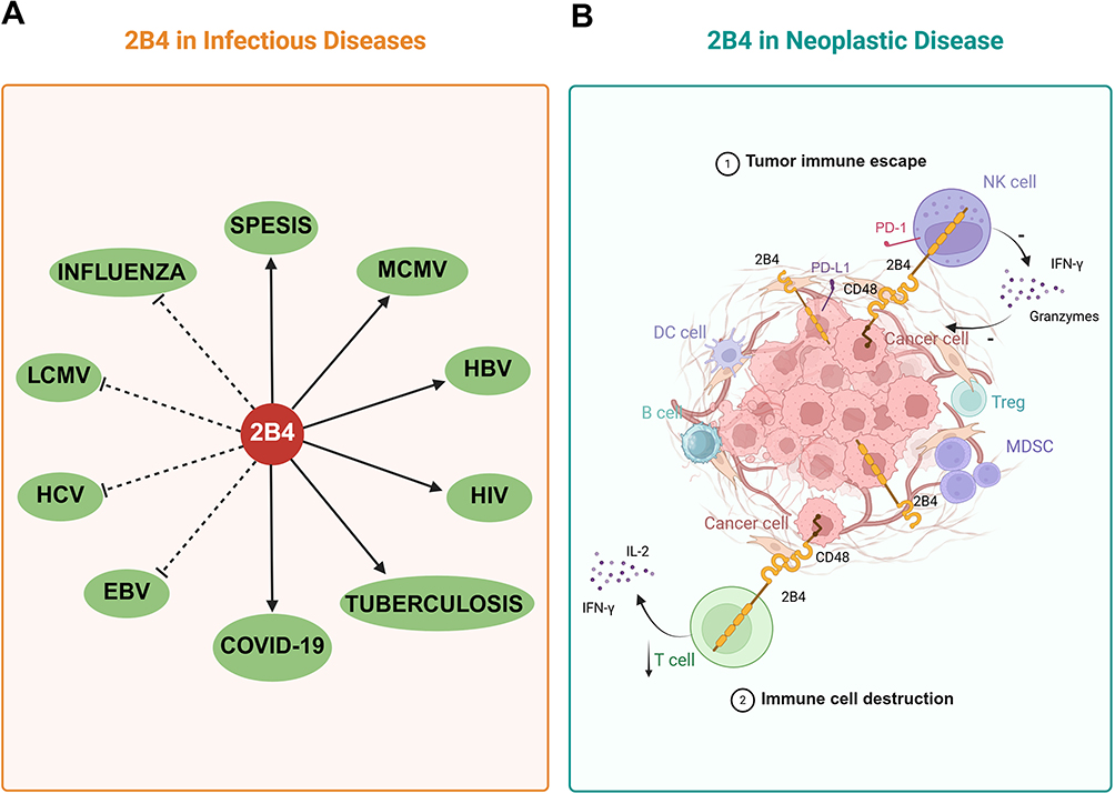 |
Figure 5 The role of 2B4/CD244 in various diseases. 2B4 regulates immune responses in a context-dependent manner. (A) In infectious diseases, 2B4 can either promote or inhibit disease progression. Solid arrows indicate a disease-promoting effect (eg, in MCMV, HIV, HBV, or COVID-19 infection), whereas dashed arrows represent a protective role (eg, in influenza virus or LCMV infection). (B) In neoplastic diseases, 2B4 contributes to tumor immune escape by interacting with CD48 and other immune checkpoint pathways (eg, PD-1), leading to reduced cytotoxicity of NK and T cells, and destruction of immune cell. |
In hepatitis C virus (HCV) infection, 2B4 expression is significantly elevated in the liver compared with normal tissue, which is correlated with the function of CD161+ perforin+ high NK cells.98 In the peripheral blood, however, a distinct CD8+ T cell subset defined by CD28+CD244+ surface phenotype secretes both IL-17 and IFN-γ. Blockade of 2B4 signalling in these cells attenuates cytokine release without affecting cytotoxic granule expression, implying that 2B4 primarily delivers an activating signal that sustains the inflammatory Tc17/IFN-γ programme.99
In XLP patients, mutations in SAP (SH2D1A) uncouple 2B4 from activating kinases and bias signaling toward inhibition. The resulting inability of CD8+ T cells to kill EBV infected, SLAM ligand expressing B cells underlies the exquisite susceptibility to fatal infectious mononucleosis.100,101 In 2B4-deficient mice, persistent infection with lymphocytic choriomeningitis virus (LCMV) leads to diminished responsiveness of LCMV specific CD8+ T cells and prolonged viral persistence. These changes are attributed to regulation by 2B4-deficient NK cells early in infection rather than intrinsic alterations in CD8+ T cells.102
In influenza virus infection, upregulation of 2B4 on NK cells modulates NKp46 function, enhancing NK cell activity against the virus. In vitro stimulation with influenza virus increases 2B4 expression on NK cells, leading to increased production of CD107a and IFN-γ. In vivo, influenza virus vaccination elevates 2B4 expression on NK cells, which plays a role in controlling the virus. Blockade of 2B4 or CD48 antibodies reduces CD107a and IFN-γ expression by CD8+ T cells.66
Conversely, during chronic hepatitis B virus (HBV) infection, 2B4 and PD-1 are highly co-expressed on virus specific CD8+ T cells. Functional rescue experiments demonstrated that monoclonal antibodies directed against 2B4 or its ligand CD48 restored proliferation, cytotoxicity, and cytokine production of exhausted CD8+ T cells, and-critically-this effect was achieved independently of PD-1 blockade.103 Additionally, the 2B4/SAP pathway has been implicated in the dysregulated expansion and hyperactivation of virus specific CD8+ T cells in patients with myelopathy/tropical spastic paralysis (HAM/TSP), contributing to the development of inflammatory neurological diseases.104
Similarly, in HIV infection, upregulation of 2B4 impairs invariant natural killer T (iNKT) cell responses, which have been linked to persistent HIV infection and disease progression.105 Additionally, during AIDS progression, increased 2B4 expression on CD8+ T cells is associated with reduced IFN-γ secretion and cytotoxic activity, as well as downregulation of the SAP adapter protein in 2B4+ CD8+ T cells. This finding suggests that 2B4+ CD8+ T cells may inhibit responses to specific HIV epitopes, thereby failing to control viral development during disease progression.106
In patients with severe COVID-19, increased expression of 2B4 and PD-1 on NK and CD8+ T cells is correlated with reduced intracellular perforin and granzyme A.107 These phenotypic changes are associated with profound functional exhaustion and predict adverse clinical outcomes, underscoring the inhibitory role of 2B4 in SARS-CoV-2–induced immune dysfunction.
In sepsis, genetic deletion or antibody blockade of 2B4 markedly improves murine survival and is accompanied by a selective reduction in active-caspase-3/7+ apoptotic T cells. 2B4 is critical for the apoptosis of mouse memory T cells and T cells isolated from sepsis patients.108 During active Mycobacterium tuberculosis infection, CD8+ T cells display elevated surface 2B4 that correlates with high intracellular levels of the long non-coding RNA lncRNA-CD244. Specifically, lncRNA-2B4 mediates the trimethylation of H3K27 at the IFN-γ/TNF-α locus by recruiting EZH2 to its promoter region, thereby repressing IFN-γ and TNF-α expression in CD8+ T cells and dampening anti-mycobacterial immunity.109
Recently, Li et al uncovered an additional, macrophage-centred role for the 2B4–CD48 axis during mouse cytomegalovirus (MCMV) infection. Up-regulated CD48 on antiviral Ly49H+ NK cells engages macrophage 2B4 to inhibit LFA-1–dependent phagocytosis. Consequently, this interaction safeguards the expanding pool of virus-specific adaptive NK cells from elimination by bystander macrophages. These findings position 2B4–CD48 not only as a bidirectional regulator of lymphocyte activation but also as a critical checkpoint controlling the magnitude and durability of the antiviral NK-cell response.110
2B4 in Neoplastic Disease
Recent pre-clinical data demonstrate that homozygous hormonal mouse models of pancreatic and lung carcinoma display a marked expansion of 2B4+ CD8+ T cells that parallels the magnitude of T-cell exhaustion. Elevated 2B4 expression on antigen-specific CD8+ T cells isolated from the spleens of these homozygous mice is associated with reduced production of IL-2 and IFN-γ.111 Translational studies corroborate these findings in human cancers, 2B4 is highly expressed on exhausted CD8+ T cells. For example, in melanoma, 2B4 expression is significantly higher in tumor-infiltrating lymphocytes than in peripheral blood lymphocytes.112,113 The pan-cancer analysis provides a genome-wide validation that high 2B4 expression is tightly linked to CD8+ T-cell infiltration yet paradoxically portends a favorable prognosis in multiple human tumors, including cutaneous melanoma. They demonstrated that elevated 2B4 mRNA correlates positively with the abundance of CD8+ T cells, dendritic cells, and monocytes across 33 cancer types, and that this association is most pronounced in uterine corpus endometrial carcinoma (UCEC).114
In multiple myeloma, high 2B4 expression on CD8+ T cells induces a depleted state, characterized by reduced expression of both CD28 and CD107a, alongside diminished proliferative capacity.115 Female 2B4−/− mice demonstrate a reduced ability to reject both CD48+ and CD48− melanoma cells, highlighting the CD48-independent specificity of the immune response to melanoma in the absence of 2B4.71 Additionally, 2B4 receptors predominantly exhibit inhibitory signaling in tumor-associated immune cells. The balance between signal activation and inhibition is influenced by the density of 2B4 on the cell surface and the availability of the signaling adaptor SAP.46
Zhang et al reported that 2B4 exerts differential effects on leukemia-initiating cells (LICs) compared with normal hematopoietic stem cells (HSCs). Specifically, 2B4 interacts with c-Kit to regulate LIC proliferation and self-renewal via the SHP-2/p27 signaling pathway.116 In glioma patients, CD48 expression is linked to malignant progression, poor prognosis, immunosuppression, and inflammatory responses.117
Therapeutic interventions targeting 2B4 have shown promise in enhancing immune responses against tumors. Alloferon, an immunomodulatory peptide, upregulates 2B4 expression, thereby increasing IFN-γ and TNF-α production and promoting extracellular granule secretion from NK cells, which enhances cytotoxicity and antiviral effect.118 In an ovarian cancer xenograft model, chimeric antigen receptor NK (CAR-NK) cells engineered with the NKG2D transmembrane domain, the 2B4 costimulatory domain, and the CD3ζ signaling domain significantly inhibited tumor growth and prolonged survival.45 Similarly, in colorectal cancer, elevated 2B4 mRNA expression in early and advanced stages correlates with T-cell depletion, suggesting its role as an immune checkpoint.119
In acute myeloid leukemia (AML), 2B4 expression is markedly increased on both CD4+ and CD8+ T cells, indicating a depleted state. Blocking the 2B4 immune checkpoint promotes the proliferation of these T-cell subsets.120 Furthermore, in head and neck squamous cell carcinoma (HNSCC), compared with healthy tissues, tumor-infiltrating CD8+ T cells exhibit significantly elevated 2B4 expression, which is correlated with PD-1 expression. Notably, 2B4−/− mice demonstrate impaired tumor growth in HNSCC models, an effect that is mitigated by monoclonal antibody blockade of 2B4.121
These findings collectively highlight the multifaceted role of 2B4 in neoplastic diseases, where it contributes to immune cell exhaustion and tumor immune evasion. Targeting 2B4 signaling pathways represents a promising avenue for enhancing antitumor immunity and improving therapeutic outcomes in various cancers.
Therapeutic Strategies
2B4 as a Therapeutic Target in Cancer
Immunotherapy has emerged as a pivotal strategy in cancer treatment, with monoclonal antibodies that block PD-1, CTLA-4, TIGIT or LAG-3 already transforming clinical practice122–129 Despite these developments, the heterogeneity and immune complexity of tumors mean that a substantial proportion of patients do not benefit from existing immunotherapeutic agents. Consequently, identifying novel therapeutic targets and prognostic biomarkers is essential to improve patient outcomes.114
Recent studies indicate that 2B4 is a common feature across various cancers and is a promising target for immunotherapy. Antagonizing 2B4 expression has been shown to inhibit tumor growth.114 Unlike the other immune checkpoint markers, 2B4 has a dual function as stimulatory as well as inhibitory receptors depending on the context of available ligand A synergistic effect of 2B4 and PD-1 or other checkpoint in tumor was found.130 But, there was no specific comparison of 2B4 with other checkpoint molecules, which needs further investigation.
Emerging evidence suggests that the modulation of 2B4 signaling directly impacts tumor cells with high 2B4 expression. For example, the knockdown of the 2B4 gene in a human leukemia cell line significantly impaired proliferation both in vitro and in vivo without affecting the regenerative capacity of hematopoietic stem cells.131 Furthermore, deletion of 2B4 delays leukemogenesis in an AML mouse model, underscoring its potential as a unique therapeutic target. These findings support the hypothesis that 2B4 not only acts as a direct antileukemic target but also mediates immunosuppressive functions within the tumor microenvironment. Consequently, targeting 2B4 on tumor cells with monoclonal antibodies may effectively inhibit tumor cell proliferation.
Given that 2B4 is widely expressed on various hematopoietic cells, its expression is critical for the functional regulation and depletion of immune cells, particularly within the tumor microenvironment. High 2B4expression on immune cells is associated with a depleted immune state and facilitates immune escape from tumors.46 For example, blocking 2B4 signaling in exhausted CD8+ T cells may alleviate their depletion and reactivate memory CD8+ T cells in cancer. Additionally, inhibiting the 2B4 pathway in DCs enhances the release of proinflammatory cytokines and activates NK cells and CD4+ T cells. Similarly, blocking 2B4 signaling in MDSCs can reduce their immunosuppressive capacity, thereby preventing tumor immune escape and overcoming drug resistance.132
Moreover, high 2B4 expression impedes the generation of Ly6C+ oligomacrophages, which possess antitumor properties. 2B4-deficient macrophages have been shown to increase the efficacy of anti-PD-L1 therapy and promote the activation of tumor-specific CD8+ T cells, including the expansion of memory T cells.133 Notably, 2B4−/− mice exhibit a normal phenotype, with unaltered immune cell maturation and lifespan, suggesting that therapeutic blockade of 2B4 is feasible with minimal side effects. Consequently, targeted anti-2B4 therapies could serve as adjunctive treatments to existing checkpoint inhibitors or as standalone strategies for managing myelosuppression.
Academician Tian Zhigang reported decreased 2B4 expression on NK cells in certain tumor patients.114 However, both high 2B4 expression leading to NK cell depletion and low 2B4 expression resulting in reduced NK cell function can facilitate immune escape from tumors. As an activating receptor, 2B4 plays a crucial role in enhancing immune cell function and preventing apoptosis, highlighting its dual role in immune regulation.
2B4 as a Therapeutic Target in Infectious Diseases
The role of 2B4 (CD244) in infectious diseases is increasingly recognized. 2B4 modulates the immune response to pathogens, which involves complex interactions between immune cell populations. In viral infections, 2B4 expression on immune cells influences disease progression. High 2B4 expression in certain immune subsets may lead to an overactive immune response, causing immunopathology, whereas insufficient 2B4 signaling can impair virus clearance. In chronic viral infections such as HIV, immune exhaustion occurs over time. Elevated 2B4 levels on CD8+ T cells contribute to their functional decline and reduced viral control. Targeting 2B4 may reverse immune exhaustion and restore antiviral responses by reactivating these T cells.134
2B4 also has therapeutic potential in bacterial infections. The 2B4-CD48 interaction modulates the activation of macrophages and neutrophils, which are key layers in antibacterial immunity. Modulating 2B4 signaling may increase macrophage phagocytosis and bacterial killing and improve immune cell recruitment to infection sites. In parasitic infections such as malaria, 2B4 may influence the immune response against the parasite.135 Targeting 2B4 could enhance immune control by promoting the elimination of infected red blood cells or modulating the inflammatory response to reduce disease severity. The immune dysregulation observed in both infectious diseases and cancer suggests that targeting 2B4 in oncology could inform similar strategies in infectious diseases. For example, approaches developed to block 2B4 in cancer to enhance immune function could be adapted to increase immune responses against pathogens.66,102
In conclusion, 2B4 holds promise as a therapeutic target in both oncology and infectious diseases. Its role in immune modulation and association with disease severity makes it an attractive candidate for novel immunotherapies. Targeting 2B4 could improve patient outcomes, either as an adjunct to current treatments or as a standalone therapy. Future research should focus on elucidating the precise mechanisms of 2B4 action, optimizing therapeutic strategies, and conducting clinical trials to assess the safety and efficacy of 2B4-targeted therapies, potentially offering new treatments for challenging diseases.
2B4 as a Biomarker for Disease Monitoring
2B4 and its ligand CD48 also hold potential as biomarkers for disease state monitoring. In patients with mild asthma, peripheral soluble CD48 levels are significantly elevated compared with those in healthy individuals, whereas patients with severe asthma present markedly lower soluble CD48 levels.136,137 This variation is correlated with disease severity, making soluble CD48 a useful indicator for mild asthma. Given the differential expression patterns of 2B4 and CD48 across various disease states, monitoring peripheral CD48 levels could provide valuable insights into disease progression and severity.
Tracking these biomarkers over time could help clinicians better understand dynamic changes in disease states, allowing timely adjustments to treatment strategies and potentially improving patient outcomes. Future research should focus on validating the utility of CD48 and 2B4 as biomarkers in larger patient cohorts and exploring their clinical applicability across different disease contexts. Additionally, investigating the mechanisms that regulate the expression of 2B4 and CD48 in various diseases could further enhance their role in disease monitoring and management.
Conclusions
Over the past decade, 2B4 (CD244) has emerged as a context-dependent rheostat whose output-activation or inhibition-is dictated by a combinatorial code comprising isoform identity, glycosylation density, SAP/EAT-2 stoichiometry, and CD48 encounter frequency. Mechanistically, SAP-mediated docking of Fyn licenses PI3K–ERK and PKC-θ cascades that culminate in perforin/granzyme release and IFN-γ/TNF-α transcription, whereas SHP-1/2 and SHIP recruitment extinguishes these signals, enforcing peripheral tolerance, tumor escape, and virus-induced exhaustion across NK, CD8+ T, DC, MDSC, and B-cell compartments. Pre-clinical studies demonstrated that monoclonal antibodies and 2B4-costimulated CAR-NK cells can reinvigorate exhausted antitumor and antiviral responses without precipitating systemic autoimmunity. These data position 2B4 as a tractable next-generation checkpoint, complementary to PD-1/LAG-3/TIGIT blockade.
Nevertheless, urgent knowledge gaps must be bridged before clinical deployment: (i) Both human 2B4 splice isoforms (h2B4-A vs h2B4-B) and their differential glycosylation patterns profoundly alter ligand affinity and downstream signaling; however, the in vivo spatiotemporal regulation of these isoforms and their glycoforms during NK-cell or CD8+ T-cell differentiation remains undefined-what are the molecular switches that govern this plasticity? (ii) Pre-clinical blockade of 2B4 reverses exhaustion of tumor-infiltrating and virus-specific CD8+ T cells without overt autoimmunity in murine models; can we prospectively identify human patient cohorts in whom 2B4-targeted therapy will synergize with PD-1/PD-L1 inhibition while avoiding hyper-inflammatory toxicities?
Abbreviations
Ig, Immunoglobulin; NK, Natural killer; MDSCs, Myeloid-derived suppressor cells; SLAM, Signaling lymphocyte activation molecule; ITSMs, Immunoreceptor tyrosine-based switch motifs; SAP, SLAM-associated protein; EAT-2, Ewing’s sarcoma-associated transcript 2; Ig-SF, Immunoglobulin superfamily; CD2AP, CD2-associated protein; hnRNAs, Heterogeneous nuclear RNAs; h2B4, Human 2B4; NTB-A, NK-T-B antigen; DCs, Dendritic cells; Treg, Regulatory T; IFN-γ, Interferon-gamma; HBV, Hepatitis B virus; XLP, X-linked lymphoproliferative disease; BMMCs, Bone marrow-derived mast cells; TNF-α, Tumor necrosis factor-α; MHC, Major histocompatibility complex; β2 m, β2-microglobulin; dNK, Decidual NK; IL-2, Interleukin-2; CTL, Cytotoxic T lymphocyte; TLR, Toll-like receptor; WT, Wild-type; IL-12p70, Interleukin-12 p70; ROS, Reactive oxygen species; IL-13, Interleukin-13; IgM, Immunoglobulin M; IgG, Immunoglobulin G; TCR, T-cell receptor; SH2, Src homology 2; PIX, PAK-interacting exchange factor; NFAT, Nuclear factor for activated T cells; MAPK, Mitogen-activated protein kinase; PKC, Protein kinase C; AP-1, Activator protein 1; TB, Tuberculosis; HCV, Hepatitis C virus; iNKT, Invariant natural killer T; LCMV, Lymphocytic choriomeningitis virus; LICs, Leukemia-initiating cells; HSCs, Hematopoietic stem cells; CAR-NK, Chimeric antigen receptor NK; AML, Acute myeloid leukemia.
Data Sharing Statement
No datasets were generated or analysed during the current study.
Author Contributions
All authors contributed to data analysis, drafting or revising the article, have agreed on the journal to which the article will be submitted, gave final approval of the version to be published, and agree to be accountable for all aspects of the work.
Funding
This work was supported by funds from the National Natural Science Foundation of China (no. 81702029) and the Yantai Science and Technology Innovation Development Program (2024XDRH115).
Disclosure
The authors declare that they have no known competing financial interests or personal relationships that could have appeared to influence the work reported in this paper.
References
1. Latchman Y, McKay PF, Reiser H. Cutting edge: identification of the 2B4 molecule as a counter-receptor for CD48. J Immunol. 1998;161(11):5809–5812. doi:10.4049/jimmunol.161.11.5809
2. Brown MH, Boles K, van der Merwe PA, Kumar V, Mathew PA, Barclay AN. 2B4, the natural killer and T cell immunoglobulin superfamily surface protein, is a ligand for CD48. J Exp Med. 1998;188(11):2083–2090. doi:10.1084/jem.188.11.2083
3. Fouquet G, Marcq I, Debuysscher V, et al. Signaling lymphocytic activation molecules Slam and cancers: friends or foes? Oncotarget. 2018;9(22):16248–16262. doi:10.18632/oncotarget.24575
4. Cocks BG, Chang CC, Carballido JM, Yssel H, de Vries JE, Aversa G. A novel receptor involved in T-cell activation. Nature. 1995;376(6537):260–263. doi:10.1038/376260a0
5. Garniwagner BA, Purohit A, Mathew PA, Bennett M, Kumar V. A novel function-associated molecule related to non-mhc-restricted cytotoxicity mediated by activated natural-killer-cells and t-cells. J Immunol. 1993;151(1):60–70. doi:10.4049/jimmunol.151.1.60
6. Shimasaki N, Jain A, Campana D. NK cells for cancer immunotherapy. Nat Rev Drug Discov. 2020;19(3):200–218. doi:10.1038/s41573-019-0052-1
7. Xu Y, Liu Q, Zhong M, et al. 2B4 costimulatory domain enhancing cytotoxic ability of anti-CD5 chimeric antigen receptor engineered natural killer cells against T cell malignancies. J Hematol Oncol. 2019;12(1):49. doi:10.1186/s13045-019-0732-7
8. Vacca P, Pietra G, Falco M, et al. Analysis of natural killer cells isolated from human decidua: evidence that 2B4 (CD244) functions as an inhibitory receptor and blocks NK-cell function. Blood. 2006;108(13):4078–4085. doi:10.1182/blood-2006-04-017343
9. Xu X, Zheng G, Ren Y, et al. A novel 2B4 receptor leads to worse pregnancy outcomes by facilitating TNF-α and IFN-γ production in dNK cells during Toxoplasma gondii infection. Parasit Vectors. 2022;15(1):337. doi:10.1186/s13071-022-05455-9
10. Sivori S, Falco M, Marcenaro E, et al. Early expression of triggering receptors and regulatory role of 2B4 in human natural killer cell precursors undergoing in vitro differentiation. Proc Natl Acad Sci U S A. 2002;99(7):4526–4531. doi:10.1073/pnas.072065999
11. Pahima H, Puzzovio PG, Levi-Schaffer F. 2B4 and CD48: a powerful couple of the immune system. Clin Immunol. 2019;204:64–68. doi:10.1016/j.clim.2018.10.014
12. Bloch-Queyrat C, Fondanèche MC, Chen R, et al. Regulation of natural cytotoxicity by the adaptor SAP and the Src-related kinase Fyn. J Exp Med. 2005;202(1):181–192. doi:10.1084/jem.20050449
13. Eissmann P, Beauchamp L, Wooters J, Tilton JC, Long EO, Watzl C. Molecular basis for positive and negative signaling by the natural killer cell receptor 2B4 (CD244). Blood. 2005;105(12):4722–4729. doi:10.1182/blood-2004-09-3796
14. Vaidya SV, Mathew PA. Of mice and men: different functions of the murine and human 2B4 (CD244) receptor on NK cells. Immunol Lett. 2006;105(2):180–184. doi:10.1016/j.imlet.2006.02.006
15. Mathew PA, Garni-Wagner BA, Land K, et al. Cloning and characterization of the 2B4 gene encoding a molecule associated with non-MHC-restricted killing mediated by activated natural killer cells and T cells. J Immunol. 1993;151(10):5328–5337. doi:10.4049/jimmunol.151.10.5328
16. Tangye SG, Lazetic S, Woollatt E, Sutherland GR, Lanier LL, Phillips JH. Cutting edge: human 2B4, an activating NK cell receptor, recruits the protein tyrosine phosphatase SHP-2 and the adaptor signaling protein SAP. J Immunol. 1999;162(12):6981–6985. doi:10.4049/jimmunol.162.12.6981
17. Boles KS, Nakajima H, Colonna M, et al. Molecular characterization of a novel human natural killer cell receptor homologous to mouse 2B4. Tissue Antigens. 1999;54(1):27–34. doi:10.1034/j.1399-0039.1999.540103.x
18. Nakajima H, Cella M, Langen H, Friedlein A, Colonna M. Activating interactions in human NK cell recognition: the role of 2B4-CD48. Eur J Immunol. 1999;29(5):1676–1683. doi:10.1002/(SICI)1521-4141(199905)29:05<1676::AID-IMMU1676>3.0.CO;2-Y
19. Malaer JD, Marrufo AM, Mathew PA. 2B4 (CD244, SLAMF4) and CS1 (CD319, SLAMF7) in systemic lupus erythematosus and cancer. Clin Immunol. 2019;204:50–56. doi:10.1016/j.clim.2018.10.009
20. Nakajima H, Colonna M. 2B4: an NK cell activating receptor with unique specificity and signal transduction mechanism. Hum Immunol. 2000;61(1):39–43. doi:10.1016/S0198-8859(99)00170-6
21. Dustin ML, Olszowy MW, Holdorf AD, et al. A novel adaptor protein orchestrates receptor patterning and cytoskeletal polarity in T-cell contacts. Cell. 1998;94(5):667–677. doi:10.1016/S0092-8674(00)81608-6
22. Schatzle JD, Sheu S, Stepp SE, Mathew PA, Bennett M, Kumar V. Characterization of inhibitory and stimulatory forms of the murine natural killer cell receptor 2B4. Proc Natl Acad Sci U S A. 1999;96(7):3870–3875. doi:10.1073/pnas.96.7.3870
23. Ames JB, Vyas V, Lusin JD, Mariuzza R. NMR structure of the natural killer cell receptor 2B4 (CD244): implications for ligand recognition. Biochemistry. 2005;44(17):6416–6423. doi:10.1021/bi050139s
24. Stepp SE, Schatzle JD, Bennett M, Kumar V, Mathew PA. Gene structure of the murine NK cell receptor 2B4: presence of two alternatively spliced isoforms with distinct cytoplasmic domains. Eur J Immunol. 1999;29(8):2392–2399. doi:10.1002/(SICI)1521-4141(199908)29:08<2392::AID-IMMU2392>3.0.CO;2-R
25. Kumaresan PR, Mathew PA. Structure of the human natural killer cell receptor 2B4 gene and identification of a novel alternative transcript. Immunogenetics. 2000;51(11):987–992. doi:10.1007/s002510000237
26. Mathew SO, Rao KK, Kim JR, Bambard ND, Mathew PA. Functional role of human NK cell receptor 2B4 (CD244) isoforms. Eur J Immunol. 2009;39(6):1632–1641. doi:10.1002/eji.200838733
27. Rudd PM, Wormald MR, Stanfield RL, et al. Roles for glycosylation of cell surface receptors involved in cellular immune recognition. J Mol Biol. 1999;293(2):351–366. doi:10.1006/jmbi.1999.3104
28. Engel P, Pérez-Carmona N, Albà MM, Robertson K, Ghazal P, Angulo A. Human cytomegalovirus UL7, a homologue of the SLAM-family receptor CD229, impairs cytokine production. Immunol Cell Biol. 2011;89(7):753–766. doi:10.1038/icb.2011.55
29. Sintes J, Romero X, de Salort J, Terhorst C, Engel P. Mouse CD84 is a pan-leukocyte cell-surface molecule that modulates LPS-induced cytokine secretion by macrophages. J Leukoc Biol. 2010;88(4):687–697. doi:10.1189/jlb.1109756
30. Slomiany A, Okazaki K, Slomiany BL. Synthesis and macromolecular organization of gastrointestinal mucin: evidence for the origin of mucin “link protein”. J Clin Gastroenterol. 1992;14 Suppl 1:S71–81. doi:10.1097/00004836-199206001-00013
31. Margraf-Schönfeld S, Böhm C, Watzl C. Glycosylation affects ligand binding and function of the activating natural killer cell receptor 2B4 (CD244) protein. J Biol Chem. 2011;286(27):24142–24149. doi:10.1074/jbc.M111.225334
32. Velikovsky CA, Deng L, Chlewicki LK, Fernández MM, Kumar V, Mariuzza RA. Structure of natural killer receptor 2B4 bound to CD48 reveals basis for heterophilic recognition in signaling lymphocyte activation molecule family. Immunity. 2007;27(4):572–584. doi:10.1016/j.immuni.2007.08.019
33. Mathew SO, Kumaresan PR, Lee JK, Huynh VT, Mathew PA. Mutational analysis of the human 2B4 (CD244)/CD48 interaction: lys68 and Glu70 in the V domain of 2B4 are critical for CD48 binding and functional activation of NK cells. J Immunol. 2005;175(2):1005–1013. doi:10.4049/jimmunol.175.2.1005
34. Kumaresan PR, Huynh VT, Mathew PA. Polymorphism in the 2B4 gene of inbred mouse strains. Immunogenetics. 2000;51(8–9):758–761. doi:10.1007/s002510000198
35. Waggoner SN, Kumar V. Evolving role of 2B4/CD244 in T and NK cell responses during virus infection. Front Immunol. 2012;3:377. doi:10.3389/fimmu.2012.00377
36. Bida AT, Upshaw Neff JL, Dick CJ, et al. 2B4 utilizes ITAM-containing receptor complexes to initiate intracellular signaling and cytolysis. Mol Immunol. 2011;48(9–10):1149–1159. doi:10.1016/j.molimm.2011.02.008
37. Veillette A. SAP: a molecular switch regulating the immune response through a unique signaling mechanism. Eur J Immunol. 2003;33(5):1141–1144. doi:10.1002/eji.200323959
38. Pérez-Quintero LA, Roncagalli R, Guo H, Latour S, Davidson D, Veillette A. EAT-2, a SAP-like adaptor, controls NK cell activation through phospholipase Cγ, Ca++, and Erk, leading to granule polarization. J Exp Med. 2014;211(4):727–742. doi:10.1084/jem.20132038
39. Endt J, Eissmann P, Hoffmann SC, Meinke S, Giese T, Watzl C. Modulation of 2B4 (CD244) activity and regulated SAP expression in human NK cells. Eur J Immunol. 2007;37(1):193–198. doi:10.1002/eji.200636341
40. Romero X, Benítez D, March S, Vilella R, Miralpeix M, Engel P. Differential expression of SAP and EAT-2-binding leukocyte cell-surface molecules CD84, CD150 (SLAM), CD229 (Ly9) and CD244 (2B4). Tissue Antigens. 2004;64(2):132–144. doi:10.1111/j.1399-0039.2004.00247.x
41. Gu C, Tangye SG, Sun X, Luo Y, Lin Z, Wu J. The X-linked lymphoproliferative disease gene product SAP associates with PAK-interacting exchange factor and participates in T cell activation. Proc Natl Acad Sci U S A. 2006;103(39):14447–14452. doi:10.1073/pnas.0606624103
42. Clarkson NG, Simmonds SJ, Puklavec MJ, Brown MH. Direct and indirect interactions of the cytoplasmic region of CD244 (2B4) in mice and humans with FYN kinase. J Biol Chem. 2007;282(35):25385–25394. doi:10.1074/jbc.M704483200
43. Cannons JL, Yu LJ, Hill B, et al. SAP regulates T(H)2 differentiation and PKC-theta-mediated activation of NF-kappaB1. Immunity. 2004;21(5):693–706. doi:10.1016/j.immuni.2004.09.012
44. Li Y, Hermanson DL, Moriarity BS, Kaufman DS. Human iPSC-derived natural killer cells engineered with chimeric antigen receptors enhance anti-tumor activity. Cell Stem Cell. 2018;23(2):181–192.e5. doi:10.1016/j.stem.2018.06.002
45. Chuang SS, Lee JK, Mathew PA. Protein kinase C is involved in 2B4 (CD244)-mediated cytotoxicity and AP-1 activation in natural killer cells. Immunology. 2003;109(3):432–439. doi:10.1046/j.1365-2567.2003.01662.x
46. Agresta L, Hoebe KHN, Janssen EM. The emerging role of cd244 signaling in immune cells of the tumor microenvironment. Front Immunol. 2018;9:2809. doi:10.1038/ni.3369
47. Anderson SK, Ortaldo JR, McVicar DW. The ever-expanding Ly49 gene family: repertoire and signaling. Immunol Rev. 2001;181:79–89. doi:10.1034/j.1600-065X.2001.1810106.x
48. Wu N, Zhong MC, Roncagalli R, et al. A hematopoietic cell-driven mechanism involving SLAMF6 receptor, SAP adaptors and SHP-1 phosphatase regulates NK cell education. Nat Immunol. 2016;17(4):387–396.
49. Lanier LL. Up on the tightrope: natural killer cell activation and inhibition. Nat Immunol. 2008;9(5):495–502. doi:10.1038/ni1581
50. Latour S, Gish G, Helgason CD, Humphries RK, Pawson T, Veillette A. Regulation of SLAM-mediated signal transduction by SAP, the X-linked lymphoproliferative gene product. Nat Immunol. 2001;2(8):681–690. doi:10.1038/90615
51. Kashiwada M, Lu P, Rothman PB. PIP3 pathway in regulatory T cells and autoimmunity. Immunol Res. 2007;39(1–3):194–224. doi:10.1007/s12026-007-0075-2
52. Saborit-Villarroya I, Del Valle JM, Romero X, et al. The adaptor protein 3BP2 binds human CD244 and links this receptor to Vav signaling, ERK activation, and NK cell killing. J Immunol. 2005;175(7):4226–4235. doi:10.4049/jimmunol.175.7.4226
53. Saborit-Villarroya I, Martinez-Barriocanal A, Oliver-Vila I, Engel P, Sayos J, Martin M. The adaptor 3BP2 activates CD244-mediated cytotoxicity in PKC- and SAP-dependent mechanisms. Mol Immunol. 2008;45(12):3446–3453. doi:10.1016/j.molimm.2008.03.021
54. König S, Nimtz M, Scheiter M, Ljunggren HG, Bryceson YT, Jänsch L. Kinome analysis of receptor-induced phosphorylation in human natural killer cells. PLoS One. 2012;7(1):e29672. doi:10.1371/journal.pone.0029672
55. Moretta A, Bottino C, Vitale M, et al. Activating receptors and coreceptors involved in human natural killer cell-mediated cytolysis. Annu Rev Immunol. 2001;19:197–223. doi:10.1146/annurev.immunol.19.1.197
56. Watzl C, Claus M. WhatSAP - 2B4 sends mixed messages in the absence of SAP. Eur J Immunol. 2014;44(5):1281–1284. doi:10.1002/eji.201444562
57. Meazza R, Tuberosa C, Cetica V, et al. XLP1 inhibitory effect by 2B4 does not affect DNAM-1 and NKG2D activating pathways in NK cells. Eur J Immunol. 2014;44(5):1526–1534. doi:10.1002/eji.201344312
58. Bottino C, Castriconi R, Moretta L, Moretta A. Cellular ligands of activating NK receptors. Trends Immunol. 2005;26(4):221–226. doi:10.1016/j.it.2005.02.007
59. Sanchez-Correa B, Morgado S, Gayoso I, et al. Human NK cells in acute myeloid leukaemia patients: analysis of NK cell-activating receptors and their ligands. Cancer Immunol Immunother. 2011;60(8):1195–1205. doi:10.1007/s00262-011-1050-2
60. Valiante NM, Trinchieri G. Identification of a novel signal transduction surface molecule on human cytotoxic lymphocytes. J Exp Med. 1993;178(4):1397–1406. doi:10.1084/jem.178.4.1397
61. Schuhmachers G, Ariizumi K, Mathew PA, Bennett M, Kumar V, Takashima A. 2B4, a new member of the immunoglobulin gene superfamily, is expressed on murine dendritic epidermal T cells and plays a functional role in their killing of skin tumors. J Invest Dermatol. 1995;105(4):592–596. doi:10.1111/1523-1747.ep12323533
62. Eissens DN, Spanholtz J, van der Meer A, et al. Defining early human NK cell developmental stages in primary and secondary lymphoid tissues. PLoS One. 2012;7(2):e30930. doi:10.1371/journal.pone.0030930
63. Chlewicki LK, Velikovsky CA, Balakrishnan V, Mariuzza RA, Kumar V. Molecular basis of the dual functions of 2B4 (CD244). J Immunol. 2008;180(12):8159–8167. doi:10.4049/jimmunol.180.12.8159
64. Costello RT, Sivori S, Mallet F, et al. A novel mechanism of antitumor response involving the expansion of CD3+/CD56+ large granular lymphocytes triggered by a tumor-expressed activating ligand. Leukemia. 2002;16(5):855–860. doi:10.1038/sj.leu.2402488
65. Kim EO, Kim TJ, Kim N, Kim ST, Kumar V, Lee KM. Homotypic cell to cell cross-talk among human natural killer cells reveals differential and overlapping roles of 2B4 and CD2. J Biol Chem. 2010;285(53):41755–41764. doi:10.1074/jbc.M110.137976
66. Jost S, Reardon J, Peterson E, et al. Expansion of 2B4+ natural killer (NK) cells and decrease in NKp46+ NK cells in response to influenza. Immunology. 2011;132(4):516–526. doi:10.1111/j.1365-2567.2010.03394.x
67. Sun C, Fu B, Gao Y, et al. TGF-β1 down-regulation of NKG2D/DAP10 and 2B4/SAP expression on human NK cells contributes to HBV persistence. PLoS Pathog. 2012;8(3):e1002594. doi:10.1371/journal.ppat.1002594
68. Parolini S, Bottino C, Falco M, et al. X-linked lymphoproliferative disease. 2B4 molecules displaying inhibitory rather than activating function are responsible for the inability of natural killer cells to kill Epstein-Barr virus-infected cells. J Exp Med. 2000;192(3):337–346. doi:10.1084/jem.192.3.337
69. Mooney JM, Klem J, Wülfing C, et al. The murine NK receptor 2B4 (CD244) exhibits inhibitory function independent of signaling lymphocytic activation molecule-associated protein expression. J Immunol. 2004;173(6):3953–3961. doi:10.4049/jimmunol.173.6.3953
70. Lee KM, McNerney ME, Stepp SE, et al. 2B4 acts as a non-major histocompatibility complex binding inhibitory receptor on mouse natural killer cells. J Exp Med. 2004;199(9):1245–1254. doi:10.1084/jem.20031989
71. Vaidya SV, Stepp SE, McNerney ME, et al. Targeted disruption of the 2B4 gene in mice reveals an in vivo role of 2B4 (CD244) in the rejection of B16 melanoma cells. J Immunol. 2005;174(2):800–807. doi:10.4049/jimmunol.174.2.800
72. Elishmereni M, Fyhrquist N, Singh Gangwar R, Lehtimäki S, Alenius H, Levi-Schaffer F. Complex 2B4 regulation of mast cells and eosinophils in murine allergic inflammation. J Invest Dermatol. 2014;134(12):2928–2937. doi:10.1038/jid.2014.280
73. Johnson LA, Vaidya SV, Goldfarb RH, Mathew PA. 2B4(CD244)-mediated activation of NK cells reduces metastases of B16F10 melanoma in mice. Anticancer Res. 2003;23(5a):3651–3655.
74. Wu Y, Kuang DM, Pan WD, et al. Monocyte/macrophage-elicited natural killer cell dysfunction in hepatocellular carcinoma is mediated by CD48/2B4 interactions. Hepatology. 2013;57(3):1107–1116. doi:10.1002/hep.26192
75. McNerney ME, Guzior D, Kumar V. 2B4 (CD244)-CD48 interactions provide a novel MHC class I-independent system for NK-cell self-tolerance in mice. Blood. 2005;106(4):1337–1340. doi:10.1182/blood-2005-01-0357
76. Fortenbery NR, Paraiso KH, Taniguchi M, Brooks C, Ibrahim L, Kerr WG. SHIP influences signals from CD48 and MHC class I ligands that regulate NK cell homeostasis, effector function, and repertoire formation. J Immunol. 2010;184(9):5065–5074. doi:10.4049/jimmunol.0901862
77. González-Alvaro I, Domínguez-Jiménez C, Ortiz AM, et al. Interleukin-15 and interferon-gamma participate in the cross-talk between natural killer and monocytic cells required for tumour necrosis factor production. Arthritis Res Ther. 2006;8(4):R88. doi:10.1186/ar1955
78. Fang J, Chen F, Liu D, Gu F, Chen Z, Wang Y. Prognostic value of immune checkpoint molecules in breast cancer. Biosci Rep. 2020;40:7. doi:10.1042/BSR20201054
79. Messmer B, Eissmann P, Stark S, Watzl C. CD48 stimulation by 2B4 (CD244)-expressing targets activates human NK cells. J Immunol. 2006;176(8):4646–4650. doi:10.4049/jimmunol.176.8.4646
80. Lee KM, Forman JP, McNerney ME, et al. Requirement of homotypic NK-cell interactions through 2B4(CD244)/CD48 in the generation of NK effector functions. Blood. 2006;107(8):3181–3188. doi:10.1182/blood-2005-01-0185
81. Assarsson E, Kambayashi T, Schatzle JD, et al. NK cells stimulate proliferation of T and NK cells through 2B4/CD48 interactions. J Immunol. 2004;173(1):174–180. doi:10.4049/jimmunol.173.1.174
82. Kambayashi T, Assarsson E, Chambers BJ, Ljunggren HG. Cutting edge: regulation of CD8(+) T cell proliferation by 2B4/CD48 interactions. J Immunol. 2001;167(12):6706–6710. doi:10.4049/jimmunol.167.12.6706
83. Stark S, Watzl C. 2B4 (CD244), NTB-A and CRACC (CS1) stimulate cytotoxicity but no proliferation in human NK cells. Int Immunol. 2006;18(2):241–247. doi:10.1093/intimm/dxh358
84. Ezinne CC, Yoshimitsu M, White Y, Arima N. HTLV-1 specific CD8+ T cell function augmented by blockade of 2B4/CD48 interaction in HTLV-1 infection. PLoS One. 2014;9(2):e87631. doi:10.1371/journal.pone.0087631
85. Rey J, Giustiniani J, Mallet F, et al. The co-expression of 2B4 (CD244) and CD160 delineates a subpopulation of human CD8+ T cells with a potent CD160-mediated cytolytic effector function. Eur J Immunol. 2006;36(9):2359–2366. doi:10.1002/eji.200635935
86. Schnorfeil FM, Lichtenegger FS, Emmerig K, et al. T cells are functionally not impaired in AML: increased PD-1 expression is only seen at time of relapse and correlates with a shift towards the memory T cell compartment. J Hematol Oncol. 2015;8:93. doi:10.1186/s13045-015-0189-2
87. Munitz A, Bachelet I, Fraenkel S, et al. 2B4 (CD244) is expressed and functional on human eosinophils. J Immunol. 2005;174(1):110–118. doi:10.4049/jimmunol.174.1.110
88. El-Shazly AE, Henket M, Lefebvre PP, Louis R. 2B4 (CD244) is involved in eosinophil adhesion and chemotaxis, and its surface expression is increased in allergic rhinitis after challenge. Int J Immunopathol Pharmacol. 2011;24(4):949–960. doi:10.1177/039463201102400413
89. Berger AE, Durrieu C, Dzviga C, Perrot JL, Lambert C. Human peripheral basophils extended phenotype shows a high expression of CD244 immuno-regulatory receptor. J Immunol Methods. 2021;492:112951. doi:10.1016/j.jim.2020.112951
90. Vivanco Gonzalez N, Oliveria JP, Tebaykin D, et al. Mass cytometry phenotyping of human granulocytes reveals novel basophil functional heterogeneity. iScience. 2020;23(11):101724. doi:10.1016/j.isci.2020.101724
91. Georgoudaki AM, Khodabandeh S, Puiac S, et al. CD244 is expressed on dendritic cells and regulates their functions. Immunol Cell Biol. 2015;93(6):581–590. doi:10.1038/icb.2014.124
92. Bronte V, Brandau S, Chen SH, et al. Recommendations for myeloid-derived suppressor cell nomenclature and characterization standards. Nat Commun. 2016;7:12150.
93. Yang B, Wang X, Jiang J, Zhai F, Cheng X. Identification of CD244-expressing myeloid-derived suppressor cells in patients with active tuberculosis. Immunol Lett. 2014;158(1–2):66–72. doi:10.1016/j.imlet.2013.12.003
94. Youn JI, Collazo M, Shalova IN, Biswas SK, Gabrilovich DI. Characterization of the nature of granulocytic myeloid-derived suppressor cells in tumor-bearing mice. J Leukoc Biol. 2012;91(1):167–181. doi:10.1189/jlb.0311177
95. De Salort J, Sintes J, Llinàs L, Matesanz-Isabel J, Engel P. Expression of SLAM (CD150) cell-surface receptors on human B-cell subsets: from pro-B to plasma cells. Immunol Lett. 2011;134(2):129–136. doi:10.1016/j.imlet.2010.09.021
96. Gao N, Schwartzberg P, Wilder JA, Blazar BR, Yuan D. B cell induction of IL-13 expression in NK cells: role of CD244 and SLAM-associated protein. J Immunol. 2006;176(5):2758–2764. doi:10.4049/jimmunol.176.5.2758
97. Ray A, Yuan CY, Miller NM, Mei H, Dittel BN. 2B4 is dispensable for t-dependent b cell immune responses, but its deficiency leads to enhanced t-independent responses due to an increase in peritoneal cavity b1b cells. PLoS One. 2015;10(8):e0137314. doi:10.1371/journal.pone.0137314
98. Cosgrove C, Berger CT, Kroy DC, et al. Chronic HCV infection affects the NK cell phenotype in the blood more than in the liver. PLoS One. 2014;9(8):e105950. doi:10.1371/journal.pone.0105950
99. Han W, Li J, Zhou H, et al. Identification of the association of CD28(+) CD244(+) Tc17/IFN-γ cells with chronic hepatitis C virus infection. J Med Virol. 2020;92(12):3534–3544. doi:10.1002/jmv.26205
100. Hislop AD, Palendira U, Leese AM, et al. Impaired Epstein-Barr virus-specific CD8+ T-cell function in X-linked lymphoproliferative disease is restricted to SLAM family-positive B-cell targets. Blood. 2010;116(17):3249–3257. doi:10.1182/blood-2009-09-238832
101. Chatterjee B, Deng Y, Holler A, et al. CD8+ T cells retain protective functions despite sustained inhibitory receptor expression during Epstein-Barr virus infection in vivo. PLoS Pathog. 2019;15(5):e1007748. doi:10.1371/journal.ppat.1007748
102. Waggoner SN, Taniguchi RT, Mathew PA, Kumar V, Welsh RM. Absence of mouse 2B4 promotes NK cell-mediated killing of activated CD8+ T cells, leading to prolonged viral persistence and altered pathogenesis. J Clin Invest. 2010;120(6):1925–1938. doi:10.1172/JCI41264
103. Raziorrouh B, Schraut W, Gerlach T, et al. The immunoregulatory role of CD244 in chronic hepatitis B infection and its inhibitory potential on virus-specific CD8+ T-cell function. Hepatology. 2010;52(6):1934–1947. doi:10.1002/hep.23936
104. Enose-Akahata Y, Matsuura E, Oh U, Jacobson S. High expression of CD244 and SAP regulated CD8 T cell responses of patients with HTLV-I associated neurologic disease. PLoS Pathog. 2009;5(12):e1000682. doi:10.1371/journal.ppat.1000682
105. Ahmad F, Shankar EM, Yong YK, et al. Negative checkpoint regulatory molecule 2B4 (CD244) upregulation is associated with invariant natural killer t cell alterations and human immunodeficiency virus disease progression. Front Immunol. 2017;8:338. doi:10.3389/fimmu.2017.00338
106. Aldy KN, Horton NC, Mathew PA, Mathew SO. 2B4+ CD8+ T cells play an inhibitory role against constrained HIV epitopes. Biochem Biophys Res Commun. 2011;405(3):503–507. doi:10.1016/j.bbrc.2011.01.062
107. Li M, Guo W, Dong Y, et al. Elevated Exhaustion Levels of NK and CD8(+) T cells as indicators for progression and prognosis of COVID-19 Disease. Front Immunol. 2020;11:580237. doi:10.3389/fimmu.2020.580237
108. Xie J, Chen CW, Sun Y, et al. Increased attrition of memory T cells during sepsis requires 2B4. JCI Insight. 2019;4(9). doi:10.1172/jci.insight.126030
109. Wang Y, Zhong H, Xie X, et al. Long noncoding RNA derived from CD244 signaling epigenetically controls CD8+ T-cell immune responses in tuberculosis infection. Proc Natl Acad Sci U S A. 2015;112(29):E3883–92. doi:10.1073/pnas.1501662112
110. Li R, Galindo CC, Davidson D, et al. Suppression of adaptive NK cell expansion by macrophage-mediated phagocytosis inhibited by 2B4-CD48. Cell Rep. 2024;43(3):113800. doi:10.1016/j.celrep.2024.113800
111. Mittal R, Wagener M, Breed ER, et al. Phenotypic T cell exhaustion in a murine model of bacterial infection in the setting of pre-existing malignancy. PLoS One. 2014;9(5):e93523. doi:10.1371/journal.pone.0093523
112. Chen Y, Feng Z, Kuang X, et al. Increased lactate in AML blasts upregulates TOX expression, leading to exhaustion of CD8(+) cytolytic T cells. Am J Cancer Res. 2021;11(11):5726–5742.
113. Baitsch L, Baumgaertner P, Devêvre E, et al. Exhaustion of tumor-specific CD8+ T cells in metastases from melanoma patients. J Clin Invest. 2011;121(6):2350–2360. doi:10.1172/JCI46102
114. Deng Z, Liu Y, Zhou H. Distinct roles of CD244 expression in cancer diagnosis and prognosis: a pan-cancer analysis. Heliyon. 2024;10(7):e28928. doi:10.1016/j.heliyon.2024.e28928
115. Zelle-Rieser C, Thangavadivel S, Biedermann R, et al. T cells in multiple myeloma display features of exhaustion and senescence at the tumor site. J Hematol Oncol. 2016;9(1):116. doi:10.1186/s13045-016-0345-3
116. Zhang F, Liu X, Chen C, et al. CD244 maintains the proliferation ability of leukemia initiating cells through SHP-2/p27(kip1) signaling. Haematologica. 2017;102(4):707–718. doi:10.3324/haematol.2016.151555
117. Zou C, Zhu C, Guan G, et al. CD48 is a key molecule of immunomodulation affecting prognosis in glioma. Onco Targets Ther. 2019;12:4181–4193. doi:10.2147/OTT.S198762
118. Bae S, Oh K, Kim H, et al. The effect of alloferon on the enhancement of NK cell cytotoxicity against cancer via the up-regulation of perforin/granzyme B secretion. Immunobiology. 2013;218(8):1026–1033. doi:10.1016/j.imbio.2012.12.002
119. Saleh R, Taha RZ, Toor SM, et al. Expression of immune checkpoints and T cell exhaustion markers in early and advanced stages of colorectal cancer. Cancer Immunol Immunother. 2020;69(10):1989–1999. doi:10.1007/s00262-020-02593-w
120. Abdolmaleki M, Mojtabavi N, Zavvar M, Vaezi M, Noorbakhsh F, Nicknam MH. Scrutinizing the expression and blockade of inhibitory molecules expressed on t cells from acute myeloid leukemia patients. Iran J Allergy Asthma Immunol. 2018;17(3):265–273.
121. Agresta L, Lehn M, Lampe K, et al. CD244 represents a new therapeutic target in head and neck squamous cell carcinoma. J Immunother Cancer. 2020;8:1. doi:10.1136/jitc-2019-000245
122. Chu X, Tian W, Wang Z, Zhang J, Zhou R. Co-inhibition of TIGIT and PD-1/PD-L1 in cancer immunotherapy: mechanisms and clinical trials. Mol Cancer. 2023;22(1):93. doi:10.1186/s12943-023-01800-3
123. Gou Q, Dong C, Xu H, et al. PD-L1 degradation pathway and immunotherapy for cancer. Cell Death Dis. 2020;11(11):955. doi:10.1038/s41419-020-03140-2
124. Chuah S, Lee J, Song Y, et al. Uncoupling immune trajectories of response and adverse events from anti-PD-1 immunotherapy in hepatocellular carcinoma. J Hepatol. 2022;77(3):683–694. doi:10.1016/j.jhep.2022.03.039
125. Cheng W, Kang K, Zhao A, Wu Y. Dual blockade immunotherapy targeting PD-1/PD-L1 and CTLA-4 in lung cancer. J Hematol Oncol. 2024;17(1):54. doi:10.1186/s13045-024-01581-2
126. Boutros C, Tarhini A, Routier E, et al. Safety profiles of anti-CTLA-4 and anti-PD-1 antibodies alone and in combination. Nat Rev Clin Oncol. 2016;13(8):473–486. doi:10.1038/nrclinonc.2016.58
127. Agarwal S, Aznar MA, Rech AJ, et al. Deletion of the inhibitory co-receptor CTLA-4 enhances and invigorates chimeric antigen receptor T cells. Immunity. 2023;56(10):2388–2407.e9. doi:10.1016/j.immuni.2023.09.001
128. Cai L, Li Y, Tan J, Xu L, Li Y. Targeting LAG-3, TIM-3, and TIGIT for cancer immunotherapy. J Hematol Oncol. 2023;16(1):101. doi:10.1186/s13045-023-01499-1
129. Yang R, Sun L, Li CF, et al. Galectin-9 interacts with PD-1 and TIM-3 to regulate T cell death and is a target for cancer immunotherapy. Nat Commun. 2021;12(1):832. doi:10.1038/s41467-021-21099-2
130. Ma X, Bi E, Lu Y, et al. Cholesterol Induces CD8(+) T Cell Exhaustion in the Tumor Microenvironment. Cell Metab. 2019;30(1):143–156.e5. doi:10.1016/j.cmet.2019.04.002
131. Long AH, Highfill SL, Cui Y, et al. Reduction of MDSCs with all-trans retinoic acid improves car therapy efficacy for sarcomas. Cancer Immunol Res. 2016;4(10):869–880. doi:10.1158/2326-6066.CIR-15-0230
132. Myers JA, Miller JS. Exploring the NK cell platform for cancer immunotherapy. Nat Rev Clin Oncol. 2021;18(2):85–100. doi:10.1038/s41571-020-0426-7
133. Kim J, Kim TJ, Chae S, et al. Targeted deletion of CD244 on monocytes promotes differentiation into anti-tumorigenic macrophages and potentiates PD-L1 blockade in melanoma. Mol Cancer. 2024;23(1):45. doi:10.1186/s12943-024-01936-w
134. Wang L, Zeng X, Wang Z, Fang L, Liu J. Recent advances in understanding T cell activation and exhaustion during HBV infection. Virol Sin. 2023;38(6):851–859. doi:10.1016/j.virs.2023.10.007
135. Puerta CJ, Cuellar A, Lasso P, Mateus J, Gonzalez JM. Trypanosoma cruzi-specific CD8(+) T cells and other immunological hallmarks in chronic Chagas cardiomyopathy: two decades of research. Front Cell Infect Microbiol. 2022;12:1075717. doi:10.3389/fcimb.2022.1075717
136. Zoabi Y, Rahimli Alekberli F, Minai-Fleminger Y, Eliashar R, Levi-Schaffer F. CD48 Expression on Eosinophils in Nasal Polyps of Chronic Rhinosinusitis Patients. Int Arch Allergy Immunol. 2021;182(10):962–970. doi:10.1159/000515918
137. Volkmer B, Planas R, Gossweiler E, et al. Recurrent inflammatory disease caused by a heterozygous mutation in CD48. J Allergy Clin Immunol. 2019;144(5):1441–1445.e17. doi:10.1016/j.jaci.2019.07.038
 © 2025 The Author(s). This work is published and licensed by Dove Medical Press Limited. The
full terms of this license are available at https://www.dovepress.com/terms
and incorporate the Creative Commons Attribution
- Non Commercial (unported, 4.0) License.
By accessing the work you hereby accept the Terms. Non-commercial uses of the work are permitted
without any further permission from Dove Medical Press Limited, provided the work is properly
attributed. For permission for commercial use of this work, please see paragraphs 4.2 and 5 of our Terms.
© 2025 The Author(s). This work is published and licensed by Dove Medical Press Limited. The
full terms of this license are available at https://www.dovepress.com/terms
and incorporate the Creative Commons Attribution
- Non Commercial (unported, 4.0) License.
By accessing the work you hereby accept the Terms. Non-commercial uses of the work are permitted
without any further permission from Dove Medical Press Limited, provided the work is properly
attributed. For permission for commercial use of this work, please see paragraphs 4.2 and 5 of our Terms.