Back to Journals » Drug Design, Development and Therapy » Volume 19
Zhinao Capsule Improved Sleep and Memory Disorders in APP/PS1 Mice via cAMP/PKA/CREB Signaling Pathway Modulation
Authors Xi H, Xie W ![]() , Yang Y, Li X, Zhai S
, Yang Y, Li X, Zhai S ![]() , Fang S, Yang Y, Rao Z
, Fang S, Yang Y, Rao Z ![]() , Yang W, Li H
, Yang W, Li H
Received 22 May 2025
Accepted for publication 19 September 2025
Published 13 October 2025 Volume 2025:19 Pages 9235—9252
DOI https://doi.org/10.2147/DDDT.S542074
Checked for plagiarism Yes
Review by Single anonymous peer review
Peer reviewer comments 2
Editor who approved publication: Dr Tuo Deng
Hu Xi,1,2 Wenting Xie,2 Yue Yang,2 Xiang Li,2 Shu Zhai,2 Shuzhen Fang,2 Yulong Yang,2 Zhihong Rao,2 Wenming Yang,2– 4 Hao Li1
1Wangjing Hospital, China Academy of Chinese Medical Sciences, Beijing, People’s Republic of China; 2Department of Neurology, the First Affiliated Hospital of Anhui University of Chinese Medicine, Hefei, People’s Republic of China; 3Key Laboratory of Xin’an Medicine, Ministry of Education, Hefei, People’s Republic of China; 4Center for Xin’an Medicine and Modernization of Traditional Chinese Medicine, Institute of Health and Medicine, Hefei Comprehensive National Science Center, Hefei, People’s Republic of China
Correspondence: Wenming Yang, Email [email protected] Hao Li, Email [email protected]
Objective: To evaluate the efficacy and therapeutic mechanism of Zhinao Capsule (ZNC) in alleviating memory impairment and sleep disorders through an integrated approach involving network pharmacology, molecular docking, and experimental validation with molecular biological techniques.
Materials and Methods: Fifty APP/PS1 mice were randomly divided into five groups: donepezil hydrochloride tablets group (administered with Donepezil hydrochloride tablets (0.65 mg/(kg·d)), ZNC low-, medium-, and, high-dose groups (0.234/0.468/0.936 g/(kg·d)), the model and normal groups treated with 0.9% saline at a dose of 10 mL/(kg·d). Gavage was performed once daily for 28 consecutive days after acclimatization, feeding, and grouping. Behavioral, Hematoxylin and Eosin (H&E), and Immunohistochemistry (IHC) analyses were used to verify the in vivo efficacy of ZNC in APP/PS1 mice. Network pharmacology and molecular docking were employed to predict the potential molecular mechanisms of ZNC in alleviating Alzheimer′s Disease (AD) and related sleep disorders. Enzyme-Linked Immunosorbent Assay (ELISA), Quantitative Real-Time Polymerase Chain Reaction (RT-qPCR), and Western Blot (WB) analyses were used to establish whether ZNC modulated relevant pathways to exert therapeutic effects against AD-related sleep disorders.
Results: PiezoSleep monitoring, Morris Water Maze (MWM), H&E, and IHC tests revealed that ZNC could restore impaired learning memory and sleep structures, improve the cellular morphology of neurons in the hippocampal CA1 and CA3 regions, promote neurogenesis, and increase synaptic plasticity. That neuroinflammation and the Cyclic Adenosine Monophosphate (cAMP)/Protein Kinase A (PKA)/ cAMP Response Element Binding Protein (CREB) signaling pathway were ZNC’s key avenues in alleviating AD-related sleep disorders. On the other hand, the IHC, WB, ELISA, and RT-qPCR tests confirmed that ZNC significantly upregulated Brain-Derived Neurotrophic Factor (BDNF), cAMP, PKA mRNA, CREB mRNA, p-PKA, p-CREB, Synaptophysin (SYN), and Postsynaptic Density Protein 95 (PSD-95), significantly downregulated Amyloid Beta 1– 42 (Aβ 1-42) and inflammatory factors such as Glial Fibrillary Acidic Protein (GFAP), Interleukin-6 (IL6), IL-1β, and Tumor Necrosis Factor-α (TNF-α), and upregulated neurotransmitters such as 5-Hydroxyindole-3-Acetic Acid (5-HIAA), 5-Hydroxy Tryptamine (5-HT), and Gamma-Aminobutyric Acid (GABA)(p< 0.01, p< 0.05).
Conclusion: The present results show that ZNC can improve sleep and memory disorders in APP/PS1 mice via modulating the cAMP/PKA/CREB signaling pathway, attenuating Aβ and neuroinflammation, and improving synaptic plasticity.
Keywords: Zhinao Capsule, cAMP/PKA/CREB signaling pathway, Alzheimer’s disease, sleep disorders, APP/PS1 mice, neuroinflammation
Introduction
Alzheimer’s Disease (AD) is a chronic degenerative illness of the Central Nervous System (CNS) that causes cognitive, mental, behavioral, and physical disorders, with sporadic and late-onset AD predominating.1 Synaptic dysfunction, along with extracellular Amyloid Beta (Aβ) and intracellular Phosphorylated tau (P-tau), is a key pathological hallmark and contributing factor in the etiology of AD.2 Sleep is an essential physiological mechanism for sustaining normal life activities and supporting brain activity. During sleep, brain metabolism and cortical interstitial activity are enhanced, accelerating convective exchange between Cerebrospinal Fluid (CSF) and Interstitial Fluid (IF) and facilitating the removal of toxic substances such as Aβ and P-tau, ultimately enhancing learning and memory functions.3 Compared to normal individuals, patients with sleep disorders are 1.55–3.78 times more likely to suffer from AD, Mild Cognitive Impairment (MCI), or prodromal AD.4 Moreover, the prevalence of sleep disorders in up to 70% of AD patients could be attributed to impaired cyclic clearance of toxic substances such as Aβ and P-tau, inducing hippocampal neuronal cell atrophy, reducing neurogenesis, and disrupting the consolidation of long-lasting memories and protein synthesis. These phenomena could impair learning and memory function, thus aggravating cognitive and sleep impairment. There is a bidirectional physiological and pathological relationship between AD and sleep disorders, an important negative regulator of healthy living in AD patients and a persistent risk predictor for AD progression.5,6 Considering the increasing ageing of the global population, a breakthrough in medical research in terms of prevention, delay, or cure of AD would be crucial, helping to avoid a potential burden to individuals, families, and society. Overall, early AD detection and prevention could significantly improve the Quality of Life (QoL) of AD patients. Therefore, elucidating the pathogenesis of AD-related sleep disorders and actively exploring new therapeutic targets and interventions would be imperative for improved patient outcomes.
There are currently no specific treatments for AD-related sleep disorders, with current clinical interventions mainly focused on slowing down AD progression, highlighting the fact that sleep disorders symptoms are often overlooked. Commonly used drugs for managing AD include cholinesterase inhibitors and glutamate receptor antagonists. Other interventions include AD modification therapies and treatments aimed at improving related sleep disorders, including melatonin, hypnotic drugs, bright light therapy, and other pharmacological and non-pharmacological interventions, which could be used either in combination or single application. However, these drugs have been associated with certain limitations, including Gastrointestinal (GI) reactions, cardiovascular side effects, withdrawal reactions, and a short honeymoon period, among others. Furthermore, long-term use of melatonin and hypnotic drugs could reduce the concentration of endogenously secreted melatonin peaks, resulting in drug dependence, addiction, and other adverse consequences.7,8 Therefore, an in-depth exploration of the pathogenesis of AD-related sleep disorders and developing novel effective preventive and curative medications would be imperative for improved clinical outcomes. Based on the theory of “managing different diseases with the same treatment”, Traditional Chinese Medicine (TCM) has demonstrated extensive utility in the prevention and treatment of AD-related sleep disorders. It offers the advantages of multi-component, multi-target, multi-session, and multi-pathway treatments, especially in complicated cases involving multiple systems, organs, and targets. In this regard, focusing on Zhinao Capsule (ZNC), we sought to establish the clinical therapeutic effects of TCM formulas in AD-related sleep disorders prevention and treatment.
Originally prepared pharmacologically for AD treatment at the First Affiliated Hospital of Anhui University of Chinese Medicine (Patent No. ZL200310161324.9), ZNC was approved by the Anhui Provincial Drug Administration in 2024 (Anhui Pharmaceutical Preparation Z20240001) and its key components include Codonopsis Radix, Astragali Radix, Polygonati Rhizoma, Cistanches Herba, Curcumae Radix, Acori Tatarinowii Rhizoma, Chuanxiong Rhizoma, and Pheretima, among others (Proportion: 15:15:12:15:10:8:10:8) (Table 1). Through mechanisms such as apoptosis attenuation, ZNC was previously reported to significantly improve memory and hippocampal neuron morphology in AD patients and APP/PS1 mice.9 The APP/PS1 mice have been established as robust animal models of AD. Disorders in the sleep-wake cycle and increased sleep arousal in APP/PS1 mice were reported in several animal experiments, including running-wheel experiments, electroencephalography, clock genes, and assessment of sleep-wake-related neurotransmitter expression, and results were compared with those of wild-type controls. Furthermore, an increased number of arousals was reported in APP/PS1 mice, indicating disrupted sleep-wake cycle pathology.10,11 Nonetheless, it remains unclear whether the precise molecular mechanism of ZNC-mediated cognitive enhancement correlates with improved sleep. The cAMP/PKA/CREB signaling pathway plays a crucial role in neuronal genesis, maturation, apoptosis, and synaptic plasticity reestablishment, highlighting its potential involvement in the onset, development, and clinical regression of sleep disturbances, as well as in learning and memory processes.12,13
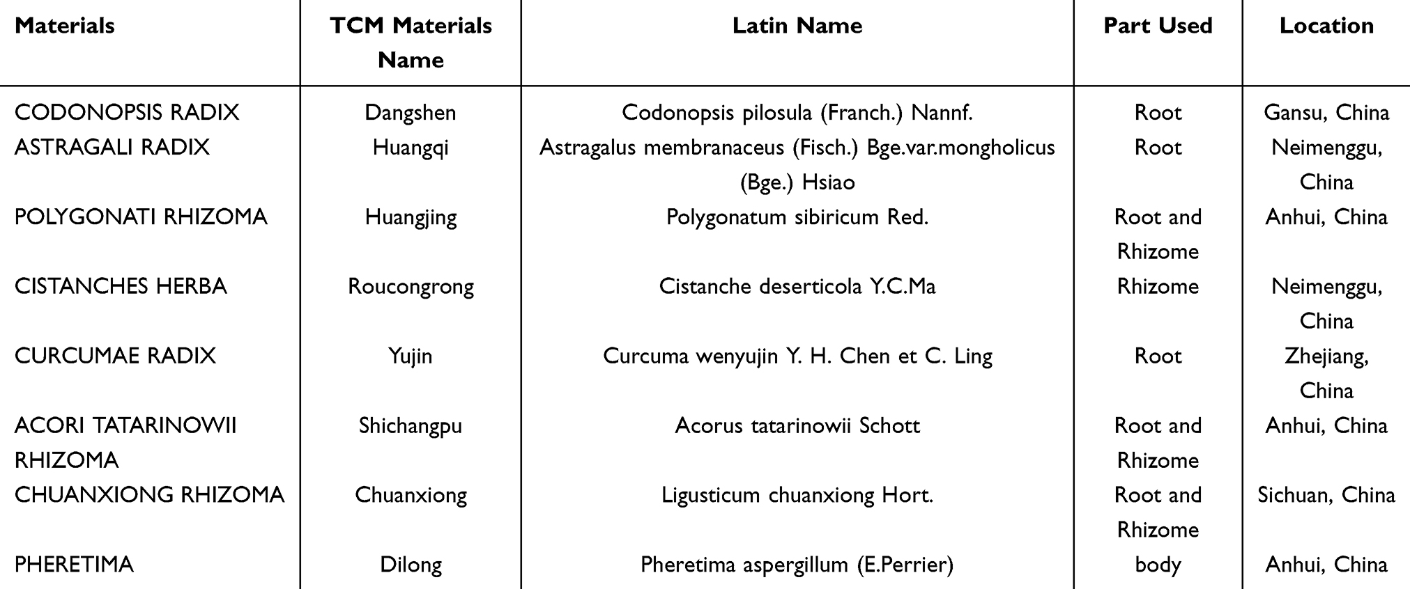 |
Table 1 The Compositions of ZNC |
Clinical studies have demonstrated that ZNC can significantly restore the patients’ sleep structure and improve learning and memory abilities, but the underlying mechanisms are not fully understood. Therefore, we employed the APP/PS1 mice as model animals to receive ZNC treatment, and performed network pharmacology, molecular docking, and molecular biology experiments to deeply investigate whether ZNC can delay the onset of AD-related cognitive and sleep dysfunctional behaviors via cAMP/PKA/CREB signaling pathway modulation, and to explore the possible molecular mechanisms.
Materials and Methods
Experimental Animals and Groupings
Animal experiments were performed using 50 Specific Pathogen Free (SPF)-grade male APP/PS1 double transgenic mice and 10 male C57BL/6J mice (age=6 months) (Certificate of Compliance No. 20210516Abbb05000259). The 50 APP/PS1 mice were randomly divided into five groups: Model, Donepezil hydrochloride tablets, low-dose ZNC, medium-dose ZNC, and high-dose ZNC (N=10/group). Meanwhile, 10 C57BL/6J wild-type mice of the same age formed the normal group. All animals were kept under standard SPF conditions at Anhui Agricultural University (Experimental Animal Use License: SYXK(Anhui) 2021–009), and the university’s Ethics Committee approved the study protocol (Approval number: AHAUB2023024).
Drug Administration
The model and normal groups received 0.9% saline at a dose of 10 mL/(kg·d). Meanwhile, the ZNC low-, medium-, and, high-dose groups received 0.234 g/(kg·d), 0.468 g/(kg·d), and 0.936 g/(kg·d) of ZNC (0.4g/capsule; First Affiliated Hospital of Anhui University of Chinese Medicine, Batch number: 20221129), respectively. On the other hand, the Donepezil hydrochloride tablets group received Donepezil hydrochloride tablets at a dose of 0.65 mg/(kg·d) (5mg/tablet; Zhejiang Huahai Pharmaceutical Co., Lot number: 0000018324). Gavage was administered once per day for 28 days consecutively after acclimatization feeding and grouping (Figure 1A).
 |
Figure 1 ZNC improves learning memory and sleep disorders in APP/PS1 mice. (A) Schematic diagram showing the experimental process in APP/PS1 mice. (B) Effects of ZNC on sleep in the indicated groups. (C) Effects of ZNC on sleep structure in the indicated groups. (D) Effects of ZNC on the first 5 days of spatial localization navigation training in all mice. (E) Effect of ZNC on target quadrant swimming time of the space exploration experiment on day 6 in all mice. (F) Effect of ZNC on the number of crossings of target platforms on day 6 of the space exploration experiment in the indicated groups. (G) Effect of ZNC on the resident platform incubation period in day 6 space exploration experiments in groups shown. (H) Swimming trajectories of day 6 space exploration experiments. Statistical significance of all plots was determined by one-way ANOVA. Data are expressed as the mean SD (n = 8). Compared with the normal group, ΔΔP<0.01; compared with the model group, *P<0.05, **P<0.01; compared with the donepezil hydrochloride tablets group, ▲P<0.05, ▲▲P<0.01; compared with the ZNC low and medium dose groups and the ZNC high dose group, #P<0.05, ##P<0.01; Red colored upward arrows mean to start the Behavior test. |
Behavioral Testing
PiezoSleep Monitoring
The PiezoSleep non-invasive monitoring system (Signal Solutions, LLC, Lexington, KY, USA) was used for complete sleep/wakefulness tracking in unrestrained animals. The system integrates high-precision sensors, data acquisition techniques, and automatic sleep/wakefulness analysis. The experimental platform was a rectangular feeding cage (15cm*22cm*20cm), with a 14cm*20cm piezoelectricity sensor at the bottom. These dimensions allow for the concurrent monitoring of autonomous activity and sleep patterns of multiple mice. The piezoelectric system detects the pressure changes resulting from movement, including minute respiratory movements. Notably, based on EEG data and human observations, the classification accuracy of this piezoelectric system was previously estimated at ⁓95%.14,15 The sleep parameters examined included average total sleep % over 24 h (total sleep proportion), average sleep % at night (dark-phase sleep proportion), and average sleep % during the day (light-phase sleep proportion).
Morris Water Maze (MWM) Test
Experimental data from orientation navigation, spatial exploration, and the visual platform tasks were analyzed using the MWM test to assess long-term spatial learning and memory abilities. The MWM video analysis system has four elements: A circular pool, a platform, a video recording device, and a software analysis system. A 90-s training time was set in the localization and navigation training experiments on days 1–5, with the mice artificially trained to station for 15s if the stationing action could not be completed within the specified time. Each experimental mouse was trained 4×/day, with the swimming trajectory and resident platform incubation period recorded. The platform was removed on the 6th day of the space exploration experiment, with the same experimental method used to conduct experiments from other quadrants besides the target platform. In the process, swimming trajectory and number of target platform crossings, target quadrant swimming time, and resident platform incubation period were recorded.
Hematoxylin and Eosin (H&E) Staining
First, APP/PS1 mouse hippocampal tissues were fixed in a 4% Paraformaldehyde (PFA) solution, paraffin-embedded, and ultimately sliced into 2 μm-thick sections for H&E staining and microscopic observation (CX 41, Olympus, JPN).
Immunohistochemistry (IHC) Analysis
After embedding in paraffin, APP/PS1 mouse hippocampal tissues were cut into 2 μm-thick slices, deparaffinized with xylene 3 times (5 min/time), and subjected to gradient dehydration using 100%, 95%, and 80% ethanol (5 min/time). After rinsing with running water, the tissues were subjected to antigen high-pressure repair, preserved, and sealed. Following that, they were incubated with primary antibodies against Aβ1-42, BDNF and GFAP in a sequence (37 °C, 60 min, ab201061\GR3319825-10\GR286628-10, Abcam, China), followed by a secondary antibody (37 °C, 30 min). Subsequently, a DAB colorant was developed, and hematoxylin re-staining was performed. Finally, the tissues were sealed for microscopic observation (CX 41, Olympus, JPN).
Network Pharmacology Analysis
Using PubChem (https://pubchem.ncbi.nlm.nih.gov/), several databases including the Traditional Chinese Medicine Systems Pharmacology (TCMSP) (https://tcmspw.com/), High-throughput Experiment- and Reference-guided database of TCM (HERB) (http://herb.ac.cn/), The Encyclopedia of Traditional Chinese Medicine (ETCM) (http://www.tcmip.cn/ETCM/), Traditional Chinese Medicine Information (TCMID) database (https://ngdc.cncb.ac.cn/databasecommons/), and the Bioinformatics Annotation Database for Molecular Mechanisms of TCM (BATMAN-TCM) (http://bionet.ncpsb.org.cn/batman-tcm/), among others were mined to determine the herbs in ZNC, along with their chemical compositions. SwissTargetPrediction (http://www.swisstargetprediction.ch/index.php), TargetNet (http://targetnet.scbdd.com/home/index/), PharmMapper (http://www.lilab-ecust.cn/pharmmapper/), and other databases were used in combination with ADME properties and Lipinski’s rule for complementary screening of ZNC pharmacodynamic active ingredients and potential protein targets. Furthermore, UniProt (https://www.uniprot.org/) and STRING (https://cn.string-db.org/) were used for gene standardization to construct a ZNC-pharmacodynamic active ingredient-target database. The Online Catalog of Human Genes and Genetic Disorders (OMIM) database (https://omim.org/) and GeneCards (https://www.genecards.org/), among other related databases, were further used to identify and convert gene-protein information to obtain the potential targets of AD sleep disorders. The Venny-2.1.0 online platform (https://bioinfogp.cnb.csic.es/tools/venny/) was used to obtain the intersection targets of ZNC and disease-related genes, and to draw Venn diagrams. The intersecting targets were then inputted into the STRING database (https://cn.string-db.org/), with the species and confidence level set to Homo sapiens and 0.900, respectively. After removing the free nodes, the resulting Protein-Protein Interactions (PPIs) were imported into Cytoscape-3.10.2 for visualization. The intersecting targets of ZNC and AD sleep disorders were further subjected to Gene Ontology (GO) and Kyoto Encyclopedia of Genes and Genomes (KEGG) enrichment analyses using the “clusterprofile” and “ggplot2” packages in R-4.4.1. A ZNC-active component-intersecting target-KEGG pathway map was then constructed. Core targets were identified using CytoHubba plugin and visualized using the Transcriptional Regulatory Relationships Unraveled by Sentence-based Text mining (TRRUST) database (https://www.grnpedia.org/trrust/). Meanwhile, the GeneMANIA database (https://genemania.org/) was used to screen the core Transcription Factors (TFs) and their related pathways. The results were imported into Cytoscape-3.10.2 to draw the core target-TF-pathway maps. The binding energies were calculated using AutodockTolls-1.5.6 and visualized with PyMol-3.0.4 and Discovery Studio-4.5.
Enzyme-Linked Immunosorbent Assay (ELISA)
The expression levels of mouse cAMP, IL-6, IL-1β, TNF-α, 5-HIAA, 5-HT, and GABA, among other markers were assessed using appropriate ELISA kits (JYM, China; Catalogue numbers: JYM0628Mo, JYM0012Mo, JYM0531Mo, JYM0218Mo, JYM0762Mo, JYM0433Mo, and JYM0432Mo). After rinsing with PBS, APP/PS1 mouse hippocampal tissues were homogenized in buffer at a pH of 7.4. The homogenates were then transferred to 96-well plates and shaken gently for 1 h. Following that, enzyme reagents were added and shaken gently for 2 h. After incubation (at 37°C, for 30 min), the samples were washed five times for 30s/s before adding the color development solution. Finally, Optical Density (OD) was measured at 450 nm using a plate reader.
Real-Time Quantitative Polymerase Chain Reaction (RT-qPCR)
First, APP/PS1 mouse hippocampal tissues were ground and homogenized in buffer. Total RNA was then extracted and reversed-transcribed into complementary DNA (cDNA) using a reverse transcription kit (TaKaRa, RR047A, China). Following that, mRNA levels were quantified using the SYBR qPCR Mix (Novoprotein, E096-01B, China). The relative mRNA expression of PKA, CREB, IL-6, IL-1β, and TNF-α was calculated using the 2−ΔΔCt method, with β-actin as an internal reference (Table 2).
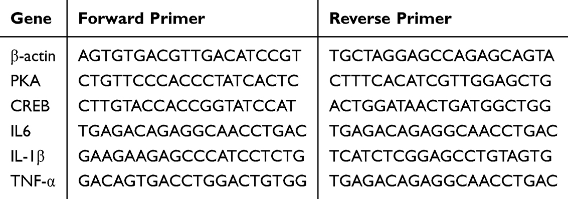 |
Table 2 Forward and Reverse Primer Sequences for RT-qPCR |
Western Blot (WB) Analysis
After lysing in ice-cold RIPA buffer, APP/PS1 mouse hippocampal tissues were subjected to Sodium Dodecyl Sulfate Polyacrylamide Gel Electrophoresis (SDS-PAGE) supersampling. The tissues were then transferred to Polyvinylidene Difluoride (PVDF) transmembranes (Millipore, IPVH00010, Germany), which were immersed in 5% skimmed milk powder. Subsequently, membranes were incubated with primary antibodies (PKA, bs-0520R, Bioss, China; p-PKA, bs-3725R, Bioss, China; CREB. 9197, Cell Signaling Technology (CST), China; p-CREB, 9198, CST, China; SYN, bs-8845R, Bioss, China; PSD95, ab238135, Abcam, China; GAPDH, TA-08, Zsbio, China), followed by secondary antibodies. Following that, protein bands were visualized using an ECL luminescence kit (Biosharp, BL520A, Canada). Finally, PKA, p-PKA, CREB, p-CREB, SYN, and PSD-95 protein expressions were quantified using ImageJ software, with GAPDH as an internal reference.
Data Analysis
Data analysis and visualization were performed using SPSS 26.0 (SPSS Inc., Chicago, IL, USA) and Graphpad Prism 9.0 (GraphPad Software Inc., San Diego, CA, USA) software. Measurement data were expressed as mean ±standard deviation (X±s). To minimize experimental bias, behavioral assessments, histopathological analysis, and data processing were conducted by blinded researchers, and the groups were anonymized using animal identifiers and specimens. The sample size was calculated using pre-specified parameters: 80% power, α=0.05 (two-tailed), medium effect size (Cohen’s d=0.5), and 20% estimated mortality rate. The chi-square test or one-way Analysis of Variance (ANOVA) was used for comparisons between multiple groups when the data met the assumptions of independence, normality, and homogeneity of variance. Pairwise comparisons between groups were performed with the Least Significant Difference (LSD) test after confirming normal distribution and homogeneity of variance. A non-parametric test Dunnett was used if the above conditions were not met. Results or differences with P<0.05 were considered statistically significant.
Results
ZNC Improved Sleep and Learning Memory in APP/PS1 Mice
PiezoSleep Monitoring
The sleep structure of APP/PS1 mice was assessed through PiezoSleep monitoring. Environmental acclimatization was performed in the first 24 h, followed by sleep monitoring in the subsequent 48 h. Compared to normal group mice, model group mice showed significantly lower percentages of day and total sleep, and a significantly higher percentage of dark sleep. Furthermore, compared to normal group mice, mice in the ZNC groups exhibited significantly higher percentages of day and total sleep and a significantly lower dark-phase sleep percentage (p<0.01, p<0.05). These findings suggest that ZNC improved sleep quality in APP/PS1 mice, particularly optimizing the sleep structure (Figure 1B and C).
MWM Test
The MWM experiment began with five consecutive days of spatial localization navigation training to assess the spatial learning ability of APP/PS1 mice. A spatial exploration experiment was performed on the last day to assess the memory ability of the animals. During spatial localization learning and memory navigation training, all experimental group mice exhibited a shortened resident platform incubation period and seeking trajectory compared to the model group, albeit to varying degrees. In the spatial exploration experiment, compared to the normal group, the model group showed significantly fewer target platform crossings and spent less time in target quadrant swimming. Conversely, the ZNC medium- and high-dose groups and the Donepezil Hydrochloride Tablet group had significantly more target platform crossings and spent more time in target quadrant swimming, with a clear seeking trajectory compared with the model group (p<0.01) (Figure 1D–H). These findings suggest that ZNC improved learning and memory impairment in APP/PS1 mice.
Effect of ZNC on Hippocampal Tissue Morphology
According to the HE staining results, neuronal cells in the hippocampal CA1 and CA3 regions in normal group mice were large, uniform, and structurally intact, with a thickness of about “3–5 layers”. Notably, the cells were also distributed in a sequential order. Compared to the normal group, most neuronal cells in the hippocampal CA1 and CA3 regions of model group mice exhibited an abnormal structure. The nuclei were distended and deeply blue-stained, and the cell outlines were irregularly altered, with large spacings. Furthermore, compared to the model group, which had large spacing between cells, the ZNC (High) and donepezil hydrochloride tablet groups exhibited irregular cell outlines, with a scattered arrangement. Moreover, compared to the model group, the above-mentioned pathological changes in the hippocampus of the mice were significantly improved in the ZNC (high) and Donepezil Hydrochloride tablet groups (Figure 2A and B).
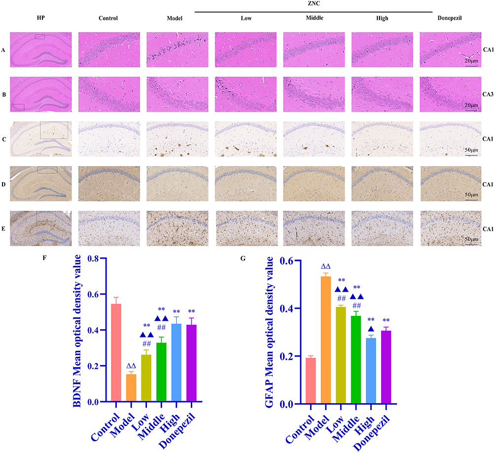 |
Figure 2 Effect of ZNC on hippocampal tissue morphology in APP/PS1 mice. (A) Representative HE-stained sections of hippocampal CA 1 region (scale bar = 20 μm). (B) Representative HE-stained sections of hippocampal CA 3 region (scale bar = 20 μm). (C) Effect of ZNC on Aβ1-42 changes in hippocampal (scale bar = 50 μm). (D) Effect of ZNC on BDNF changes in hippocampus (scale bar = 50 μm). (E) Effect of ZNC on GFAP changes in hippocampal (scale bar = 50 μm). (F and G) The mean optical density value showing the results of D and E, which were consistent with changes in BDNF and GFAP in the indicated groups. (n=3). Compared with the normal group, ΔΔP<0.01; compared with the model group, **P<0.01; compared with the donepezil hydrochloride tablets group, ▲P<0.05, ▲▲P<0.01; compared with the ZNC low and medium dose groups and the ZNC high dose group, ##P<0.01. |
Effects of ZNC on Hippocampal Aβ1-42 and Neurotransmission in APP/PS1 Mice
After 4 weeks of ZNC treatment, the effects of Aβ1-42 deposition, BDNF on neuronal regeneration and GFAP on neuroinflammation in the hippocampal CA1 region of APP/PS1 mice were detected using IHC analysis. The nucleus of the CA1 region of the mouse hippocampus was blue-stained. Meanwhile, BDNF, GFAP and Aβ1-42 were mainly distributed in the cytoplasm, which was brown-yellow-stained. Notably, GFAP exhibited a “fireworks-like” morphology, Aβ1-42 displayed a “plaque” morphology. Specifically, regarding the deposition of Aβ1-42, mice in the model group had marked accumulation in the hippocampal CA1 region compared with those in the normal group. The ZNC (high) and donepezil hydrochloride tablet groups had significantly fewer plaque in the hippocampal CA1 region of APP/PS1 mice (Figure 2C). A comparison of the results based on average OD values further revealed that compared to normal group mice, model group mice exhibited a significantly lower BDNF protein expression in the hippocampal CA1 region and significantly higher GFAP protein expression. On the other hand, the ZNC (high) and donepezil hydrochloride tablet groups exhibited significantly reduced neuroinflammation and increased neuronal regeneration in the hippocampal CA1 region of APP/PS1 mice (p<0.01). The significant Aβ1-42, GFAP protein downregulation and the significant BDNF protein upregulation following ZNC treatment suggest that it decreased the deposition of Aβ and mitigate its toxic effects, exerted an anti-neuroinflammation effect, protected the hippocampal neurons of mice from inflammatory injury, and promoted neuronal regeneration. These effects could help reverse neuroinflammation-induced cognitive function impairments and sleep disorders related to AD (Figure 2D–G).
ZNC Targeted the cAMP/PKA/CREB Pathway and Neuroinflammation for AD Sleep Disorders Alleviation
The initial search of relevant databases for Chinese herbal compounds in ZNC yielded 950 chemical components (Figure 3A). After supplementing the relevant databases with ADME properties and performing Lipinski’s rule screening and de-emphasis, potential pharmacodynamically active components of ZNC were screened out, including Quercetin, Kaempferol, Luteolin, 7-O- methylisomucronulatol, Naringenin, and 70 other potential active ingredients. Moreover, 350 corresponding targets were identified. The analysis using the VENN 2.1 online platform yielded 299 intersecting genes of ZNC and AD sleep disorders pathophysiology (Figure 3B), which were imported into the STRING database to construct the PPI clustering network. The main clusters were response to oxygen levels, inflammatory responses, and phasel-functionalization of compounds (Figure 3C). Moreover, GO enrichment analysis yielded 2017 biological functional items, of which Biological Processes (BPs) were mainly related to inflammation, Oxidative Stress (OS), and response to xenobiotic stimuli, among other items; Cellular Components (CCs) were mainly related to neuronal projection membranes, cytosol, presynaptic membranes, postsynaptic membranes, and so on; and Molecular Functions (MFs) were mainly related to transcription co-regulation factor binding, postsynaptic neurotransmitter receptor activity, and neurotransmitter receptor activity (Figure 3D and E). On the other hand, KEGG enrichment analysis revealed that the highly enriched pathways were PI3K-Akt signaling, neurodegeneration-multiple diseases, AD, cAMP signaling, IL17 signaling, and MAPK signaling. Cytoscape-3.10.2 software was utilized to construct a ZNC-AD sleep disorder-intersecting target-KEGG visualization network (Figure 3F), with the cytoHubba plug-in used to identify the top ten core targets: TP53, JUN, IL6, IL1B, CXCL8, IL1A, MYC, AKT1, IFNG, IL10 (Figure 3G). The GeneMANIA database identified IL20, IL26, IL19, and other potentially relevant inflammatory target genes (Figure 3H). The TRRUST database identified JUN, RELA, NFKB1, STAT3, STAT1, ZNF300, EP300, FOS, CEBPB, EGR1, and CREB1 as core TFs. The core target-transcription factor-pathway network included the JUN proto-oncogene; RELA (v-rel Reticuloendotheliosis Viral Oncogene Homolog A, Avian), NFKB1 (Nuclear Factor of Kappa Light Polypeptide Gene Enhancer in B-cells 1), STAT3 (Signal Transducer and Activator of Transcription 3, Acute-Phase Response Factor), STAT1 (Signal Transducer and Activator of Transcription 1), ZNF300 (Zinc Finger Protein 300, 91Kd), EP300 (E1A Binding Protein p300), FOS (FBJ Murine Osteosarcoma Viral Oncogene Homolog), C/EBPB (CCAAT/Enhancer Binding Protein Beta), EGR1 (Early Growth Response 1), REB1 (cAMP Responsive Element Binding Protein 1), and other related pathways, and was visualized using Cytoscape-3.10.2 software (Figure 3I). The core targets and the top five small molecule compounds among the pharmacodynamic active ingredients were docked using AutodockTolls-1.5.6 to better understand the correlation between ZNC pharmacodynamic active ingredients and AD-related sleep disorders. The binding energies were ≤-5kJ/mol (Figure 3J), suggesting a strong correlation between the two. Lower binding energies of the core targets were further visualized using PyMol-3.0.4 and Discovery Studio-4.5 (Figure 3K). Overall, ZNC-mediated cAMP/PKA/CREB pathway modulation that suppressed cellular inflammatory factors IL-6, IL-1β, and TNF-α and promoted neurotransmitter expression could be its mechanism in AD sleep disorders treatment.
 |
Figure 3 cAMP/PKA/CREB mediates the effects of ZNC on AD sleep disorders. (A) ZNC drug composition. (B) VENN plot of the genes intersecting ZNC (yellow) with AD sleep disorders (purple). (C) PPI network, clustered as Response to oxygen levels (red), Inflammatory response (green), Phasel-Functionalization of compounds(blue). (D) The top 10 enriched GO terms in the GO enrichment analysis (BP, CC, MF). (E) The top 30 enriched KEGG terms. The horizontal axis is rich factor (F) Network diagram of ZNC-AD sleep disorder-intersecting target-KEGG pathway visualization. (G) HUB core genes in the PPI network. (H) HUB core gene-related target genes. (I) HUB core target gene-transcription factor-pathway visualization network. (J) Docking binding energy of HUB core target genes with ZNC pre-5 pharmacodynamic active ingredient molecules. (K) Docking visualization of HUB core target genes with ZNC pre-5 pharmacodynamic active ingredient molecules. |
ZNC Regulated Hippocampal Inflammatory Cytokines and Neurotransmitters in the APP/PS1 Mice Model
After 4 weeks of ZNC treatment, the expressions of inflammatory cytokines (such as IL-6, IL-1β, and TNF-α) and neurotransmitters (such as 5-HIAA, 5-HT, and GABA) in the hippocampus of APP/PS1 mice were detected using ELISA and RT-qPCR. Compared to the normal group, model group mice exhibited a significantly higher hippocampal expression of IL-6, IL-1β, and TNF-α, and a significantly lower expression of 5-HIAA, 5-HT, and GABA. Furthermore, compared to the model group, the ZNC (high) and Donepezil hydrochloride tablet groups exhibited significantly lower expression levels of inflammatory factors such as IL-6, IL-1β, and TNF-α, and a higher 5-HIAA, 5-HT, and GABA expression (p<0.01, p<0.05) (Figure 4A–I). Furthermore, consistent with the HE results, 5-HT production was significantly greater than 5-HIAA synthesis. Overall, ZNC could exert anti-neuroinflammation effects and upregulate neurotransmitters, potentially reversing AD-related sleep disorders.
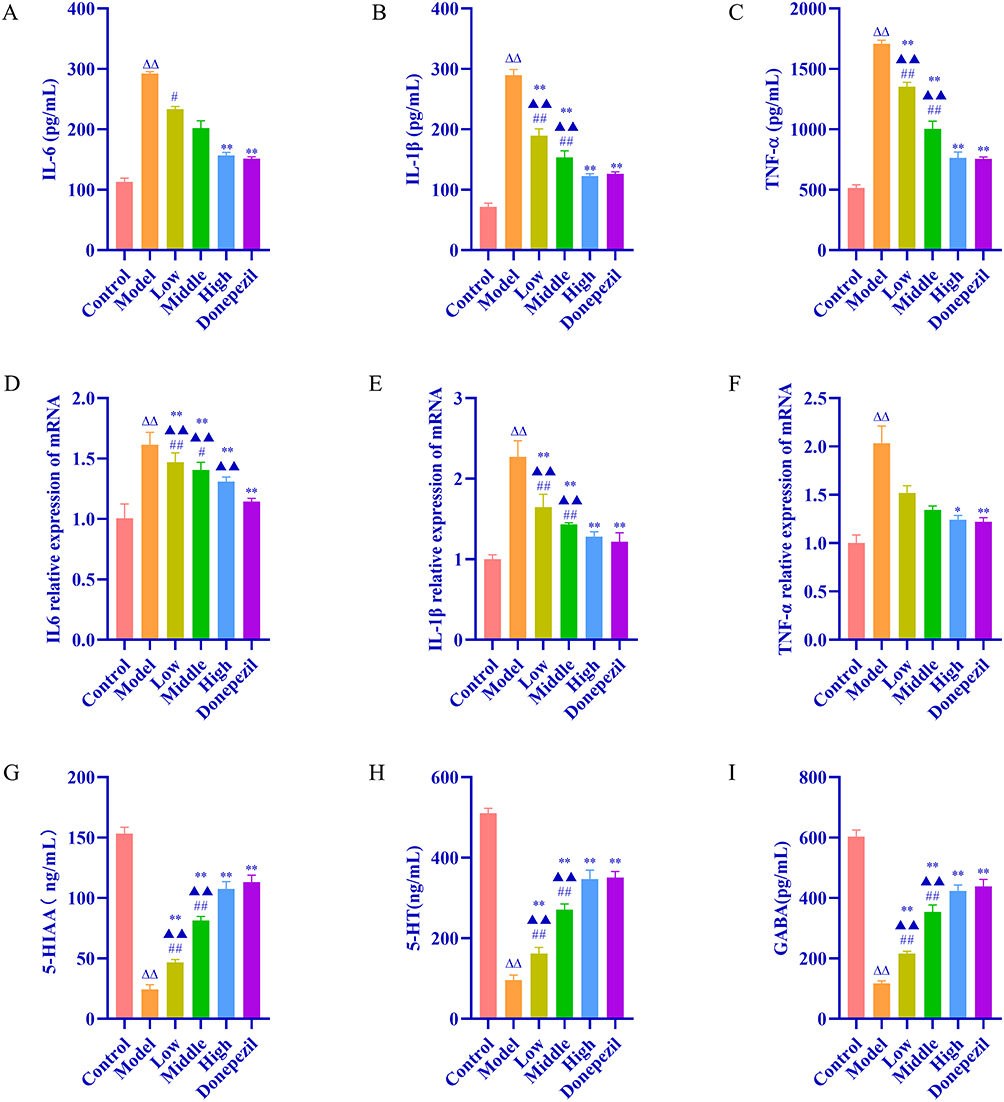 |
Figure 4 ZNC treats AD sleep disorders by inhibiting cellular inflammatory factors and increasing neurotransmitter expression. (A–I) Representative ELISA of IL-6, IL-1β, TNF-α, 5-HIAA, 5-HT, GABA, and representative RT-qPCR of IL-6, IL-1β, and TNF-α in the hippocampus of APP/PS1 mice. Data are expressed as the mean SD (n = 6). Compared with the normal group, ΔΔP<0.01; compared with the model group, *P<0.05, **P<0.01; compared with the donepezil hydrochloride tablets group, ▲▲P<0.01; compared with the ZNC low and medium dose groups and the ZNC high dose group, #P<0.05, ##P<0.01. |
ZNC Mediated cAMP/PKA/CREB Signaling Pathway Activation
After 4 weeks of ZNC treatment, hippocampal cAMP, PKA and, CREB signaling pathway-related protein expressions in APP/PS1 mice were detected using ELISA, RT-qPCR and, WB, respectively. According to the results, compared to the normal group, model group mice exhibited significantly lower hippocampal cAMP, PKA mRNA, and CREB mRNA expression levels. Furthermore, compared to the model group, the ZNC (high) and donepezil hydrochloride tablet groups showed significantly higher cAMP, PKA mRNA, and CREB mRNA expression levels (p<0.01, p<0.05) (Figure 5A–C). Moreover, compared to the model group, the ZNC (high) and donepezil hydrochloride tablet groups showed significantly higher p-PKA, p-CREB, SYN, and PSD-95 expression levels (p<0.01) (Figure 5D–K). These findings suggest that APP/PS1 model mice expressed key proteins of the cAMP/PKA/CREB signaling pathway and ZNC upregulated their phosphorylation levels, regulating neuron and synapse genesis and plasticity, and ultimately effectively improving learning, memory and sleep disorders (Figure 6).
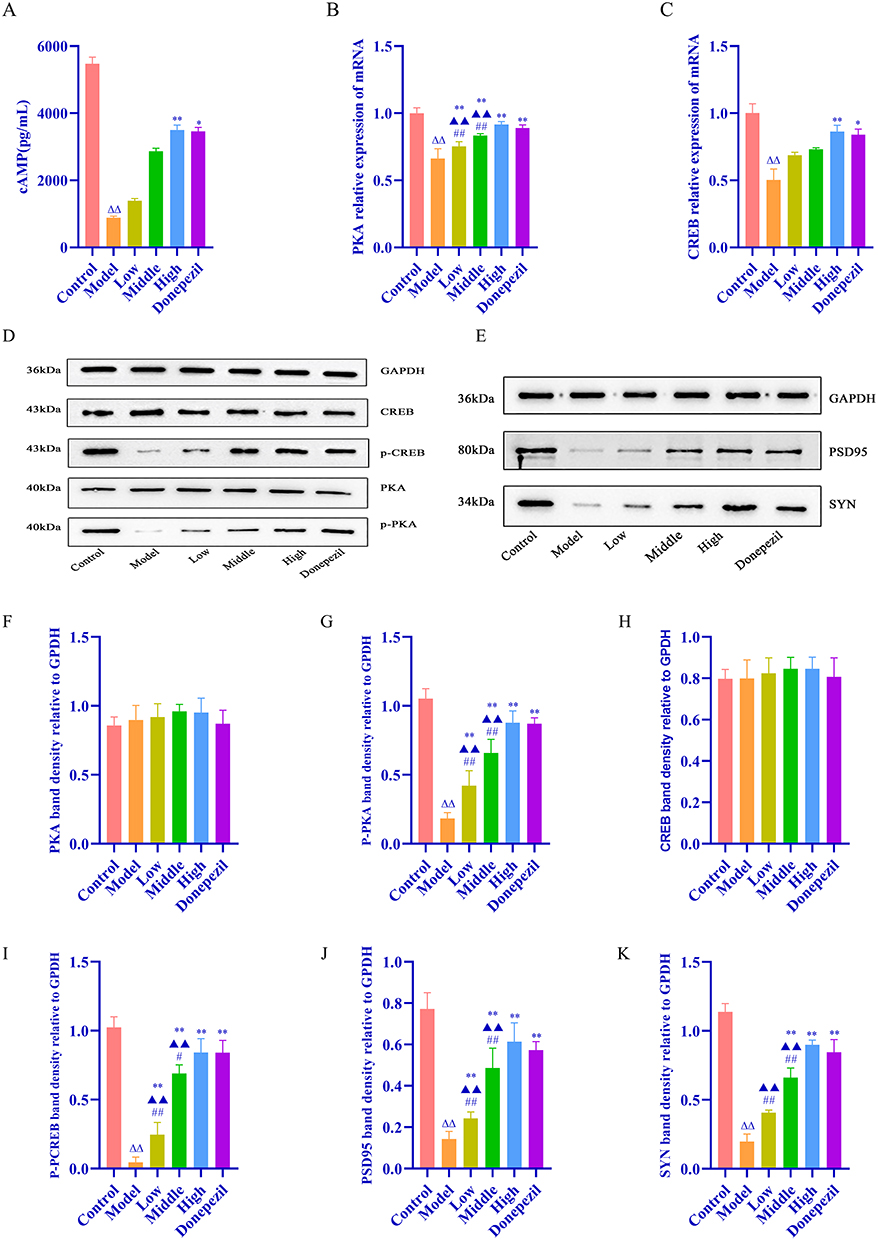 |
Figure 5 ZNC cAMP/PKA/CREB signaling pathway that regulates neuronal and synaptogenesis and its plasticity in AD sleep disorders. (A) Representative ELISA of cAMP in the hippocampus of APP/PS1 mice. (B and C) Representative RT-qPCR of PKA and CREB in the hippocampus of APP/PS1 mice. (D–K) Representative Western blot of PKA, p-PKA, CREB, p-CREB, SYN, PSD-95 in the hippocampus of APP/PS1 mice. Data are expressed as the mean SD (n = 6). Compared with the normal group, ΔΔP<0.01; compared with the model group, *P<0.05, **P<0.01; compared with the donepezil hydrochloride tablets group, ▲▲P<0.01; compared with the ZNC low and medium dose groups and the ZNC high dose group, #P<0.05, ##P<0.01. |
 |
Figure 6 A diagram showing the mechanism by which ZNC improves sleep and memory impairment in APP/PS1 mice via cAMP/PKA/CREB signaling pathway. A red upward arrow indicates an increase, and a blue downward arrow indicates a decrease or relief. Drawn with Figdraw. |
Discussion
According to reports, AD, a chronic progressive disease primarily characterized by a significant decline in recent memory, ranks first among chronic neurodegenerative diseases. Besides memory loss, AD also features varying degrees of spatial, linguistic, computational, and non-cognitive symptoms, which might cause severe disability or even death. These clinical complications not only severely jeopardize the patient’s well-being in his or her later years, but also place a heavy burden on the state, society, and families, especially with the increasing ageing population.1 Sleep disorders, on the other hand, include difficulties in falling asleep and/or insufficient sleep depth (≥ 3 times per week), and could result in a poor mental state, impacting various aspects of life, including work.16
It has been established that AD-related sleep disorders are prevalent in ⁓45% of normal middle-aged and elderly people. Moreover, AD-related sleep disorders often exhibit more complex and severe clinical symptoms, mostly manifesting as prolonged sleep latency, an increased number of nighttime awakenings, difficulties in sleep maintenance, and daytime drowsiness, among others. Other symptoms might include a reversed day and night sleep pattern, ie, daytime sleep and nighttime wakefulness.5 Sleep disorder-induced cognitive function deficits have been widely reported. In AD patients, these disorders may occur years before a significant decline in learning and memory abilities is observed.17,18 It is also noteworthy that the pathogenesis of AD-related sleep disorders was unclear, with possible mechanisms including abnormalities in circadian rhythm gene expression,19 decreased melatonin levels,20 an imbalance in the expression of sleep-wake-related neurotransmitters,21 Aβ deposition,22 orexigenic system disorders,23 and genetic factors,24 among others. Sleep disorders are not only a clinical symptom of AD, but are also among its leading risk factors. Furthermore, AD-sleep disorders interaction could result in a vicious circle. Moreover, the symptoms of sleep disorders are often neglected. These phenomena have been linked to a short honeymoon period, dependence, and other drawbacks associated with relevant therapeutic interventions, highlighting the need for in-depth research on sleep disorders as a controllable target and to develop novel, effective prevention and treatment agents. Such efforts would be crucial for the early prevention and treatment of AD and other neurodegenerative diseases, and even for delaying and reversing the disease.
Notably, ZNC, whose formulation is based on the ancient Huizhou Xin’an medical principle of solidifying the root and cultivating vitality, comprises eight kinds of Chinese herbs, including Cistanches Herba and Astragali Radix. The formulation leverages sweet and warm cultivation and supplementation to support yang and benefit yin. It could also help in expelling phlegm, removing blood stasis, and unblocking meridians, aligning with the therapeutic principle of using the passage both as a tonic and as a tool. Overall, ZNC may be effective in tonifying the spleen and kidneys, excluding phlegm, opening orifices, and revitalizing the spirit, ultimately improving cognitive function. Although ZNC was previously reported to improve learning and memory function and hippocampal histopathology in APP/PS1 mice, reports on its potential to improve learning and memory by alleviating sleep disorders, as well as the underlying mechanisms, are quite limited. Consequently, using an APP/PS1 mice AD-related sleep disorders model, we evaluated the pharmacological effects of ZNC on sleep and learning memory using PiezoSleep monitoring and the MWM test. Meanwhile, HE staining was used to observe the morphology of hippocampal tissues in APP/PS1 mice. Furthermore, to evaluate ZNC’s effects on neurological function and learning memory, as well as the degree of neuronal damage and the pharmacodynamic effects, the expression of the Aβ1-42, neurotransmitter BDNF and the inflammatory marker GFAP was detected using IHC analysis. The possible mechanisms of ZNC in AD sleep disorders treatment were assessed using network pharmacology, and the expression of neurotransmitters 5-HIAA, 5-HT, and GABA, and inflammatory cytokines IL-6, IL-1β, and TNF-α in hippocampal tissues was detected using ELISA and RT-PCR tests. Finally, the potential involvement of the cAMP/PKA/CREB pathway and synaptic plasticity-related mRNAs was examined using RT-PCR and WB, respectively. We found that ZNC may improve sleep, learning, and memory disorders in APP/PS1 mice via cAMP/PKA/CREB pathway modulation.
Notably, APP/PS1 mice are often used to construct pathological mouse models for AD, often exhibiting sleep disorder symptoms after induction. On the other hand, PiezoSleep monitoring is valuable in verifying the success of AD-related sleep disorders disease modeling.In previous studies, researchers adopted brain stereotaxic cranial perforation, implantation of electroencephalogram/electromyogram (EEG/EMG) chips, and analysis of neuroelectric signals to monitor the sleep and muscle tone in animals. Although the accuracy of this experiment is relatively high, this operation runs against the ethics of animal experimentation. Moreover, invasive and unhygienic operation as well as breeding environment will have fatal and serious consequences for the experimental animals, and the surgery itself will take a certain time and postoperative pain. For instance, the surgery itself and postoperative pain will take a long time to recover, which can potentially increase the lethality and aggravate the sleep disorder for APP/PS1 mice, yielding biased results. Therefore, we selected the non-invasive and non-invasive piezo sleep system, which is more convenient and comfortable compared with traditional monitoring methods. The proposed experimental method can replace the invasive EEG/EMG chip with a piezoelectric film, placed under a thin layer of padding, and outputs an electrical signal by converting the pressure and voltage generated through the movement, facilitating the monitoring of fine movements generated by experimental mice in a highly sensitive manner. It also records the fine movements generated by experimental mice, such as feeding, scratching, and more delicate heartbeat and respiratory movements, as well as monitoring wakefulness and sleep. In addition, it can distinguish between rapid eye movement (REM) and non-rapid eye movement (NREM) sleep in experimental mice with high precision.15 In this study, we found, for the first time, that APP/PS1 mice developed sleep disorders, as indicated by the decreased day sleep, increased night sleep, and decreased total sleep status, making it ideal for studying sleep disorders. Research has indicated that ZNC can optimize the sleep structure and improve sleep disorders by increasing day sleep, decreasing night sleep, and the proportion of total sleep, which improves the learning and memory disorders.
Network pharmacology combined with molecular docking studies have revealed that ZNC regulates AD sleep disorders by inhibiting the cellular inflammatory factors such as IL-6, IL-1β, and TNF-α and promoting the expression of neurotransmitters by modulating the cAMP/PKA/CREB signaling pathway. In vivo experiments in APP/PS1 mice verified the above predictions, showing that ZNC decreased the expression of inflammatory indicators such as GFAP, IL-6, IL-1β, TNF-α, and increased the levels of neurotransmitters (BDNF, 5-HIAA, 5-HT, GABA, etc.) in the hippocampus of APP/PS1 mice. It has been reported that inflammatory mediators such as GFAP, IL-6, IL-1β, and TNF-α further upregulate the expression of Aβ load and disrupt sleep regulation, promoting the occurrence and AD sleep disorders.25–27 The 5-HT expression level is positively correlated with sleep and actively participate in regulation of the sleep-wake cycle through the dynamic changes its metobilite 5-HIAA. The ratio of the two (5-HIAA/5-HT) serves as an indicator of the metabolic rate.28 As a recognized neurotransmitter that regulates the sleep-wake, GABA is considered one of the most important inhibitory neurotransmitters, exhibiting a sedative-hypnotic effect when it binds to the receptor. The GABA mainly exerts its biological effects via acting on two receptors, GABAA and GABAB. Research has shown that activation of GABAB receptor is a key factor modulating the development of sleep. Moreover, activation of the GABAB receptor influences the sleep and circadian regulation via the cAMP/ CREB signaling pathway.29
As a second messenger, cAMP is widely involved in the regulation of cellular material metabolism and biological functions. Notably, the cAMP signaling controls circadian pacing signals and determines their amplitude, phase, and period, and the cAMP signaling itself exhibits a circadian rhythm.30 Following the synthesis of cAMP, the cAMP-dependent PKA phosphorylation has been shown to participate in the transmission of neurological signals, such as learning and memory, through p-PKA, which further binds to CREB, stimulating the phosphorylation of p-CREB to promote the expression of BDNF neurotransmitter, which is involved in neuronal genesis, differentiation, and regulation of synaptic plasticity processes, thereby enhancing learning, memory, and optimizing sleep architecture.31 Notably, targeting circadian cAMP signaling alone was found to similarly improve the long-term memory in mice. Inhibition of cAMP/ PKA/CREB exacerbated postoperative memory loss in aged rats.32 Sleep disorders can significantly reduce the concentration levels of neurotransmitters in the brain, including GABA, with a concomitant loss of neurons and synaptic damage, while Aβ and Tau proteins exacerbate synaptic dysfunction, synaptic plasticity, and loss of synapses in the cortex and hippocampus. Notably, the cAMP/PKA/CREB signaling pathway can modulate neurotransmitters to promote postsynaptic long term potentiation (LTP), which is implicated in learning and memory, and inhibition of which exacerbates cognitive deficits.33 The cAMP/PKA/CREB signaling confers neuroprotection mainly by altering the downstream expression of BDNF, etc.34 BDNF plays a key role in neuronal genesis, differentiation and, synaptic function, and can promote neurogenesis as well as replenish lost neurons and promote synaptic development, to improve cognitive function, alleviate neuronal-synaptic damage and other pathological changes in AD. In this study, we found that ZNC ameriolated learning, memory, and sleep disorders by increasing the expression of neurotransmitter-related proteins.35 Synaptogenesis, plasticity regulation, and neural signaling are mainly regulated by SYN, a membrane protein expressed in the presynaptic membrane, and PSD-95, a structural protein found in the postsynaptic membrane, which are synergistically regulated by both. Researchers have found that SYN and PSD-95 expression are downregulated in the hippocampus of APP/PS1 mice.36–38 In this study, we found that the expression of cAMP, PKA mRNA, CREB mRNA, p-PKA, p-CREB, SYN and PSD-95 was up-regulated in the high-dose group of ZNC and the group treated with Donepezil Hydrochloride Tablets, which demonstrates that there are key proteins of cAMP/PKA/CREB signaling pathway involved in the pathological processes in APP/PS1 mice, and ZNC can improve learning and memory ability by enhancing the phosphorylation level of key proteins of cAMP/PKA/CREB signaling pathway, significantly downregulated Aβ1-42, regulating the neuron, synaptogenesis and its plasticity, and improving learning and memory ability. TCM adopts a holistic approach, emphasizing individualized treatment based on the principle of heterogeneous therapy. In the context of AD, this approach targets sleep disorders with shared underlying causes, aiming to optimize sleep structure and thereby slow disease progression.
Although we integrated network pharmacology, molecular docking, and in vivo validation experiments to investigate the therapeutic effects of ZNC on AD-related sleep disorders and its molecular mechanisms, there are several limitations that should be acknowledged. First, the regulatory effects on the cAMP/PKA/CREB pathway were validated solely in APP/PS1 mice (an Aβ-related AD model), and thus whether similar outcomes occur in 3xTg-AD mice that exhibit dual Aβ/Tau pathology is not known. Second, we did not perform in vitro cell-based experiments, which limited our ability to clarify the cellular effects. Third, we adopted a 28-day intervention period, and hence, the long-term efficacy could not be explored. Fourth, we investigated the activity and breathing in sleep behavior tracking, but did not comprehensively cover established sleep behaviors such as bruxism, sleep-related muscle activity, and stress/emotion-associated phenotypes.39–41 To address this, future investigations using 3xTg-AD mice with enhanced pathological realism and primary neuronal cells are needed to explore bruxism, muscle activity, and stress/emotion-linked phenotypes, while also conducting long-term observations. In vitro experiments in which these factors are silenced/overexpressed are advocated to verify the mechanisms and enhance the clinical translation of the findings.
Conclusion
In conclusion, ZNC improves hippocampal neuronal damage and learning and memory functions as well as sleep disorders associated with AD sleep disorders, may by downregulating Aβ1-42, inhibiting cellular inflammatory factors, such as IL-6, IL-1β, and TNF-α, and promoting neurotransmitter expression, such as BDNF, 5-HIAA, 5-HT, and GABA, via modulating the cAMP/PKA/CREB signaling pathway. Our results provide an important basis that will guide further prospective studies exploring the mechanisms of ZNC in the treatment of AD sleep disorders, to uncover new treatment strategies for AD sleep disorders. In future, we will perform in vivo and in vitro investigations to elucidate the mechanistic basis of ZNC therapeutic effects, as well as its clinical applicability and translational potential.
Abbreviations
ZNC, Zhinao Capsule; HE, Hematoxylin and eosin; IHC, Immunohistochemistry; AD, Alzheimer′s Disease; RT-qPCR, Quantitative real-time polymerase chain reaction; MWM, Morris water maze test; CNS, Central Nervous System; Aβ Amyloid Beta; P-tau, intracellular Phosphorylated tau; CSF, Cerebrospinal Fluid; IF, Interstitial Fluid; MCI, Mild Cognitive Impairment; GI, Gastrointestinal; cAMP, cyclic adenosine monophosphate; PKA, protein kinase A; CREB, cAMP response element binding protein; SYN, Synaptophysin; PSD-95, Postsynaptic density protein 95; GFAP, Glial Fibrillary Acidic Protein; IL6, Interleukin-6; IL-1β, Interleukin-1β; TNF-α, Tumor necrosis factor-α; 5-HIAA, 5-Hydroxyindole-3-acetic acid; 5-HT, 5-hydroxy tryptamine; GABA, Gamma-aminobutyric acid; CSF, cerebrospinal fluid; IF interstitial fluid; MCI, Mild cognitive impairment; TCM, Traditional Chinese medicine; TCMSP, The Traditional Chinese Medicine Systems Pharmacology Database and Analysis Platform; HERB, A high-throughput experiment- and reference-guided database of traditional Chinese medicine; ETCM, The Encyclopedia of Traditional Chinese Medicine; TCMID, Traditional Chinese Medicine Information; BATMAN-TCM, A Bioinformatics Annotation database for Molecular mechANism of Traditional Chinese Medicine; OMIM, An Online Catalog of Human Genes and Genetic Disorders; KEGG, Kyoto Encyclopedia of Genes and Genomes; TRRUST, Transcriptional Regulatory Relationships Unraveled by Sentence-based Text mining; REM, rapid eye movement; NREM, Non-rapid eye movement; LTP, long term potentiation; EEG, electroencephalogram; EMG, electromyogram.
Data Sharing Statement
All original data for this manuscript are demonstrated in the article, for further information you can inquire with the corresponding author Wenming Yang.
Ethics Approval and Consent to Participate
The authors bear sole responsibility for ensuring the veracity and integrity of the work in its entirety, and for addressing any concerns pertaining to its accuracy or integrity. The experimental protocol was approved by the Animal Research Ethics Committee of Anhui Agricultural University (Approval no. AHAUB2023024). All animal care and experimental procedures follow the Guidelines for the Care and Use of Laboratory Animals (Ministry of Science and Technology of China, Animal Research Ethics Committee of Anhui Agricultural University) and ARRIVE guidelines (Animal Research: Reporting of Invasive Veterinary Experiments).
Acknowledgments
We gratefully acknowledge Anhui Agricultural University for providing SPF grade animal house for APP/PS1 mice breeding, and all the experts of the First Affiliated Hospital of Anhui University of Chinese Medicine and Wangjing Hospital, China Academy of Chinese Medical Sciences for their help and providing laboratories and platforms for the conduct of this experiment.
Author Contributions
All authors made a significant contribution to the work reported, whether that is in the conception, study design, execution, acquisition of data, analysis and interpretation, or in all these areas; took part in drafting, revising or critically reviewing the article; gave final approval of the version to be published; have agreed on the journal to which the article has been submitted; and agree to be accountable for all aspects of the work.
Funding
This work was financially supported by the National Science and Technology Major Project (No. 2023ZD0505801), Institute of Xin’an Medicine and Modernization of Traditional Chinese Medicine, Institute of Health and Medicine, Hefei Comprehensive National Science Center (No. 2024CXMMTCM001); Anhui Provincial Natural Science Foundation Youth Project (No. 2108085QH368); Anhui Provincial Health and Wellness Research Project (No. AHWJ2022b086).
Disclosure
None of the authors have any conflicts of interest associated with this study, and all of the authors have read and approved this manuscript.
References
1. Better MA. Alzheimer’s disease facts and figures. Alzheimer’s Dementia. 2024;20(5):3708–3821. doi:10.1002/alz.13809
2. Zheng Q, Wang X. Alzheimer’s disease: insights into pathology, molecular mechanisms, and therapy. Protein Cell. 2025;16(2):83–120. doi:10.1093/procel/pwae026
3. Yuan M, Hong B, Zhang W, et al. Late-life sleep duration associated with amnestic mild cognitive impairment. Inter Psychogeriatr. 2023;35(8):439–448. doi:10.1017/s1041610221000466
4. Bubu OM, Brannick M, Mortimer J, et al. Sleep, cognitive impairment, and Alzheimer’s disease: a systematic review and meta-analysis. Sleep. 2017;40(1). doi:10.1093/sleep/zsw032
5. Lacerda RAV, Desio JAF, Kammers CM, et al. Sleep disorders and risk of’alzheimer’s disease: a two-way road. Ageing Res Rev. 2024;101:102514. doi:10.1016/j.arr.2024.102514
6. Joiner WJ. Neuroscience: sleep fragmentation impairs memory formation. Curr Biolog. 2019;29(22):R1181–r1184. doi:10.1016/j.cub.2019.09.060
7. Karimi Tari P, Parsons CG, Collingridge GL, Rammes G. Memantine: updating a rare success story in pro-cognitive therapeutics. Neuropharmacology. 2024;244:109737. doi:10.1016/j.neuropharm.2023.109737
8. Ajenikoko MK, Ajagbe AO, Onigbinde OA, Okesina AA, Tijani AA. Review of Alzheimer’s disease drugs and their relationship with neuron-glia interaction. IBRO Neurosci Rep. 2023;14:64–76. doi:10.1016/j.ibneur.2022.11.005
9. Ma Y, Huang S, Jiang H, Yang W, Nilashi M. Mechanism of Zhinao capsule in treating Alzheimer’s Disease based on network pharmacology analysis and molecular docking validation. J Healthcare Engine. 2022;2022:708769. doi:10.1155/2022/5708769
10. Kent BA, Michalik M, Marchant EG, et al. Delayed daily activity and reduced NREM slow-wave power in the APPswe/PS1dE9 mouse model of’Alzheimer’s disease. Neurobiol Aging. 2019;78:74–86. doi:10.1016/j.neurobiolaging.2019.01.010
11. Tabassum S, Misrani A, Tang BL, Chen J, Yang L, Long C. Jujuboside A prevents sleep loss-induced disturbance of hippocampal neuronal excitability and memory impairment in young APP/PS1 mice. Sci Rep. 2019;9(1):4512. doi:10.1038/s41598-019-41114-3
12. Ocalan B, Cakir A, Koc C, Suyen GG, Kahveci N. Uridine treatment prevents REM sleep deprivation-induced learning and memory impairment. Neurosci Res. 2019;148:42–48. doi:10.1016/j.neures.2019.01.003
13. Xie G, Huang X, Li H, Wang P, Huang P. Caffeine-related effects on cognitive performance: roles of apoptosis in rat hippocampus following sleep deprivation. Biochem Biophys Res Commun. 2021;534:632–638. doi:10.1016/j.bbrc.2020.11.029
14. Vanneau T, Quiquempoix M, Trignol A, et al. Determination of the sleep–wake pattern and feasibility of NREM/REM discrimination using the non-invasive piezoelectric system in rats. J Sleep Res. 2021;30(6):e13373. doi:10.1111/jsr.13373
15. Topchiy I, Fink AM, Maki KA, Calik MW. Validation of piezosleep scoring against EEG/EMG sleep scoring in rats. Nat Sci Sleep. 2022;14:1877–1886. doi:10.2147/nss.S381367
16. Gauld C, Wakefield JC, Micoulaud-Franchi JA. Proposing a definition for sleep disorders: an epistemological review. Sleep Med Rev. 2025;79:102028. doi:10.1016/j.smrv.2024.102028
17. Wang C, Holtzman DM. Bidirectional relationship between sleep and Alzheimer’s disease: role of amyloid, tau, and other factors. Neuropsychopharmacology. 2020;45(1):104–120. doi:10.1038/s41386-019-0478-5
18. Kusztor A, Raud L, Juel BE, Nilsen AS, Storm JF, Huster RJ. Sleep deprivation differentially affects subcomponents of cognitive control. Sleep. 2019;42(4). doi:10.1093/sleep/zsz016
19. Hoyt KR, Obrietan K. Circadian clocks, cognition, and Alzheimer’s disease: synaptic mechanisms, signaling effectors, and chronotherapeutics. Mol Neurodegener. 2022;17(1):35. doi:10.1186/s13024-022-00537-9
20. Andrade MK, Souza LC, Azevedo EM, et al. Melatonin reduces β-amyloid accumulation and improves short-term memory in streptozotocin-induced sporadic Alzheimer’s disease model. IBRO Neuroscience Rep. 2023;14:264–272. doi:10.1016/j.ibneur.2023.01.005
21. Van Erum J, Van Dam D, De Deyn PP. Alzheimer’s disease: neurotransmitters of the sleep-wake cycle. Neurosci Biobehav Rev. 2019;105:72–80. doi:10.1016/j.neubiorev.2019.07.019
22. Lucey BP, Hicks TJ, McLeland JS, et al. Effect of sleep on overnight cerebrospinal fluid amyloid β kinetics. Ann Neurol. 2018;83(1):197–204. doi:10.1002/ana.25117
23. Carpi M, Mercuri NB, Liguori C. Orexin receptor antagonists for the prevention and treatment of Alzheimer’s disease and associated sleep disorders. Drugs. 2024;84(11):1365–1378. doi:10.1007/s40265-024-02096-3
24. Yu X, Zhou X, He Z, et al. Sleep and APOE -ε4 have a synergistic effect on plasma biomarkers and longitudinal cognitive decline in older adults. CNS Neurosci Ther. 2024;30(2):e14558. doi:10.1111/cns.14558
25. Sánchez Romero EA, Martínez-Pozas O, García-González M, et al. Association between sleep disorders and sleep quality in patients with temporomandibular joint osteoarthritis: a systematic review. Biomedicines. 2022;10(9):2143. doi:10.3390/biomedicines10092143
26. Irwin MR, Vitiello MV. Implications of sleep disturbance and inflammation for’Alzheimer’s disease dementia. Lancet Neurol. 2019;18(3):296–306. doi:10.1016/s1474-4422(18)30450-2
27. Sharma A, Feng L, Muresanu DF, et al. Nanowired delivery of cerebrolysin together with antibodies to amyloid beta peptide, phosphorylated tau, and tumor necrosis factor alpha induces superior neuroprotection in Alzheimer’s disease brain pathology exacerbated by sleep deprivation. Adv Neurobiol. 2023;32:3–53. doi:10.1007/978-3-031-32997-5_1
28. Oikonomou G, Altermatt M, Zhang R-W, et al. The serotonergic raphe promote sleep in zebrafish and mice. Neuron. 2019;103(4):686–701.e8. doi:10.1016/j.neuron.2019.05.038
29. Bao H, Peng Z, Cheng X, et al. GABA induced by sleep deprivation promotes the proliferation and migration of colon tumors through miR-223-3p endogenous pathway and exosome pathway. J Exper Clin Cancer Res. 2023;42(1):344. doi:10.1186/s13046-023-02921-9
30. Jagannath A, Varga N, Dallmann R, et al. Adenosine integrates light and sleep signalling for the regulation of circadian timing in mice. Nat Commun. 2021;12(1):2113. doi:10.1038/s41467-021-22179-z
31. Yuan L, Zhang J, Guo J-H, et al. DAla2-GIP-GLU-PAL protects against cognitive deficits and pathology in APP/PS1 mice by inhibiting neuroinflammation and upregulating cAMP/PKA/CREB signaling pathways. J’Alzheimer’s Dis. 2021;80(2):695–713. doi:10.3233/jad-201262
32. Zhang J, Wang Z, Cong K, Qi J, Sun L. Phoenixin-20 ameliorates sevoflurane inhalation-induced post-operative cognitive dysfunction in rats via activation of the PKA/CREB signaling. Aging. 2023;15(24):14666–14676. doi:10.18632/aging.205177
33. Hou Z, Yang X, Li Y, Chen J, Shang H, Wang F. Electroacupuncture enhances neuroplasticity by regulating the orexin a-mediated cAMP/PKA/CREB signaling pathway in senescence-accelerated mouse prone 8 (SAMP8) mice. Oxid Med Cell Longev. 2022;2022(1):8694462. doi:10.1155/2022/8694462
34. Tamam Y, Yokuş B, Tamam C, et al. The effect of lidocaine on the experimental model of streptozotocin-induced’Alzheimer’s disease. Noro Psikiyatri Arsivi. 2023;60(1):68–72. doi:10.29399/npa.28112
35. Zhang Y-M, Wei R-M, Zhang J-Y, et al. Resveratrol prevents cognitive deficits induced by sleep deprivation via modulating sirtuin 1 associated pathways in the hippocampus. J Biochem Mole Toxicol. 2024;38(4):e23698. doi:10.1002/jbt.23698
36. Shen Z-C, Xia Z-X, Liu J-M, et al. APT1-mediated depalmitoylation regulates hippocampal synaptic plasticity. J Neurosci. 2022;42(13):2662–2677. doi:10.1523/jneurosci.1741-21.2022
37. Kim DY, Kim S-M, Han I-O. Chronic rapid eye movement sleep deprivation aggravates the pathogenesis of Alzheimer’s disease by decreasing brain O-GlcNAc cycling in mice. J Neuroinflammation. 2024;21(1):180. doi:10.1186/s12974-024-03179-4
38. Longhena F, Faustini G, Brembati V, Pizzi M, Benfenati F, Bellucci A. An updated reappraisal of synapsins: structure, function and role in neurological and psychiatric disorders. Neurosci Biobehav Rev. 2021;130:33–60. doi:10.1016/j.neubiorev.2021.08.011
39. Cid-Verdejo R, Chávez Farías C, Martínez-Pozas O, et al. Instrumental assessment of sleep bruxism: a systematic review and meta-analysis. Sleep Med Rev. 2024;74. 101906. doi:10.1016/j.smrv.2024.101906
40. Cid-Verdejo R, Domínguez Gordillo AA, Sánchez-Romero EA, Ardizone García I, Martínez Orozco FJ. Diagnostic accuracy of a portable electromyography and electrocardiography device to measure sleep bruxism in a sleep apnea population: a comparative study. Clocks Sleep. 2023;5(4):717–733. doi:10.3390/clockssleep5040047
41. Osses-Anguita ÁE, Sánchez-Sánchez T, Soto-Goñi XA, et al. Awake and sleep bruxism prevalence and their associated psychological factors in first-year university students: a pre-mid-post COVID-19 pandemic comparison. Int J Environ Res Public Health. 2023;20(3). doi:10.3390/ijerph20032452
 © 2025 The Author(s). This work is published and licensed by Dove Medical Press Limited. The
full terms of this license are available at https://www.dovepress.com/terms
and incorporate the Creative Commons Attribution
- Non Commercial (unported, 4.0) License.
By accessing the work you hereby accept the Terms. Non-commercial uses of the work are permitted
without any further permission from Dove Medical Press Limited, provided the work is properly
attributed. For permission for commercial use of this work, please see paragraphs 4.2 and 5 of our Terms.
© 2025 The Author(s). This work is published and licensed by Dove Medical Press Limited. The
full terms of this license are available at https://www.dovepress.com/terms
and incorporate the Creative Commons Attribution
- Non Commercial (unported, 4.0) License.
By accessing the work you hereby accept the Terms. Non-commercial uses of the work are permitted
without any further permission from Dove Medical Press Limited, provided the work is properly
attributed. For permission for commercial use of this work, please see paragraphs 4.2 and 5 of our Terms.