
Back to Journals » Clinical Ophthalmology » Volume 20
Visual Outcomes in Traumatic Aphakia Corrected with Contact Lens versus Intraocular Lens Implantation – A Retrospective Chart Review
Authors Nowroozizadeh S, Rahal DA  , Ercanbrack C, Brown KL, Sallam AB, Uwaydat SH
Received 17 February 2026
Accepted for publication 18 June 2026
Published 22 July 2026 Volume 2026:20 604003
DOI https://doi.org/10.2147/OPTH.S604003
Checked for plagiarism Yes
Review by Single anonymous peer review
Peer reviewer comments 2
Editor who approved publication: Dr Bharat Gurnani
Sara Nowroozizadeh,1 Dania A Rahal,2 Carson Ercanbrack,2 Kathryn L Brown,1 Ahmed B Sallam,1 Sami H Uwaydat1
1Harvey and Bernice Jones Eye Institute, University of Arkansas for Medical Sciences, Little Rock, AR, USA; 2College of Medicine, University of Arkansas for Medical Sciences, Little Rock, AR, USA
Correspondence: Sami H Uwaydat, Harvey and Bernice Jones Eye Institute, University of Arkansas for Medical Sciences, 4301 W. Markham Street, Little Rock, AR, 72207, USA, Tel +1 501 24 686 5822, Fax +1 501 686 7037, Email [email protected]
Purpose: To compare visual outcomes in trauma-induced aphakia after surgical intraocular lens (IOL) implantation versus contact lens correction (CL).
Methods: This study was a 7-year retrospective chart review. Patient characteristics, injury types, and lens types were documented for 42 eyes with trauma-induced aphakia, with 29% undergoing IOL implantation (n=12, mean age: 52.1 years) and 71% with CL (n=30, mean age: 41.3 years). Aphakic visual acuity (converted to logMAR scale), post-treatment visual acuity, and follow-up duration were assessed. The Wilcoxon ranked sum test was employed for outcome comparisons.
Results: Both groups demonstrated significant visual acuity improvement. Post-treatment, 33% and 37% of patients in the IOL and CL groups, respectively, reported visual acuity better than 20/40. No statistically significant difference was found between the cohorts (p = 0.5861).
Conclusion: In trauma-induced aphakia, contact lens correction is comparable to intraocular lens implantation in improving visual acuity.
Keywords: trauma, aphakia, contact lens, intraocular lens, implantation
Introduction
Ocular trauma is a major cause of visual impairment with an approximate annual incidence of fifty million cases, 1.5% of which necessitate hospitalization.1,2 An estimated 24 million people in the United States have sustained an eye injury, with 1.5 million experiencing resultant visual impairment and 1.7 million sustaining at least some degree of blindness.3 Many studies have focused on identifying the cause of these injuries in hopes of recognizing possible prevention strategies.4 Among the array of factors leading to ocular trauma, the most frequent mechanisms involve falls, motor vehicle accidents, and blunt force.1
Traumatic ocular injuries can lead to aphakia. Aphakia poses refractive and visual challenges, which are often managed with intraocular lens implants or other corrective measures to restore optimal vision. Two primary techniques address vision loss arising from aphakia. The first involves surgically placing a secondary intraocular lens (IOL).5 Studies demonstrate that IOL implantation can restore baseline best corrected visual acuity (BCVA) by postoperative week one.5 The second approach employs scleral contact lenses (CL),6 a longstanding choice for aphakia correction due to their extended history of safe use and straightforward implementation.7
The main advantage of scleral contact lenses is in their ability to correct vision even in cases where irregular astigmatism and other corneal pathologies can cause impairment,6 whereas their disadvantage is the daily requirement to handle the CL. Rigid gas permeable CL (which holds a spherical shape) also remains a competitive alternative to surgery. CL may be used in cases where trauma has damaged the capsular bag, making IOL placement a more complex process. Conversely, IOL placement does not require daily lens changes, but the surgery to implant an IOL carries both intra- and post-operative risks that must be considered when deciding on this approach.2 A direct comparison between the two methods has not yet been assessed; thus, this descriptive study aims to contrast IOL implementation with scleral contact lens usage in trauma-induced aphakia patients with regard to post-interventional visual acuity. Given the significant impact of ocular trauma on patients’ lives, optimizing treatment has the potential to substantially improve overall patient outcomes.1
Method
We conducted a retrospective chart review spanning 7 years (January 1st, 2014, to November 30th, 2021) at the Jones Eye Institute, University of Arkansas for Medical Sciences, of patients identified with trauma-induced aphakia. Institutional Review Board (IRB) approval was obtained from the University of Arkansas for Medical Sciences, and the described study adhered to the tenets of the Declaration of Helsinki. Inclusion criteria required a diagnosis of aphakia resulting from trauma. Each patient was given the choice of either undergoing another surgery to correct the aphakia, or to instead wear contact lenses. The decision was not made by the surgeon and did not depend on the type of injury, presence or absence of capsular support, or presence or absence of corneal scars. Factors considered by the patients in their decision making include the patient’s occupation and dexterity in handling CL due to factors such as tremors or arthritis. Individuals under 18 years old and those with less than 6 months follow-up post-IOL implantation were excluded, independent of whether the implantation was primary or secondary. Various patient attributes were documented and quantified, including age, gender, injury type (blunt, sharp, intra-ocular foreign body – IOFB, gunshot), and contact lens/intraocular lens (IOL) types. Parameters such as aphakic visual acuity, post-treatment visual acuity, and post-surgery follow-up duration were also recorded. Visual acuity was converted to logMAR scale (logMAR = -log10(Snellen decimal)). To compare visual acuity outcomes after contact lens usage or IOL implantation, a Wilcoxon ranked sum test was conducted.
Results
Forty two patients (forty two eyes) were identified (thirty six males, six females). Twelve individuals (29%) underwent IOL implantation to correct vision (two AC, four sulcus, four Akreos, two PC) with an average age of 52.1 years, while thirty individuals (71%) were fitted with contact lenses (CL) (18 scleral, one rigid gas permeable, eight spheric soft, three Toric soft) and had an average age of 41.3 years. Within the IOL group, three cases resulted from blunt trauma, two from sharp trauma, and seven from intraocular foreign bodies (IOFB). Conversely, the CL group comprised twelve cases from blunt trauma, six from sharp trauma, eleven from IOFB, and one from gunshot injury (Table 1).
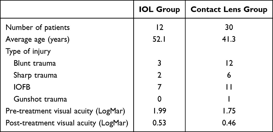 |
Table 1 Summary of Results |
Prior to treatment, the mean LogMAR uncorrected visual acuity was 1.99 for the IOL group and 1.75 for the CL group. Following treatment, the LogMAR best corrected visual acuity (BCVA) improved to 0.53 for the IOL group and 0.46 for the CL group (Figures 1 and 2). Notably, four patients (33%) in the IOL group and eleven patients (37%) in the CL group reported a visual acuity better than 20/40. However, the comparison of visual outcomes between the two cohorts did not yield a statistically significant difference (p = 0.5861). Within the IOL group, there were two instances of complications: one patient required a corneal graft due to exposure of the GORE-TEX suture, and one developed opacification of the Akreos lens. There were no complications observed in the CL group following intervention.
 |
Figure 1 Pre-treatment and Post-treatment Visual Acuity in IOL Cohort. Y-axis designates each patient within the cohort. |
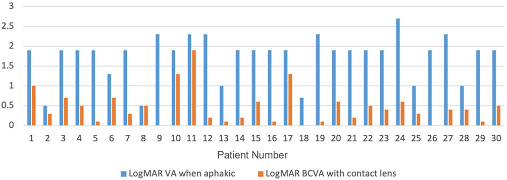 |
Figure 2 Pre-treatment and Post-treatment Visual Acuity in Contact Lens Cohort. Y-axis designates each patient within the cohort. |
Discussion
In comparing contact lenses with intraocular lenses for the correction of traumatic aphakia, it is essential to consider multiple factors. Intraocular lenses, while offering a permanent solution that eliminates the need for daily care, come with a noteworthy downside. The surgical procedure required for their implantation can be associated with a spectrum of complications, including corneal decompensation, retinal detachment, endophthalmitis, and potential decentralization of the intraocular lens.2,4,5 Contact lenses offer certain advantages including the ability to correct corneal astigmatism, which may not be optimally addressed with intraocular lenses, especially when irregular.6,7 In the present study, no significant difference was found in visual acuity outcomes between IOLs and CLs. Given this outcome, the complications of IOL implementation demand additional consideration when tailoring intervention for post-traumatic aphakia.
The long-term outcome of scleral fixated lenses (sutured Akreos lenses or 3-piece IOLs fixated using the Yamani technique) has not been established, with recent reports of lens tilt, as well as exposure of the lens haptic or the GORE-TEX suture.8,9 Although the long-term stability and positioning of scleral fixation have not been comprehensively assessed, some studies have sought to characterize its complications. A study conducted by Deb et al, involved 45 cases of traumatic aphakia treated with scleral fixation, in which a significant rate of complications was observed.2 These complications included hypotony, choroidal detachment, elevated intraocular pressure, corneal edema, vitreous hemorrhage, glaucoma, and cystoid macular edema. Another study by Mahapatra et al evaluated the outcomes of using an anterior chamber lens in 88 patients with traumatic and surgical aphakia.10 The study revealed several complications, including cystoid macular edema occurring in 8.8% of patients, elevated intraocular pressure in 4.4%, persistent uveitis in 2.2%, retinal detachment in 2.2% of cases, and intraocular lens tilt in 1.1% of cases. In our own investigation, we observed two instances of complications within the Akreos group. One patient experienced exposure of the GORE-TEX suture, necessitating a corneal graft. Another patient developed opacification of the Akreos lens. It is also important to note that these lenses are typically repurposed from designs intended for in-bag placement.
This study had some limitations. The study design allowed patients to choose their own treatment. This has the potential to allow a degree of self-selection bias. Health and physical limitations, occupation environment, and age were used by the patients to guide their decision. As a result, older patients with less dexterity tended to choose IOL placement compared to younger patients with more dexterity and a greater aversion to undergoing surgery. This may explain the difference in the average age between the two groups (52 years for the IOL group vs 41 years for the CL group). The complications observed in the IOL group were limited to lens opacification and the exposure of a GORE-TEX suture. It is important to recognize that different IOL placements may influence risk of different potential complications. Because the CL group only had 12 patients, larger studies are needed to further validate these findings and increase statistical power; however, the findings of this descriptive study remain relevant as they reflect real-world observations.
Conclusion
The patient cohort reported in this descriptive study was selected from individuals with traumatic aphakia associated with other ocular injuries, including corneal irregularities, and traumatic optic nerve or retinal damage, which can all impact visual recovery. Despite our small patient cohort, we demonstrate that outcomes with contact lenses remain on par with those achieved with alternative approaches, including scleral fixation or other modalities for secondary intraocular lens implantation. This finding underscores the viability of contact lens correction and may guide the discussion with patients when considering correction of traumatic aphakia.
Abbreviations
IOL, Intraocular Lens; CL, Contact Lens; BCVA, Best Corrected Visual Acuity; IOFB, intraocular foreign body.
Ethics Approval and Informed Consent
This study was approved by the Institutional Review Board at the University of Arkansas for Medical Sciences (approval number: 273739). The requirement for informed patient consent was waived by the IRB due to the retrospective nature of the study. All patient data were anonymized and handled in accordance with confidentiality protocols.
Disclosure
The authors report no other conflicts of interest in this work.
References
1. He CH, Poulsen DM, Parsikia A, Mbekeani JN. Characteristics of ocular trauma in the United States. Arquivos Brasileiros de Oftalmologia. 2022;85(3). doi:10.5935/0004-2749.20220035
2. Deb AK, Sarkar S, Natarajan K, et al. Complications and visual outcome of sutureless, scleral fixated intraocular lens in cases with traumatic aphakia. Therap Adv Ophthalmol. 2021;13:25158414211009095. doi:10.1177/25158414211009095
3. Swain T, McGwin G. The prevalence of eye injury in the United States, estimates from a meta-analysis. Ophthal Epidemiol. 2019;27(3):186–5. doi:10.1080/09286586.2019.1704794
4. Hoskin AK, Watson SL. Ocular trauma and prevention measures. Clin Exp Ophthalmol. 2020;48(7):875–876. doi:10.1111/ceo.13850
5. Mustafi D, Tom E, Messenger WB, Banitt MR, Rezaei KA. Outcomes of sutureless secondary intraocular lens fixation with haptic flanging in a cohort of surgically complex eyes. Graefes Arch Clin Exp Ophthalmol. 2021;259(5):1357–1363. doi:10.1007/s00417-020-04937-9
6. Severinsky B, Millodot M. Current applications and efficacy of scleral contact lenses—A retrospective study. J Optometry. 2010;3(3):158–163. doi:10.1016/S1888-4296(10)70022-4
7. Jacobs DS, Carrasquillo KG, Cottrell PD, et al. BCLA CLEAR – medical use of contact lenses. Contact Lens Anterior Eye. 2021;44(2):289–329. doi:10.1016/j.clae.2021.02.002
8. Tsai YY, Tseng SH. Transscleral fixation of foldable intraocular lens after pars plana lensectomy in eyes with a subluxated lens. J Cataract Refract Surg. 1999;25(5):722–724. doi:10.1016/S0886-3350(99)00019-X
9. Parikakis E, Batsos G, Kontomichos L, Peponis V, Christodoulou E, Karagiannis D. Traumatic aniridia and aphakia management with Iris reconstruction lens using gore-tex sutures, an ab-externo approach. Am J Case Rep. 2020;21. doi:10.12659/AJCR.924706
10. Mahapatra S, Mannem N. Anterior chamber intraocular lens—An effective alternative in traumatic and surgical aphakia in the era of scleral-fixated intraocular lens. Indian J Ophthalmol. 2021;69(6):1404. doi:10.4103/ijo.IJO_2192_20
 © 2026 The Author(s). This work is published and licensed by Dove Medical Press Limited. The
full terms of this license are available at https://www.dovepress.com/terms
and incorporate the Creative Commons Attribution
- Non Commercial (unported, 4.0) License.
By accessing the work you hereby accept the Terms. Non-commercial uses of the work are permitted
without any further permission from Dove Medical Press Limited, provided the work is properly
attributed. For permission for commercial use of this work, please see paragraphs 4.2 and 5 of our Terms.
© 2026 The Author(s). This work is published and licensed by Dove Medical Press Limited. The
full terms of this license are available at https://www.dovepress.com/terms
and incorporate the Creative Commons Attribution
- Non Commercial (unported, 4.0) License.
By accessing the work you hereby accept the Terms. Non-commercial uses of the work are permitted
without any further permission from Dove Medical Press Limited, provided the work is properly
attributed. For permission for commercial use of this work, please see paragraphs 4.2 and 5 of our Terms.