Back to Journals » Clinical, Cosmetic and Investigational Dermatology » Volume 15
VISIA Skin Analysis System as a Tool to Evaluate the Reduction of Pigmented Skin and Vascular Lesions Using the 532 Nm Laser
Authors Zawodny P, Stój E, Kulig P ![]() , Skonieczna-Żydecka K
, Skonieczna-Żydecka K ![]() , Sieńko J
, Sieńko J ![]()
Received 19 July 2022
Accepted for publication 22 September 2022
Published 14 October 2022 Volume 2022:15 Pages 2187—2195
DOI https://doi.org/10.2147/CCID.S380388
Checked for plagiarism Yes
Review by Single anonymous peer review
Peer reviewer comments 2
Editor who approved publication: Dr Jeffrey Weinberg
Piotr Zawodny,1 Elżbieta Stój,1 Piotr Kulig,2 Karolina Skonieczna-Żydecka,3 Jerzy Sieńko4
1Zawodny Esthetic Medicine Clinic, Szczecin, Poland; 2Department of General Pathology, Pomeranian Medical University in Szczecin, Szczecin, Poland; 3Department of Biochemical Science, Pomeranian Medical University in Szczecin, Szczecin, Poland; 4Department of General and Transplantation Surgery, Pomeranian Medical University in Szczecin, Szczecin, 70-111, Poland
Correspondence: Jerzy Sieńko, Department of General and Transplantation Surgery, Pomeranian Medical University, Szczecin, Poland, Tel +48 91 466-11-36, Fax +48 91 466 11 30, Email [email protected]
Purpose: Esthetic medicine is a rapidly developing field of medicine that is not only beneficial in terms of external appearance, but also significantly improves overall quality of life. Currently, pigmented and vascular skin lesions are more prevalent due to multiple environmental factors and are a characteristic manifestation of skin aging. The development of modern laser therapy has contributed to the successful management of multiple skin conditions. The aim of our study was to show the effect of concomitant reduction of both vascular and hyperpigmented skin lesions located on the facial area after repetitive 532 nm laser therapy and to emphasize that the detection of such observation was possible due to the implementation of System of Skin Analysis and Assessment.
Patients and Methods: We retrospectively analyzed 100 patients’ records with “VISIA” Skin Analysis System after 532nm laser therapy.
Results: Laser therapy significantly decreased VISIA scores for all tested lesions, ie, macules, pigmented and vascular lesions (p< 0.0001 for all). The efficacy of laser treatment was not significantly different regarding skin phototype (p> 0.05) and did not correlate with age of participants (p> 0.05). The more laser sessions were performed, the higher improvement in vascular lesion VISIA scores was observed (r=0.26, p=0.0097).
Conclusion: 532 nm laser therapy is effective regarding vascular and hyperpigmented skin lesions located on the facial area. The System of Skin Analysis and Assessment is a good tool to test the treatment efficacy during regular follow-up procedure.
Keywords: laser therapy, aesthetic medicine, cosmetic dermatology, VISIA, pigmented skin lesions, vascular skin lesions, skin aging
Foreword
Esthetic medicine is a flourishing branch of medicine. Every year the number of patients who undergo cosmetic procedures significantly increases. World Health Organization established the following definition of health: Health is a state of complete physical, mental and social well-being and not merely the absence of disease or infirmity.1 Esthetic medicine does not only contribute to the physical aspect of health, but also addresses mental and social aspects, since physical appearance affects multiple facets of human life. Vascular and pigmented skin lesions are a characteristic manifestation of ageing2 and due to implementation of laser therapy may no longer be the cause of complexes, depression and low self-esteem. Skin is one of the biggest organs in the human body and has many vital functions. It acts as a physical barrier between the external environment and an organism. As a result, skin protects against external physical, chemical and biological factors. Moreover, skin provides proper internal conditions and prevents the body from losing vital molecules.3 Pigmented and vascular skin lesions are predominantly esthetic defects, the prevalence of which increases with age. Among the causes of localized skin hyperpigmentation are physical and chemical factors, medications, endocrine abnormalities, inflammatory states, metabolic disturbances and vitamin insufficiency.4
Prevalence of dermatological diseases constantly increases due to harmful environmental factors, in particular UV radiation and pollution.5 Ongoing development of modern technology, with a special emphasis on those based on highly specialized laser therapy, has led to the development of highly effective therapeutic methods for a vast variety of skin disorders.6 Broad application of laser therapy in esthetic medicine and dermatology started off a new era of procedures and pushed the boundaries of the treatment for multiple skin lesions. This phenomenon is associated with laser qualities and features. In the first place one should mention the high efficacy combined with minimal interference with adjacent tissues and therefore limited number of complications. As a result, good esthetic results are achieved less invasively compared to the past.7 Rapid development of laser therapy and common awareness of its high efficacy has led, over a very short period of time, to a remarkable increase in interest in therapeutic options offered by esthetic medicine.
Skin dyspigmentation is one of the main complaints in dermatology and esthetic medicine. A great variety of lesions that manifest themselves in this manner exist. Moreover, their classification and further evaluation is often too abstract for patients who frequently do not understand their skin disorders. There are several tools to objectively assess skin dyspigmentation, yet are not very convenient in clinical setting and are predominantly used in research.8 In order to objectively assess skin lesions, VISIA Skin Analysis System (further in text: System of Skin Analysis and Assessment) was developed. A rotating capture module takes a series of images in vertical, horizontal and frontal views. Not only does it differentiate the number of skin lesions, but it also enables tracking and documenting of therapy efficacy over time.9 Moreover, the System of Skin Analysis and Assessment facilitates the knowledge of skin disorders among patients and therefore aids in their education.8
The aim of our study was to show the effect of concomitant reduction of both vascular and hyperpigmented skin lesions located on the facial area after repetitive 532 nm laser therapy, despite the fact that treatment protocol was dedicated to vascular skin lesions. Moreover, we wished to emphasize that the detection of such observation was possible due to the implementation of the System of Skin Analysis and Assessment in the regular follow-up procedure.
Materials and Methods
Study Group
The study was designed as a retrospective trial without a control group. Analyzed data were obtained from standard medical documentation recorded for every patient in our center. Records for a total of 100 patients with vascular lesions located in facial area were analyzed, among them 86 women (19–65 years old) and 14 men (30–67 years old). In every patient gender, age, skin phototype, number of procedures, left, right and frontal view were assessed. Moreover, in every patient the quantity of skin lesions defined as macules, pigmented lesions, and vascular lesions was determined before and after each laser procedure.
Methodology
In order to avoid any bias during the study the same conditions were provided for every patient. Before the examination skin was cleaned using the same make-up remover. The same light was provided in the consulting office regardless of time of day. In addition to that, clinical evaluation was performed by the same, experienced physician who also conducted laser therapy on all of the patients analyzed in the study.
The exact treatment protocol was tailored for each patient’s needs. In every case only one pass was performed. The number of shots was dependent on the size of the treated skin surface. In some cases treatment was performed on the whole face, whilst in other patients only on selected area eg, on cheek or nose. For the post-treatment period sun protection was always recommended, more specifically sun cream with UV filter.
The measurement of vascular and pigmented lesion reduction was performed using the System of Skin Analysis and Assessment which objectively and repetitively assesses skin before and after the procedure. The capture module of the System of Skin Analysis and Assessment provides proper light, adjusts patient’s position, and rotates around patient’s face capturing left, right, and frontal view. Vascular lesions are identified by the System of Skin Analysis and Assessment since hemoglobin emits red color which is detected by the system via RBX technology (Red/Brown Subsurface Analysis).8
532 nm Cutera Excel V laser was used for every patient. Laser with a wavelength of 532 nm has high affinity for oxyhemoglobin which facilitates targeting vascular lesions with a very small diameter. The pulse duration measured in milliseconds is optimal for achieving selective thermal destruction of blood vessels.
All procedures were performed at the esthetic medicine center in Szczecin, Poland. Effects of treatment over the period of time were photographically documented with 365 nm filter.
To test the efficacy, we compared the VISIA scores before and after the treatment for each tested lesion. Also, we calculated the VISIA change score (delta: VISIA score after – VISIA score before) for every parameter.
Statistical Analysis
In order to asses normality, Shapiro–Wilk test was utilized. As the majority of variable distribution was significantly different from normality (7 out of 9 tested parameters), Wilcoxon rank test was used to evaluate the efficacy of treatment at endpoint compared to baseline and Mann–Whitney test to evaluate whether the 532 laser efficacy differs regarding skin phototype. For correlation analysis, Spearman test was adopted. All calculations were performed in MedCalc software (version 20.110, Ostend, Belgium). The significance level was established at p < 0.05.
Results
In our retrospective study, 100 patients were enrolled. 86 participants were women (19–65 years old) and 14 men (30–67 years old). Overall, the mean age was 43.6 ± 11.51 years. We were able to detect three different Fitzpatrick skin types: I (76 patients), II (23 patients) and III (1 patient).
In order to achieve a satisfactory outcome, up to 8 laser procedures were performed for each study participant. The mean number of laser sessions was 1.76 ±1.30. No adverse effects were reported. After the intervention, a reduction of vascular skin lesions occurred (p = 0.0001). Although the 532 nm laser is used for treatment of vascular skin lesions, we also observed the parallel reduction in pigmented skin lesions as a result of the repetitive therapy with the 532 nm laser (p < 0.0001). We also observed a reduction in macules’ VISIA score (this refers to pigmented and vascular lesions combined, p = 0.0001). Results are depicted in Table 1 and Figures 1–3.
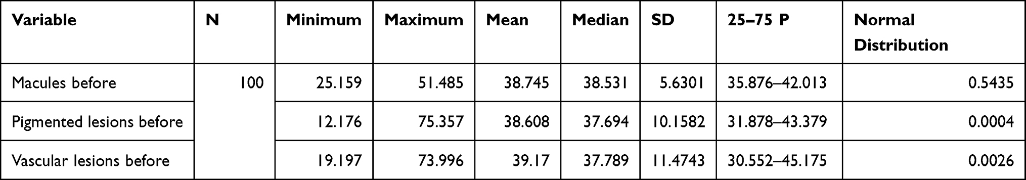 |
Table 1 VISIA Score for Different Type of Lesions at Baseline |
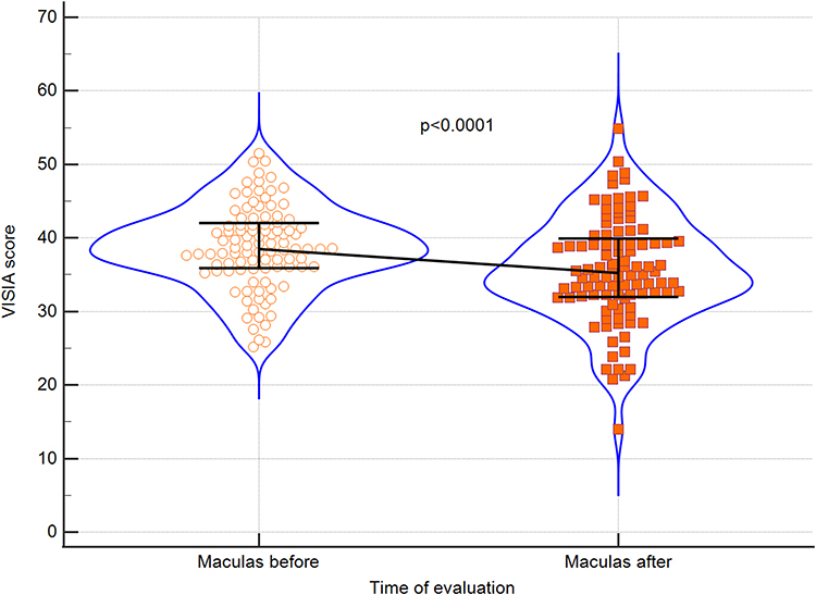 |
Figure 1 Violin plots for VISIA scores before and after treatment regarding macules. Medians and IQRs are shown. Circles and squares depict individual cases. |
 |
Figure 2 Violin plots for VISIA scores before and after treatment regarding pigmented lesions. Medians and IQRs are shown. Circles and squares depict individual cases. |
 |
Figure 3 Violin plots for VISIA scores before and after treatment regarding vascular lesions. Medians and IQRs are shown. Circles and squares depict individual cases. |
During the second step of our analysis, we tested whether laser efficacy is somehow dependent on skin phototype. There were no significant differences regarding this variable in laser efficacy, although highest reductions in VISIA scores were demonstrated for skin phototypes 1 and 2. Results are shown in Table 2.
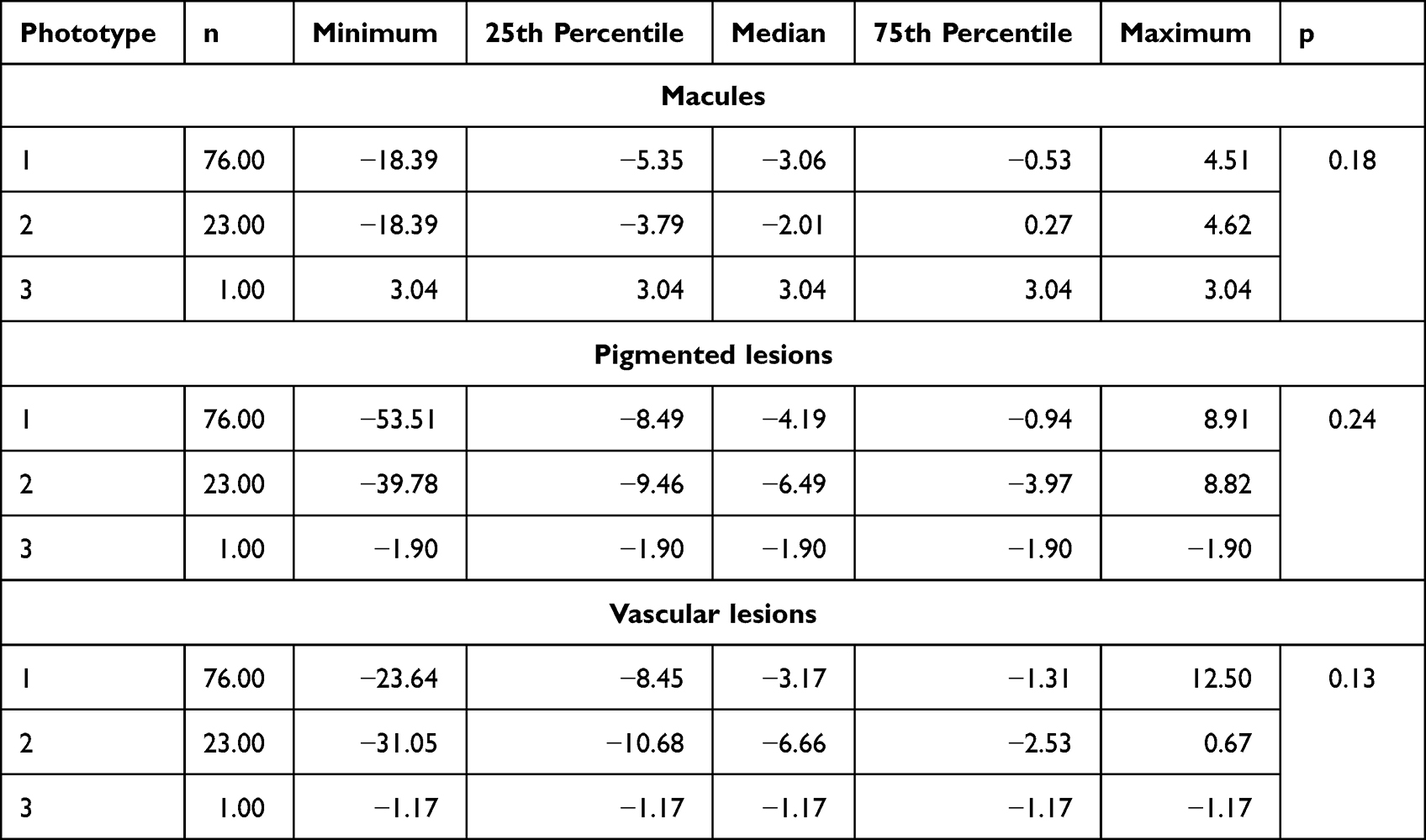 |
Table 2 Laser Efficacy Regarding Skin Phototype |
In the last part of our analysis, we correlated laser efficacy parameters (deltas) with age of participants and number of laser procedures. As expected, we found that the more laser sessions were performed, the higher the efficacy, but only with regard to vascular lesions. The results are presented in Table 3 and Figure 4.
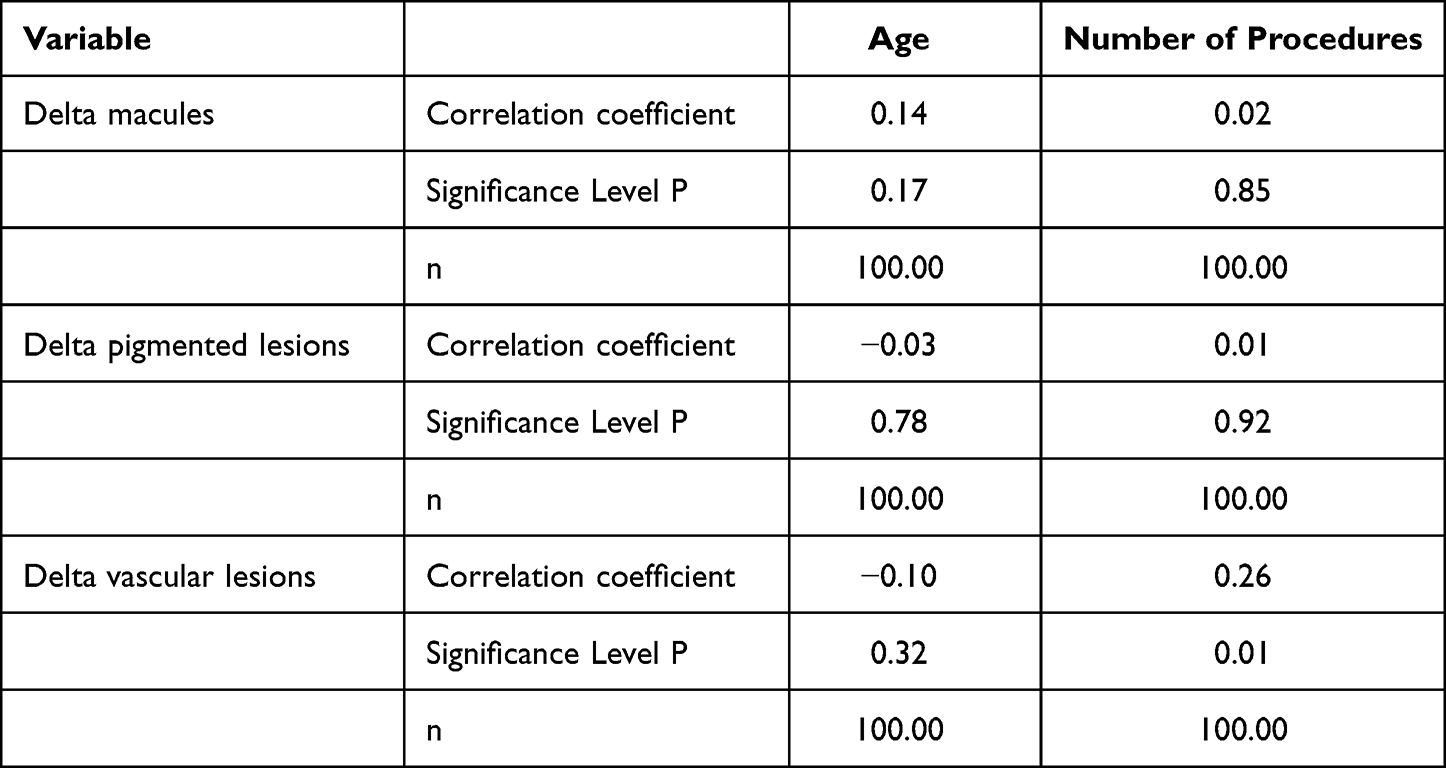 |
Table 3 Correlation Analysis of Laser Efficacy and Age of Study Participants and Number of Laser Sessions |
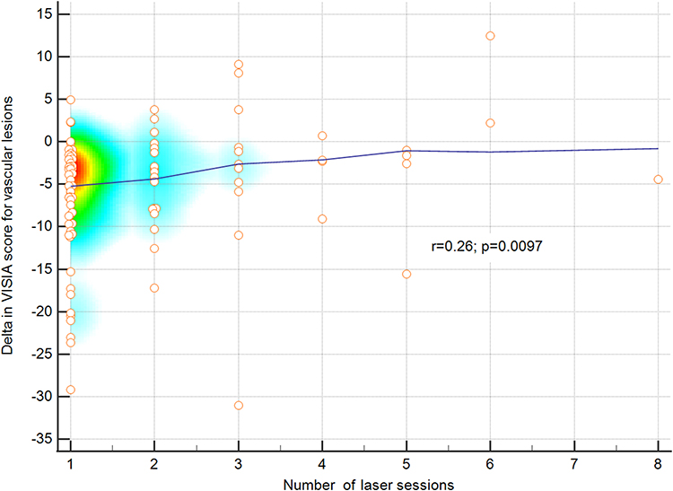 |
Figure 4 Correlation between number of laser session and VISIA score decrease for vascular lesions. Colors depict a heatmap. |
Discussion
Vascular lesions located in facial area are easy to spot and therefore make patients seek medical help. Every individual perceives aforementioned skin dyspigmentations differently and therefore they are very subjective. Therefore, not only is patients’ judgement prone to being inaccurate, even evaluation performed by a trained physician might be biased as multiple factors could affect a clinical assessment.10 Although the patients’ personal feelings about their own condition are of a pivotal importance, methods that objectively evaluate skin lesions should be implemented in everyday clinical practice.
There are a couple of methods to evaluate skin lesions, among them semi-quantitative evaluation performed by investigator with the advent of scales/questionnaires. This however lacks the comparability and repeatability due to its subjective nature. Objective and noninvasive methods used by academics and practitioners to investigate skin structure include Mexameter, reviscometer, laser Doppler flowmetry and others, but are strongly dependent on environmental factors, thus not optimal.11,12 Novel methods created by skin engineers using digital photographs have recently been acknowledged.13–16
Laser therapy should always be preceded by a thorough evaluation of the skin. In addition, during the post-treatment follow-up, skin condition ought to be analyzed as well. There are several scales and tools that could be applied in clinical practice in order to evaluate skin lesions. For instance, Bossart et al developed a plugin for Image J17 software that enables objective and fully automated hyperpigmentation assessment. The proposed tool is easy to implement in everyday clinical practice and therefore may facilitate assessment, follow‐up, and therapy. However, its application is limited exclusively to hyperpigmentation. Skin analysis tools can also be applied to scar assessment. Beausang et al designed a scale that enables clinical scar analysis. Scars were assessed clinically, photographically, as well as histologically. Results revealed that clinical evaluation was highly correlated with photographs and microscopic findings which in turn implies its utility. The proposed scale is easy to use, not time-consuming, and does not require specialized nor expensive equipment.18 On the other hand, since the evaluation relies solely upon the assessing physician, results could potentially be slightly biased.
The System of Skin Analysis and Assessment is easily adopted into clinical practice apparatus that precisely and objectively assesses multiple skin lesions. The device uses different light types to detect and evaluate different skin characteristics. In standard light, spots and rhytides are identified. Moreover, in the same light skin texture and pores are assessed. In ultraviolet light, the device evaluates spots and porphyrin in the image. This is relevant, since porphyrins in the skin are produced by bacteria, namely Propionibacterium acnes.19 Finally, in the cross polarized light, brown spots and red areas are detected. Vascular lesions are identified by the the VISIA system since hemoglobin emits red color which is detected by the System via RBX technology (Red/Brown Subsurface Analysis).8 The System of Skin Analysis and Assessment has been tested in several trials and is proven to be a reliable tool. Chen et al assessed twenty healthy women with Fitzpatrick skin type IV with melasma. Patients were also asked to self-evaluate the degree of improvement with the same scale rated as mild, moderate, good, and excellent. Before and after laser therapy, an objective assessment was conducted using the System of Skin Analysis and Assessment under the same conditions. Eight skin characteristics detectable by the system were evaluated on the facial region. Subjective and objective (using System of Skin Analysis and Assessment) assessments were conducted at baseline and 4 weeks after the third session of the treatment. Authors concluded that the objective assessment using the System of Skin Analysis and Assessment showed significant improvement in skin conditions.20 Kuo et al assessed, using the same device, a change in facial skin texture in patients with hyperthyroidism after thyroidectomy. They intended to evaluate and quantify the skin characters of patients with hyperthyroidism before and after thyroidectomy. Although their study revealed that surgical removal of the thyroid gland in patients with hyperthyroidism did not improve the skin quality and texture, the VISIA system proved to be a highly reliable tool in the objective evaluation of the skin condition.21 As compared to other imaging analysis software, for instance Image- Pro® Plus 7.0 (IPP®, Media Cybernetics Inc), VISIA correlated with the detection of skin spots, UV spots, wrinkles, red areas, and pores. Of note, VISIA detected spots with high sensibility on forehead area.13 Also, when compared to CSKIN®, facial redness was detected by VISIA with similar rates.10 VISIA® is RGB-based software using Windows operating platform, whilst CSKIN® is based on LAB color channel and utilizes iOS system. The latter one has the ability to detect acne, wrinkles, pores, red, spots, UV spots, and blackheads but not porphyrins and brown spots.10 Despite multiple advantages, System of Skin Analysis and Assessment has several limitations. It could only be applied to evaluate facial area and requires special equipment and software, which renders it less accessible than scales relying only upon clinical evaluation. Moreover, it is not suitable for assessing scars.
Laser therapy is a well-established therapeutic method, particularly in esthetic dermatology. Its application in esthetic medicine is broad and it is a viable therapeutic option for multiple conditions. Laser therapy proved to be a safe and effective treatment method for hair loss due to androgenic alopecia.22 In a study conducted by Kalashnikova et al an Er: YAG laser (2,940 nm) and a special treatment head SMA turned out to be highly effective for the treatment of skin manifestations of psoriasis vulgaris. Their results also demonstrated an improvement in the quality of life of patients after the course of treatment.23 Results of a double center retrospective study depicted that laser therapy was an effective method of treatment for skin hyperpigmentation and up to five laser sessions were necessary to achieve the clinical endpoint, ie, complete hyperpigmentation regression.24 Although in the aforementioned study, clinical results were similar to the literature data, authors applied a Visual Analog Scale (VAS) to the patients at the three-month follow-up to measure their contentment. They also reported as limitation of their study, that no digital skin analysis devices were used.24 In order to objectify the results we aimed to test 532nm laser efficacy for both vascular and pigmented lesions and to see whether this efficacy might be evaluated by the System of Skin Analysis and Assessment in an esthetic practice.
In our study we demonstrated that patients who undergo treatment dedicated to vascular lesions also benefit in terms of hyperpigmentation. We also observed that reduction of pigmented skin lesions occurs as an indirect result of laser therapy dedicated to vascular lesions. We found that skin phototype is not significantly linked to 532nm laser efficacy and that vascular lesions were reduced with increasing number of laser sessions. Furthermore, our results show that objective assessment of treatment progress before and after each procedure with the VISIA system has multiple benefits. The patients serve as partners in the therapeutic process and can discuss results with the consulting doctor. Besides, photographic documentation gives insight into the disease itself and patients become more aware of their condition. From a clinical standpoint, optimal number of laser procedures can be adjusted to every single patient and therefore the medical team can provide their patients with the best possible care. Therefore, we argue that the System of Skin Analysis and Assessment should be implemented in every esthetic center, since it enables tailoring therapy according to patients’ needs.
Conclusion
Repetitive 532 nm laser therapy effectively reduces pigmented skin and vascular lesions. The System of Skin Analysis and Assessment is a good tool to test the treatment efficacy during regular follow-up procedure.
Institutional Review Board Statement
The study was conducted in accordance with the Declaration of Helsinki and approved by the Institutional Review Ethics Committee of Regional Medical Chamber in Szczecin (OIL/Sz/KB/PH/425/2022).
Author Contributions
All authors contributed to data analysis, drafting or revising the article, have agreed on the journal to which the article will be submitted, gave final approval of the version to be published, and agree to be accountable for all aspects of the work.
Informed Consent Statement
Patient consent was waived due to the retrospective nature of the study. Confidentiality of patients was ensured.
Funding
This research received no external funding.
Disclosure
The authors declare no conflicts of interest.
References
1. World Health Organization. Constitution of the World Health Organization; 2006. Available from: www.who.int/governance/eb/who_constitution_en.pdf.
2. Khavkin J, Ellis DAF. Aging skin: histology, physiology, and pathology. Facial Plast Surg Clin North Am. 2011;19(2):229–234. doi:10.1016/j.fsc.2011.04.003
3. Dąbrowska AK, Spano F, Derler S, Adlhart C, Spencer ND, Rossi RM. The relationship between skin function, barrier properties, and body-dependent factors. Skin Res Technol. 2018;24(2):165–174. doi:10.1111/srt.12424
4. Cestari TF, Dantas LP, Boza JC. Acquired hyperpigmentations. An Bras Dermatol. 2014;89(1):11–25. doi:10.1590/abd1806-4841.20142353
5. Effects of air pollution on the skin: A review. Indian journal of dermatology, venereology and leprology; 2017. Available from: https://ijdvl.com/effects-of-air-pollution-on-The-skin-A-review/.
6. Omi T, Numano K. The Role of the CO2 Laser and Fractional CO2 Laser in Dermatology. Laser Ther. 2014;23(1):49–60. doi:10.5978/islsm.14-RE-01
7. Beasley KL, Weiss RA. Radiofrequency in cosmetic dermatology. Dermatol Clin. 2014;32(1):79–90. doi:10.1016/j.det.2013.09.010
8. Goldsberry A, Hanke CW, Hanke KE. VISIA system: a possible tool in the cosmetic practice. J Drugs Dermatol. 2014;13(11):1312–1314.
9. VISIA Skin Analysis. Canfield scientific. Available from: https://www.canfieldsci.com/imaging-systems/visia-complexion-analysis/.
10. Chen Y, Hua W, Li A, He H, Xie L, Li L. Analysis of facial redness by comparing VISIA® from Canfield and CSKIN® from Yanyun Technology. Skin Res Technol. 2020;26(5):696–701. doi:10.1111/srt.12856
11. Stefaniak AB, du Plessis J, John SM, et al. International guidelines for the in vivo assessment of skin properties in non-clinical settings: part 1. pH. Skin Res Technol. 2013;19(2):59–68. doi:10.1111/srt.12016
12. du Plessis J, Stefaniak A, Eloff F, et al. International guidelines for the in vivo assessment of skin properties in non-clinical settings: part 2. transepidermal water loss and skin hydration. Skin Res Technol. 2013;19(3):265–278. doi:10.1111/srt.12037
13. Wang X, Shu X, Li Z, et al. Comparison of two kinds of skin imaging analysis software: VISIA® from Canfield and IPP® from Media Cybernetics. Skin Res Technol. 2018;24(3):379–385. doi:10.1111/srt.12440
14. Zorin V, Zorina A, Cherkasov V, Deev R, Kopnin P, Isaev A. Clinical-instrumental and morphological evaluation of the effect of autologous dermal fibroblasts administration. J Tissue Eng Regen Med. 2017;11(3):778–786. doi:10.1002/term.1976
15. Takahashi Y, Fukushima Y, Kondo K, Ichihashi M. Facial skin photo-aging and development of hyperpigmented spots from children to middle-aged Japanese woman. Skin Res Technol. 2017;23(4):613–618. doi:10.1111/srt.12380
16. Xu DT, Yan JN, Cui Y, Liu W. Quantifying facial skin erythema more precisely by analyzing color channels of The VISIA Red images. J Cosmet Laser Ther. 2016;18(5):296–300. doi:10.3109/14764172.2016.1157360
17. Bossart S, Cazzaniga S, Willenberg T, et al. Skin hyperpigmentation index: a new practical method for unbiased automated quantification of skin hyperpigmentation. J Eur Acad Dermatol Venereol. 2020;34(7):e334–e336. doi:10.1111/jdv.16312
18. Beausang E, Floyd H, Dunn KW, Orton CI, Ferguson MW. A new quantitative scale for clinical scar assessment. Plast Reconstr Surg. 1998;102(6):1954–1961. doi:10.1097/00006534-199811000-00022
19. Johnson T, Kang D, Barnard E, Strain-Level LH. Differences in porphyrin production and regulation in propionibacterium acnes elucidate disease associations. mSphere. 2016;1(1):e00023–e00115. doi:10.1128/mSphere.00023-15
20. Chen YT, Lin ET, Chang CC, et al. Efficacy and safety evaluation of picosecond alexandrite laser with a diffractive lens array for treatment of melasma in Asian patients by VISIA imaging system. Photobiomodul Photomed Laser Surg. 2019;37(9):559–566. doi:10.1089/photob.2019.4644
21. Kuo SCH, Huang F, Chi SY, Lin HP, Chien PC, Hsieh CH. Investigate the improvement of facial skin texture with the VISIA system after total thyroidectomy. BMC Surg. 2021;21(1):94. doi:10.1186/s12893-021-01108-3
22. Leavitt M, Charles G, Heyman E, Michaels D. HairMax LaserComb laser phototherapy device in the treatment of male androgenetic alopecia: a randomized, double-blind, sham device-controlled, multicentre trial. Clin Drug Investig. 2009;29(5):283–292. doi:10.2165/00044011-200929050-00001
23. Kalashnikova NG, Lotti T, Urakova DS, Jafferany M. Treatment of psoriatic skin lesions with a new Er: yaglaser technology: a case series study. Dermatol Ther. 2020;33(2):e13264. doi:10.1111/dth.13264
24. Nisticò SP, Cannarozzo G, Provenzano E, et al. Nanosecond Q-switched 1064/532 nm laser to treat hyperpigmentations: a double center retrospective study. Clin Pract. 2021;11(4):708–714. doi:10.3390/clinpract11040086
 © 2022 The Author(s). This work is published and licensed by Dove Medical Press Limited. The
full terms of this license are available at https://www.dovepress.com/terms
and incorporate the Creative Commons Attribution
- Non Commercial (unported, 3.0) License.
By accessing the work you hereby accept the Terms. Non-commercial uses of the work are permitted
without any further permission from Dove Medical Press Limited, provided the work is properly
attributed. For permission for commercial use of this work, please see paragraphs 4.2 and 5 of our Terms.
© 2022 The Author(s). This work is published and licensed by Dove Medical Press Limited. The
full terms of this license are available at https://www.dovepress.com/terms
and incorporate the Creative Commons Attribution
- Non Commercial (unported, 3.0) License.
By accessing the work you hereby accept the Terms. Non-commercial uses of the work are permitted
without any further permission from Dove Medical Press Limited, provided the work is properly
attributed. For permission for commercial use of this work, please see paragraphs 4.2 and 5 of our Terms.
Recommended articles
Cellular Senescence and Anti-Aging Strategies in Aesthetic Medicine: A Bibliometric Analysis and Brief Review
Zheng H, Wu J, Feng J, Cheng H
Clinical, Cosmetic and Investigational Dermatology 2024, 17:2243-2259
Published Date: 9 October 2024
Aesthetic Medicine and Perceptual Distortion: The Role of Professional Aesthetic Drift
Armenti AF
Clinical, Cosmetic and Investigational Dermatology 2026, 19:583396
Published Date: 19 January 2026