Back to Journals » Clinical Ophthalmology » Volume 16
Utilization of Modified Induced Pluripotent Stem Cells as the Advance Therapy of Glaucoma: A Systematic Review
Authors Rizkiawan DE, Evelyn M, Tjandra KC, Setiawan B
Received 24 April 2022
Accepted for publication 22 August 2022
Published 29 August 2022 Volume 2022:16 Pages 2851—2859
DOI https://doi.org/10.2147/OPTH.S372114
Checked for plagiarism Yes
Review by Single anonymous peer review
Peer reviewer comments 3
Editor who approved publication: Dr Scott Fraser
Diondra Eka Rizkiawan,1 Malinda Evelyn,1 Kevin Christian Tjandra,1 Budi Setiawan2
1Faculty of Medicine, Diponegoro University, Semarang, Central Java, Indonesia; 2Department of Internal Medicine, Diponegoro University, Semarang, Central Java, Indonesia
Correspondence: Budi Setiawan, Department of Internal Medicine, Diponegoro University, Jl. Badak Raya 74, Kota Semarang, Central Java, Indonesia, 50167, Tel +6285865118118, Fax +622467412115, Email [email protected]
Abstract: Glaucoma is an optic neuropathy disease that causes cupping of the optic disc and decreased visual field. Glaucoma is still the second leading cause of blindness globally, with a worldwide prevalence of more than 76 million people in 2020. However, no therapy can cure glaucoma completely, especially when optic nerve damage has occurred. Available treatments only play a role in keeping the intraocular pressure stable This research aims to determine the potential use of modified stem cell therapy to treat intraocular damage in glaucoma cases. Literature research was conducted by involving seven online databases, namely Pubmed, ScienceDirect®, Proquest, EBSCOhost®, SAGE®, Clinicalkey®, and Scopus, published between 2010– 2020 with the keywords stem cells; therapy; glaucoma; optic nerve. Six articles were selected, and out of the six articles, all writings were experimental research. The entire literature states that modified stem cell therapy has the potential as a therapeutic option in treating intraocular damage in patients with glaucoma. Based on the systematic literature review that has been carried out, it is known that stem cell therapy has the potential to be a therapeutic option in treating glaucoma cases. Much more research is needed to assess the effectiveness of modified stem cell therapy in managing intraocular damage due to glaucoma.
Keywords: neuropathy, optic nerve, treatment, intraocular damage
Introduction
Glaucoma is one of the optic neuropathy disorders characterized by the progressive degeneration of retinal ganglion cells (RGC), which eventually lead to cupping of the optic disc and decreased visual field.1 Glaucoma is also closely related to an increase in intraocular pressure caused by the damage of trabecular meshwork (TBM), which results in optic nerve damage, characterized by the loss of retinal ganglion cells.2,3 Globally, in 2020, more than 76 million people are suffering from glaucoma, and it is expected to increase to 111.8 million people by 2040.4,5 Glaucoma is also a severe and complex medical problem because it often causes blindness. According to the World Health Organization (WHO), the most common causes of blindness are cataracts (51%), followed by glaucoma (8%), and age-related macular degeneration (5%).6 This data shows that glaucoma is the world’s second most common cause of blindness after cataracts. Symptoms that are often asymptomatic at an early stage and the low public awareness have contributed to the disorder’s seriousness.
Handling and treating glaucoma cases is difficult, especially because no therapy can cure glaucoma. Current treatment, both medical and surgical, is focused solely on lowering intraocular pressure. Treatment of glaucoma cases should also be carried out for life to maintain normal intraocular pressure and prevent the progression of intraocular damage due to glaucoma.7 Based on these problems, innovation is needed to handle glaucoma effectively. Besides, solutions are also required to repair the damage to retinal ganglion cells in glaucoma. One of the therapies that researchers are trying to take advantage of is stem cell therapy, a technology where cells can develop into many specific cells desired.8 In cases of glaucoma or optic neuropathies, damaged RGCs can be replaced with new ones grown from stem cells.9 Another option for RGC regeneration is to use retinal stem cells to regenerate RGCs. Indeed, stem cell therapy relies on exogenous stem cell sources due to their limited availability. Currently, many stem cell therapies for eye diseases that are created and studied are limited to treating the damage of photoreceptors and retinal pigment epithelium. iPSC-derived RGCs can serve as an excellent model for formulating approaches to promote de novo-generated RGCs to connect with their targets. Therefore, researchers have been looking into the potential use of modified stem cell therapy to treat the intraocular injury in glaucoma cases.10
This review aims to synthesize and prove the efficacy and further modification of this method so that it can be eligible for treatment and can also give data collection for the scientific community. This systematic review is expected to provide detailed information regarding the possible applications of modified stem cell therapy in treating intraocular damage in glaucoma patients.
Materials and Methods
In the present literature review, literature regarding the potential utilization of stem cells as an advanced therapy for intraocular glaucomatous damage was searched. The stages of this literature review include five steps: i) identifying the research question, ii) identifying relevant studies, iii) study selection, iv) charting the data, and v) summarizing and reporting the results.
Identifying the Research Question
This literature review was conducted to answer the following research questions:
- Does induced pluripotent stem cell therapy have the potential to treat intraocular glaucomatous damage?
- What modifications are needed in stem cell therapy for intraocular glaucomatous damage?
Identifying Relevant Studies
The literature search was carried out from January to February 2021. Keywords and synonyms used to conduct literature searches related to the research question are attached in Table 1. Boolean operators (OR, AND, NOT) combine keywords when searching for literature. The search was conducted on seven online databases, namely PubMed, ScienceDirect®, ProQuest, EBSCOhost®, SAGE®, Clinicalkey®, and Scopus.
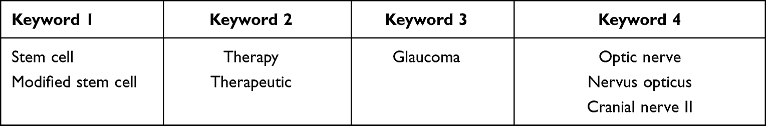 |
Table 1 Keywords That Were Used in the Database Search |
Study Selection
The inclusion criteria for the literature search consisted of journals published in English and journals published in the last ten years. The exclusion criteria for selected studies consisted of journals that were not fully accessible due to the limited facilities owned as supporting access. We thoroughly screened the titles and abstracts of the studies obtained to suit the purpose of this literature review. Abstracts that were not relevant to the research objectives were excluded. Then a full article screening was carried out from the selected abstracts to identify whether the full article was suitable for the research objectives and whether the full article could be used to answer research questions.
Charting the Data
Information obtained from all selected study articles is then displayed in the charting table The information displayed includes the author, year of publication, study objectives, location, study design, inclusion and exclusion criteria, results, and conclusions.
Summarizing and Reporting the Results
The researcher did not assess the quality of the selected articles because this study was only a literature review. The data from selected studies are reported to produce recommendations for further research regarding the use of stem cell therapy in glaucoma cases.
Results
Based on the literature search that has been conducted, a total of 2262 studies and abstracts were included in the journal screening process at an early stage. From this screening process, 362 duplicate articles were excluded from the selection. The remaining 1900 articles then entered the abstract eligibility screening stage. Only 53 articles were selected, while 1879 other articles were excluded. Of the 53 articles, 18 articles appeared relevant to the study and met the inclusion criteria for review throughout the study. Meanwhile, 35 other studies were excluded because the focus in these studies did not match the objectives of this literature review. After assessing the full articles, six studies met the inclusion criteria in this literature review (Figure 1).
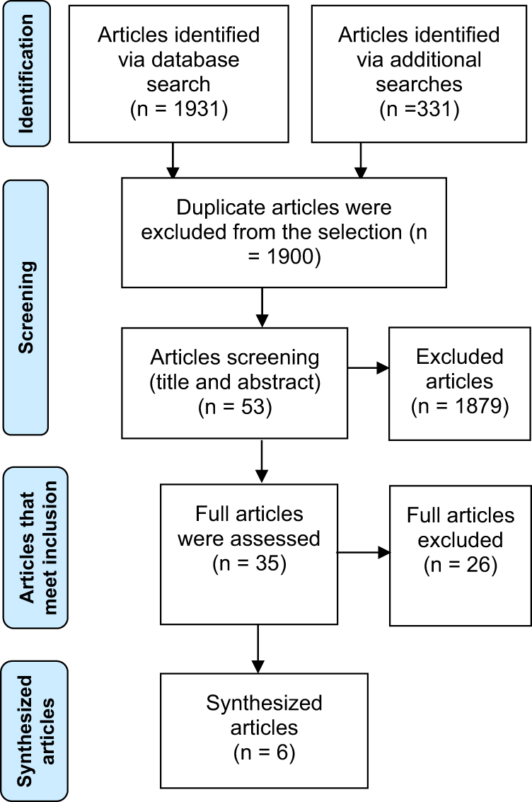 |
Figure 1 Flow diagram of the literature review process. |
In Table 2, a summary of the characteristics of the selected studies is presented. The data used from selected studies include research objectivity, study design, results, outputs, and conclusions from the study. Of all the selected studies, there were six studies that had experimental methods. Almost all studies have the aim of evaluating and proving the potential of using stem cells to replace damaged tissue and restore and restore the function of damaged eye tissue, particularly due to degenerative processes such as disease of the retina or glaucoma.
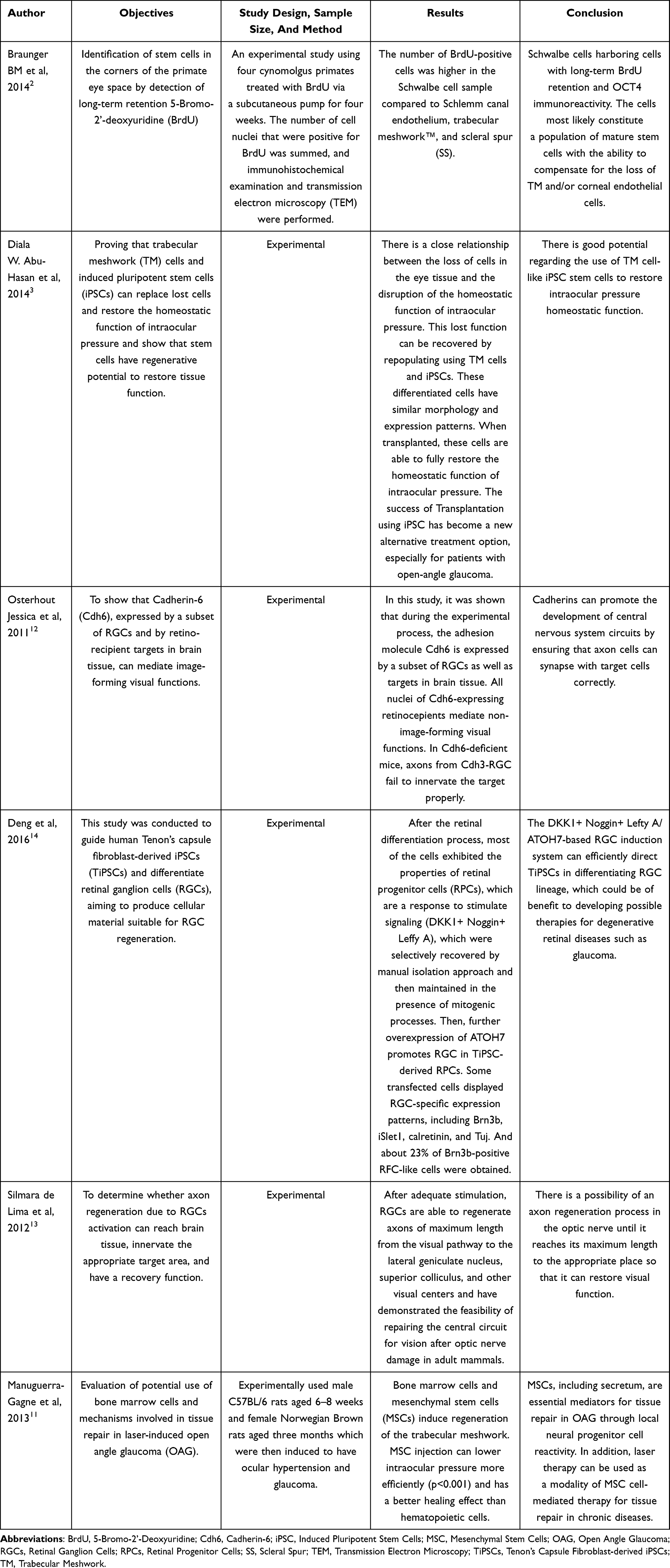 |
Table 2 Results Summary of the Characteristics of the Selected Studies |
Discussion
Pathophysiology of Glaucoma
Glaucoma is characterized by the degeneration of retinal ganglion cells. Based on the pathophysiology, glaucoma can be divided into two categories, namely open-angle glaucoma and closed-angle glaucoma. In patients with open-angle glaucoma, there is increased resistance to the aqueous humor’s outflow through the trabecular meshwork. This increased resistance is often caused by apoptosis and senescence of trabecular meshwork cells with increasing age.15 Degradation and abnormalities of the cytoskeleton arrangement of trabecular meshwork cells resulting in thickening of the drainage pathways and abnormal extracellular matrix deposition also worsen trabecular meshwork function in open-angle glaucoma.16 In closed-angle glaucoma, the aqueous humor cannot reach the trabecular meshwork due to obstruction.17 Examples of obstructions that often cause closed-angle glaucoma are anterior synechiae, the attachment of the iris to the trabecular meshwork, and posterior synechiae, where the iris is attached to the lens. This adhesion causes the aqueous humor to fail to reach the drainage system and the trabecular meshwork.18
Glaucoma is closely related to increased intraocular pressure, which is determined by the balance between the production of aqueous humor by the ciliary body and the drainage of the aqueous humor through the trabecular meshwork. The disturbance of the balance between production and drainage increases the humor Aquos, which at a later stage can increase the intraocular pressure.19 Studies have shown a link between increased intraocular pressure and retinal ganglion cell death. This study has also proven that the longer the intraocular pressure increases, the higher the degree of retinal ganglion cell damage.20 However, data show as many as 30–40% of patients with glaucoma have normal intraocular pressure. One of the causes of glaucoma at normal intraocular pressure is a decrease in neurotrophic factors needed in the maintenance of neurons in the optic nerve. Neurotrophic factors are required to maintain retinal ganglion cells, including brain-derived neurotrophic factor (BDNF), ciliary neurotrophic factor (CNTF), and cell line-derived neurotrophic factor.21 Furthermore, microcirculation disorders, changes in immune system conditions, and increased levels of oxidative stress can also cause glaucoma at normal intraocular pressure.21
Stem Cells
Stem cells are cells with the ability to differentiate and form all tissues in the human body. They are one of the potential therapies used in cases that require tissue repair and regeneration, one of which is glaucoma. For a cell to be called a stem cell, it must have two essential characteristics. The first one is the stem cell must produce offspring with the exact features the cell originates from, and the second one, the stem cell must be able to differentiate into the specific cell desired.22 There are two types of stem cells found in multicellular organisms, including humans. The first stem cells are embryonic stem cells or multipotent cells found in blastocysts, while the second stem cells are adult stem cells or pluripotent cells that can be found in a wide variety of adult tissues.23
Research has also succeeded in inducing adult cells to return to the pluripotent stage using molecular manipulation. The cells produced by this molecular manipulation are then called induced pluripotent stem cells (iPS).24 Most iPS manufacturing uses viruses such as retroviruses and lentiviruses to carry genes encoding transcription factors to adult cells to be modified. This gene will then undergo transcription and translation into a protein capable of inducing the adult cell nucleus to return to an embryonic state.25
An important concept that needs attention in stem cell therapy is how to induce stem cells to become the desired differentiated cells.26 It is necessary so that the cells can be used to treat various diseases, including glaucoma. We can further achieve differentiation of stem cells into specific desired cells by adding various growth factors and signaling pathways to resemble the conditions of their original development.27
Potential Uses of Stem Cell Therapy in Damaged Trabecular Meshwork
The research conducted successfully isolates cultures and confirms that the trabecular meshwork stem cells around the Schwalbe line are multipotent with the ability to differentiate into a wide variety of cells, including trabecular meshwork cells adipocytes osteocytes, and chondrocytes.28 Other studies have also been able to induce stem cells on the Schwalbe line trabecular meshwork to proliferate and differentiate into photoreceptors under certain conditions.29 Apart from trabecular meshwork stem cells, other stem cells that can differentiate into functional meshwork trabecular cells are adipose-derived stem cells (ADSC), mesenchymal stem cells (MSC), and iPS. iPS cells can also differentiate into trabecular meshwork cells after culturing the extracellular matrix with cell-derived trabecular meshwork. The success of a wide variety of stem cells to differentiate into functional meshwork trabecular cells provides a more effective alternative to cutting-edge therapy in treating glaucoma, especially open-angle glaucoma.3
Utilization of Stem Cells for Regeneration of Retinal Ganglion Cells
One of the stem cell therapies successfully applied and able to regenerate damaged retinal ganglion cells is iPS cell therapy. This therapy uses induced adult fibroblasts to return to pluripotent cells using four transcription factors, namely Oct3/4, Sox2, Klf4, and c-Myc. The results of the iPS are pluripotent cell colonies that are morphologically similar to ESCs, which are able to differentiate into the three germ cell layers.30
Because iPS can be programmed from the patient’s somatic cells, this therapy can maintain the unique genome of each individual. Currently, various modifications to the iPS therapy have been made to increase its acceptability and effectiveness of iPS therapy. One of them is the use of plasmid vectors and miRNA instead of retroviruses to avoid mutagenesis of the adult cells used.31,32
One of the significant challenges in stem cell therapy is to achieve the differentiation of stem cells into the desired cells, in this case, the differentiation of stem cells to retinal ganglion cells. Usually, in vivo, the differentiation of stem cells into retinal ganglion cells is regulated by several transcription factors such as Ath5, Brn3, and Notch. The transcription factors Ath5 and Brn3 play a vital role in the differentiation of retinal ganglion cells, and their levels are increased in the process of eye development.33 Meanwhile, Notch is a negative regulator of retinal ganglion cell differentiation, and its levels are decreased in normal eye development. Therefore, the addition of the transcription factors Ath5 and Brn3 and the Notch antagonist is a strategy to differentiate retinal ganglion cells from stem cells.34 Apart from transcription factors, various neurotrophic pathways and factors have been identified in the differentiation of stem cells into retinal ganglion cells. These pathways consist of fibroblast growth factor (FGF), insulin-like growth factor (IGF), bone morphogenetic protein (BMP), nodal, and Wnt signaling pathways. All of these pathways regulate retinal development, whereas FGF and IGF provide positive regulation. Meanwhile, BMP, nodal, and Wnt signaling pathways provide negative regulation.35
Modification of Stem Cell Therapy for Retinal Ganglion Cell Repair
Another major challenge in the clinical application of stem cell therapy in glaucoma sufferers is that not only do the stem cells successfully differentiate into retinal ganglion cells, but they must also be able to reach the central nervous system.36 Modifications must be made so that new retinal ganglion cells can reach the visual cortex of the cerebrum. Recent research has found that a combination of genetic modification and stimulation of the signaling pathway stimulates regeneration of the optic nerve until it reaches the central nervous system. The addition of ephrin molecules, proteoglycans, cell-adhesion molecules, and semaphorin is able to guide the axons of the developing retinal ganglion cells to reach the optic chiasm.13 Meanwhile, the addition of cadherin, ephrin, and the Wnt signaling pathway can guide and stimulate synapse formation in the superior colliculus and the visual cortex.12,37
In addition, because of the adverse intraocular environment in glaucoma, stem cell therapy needs to be combined with neuroprotective compounds. It is also associated with a decrease in neurotrophic factors required to maintain neurons and causes progression of retinal ganglion cell damage in glaucoma sufferers. Therefore, the addition of BDNF and other neurotrophic factors such as glial cell-derived neurotrophic factor (GDNF) and ciliary neurotrophic factor (CNTF) should be considered for combined stem cell therapy.38
Conclusion
The stem cells are used in cases of glaucoma, which require repair and regeneration of trabecular meshwork cells and retinal ganglion cells. iPS has been shown the ability to differentiate to replace damaged trabecular meshwork cells and retinal ganglion cells in glaucoma. Some modifications are required so that stem cells that have differentiated into trabecular meshwork cells and retinal ganglion cells can reach the central nervous system. These modifications include the addition of ephrin molecules, proteoglycans, cell-adhesion molecules, semaphorin, cadherin, and the Wnt signaling pathway. The combination of stem cells with neuroprotective factors such as BDNF, GDNF, and CNTF also needs to be considered to maintain neuronal maintenance and inhibit the progression of cell damage.
The development of new stem cell technologies not only paves the way for us to gain a better understanding of the biology associated with glaucoma and create models for the development of new drugs, but it also opens the door to the prospect of cell-based therapies that can help patients regain their vision. More specifically in relation to the field of glaucoma, there have been recent developments in the process of developing protocols for the differentiation of stem cells into trabecular meshwork and retinal ganglion cells. Further research on the effectiveness of using modified stem cells as a therapy for glaucoma and in vivo research can be carried out immediately so that clinical trials can be carried out, which in turn can be used by the community to control symptoms and reduce blindness due to glaucoma.
Disclosure
The authors report no conflicts of interest in this work.
References
1. Choudhari NS, Neog A, Fudnawala V, George R. Cupped disc with normal intraocular pressure: the long road to avoid misdiagnosis. Indian J Ophthalmol. 2011;59(6):491. doi:10.4103/0301-4738.86320
2. Braunger BM, Ademoglu B, Koschade SE, et al. Identification of adult stem cells in Schwalbe’s line region of the primate eye. Invest Ophthalmol Vis Sci. 2014;55(11):7499. doi:10.1167/iovs.14-14872
3. Abu-Hassan DW, Li X, Ryan EI, Acott TS, Kelley MJ. Induced pluripotent stem cells restore function in a human cell loss model of open-angle glaucoma. Stem Cells. 2015;33(3):751–761. doi:10.1002/stem.1885
4. Cook C, Foster P. Epidemiology of glaucoma: what’s new? Can J Ophthalmol. 2012;47(3):223–226. doi:10.1016/j.jcjo.2012.02.003
5. Hapsari DM. The Relationship Between Family Support and Adherence in Care in Glaucoma Clients in the Working Area of the Balung Health Center. Jember Regency. Uni; 2017.
6. World Health Organization. Global data on visual impairments 2010; 2012. https://www.iapb.org/wp-content/uploads/GLOBALDATAFINALforweb.pdf.
7. Harasymowycz P, Birt C, Gooi P, et al. Medical management of glaucoma in the 21st century from a canadian perspective. J Ophthalmol. 2016;2016:1–22. doi:10.1155/2016/6509809
8. Romito A, Cobellis G. Pluripotent stem cells: current understanding and future directions. Stem Cells Int. 2016;2016:1–20. doi:10.1155/2016/9451492
9. Cen L-P, Ng TK. Stem cell therapy for retinal ganglion cell degeneration. Neural Regen Res. 2018;13(8):1352. doi:10.4103/1673-5374.235237
10. Öner A. Stem cell treatment in retinal diseases: recent developments. Turk J Ophthalmol. 2018;48(1). doi:10.4274/tjo.89972
11. Manuguerra-Gagné R, Boulos PR, Ammar A, et al. Transplantation of mesenchymal stem cells promotes tissue regeneration in a glaucoma model through laser-induced paracrine factor secretion and progenitor cell recruitment. Stem Cells. 2013;31(6):1136–1148. doi:10.1002/stem.1364
12. Osterhout JA, Josten N, Yamada J, et al. Cadherin-6 mediates axon-target matching in a non-image-forming visual circuit. Neuron. 2011;71(4):632–639. doi:10.1016/j.neuron.2011.07.006
13. De lima S, Koriyama Y, Kurimoto T, et al. Full-length axon regeneration in the adult mouse optic nerve and partial recovery of simple visual behaviors. Proc Natl Acad Sci U S A. 2012;109(23):9149–9154. doi:10.1073/pnas.1119449109
14. Deng F, Chen M, Liu Y, et al. Stage-specific differentiation of iPSCs toward retinal ganglion cell lineage. Mol Vis. 2016;22:536.
15. Pulliero A, Seydel A, Camoirano A, Saccà SC, Sandri M, Izzotti A. Oxidative damage and autophagy in the human trabecular meshwork as related with ageing. PLoS One. 2014;9(6):e98106. doi:10.1371/journal.pone.0098106
16. Porter KM, Epstein DL, Liton PB. Up-regulated expression of extracellular matrix remodeling genes in phagocytically challenged trabecular meshwork cells. PLoS One. 2012;7(4):e34792. doi:10.1371/journal.pone.0034792
17. Lai J, Choy BNK, Shum JWH. Management of primary angle-closure glaucoma. Asia Pac J Ophthalmol. 2016;5(1):59–62. doi:10.1097/APO.0000000000000180
18. Sun X, Dai Y, Chen Y, et al. Primary angle closure glaucoma: what we know and what we don’t know. Prog Retin Eye Res. 2017;57:26–45. doi:10.1016/j.preteyeres.2016.12.003
19. Gemenetzi M, Yang Y, Lotery AJ. Current concepts on primary open-angle glaucoma genetics: a contribution to disease pathophysiology and future treatment. Eye. 2012;26(3):355–369. doi:10.1038/eye.2011.309
20. Khan Kareem A, Tse DY, van der Heijden ME, et al. Prolonged elevation of intraocular pressure results in retinal ganglion cell loss and abnormal retinal function in mice. Exp Eye Res. 2015;130. doi:10.1016/j.exer.2014.11.007
21. Almasieh M, Wilson AM, Morquette B, Cueva Vargas JL, Di Polo A. The molecular basis of retinal ganglion cell death in glaucoma. Prog Retin Eye Res. 2012;31(2):152–181. doi:10.1016/j.preteyeres.2011.11.002
22. Cao J, Ng ES, Mcnaughton D, et al. The characterisation of pluripotent and multipotent stem cells using Fourier transform infrared microspectroscopy. Int J Mol Sci. 2013;14(9):17453–17476. doi:10.3390/ijms140917453
23. Makhani K, Ali SM, Yousuf S, Siddiqui S. Therapeutic potential of totipotent, pluripotent and multipotent stem cells. MOJ Cell Sci Rep. 2015;2(5):00041. doi:10.15406/mojcsr.2015.02.00041
24. Zaveri L, Dhawan J. Cycling to meet fate: connecting pluripotency to the cell cycle. Front Cell Dev Biol. 2018;6. doi:10.3389/fcell.2018.00057
25. Wattanapanitch M. Recent updates on induced pluripotent stem cells in hematological disorders. Stem Cells Int. 2019;2019:1–15. doi:10.1155/2019/5171032
26. Kolios G, Moodley Y. Introduction to stem cells and regenerative medicine. Respiration. 2012;85(1):3–10. doi:10.1159/000345615
27. Biehl JK, Russell B. Introduction to stem cell therapy. J Cardiovasc Nurs. 2009;24(2):
28. Tay CY, Sathiyanathan P, Chu SWL, Stanton LW, Wong TT. Identification and characterization of mesenchymal stem cells derived from the trabecular meshwork of the human eye. Stem Cells Dev. 2012;21(9):1381–1390. doi:10.1089/scd.2011.0655
29. Nadri S, Yazdani S, Arefian E, et al. Mesenchymal stem cells from trabecular meshwork become photoreceptor-like cells on amniotic membrane. Neurosci Lett. 2013;541:43–48. doi:10.1016/j.neulet.2012.12.055
30. Omole AE, Fakoya AOJ. Ten years of progress and promise of induced pluripotent stem cells: historical origins, characteristics, mechanisms, limitations, and potential applications. PeerJ. 2018;2018. doi:10.7717/peerj.4370
31. Miyoshi N, Ishii H, Nagano H, et al. Reprogramming of mouse and human cells to pluripotency using mature microRNAs. Cell Stem Cell. 2011;8(6):633–638. doi:10.1016/j.stem.2011.05.001
32. Ikushima S, Ono R, Fukuda K, Sakayori M, Awano N, Kondo K. Trousseau’s syndrome: cancer-associated thrombosis. Jpn J Clin Oncol. 2016;46(3):204–208. doi:10.1093/jjco/hyv165
33. Ji SL, Tang SB. Differentiation of retinal ganglion cells from induced pluripotent stem cells: a review. Int J Ophthalmol. 2019;12(1):152–160. doi:10.18240/ijo.2019.01.22
34. Li L, Chen LP, Liu QH. Effect of the notch signaling pathway on retinal ganglion cells and its neuroprotection in rats with acute ocular hypertension. Int J Ophthalmol. 2018;11(2). doi:10.18240/ijo.2018.02.05
35. Ritchey ER, Zelinka CP, Tang J, Liu J, Fischer AJ. The combination of IGF1 and FGF2 and the induction of excessive ocular growth and extreme myopia. Exp Eye Res. 2012;99(1):1–16. doi:10.1016/j.exer.2012.03.019
36. Georgiev D. Photons do collapse in the retina not in the brain cortex: evidence from visual illusions. NeuroQuantology. 2011;9(2). doi:10.14704/nq.2011.9.2.403
37. He CW, Liao CP, Pan CL. Wnt signalling in the development of axon, dendrites and synapses. Open Biol. 2018;8(10):180116. doi:10.1098/rsob.180116
38. Mysona BA, Zhao J, Bollinger KE. Role of BDNF/TrkB pathway in the visual system: therapeutic implications for glaucoma. Expert Rev Ophthalmol. 2017;12(1):69–81. doi:10.1080/17469899.2017.1259566
 © 2022 The Author(s). This work is published and licensed by Dove Medical Press Limited. The
full terms of this license are available at https://www.dovepress.com/terms
and incorporate the Creative Commons Attribution
- Non Commercial (unported, 3.0) License.
By accessing the work you hereby accept the Terms. Non-commercial uses of the work are permitted
without any further permission from Dove Medical Press Limited, provided the work is properly
attributed. For permission for commercial use of this work, please see paragraphs 4.2 and 5 of our Terms.
© 2022 The Author(s). This work is published and licensed by Dove Medical Press Limited. The
full terms of this license are available at https://www.dovepress.com/terms
and incorporate the Creative Commons Attribution
- Non Commercial (unported, 3.0) License.
By accessing the work you hereby accept the Terms. Non-commercial uses of the work are permitted
without any further permission from Dove Medical Press Limited, provided the work is properly
attributed. For permission for commercial use of this work, please see paragraphs 4.2 and 5 of our Terms.