Back to Journals » Medical Devices: Evidence and Research » Volume 12
Utility Of An Automatic Limulus Amebocyte Lysate Kinetic Turbidimetric Test For Endotoxin Screening Of Dialysate Samples
Authors Uchida T, Kaku Y, Hayasaka H, Kofuji M, Momose N, Miyazawa H, Ueda Y, Ito K, Ookawara S , Morishita Y
Received 29 July 2019
Accepted for publication 17 September 2019
Published 7 October 2019 Volume 2019:12 Pages 429—433
DOI https://doi.org/10.2147/MDER.S225246
Checked for plagiarism Yes
Review by Single anonymous peer review
Peer reviewer comments 2
Editor who approved publication: Dr Scott Fraser
Takayuki Uchida,1,* Yoshio Kaku,2,* Hideyuki Hayasaka,1 Masaya Kofuji,1 Naoki Momose,1 Haruhisa Miyazawa,2 Yuichiro Ueda,2 Kiyonori Ito,2 Susumu Ookawara,2 Yoshiyuki Morishita2
1Department of Clinical Engineering, Saitama Medical Center, Jichi Medical University, Saitama-City, Saitama, Japan; 2Division of Nephrology, First Department of Integrated Medicine, Saitama Medical Center, Jichi Medical University, Saitama-City, Saitama, Japan
*These authors contributed equally to this work
Correspondence: Yoshiyuki Morishita
Division of Nephrology, First Department of Integrated Medicine, Saitama Medical Center, Jichi Medical University, 1-847 Amanuma-cho, Omiya-ku, Saitama-City, Saitama 330-8503, Japan
Tel +81-48-647-2111
Fax +81-48-647-6831
Email [email protected]
Background: Endotoxin contamination of dialysate has serious adverse effects on patients undergoing hemodialysis. Therefore, endotoxin activity in dialysate is closely monitored. Limulus amebocyte lysate (LAL) has been used as a reagent to measure endotoxin activity. Here, we investigated the efficacy of an automatic LAL kinetic turbidimetric test (Toxinometer ET-mini) for screening endotoxin activity in dialysate.
Methods: In total, endotoxin activity was measured in 110 dialysate samples obtained from several sites within hemodialysis circuits between June 2012 and March 2018. The results were compared with those from a conventional chromogenic substrate LAL test conducted by a clinical examination laboratory.
Results: Both the automatic LAL test and the chromogenic substrate LAL test had a minimum detection level of 0.001 endotoxin units (EU)/mL. Endotoxin activity levels measured via the automatic LAL test showed a strongly positive correlation (concordance correlation coefficient: 0.9933; 95% CI: 0.9902–0.9954) and good agreement (mean difference: 0.00±0.01 EU/mL) with those obtained using the chromogenic substrate LAL test.
Conclusion: The results suggest that the automatic LAL test may be useful for endotoxin activity screening in hemodialysis facilities.
Keywords: endotoxin, dialysate, hemodialysis, limulus amebocyte lysate, chromogenic substrate
Introduction
During each hemodialysis session, a patients’ blood comes into contact with 100–150 L of dialysate via a semipermeable membrane called a dialyzer. Therefore, the purity of the dialysate is crucially important for safe and better quality hemodialysis.1 As such, guidelines for hemodialysis recommend regular monitoring of dialysate purity to prevent serious biological contamination, including viable bacteria and endotoxins, or chemical contamination, including aluminum, chloride, copper, fluorine compounds, and lead.2–4 Endotoxin activity in dialysate can be measured using limulus amebocyte lysate (LAL), which is purified from the blood of amebocytes of horseshoe crabs, as a substrate.5 Endotoxin activates several components of LAL, including factor C, factor B, and pro-clotting enzyme, resulting in coagulation and gelation of LAL.5 Taking advantage of this reaction, various endotoxin activity assays using LAL as a substrate have been developed. These include measurement of LAL gel formation over time, a turbidity change-based measurement of gelation called a LAL kinetic turbidimetric test, and a chromogenic substrate LAL test in which yellow chromogen released following synthetic substrate cleavage is measured. While these assays are considered well-established methods for measurement of endotoxin activity,6 they are complex and difficult to use for screening in a hemodialysis facility. A device called a Toxinometer™ (FUJIFILM Wako Pure Chemical Corporation, Tokyo, Japan), which can measure endotoxin activity using an automatic LAL kinetic turbidimetric test, has also been developed.7 Completing measurements within 2 hrs, a Toxinometer can easily be used to measure endotoxin activity in a clinical setting.7 A previous study confirmed that an automatic LAL test using a Toxinometer was useful for measuring endotoxin activity in milk.8 However, the ability of the system to accurately evaluate endotoxin activity in dialysate has not been reported. Therefore, we investigated the utility of the automatic LAL test (Toxinometer ET-mini™) for screening endotoxin activity in dialysate samples.
Materials And Methods
Ethical Approval
This study was conducted in accordance with the Declaration of Helsinki. Because the study analyzed unlinked anonymized data from non-human samples, approval from the Ethics Committee of Saitama Medical Center, Jichi Medical University, was not required.
Study Design
This study was a single-center, prospective, cross-sectional study conducted between June 2012 and March 2018 at Saitama Medical Center, Jichi Medical University, Saitama, Japan. A total of 110 dialysate samples were obtained from several sites within hemodialysis circuits, including multiple dialysate supply tubes from 20 dialysis consoles in a central dialysate delivery system and from the outlet of the reverse osmotic water tank of the central dialysate delivery system. Endotoxin activity was immediately measured via automatic LAL test using a Toxinometer ET-mini (Figure 1A and B). All samples were also examined via chromogenic substrate LAL test, which was conducted by a clinical examination laboratory (SRL, Tokyo, Japan). Subsequently, the two values for each sample were statistically compared. If endotoxin contamination was observed, the hemodialysis circuit was washed out and sterilized with hypochlorous acid- and acetic acid-based cleaning solutions.
Principle And Procedure For Endotoxin Activity Measurement Using An Automatic LAL Test (Toxinometer ET-mini)
The Toxinometer ET-mini is compact, measuring 277 mm×176 mm×85 mm with a total weight of 2.4 kg (Figure 1A). The system can simultaneously measure four samples (Figure 1B). Dialysate (300 μL) samples were added to tubes containing LAL reagent (Limulus ES-II; FUJIFILM Wako Pure Chemical Corporation), which was obtained from blood of horseshoe crabs. Unlike standard LAL, Limulus ES-II reacts with endotoxin but does not react with β-1,3-D glucan. Carboxymethylated curdlan (carboxymethylated β-1,3-D glucan) inhibits the reaction between β-1,3-D glucan and LAL and is added to the Limulus ES-II reagent.7 Excess amounts of carboxymethylated curdlan inhibit the reaction between LAL lysate and β-1,3-D glucan because their reaction only occurs within a narrow range of β-1,3-D glucan.7 In comparison, the reaction between LAL lysate and endotoxin occurs over a wide range of concentrations and is not subject to any interference from excess concentrations of β-1,3-D glucan.7 Tubes were then placed into the Toxinometer ET-mini for measurement of endotoxin activity. The system measures endotoxin activity using a kinetic turbidimetric technique (Figure 2A and B). Briefly, endotoxin in a sample reacts with the LAL, triggering a gelation reaction. As a result of gelation, the turbidity of the reaction mixture increases, reducing the amount of light that passes through the mixture and thus decreasing light transmittance (Figure 2A). The transmitted light ratio was measured at 12-s intervals. Endotoxin activity was determined from the amount of time required for the transmitted light ratio to decrease below a certain threshold (92% of the initial value), which is defined as the gelation time (Figure 2A). Higher endotoxin activity leads to a shorter gelation time. The log value of endotoxin activity was calculated from the log–log plot of gelation time (Figure 2B).
Chromogenic Substrate LAL Test
Measurement of endotoxin activity by chromogenic substrate LAL test was conducted by a commercial clinical examination laboratory (SRL, Tokyo, Japan). Details of the procedure and measurement principles are described elsewhere.6 Briefly, the intensity of yellow chromogen released following synthetic substrate cleavage was measured and quantified.6
Statistics
Data are expressed as mean±SD. Correlations between the automatic LAL test results and those obtained via the chromogenic substrate LAL test were evaluated using Lin’s concordance correlation coefficient.9 Agreement between endotoxin activity measured by automatic LAL test and chromogenic substrate LAL test was assessed using the method of Bland and Altman. P<0.05 was considered statistically significant.
Results
Comparison Of Sensitivity Of The Automatic LAL Test And The Chromogenic Substrate LAL Test
Both the automatic LAL test and the chromogenic substrate LAL test had a minimum detection level of 0.001 endotoxin units (EU)/mL.
Comparison Of Enzyme Activity Results Obtained From The Two Testing Methods
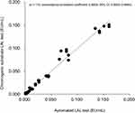
Endotoxin activity levels measured using the automatic LAL test showed a statistically significant and strongly positive correlation with those obtained using the chromogenic substrate LAL test (concordance correlation coefficient: 0.9933; 95% CI: 0.9902–0.9954) (Figure 3).
 |
Figure 3 Correlation between endotoxin activity levels measured using the automatic LAL test and the chromogenic substrate LAL test. Abbreviation: LAL, limulus amebocyte lysate. |
Analysis Of Systematic Errors Between The Automatic LAL Test And The Chromogenic Substrate LAL Test
The Bland–Altman plots for the comparative analysis showed good agreement between the two methods (n=110; mean difference=0.00±0.01 EU/mL) (Figure 4).
 |
Figure 4 Analysis of systematic errors between the automatic LAL test and the chromogenic substrate LAL test. Abbreviation: LAL, limulus amebocyte lysate. |
Discussion
In this study, endotoxin activity levels in dialysate detected using an automatic LAL test were tightly correlated with those determined using a chromogenic substrate LAL test. The sensitivity of the automatic LAL test was comparable with that of the chromogenic substrate LAL test. Additionally, the automatic LAL system was easy to operate, and results were obtained within 2 hrs. Based on these findings, we propose that the automatic LAL test could be useful for evaluating endotoxin activity in dialysate at hemodialysis facilities.
Endotoxin contamination of dialysate fluid is particularly dangerous to patient health because it can pass through the dialysis membrane, thereby entering the patient’s bloodstream.10 Endotoxins have reportedly caused serious adverse effects in patients undergoing hemodialysis, including increases in inflammatory cytokines (interleukin-6, tumor necrosis factor).11,12 Thus, monitoring endotoxin activity in dialysate is very important for maintaining the safety and quality of hemodialysis.
In this study, endotoxin activity levels measured via the automatic LAL test showed a tight correlation and good agreement with those obtained using the chromogenic substrate LAL test. Both methods had a limit of detection of 0.001 EU/mL. The Japanese Society of Dialysis and Transplantation recommends that endotoxin activity levels should be less than 0.050 EU/mL for standard dialysate and less than 0.001 EU/mL for ultra-pure dialysis fluid, which is used for online hemodiafiltration.3 Therefore, our results suggest that the sensitivity of the automatic LAL test is sufficient for monitoring endotoxin activity in dialysate. In addition, the automatic LAL test produced a result within 2 hrs, and the system was very easy to operate. This short analysis time is advantageous because it allows prompt and appropriate treatment of dialysate if contamination is detected. Additionally, the cost of measurement of one sample by automatic LAL test (USD$11) was twofold lower than that of the chromogenic substrate LAL test (USD$25). Together, these results suggest that the Toxinometer ET-mini automatic LAL kinetic test is appropriate for endotoxin screening of dialysate and may have some advantages over other methods for use in a clinical setting.
However, we note that this study was limited by the use of samples from a single center, which might influence the study results. Therefore, further large-scale, multicenter and multiethnic studies are required to confirm our findings.
Conclusion
In conclusion, endotoxin activity levels in dialysate measured using an automatic LAL test were tightly correlated with results obtained via a chromogenic substrate LAL test. The sensitivity of the automatic LAL kinetic test was comparable to the chromogenic substrate LAL test. Further, the automatic LAL test was easy to use, and results were obtained quickly. Our findings suggest that the automatic LAL kinetic test is likely to be useful for screening of endotoxin contamination of dialysate at hemodialysis facilities.
Abbreviations
LAL, limulus amebocyte lysate; EU, endotoxin units.
Acknowledgments
The authors thank all patients and clinicians who participated in the study. We also thank Tamsin Sheen, PhD, from Edanz Group for editing a draft of this manuscript.
Funding
Authors disclose no external funding sources.
Disclosure
The authors declare that they have no conflicts of interest in this work.
References
1. Fleming GM. Renal replacement therapy review: past, present and future. Organogenesis. 2011;7(1):2–12. doi:10.4161/org.7.1.13997
2. Section IV. Dialysis fluid purity. Nephrol Dial Transplant. 2002;17(Suppl 7):45–62.
3. Kawanishi H, Masakane I, Tomo T. The new standard of fluids for hemodialysis in Japan. Blood Purif. 2009;27(Suppl 1):5–10. doi:10.1159/000213490
4. Coulliette AD, Arduino MJ. Hemodialysis and water quality. Semin Dial. 2013;26(4):427–438. doi:10.1111/sdi.12113
5. Elin RJ, Hosseini J. Clinical utility of the Limulus amebocyte lysate (LAL) test. Prog Clin Biol Res. 1985;189:307–327.
6. Svensson A, Hahn-Hagerdal B. Comparison of a gelation and a chromogenic Limulus (LAL) assay for the detection of gram-negative bacteria, and the application of the latter assay to milk. J Dairy Res. 1987;54(2):267–273.
7. Oishi H, Fusamoto M, Hatayama Y, Tsuchiya M, Takaoka A, Sakata Y. An automated analysis system of Limulus amebocyte lysate (LAL)-endotoxin reaction kinetics using turbidimetric kinetic assay. Chem Pharm Bull (Tokyo). 1988;36(8):3012–3019. doi:10.1248/cpb.36.3012
8. Mottar J, De Block J, Merchiers M, Vantomme K, Moermans R. Routine limulus amoebocyte lysate (LAL) test for endotoxin determination in milk using a Toxinometer ET-201. J Dairy Res. 1993;60(2):223–228.
9. Lin LI. A concordance correlation coefficient to evaluate reproducibility. Biometrics. 1989;45(1):255–268.
10. Henrie M, Ford C, Andersen M, et al. In vitro assessment of dialysis membrane as an endotoxin transfer barrier: geometry, morphology, and permeability. Artif Organs. 2008;32(9):701–710. doi:10.1111/j.1525-1594.2008.00592.x
11. Bossola M, Sanguinetti M, Scribano D, et al. Circulating bacterial-derived DNA fragments and markers of inflammation in chronic hemodialysis patients. Clin J Am Soc Nephrol. 2009;4(2):379–385. doi:10.2215/CJN.03490708
12. Schindler R. Clinical effect of purification of dialysis fluids, evidence and experience. Blood Purif. 2009;27(Suppl 1):20–22. doi:10.1159/000213493
 © 2019 The Author(s). This work is published and licensed by Dove Medical Press Limited. The full terms of this license are available at https://www.dovepress.com/terms.php and incorporate the Creative Commons Attribution - Non Commercial (unported, v3.0) License.
By accessing the work you hereby accept the Terms. Non-commercial uses of the work are permitted without any further permission from Dove Medical Press Limited, provided the work is properly attributed. For permission for commercial use of this work, please see paragraphs 4.2 and 5 of our Terms.


