
Back to Journals » International Journal of Women's Health » Volume 17

Urogynecological Outcomes After Minimally Invasive Surgery for Deep Endometriosis: A Systematic Review
Authors Mascellino G  , Laganà AS , Anatrà M, Serra P, Mesiano G, Terrinoni M , Mascellino V, Ferrero S , Serati M, Leone M, Barra F
Received 30 August 2025
Accepted for publication 13 November 2025
Published 17 November 2025 Volume 2025:17 Pages 4577—4586
DOI https://doi.org/10.2147/IJWH.S564184
Checked for plagiarism Yes
Review by Single anonymous peer review
Peer reviewer comments 3
Editor who approved publication: Dr Everett Magann
Giuseppe Mascellino,1 Antonio Simone Laganà,1 Marco Anatrà,2 Pietro Serra,1 Giada Mesiano,3 Matteo Terrinoni,4,5 Vincenzo Mascellino,6 Simone Ferrero,2 Maurizio Serati,3 Maurizio Leone,2 Fabio Barra2,7
1Unit of Obstetrics and Gynecology, “Paolo Giaccone” Hospital - Department of Health Promotion, Mother and Child Care, Internal Medicine and Medical Specialties (PROMISE), University of Palermo, Palermo, Italy; 2Unit of Obstetrics and Gynecology, IRCCS Ospedale Policlinico San Martino, Genoa, Italy; 3Department of Obstetrics and Gynecology, Del Ponte Hospital, University of Insubria, Varese, Italy; 4Department of Medicine and Surgery, University of Perugia, Perugia, Italy; 5Department of Obstetrics and Gynecology, “Alto Tevere” Hospital - Città di Castello, Perugia, Italy; 6Department of Obstetrics and Gynecology, “Fogliani” Hospital - Milazzo, Messina, Italy; 7Department of Health Sciences (DISSAL), University of Genoa, Genoa, Italy
Correspondence: Fabio Barra, Unit of Obstetrics and Gynecology, IRCCS Ospedale Policlinico San Martino, Largo R. Benzi 10, Genoa, 16132, Italy, Tel +01139 334 9437959, Email [email protected]
Abstract: Deep endometriosis (DE) frequently affects pelvic organs and may impair urinary function through both direct involvement and surgical nerve disruption. Minimally invasive excision is the standard treatment for symptomatic DE, but its impact on lower urinary tract symptoms (LUTS) remains uncertain. We conducted a systematic review according to PRISMA 2020 guidelines to evaluate postoperative urogynecological outcomes following laparoscopic or robotic surgery for DE (PROSPERO ID: CRD420251113885). PubMed, Scopus, and the Cochrane Library were searched for studies published over the last 25 years. Eligible studies included women undergoing minimally invasive surgery for DE with postoperative urinary outcomes assessed by validated questionnaires or urodynamic testing. Methodological quality was appraised using the Newcastle–Ottawa Scale. Out of 175 records, 9 studies involving 20– 289 participants met the inclusion criteria. Six studies reported significant postoperative improvements, particularly in urgency, frequency, and dysuria, with benefits most evident in women presenting with moderate-to-severe preoperative LUTS. Improvements were observed in both subjective assessments, by using standardized questionnaires such as the International Consultation on Incontinence Questionnaire (ICIQ), Urinary Symptom Profile (USP), International Prostate Symptom Score (IPSS), and Visual Analogue Scale (VAS), and, in some cases, objective measures such as bladder capacity and uroflowmetry. Conversely, three studies documented no significant change or worsening of urinary function, predominantly in women with minimal baseline dysfunction or extensive parametrial and sacral nerve involvement. In these series, postoperative deterioration included increased voiding difficulties and worsening symptom scores. Across studies, outcome definitions, assessment tools, surgical techniques, and follow-up intervals varied substantially, introducing significant heterogeneity that prevented a formal meta-analysis from being performed. Overall, minimally invasive surgery for DE may yield meaningful symptomatic relief in selected patients, particularly when nerve-sparing techniques are applied. However, functional deterioration remains a relevant risk, especially among those with minimal baseline LUTS. Future research should prioritize prospective trials with standardized outcome measures, longer follow-up, and stratification by preoperative symptom burden to optimize patient selection and refine surgical approaches.
Keywords: deep endometriosis, urinary symptoms, nerve-sparing surgery, minimally invasive surgery, urinary function
Introduction
Endometriosis is a chronic, estrogen-dependent inflammatory disease affecting approximately 6–10% of women of reproductive age and up to 50% of those with infertility or chronic pelvic pain.1 One of its most severe manifestations is deep endometriosis (DE), defined as lesions penetrating more than 5 mm beneath the peritoneal surface.2 The pathophysiology of DE involves complex interactions among ectopic endometrial implants, neuroangiogenesis, inflammation, and fibrosis, ultimately leading not only to pelvic pain and infertility but also to functional impairment of affected pelvic organs.3
DE accounts for 15–30% of all endometriosis cases and commonly involves the uterosacral ligaments, rectovaginal septum, bladder, bowel, and ureters.4 Urinary tract involvement in DE has been reported in 0.3–12% of cases, most frequently affecting the bladder (up to 85%) and, less commonly, the ureters.5 Patients with bladder endometriosis often experience lower urinary tract symptoms (LUTS) such as urgency, frequency, dysuria, suprapubic pain, and occasionally hematuria—symptoms that frequently correlate with the menstrual cycle.5 Ureteral endometriosis, though often asymptomatic, poses a serious risk of silent ureteral obstruction, hydronephrosis, and subsequent renal function loss.6
Surgical excision remains the cornerstone of treatment for symptomatic DE, particularly when the urinary tract is involved—most notably the ureters, where obstruction and hydronephrosis may lead to silent and progressive renal impairment.7 Minimally invasive approaches such as laparoscopic excision have become standard, with nerve-sparing techniques increasingly employed to minimize postoperative complications.3,7 In cases of bladder endometriosis, partial cystectomy or bladder shaving may be performed, with symptomatic improvement reported in over 90% of patients.8 However, such procedures are not without risk: detrusor hypoactivity, urinary retention, and voiding dysfunction occur in up to 18% of cases, particularly in the absence of nerve-sparing strategies.9 In addition, surgical management of ureteral endometriosis may require ureterolysis or segmental ureteral resection with ureteroneocystostomy when obstruction is present.10
Despite technical advances, the impact of DE surgery on urinary function remains controversial. Several prospective studies have reported improvements in LUTS and urodynamic parameters following nerve-sparing laparoscopic resection with significant reductions in urgency and dysuria.9 Conversely, other studies have shown unchanged or even worsened urinary function postoperatively, particularly among patients with mild preoperative dysfunction.11
In general, these inconsistencies may be attributed to multiple factors, including heterogeneity in study design, small sample sizes, variability in surgical techniques (extent of resection, nerve-sparing versus non-nerve-sparing), and differences in outcome measures. While some studies include urodynamic testing, others rely exclusively on symptom-based questionnaires, which vary in both scope and sensitivity.12,13 Moreover, the lack of standardization in the timing of postoperative assessments—ranging from a few weeks to over a year—further complicates interpretation. Moreover, it is difficult to distinguish whether postoperative symptoms reflect the impact of the disease itself or a surgery-related functional impairment. Furthermore, only few studies have adopted objective evaluation methods such as urodynamics.
To date, few systematic reviews have specifically addressed urogynecological outcomes following DE surgery. Most reviews have focused more broadly on pain, fertility, or gastrointestinal function.6,12,13 Given the significant burden urinary symptoms place on quality of life and the risk of iatrogenic dysfunction, a targeted synthesis of the evidence is both timely and necessary.
This review therefore aims to systematically evaluate the impact of minimally invasive surgical treatment for DE on urinary symptoms, comparing pre- and postoperative outcomes using validated assessment tools. By examining studies with rigorous methodology and standardized outcome measures, we aim to clarify whether surgery provides functional benefits or poses risks to urinary health in women with DE.
Search Methods
This systematic review was conducted in accordance with the Preferred Reporting Items for Systematic Reviews and Meta-Analyses (PRISMA) 2020 statement.14 No institutional review board approval was required as only previously published, de-identified data were analyzed. All authors contributed to the development of the search strategy and to the definition of inclusion and exclusion criteria. The protocol was registered on PROSPERO (ID: CRD420251113885).
Literature Search
A systematic computerized literature search was carried out in three electronic databases—PubMed, Scopus, and the Cochrane Library—from database inception to June 2025. The search strategy included a combination of MeSH terms and free-text keywords as follows: (“endometriosis” OR “pelvic endometriosis”) AND (“surgery” OR “surgical treatment” OR “laparoscopic surgery”) AND (“urogynecological disorders” OR “urinary incontinence” OR “bladder dysfunction” OR “urinary symptoms”) AND (“postoperative” OR “after surgery”). Reference lists of all included studies and relevant reviews were also manually screened to identify additional potentially eligible articles.
All retrieved articles were reviewed by the authors. The reviewers were not blinded to the names of the authors or the sources of publication. Discrepancies were resolved by discussion and consensus. Eligibility was first assessed based on titles and abstracts; full-text versions of potentially relevant studies were then obtained and examined to determine final inclusion.
Inclusion and Exclusion Criteria
This review included peer-reviewed journal articles published in English, Italian, or French within the last 25 years. Eligible studies focused on women diagnosed with DE who underwent laparoscopic or robotic surgical excision. To be included, studies had to evaluate postoperative urinary or urogynecological function. Accepted study designs included randomized controlled trials (RCTs), prospective and retrospective cohort studies, and case-control studies involving at least five patients.
Studies were excluded if they were animal or in vitro experiments, focused on non-endometriosis gynecologic conditions, or employed open surgical approaches. Articles were also excluded if they did not report postoperative urinary outcomes or did not use validated assessment tools.
Study Eligibility and Quality Assessment
All studies retrieved from the search were independently screened by two reviewers. Any disagreement regarding inclusion was resolved through discussion or with the involvement of a third reviewer. Given the wide heterogeneity in study populations, surgical techniques, outcome measures, and follow-up duration, a quantitative synthesis (meta-analysis) was not feasible. Instead, a descriptive synthesis was performed. The quality of observational studies was assessed using the Newcastle–Ottawa Scale (NOS), evaluating selection, comparability, and outcome domains. Assessments were conducted independently by two reviewers. Details of bias assessment are available in Table 1.
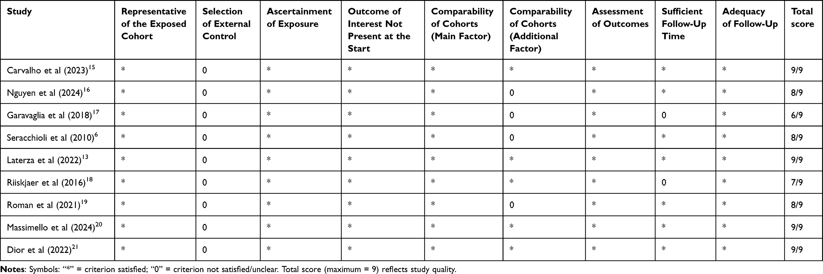 |
Table 1 Assessment of Included Studies Using the Newcastle–Ottawa Scale (NOS) |
Outcomes
Due to the diversity in methodologies and outcome reporting, the results were synthesized narratively. Trends in urinary function—whether improvement, worsening, or no change—were extracted and summarized based on validated symptom questionnaires, such as International Consultation on Incontinence Questionnaire (ICIQ), Urinary Symptom Profile (USP), International Prostate Symptom Score (IPSS) and, where available, objective urodynamic evaluations.
Results
Study Selection
A total of 175 studies were identified through database searches (PubMed = 50, Scopus = 116, Cochrane Library = 9). After removal of 54 duplicates, 121 studies were screened by title and abstract. 83 records were excluded based on automatic criteria. Full-text screening was performed on 38 articles, of which 9 met the inclusion criteria. The PRISMA flowchart in Figure 1 summarizes the selection process.22
 |
Figure 1 PRISMA 2020 flow diagram for new systematic reviews which included searches of databases and registers only. |
Study Characteristics
The nine studies included encompassed a sample size range from 20 to 289 participants. All studies involved women diagnosed with DE undergoing minimally invasive surgery (laparoscopic or robotic). Various validated tools were used to assess urinary symptoms pre- and post-operatively, including ICIQ-Urinary Incontinence Short Form (ICIQ-UI SF),23 ICIQ-Overactive Bladder module (ICIQ-OAB),24 ICIQ-Female Lower Urinary Tract Symptoms (ICIQ-FLUTS),25 ICIQ-Lower Urinary Tract Symptoms Quality of Life (ICIQ-LUTSqol),26 USP,27 IPSS,28 and formal urodynamic testing. The follow-up period ranged from 6 weeks to 12 months. Study characteristics are presented in Table 2.
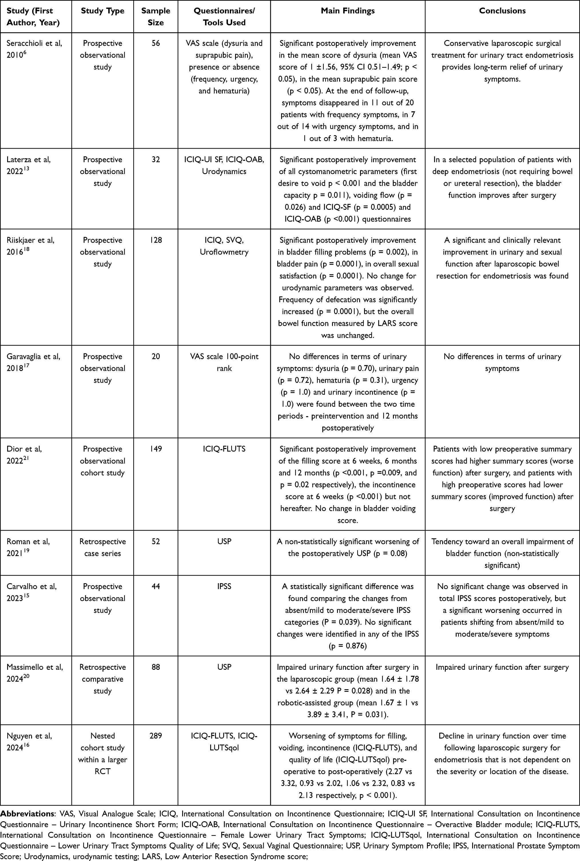 |
Table 2 Characteristics and Main Findings of Studies Evaluating Urinary Outcomes After Minimally Invasive Surgery for Deep Endometriosis |
Study Results
Among the studies reporting improvements in urinary function, several consistent trends emerged. The earliest of these, published by Seracchioli et al in 2010, analyzed a cohort of 74 women with laparoscopically and histologically confirmed urinary tract endometriosis, predominantly involving the bladder but also the ureters. At baseline, cyclical LUTS such as dysuria, suprapubic pain, urgency, frequency, and hematuria were common. Following conservative laparoscopic treatment, there was a significant reduction in dysuria and suprapubic pain, with mean Visual Analogue Scale (VAS) score for dysuria decreasing to 1.0 ± 1.56 (95% CI 0.51–1.49; p < 0.05). At the end of follow-up, symptoms resolved in over half of the patients presenting with urinary frequency or urgency.6
Similarly, Riiskjaer et al included 128 women undergoing laparoscopic bowel resection for rectosigmoid DE at a tertiary referral center. Baseline evaluations demonstrated frequent bladder filling problems and pain, in addition to intestinal dysfunction and impaired sexual function. One year after surgery, significant improvements were noted in bladder filling problems (p = 0.002) and bladder pain (p = 0.0001), accompanied by increased sexual satisfaction (p = 0.0001). However, no changes were observed in urodynamic parameters.18
In the prospective multicenter study by Laterza et al, 32 women with DE—excluding those requiring bowel or ureteral resection—were evaluated with ICIQ-UI SF, ICIQ-OAB, and urodynamic testing. At baseline, urgency and reduced bladder capacity were frequent findings. Postoperatively, patients experienced significant improvements in both subjective and objective parameters: first desire to void increased from 120 to 204 mL (p < 0.001), bladder capacity from 358 to 409 mL (p = 0.011), and maximal voiding flow from 19 to 25 mL/s (p = 0.026). Questionnaires also improved markedly, with ICIQ-UI SF decreasing from 2.5 to 0 (p = 0.0005) and ICIQ-OAB from 4.3 to 1.2 (p < 0.001).13
Consistent results were reported by Dior et al in a larger prospective cohort of 149 women with DE. Using the ICIQ-FLUTS questionnaire, the authors observed significant improvements in filling scores at 6 weeks (p < 0.001), 6 months (p = 0.009), and 12 months (p = 0.02). Incontinence scores improved at 6 weeks (p < 0.001) but not thereafter, while voiding scores remained unchanged. Notably, benefits were more pronounced among women with severe baseline symptoms, whereas those with minimal preoperative dysfunction occasionally experienced postoperative worsening.21
In contrast, several studies reported no significant change or a deterioration in urinary function. In 2018, Garavaglia et al evaluated 20 women with stage IV (according to the American Society of Reproductive Medicine) intestinal DE involving the rectosigmoid or rectum. Despite severe gynecological and gastrointestinal symptoms at baseline, no postoperative improvements in urinary function were observed: dysuria (p = 0.70), urinary pain (p = 0.72), hematuria (p = 0.31), urgency (p = 1.0), and incontinence (p = 1.0).17
In 2021 Roman et al investigated 52 women with DE involving the sacral roots and sciatic nerve, most of whom presented with sciatic pain (82.7%), pudendal neuralgia (21.2%), motor weakness (27%), and associated bladder dysfunction. At one year, there was a non-significant tendency toward worsening urinary symptoms (p = 0.08).19
One year later, Carvalho et al assessed 44 women with rectosigmoid DE treated with nerve-sparing laparoscopic resection. The median age was 39 years, and more than half were overweight (median BMI 25.8 kg/m2). At baseline, LUTS were frequent, with over 50% classified as moderate-to-severe by IPSS. Postoperatively, no significant change was observed in total IPSS scores (p = 0.876), but a significant worsening occurred in patients shifting from absent/mild to moderate/severe categories (p = 0.039).15
Similarly, in 2024 Massimello et al compared 71 women treated with conventional laparoscopy and 29 treated with robotic-assisted laparoscopy for DE involving the sacral plexus and sciatic nerve. Baseline characteristics were comparable, with frequent bladder, rectal, and sexual dysfunction. Both groups experienced worsening postoperative urinary function: USP increased from 1.64 ± 1.78 to 2.64 ± 2.29 (p = 0.028) in the laparoscopic group and from 1.67 ± 1.00 to 3.89 ± 3.41 (p = 0.031) in the robotic group.20
Finally, in the largest study included Nguyen et al (same year), followed 289 women with histologically confirmed DE nested within a randomized trial. The mean age was 32.7 years, 72% were nulliparous, and the mean BMI was 25.8 kg/m2. Disease was frequently extensive, with uterosacral, rectovaginal, and pararectal involvement. At a mean follow-up of 50 months, a consistent and statistically significant deterioration was observed across all ICIQ-FLUTS domains: filling (from 2.27 to 3.32), voiding (from 0.93 to 2.02), incontinence (from 1.06 to 2.32), and LUTS-related quality of life (from 0.83 to 2.13), all p < 0.001.16
Discussion
This systematic review evaluated the impact of minimally invasive surgery for DE on urogynecological symptoms, analyzing data from nine studies with sample sizes ranging from 20 to 289 patients. The findings reveal substantial heterogeneity in outcomes, reflecting both clinical diversity and methodological variability across studies.
A major source of heterogeneity was the variability in outcome measures. While some studies focused on specific urinary symptoms—such as dysuria, urgency, and frequency—others incorporated urodynamic parameters or patient-reported quality-of-life indices as primary endpoints. For example, Dior et al employed the ICIQ-FLUTS and ICIQ-LUTSqol to capture longitudinal changes, whereas Laterza et al combined subjective questionnaires with objective urodynamic measures, providing a more comprehensive assessment of bladder function.13,21
When looking at the symptom domains, a clear pattern emerges. Storage symptoms (urgency, frequency, dysuria) are those most consistently improved after surgery, particularly in patients with moderate-to-severe baseline dysfunction. Seracchioli et al documented a marked reduction in dysuria and suprapubic pain, with resolution of frequency and urgency in over half of symptomatic patients.6 Laterza et al showed significant improvements in first desire to void, bladder capacity, and uroflowmetry in addition to questionnaire scores,13 while Dior et al reported improvements in filling symptoms at 6 weeks, 6 months and 12 months.21 These findings suggest that surgery, especially when nerve-sparing techniques are adopted, can relieve storage complaints linked to compression of pelvic structures and local inflammation.
By contrast, voiding-related symptoms and global bladder function appear less responsive. Several studies noted no significant change in urodynamic parameters despite improvements in self-reported symptoms.6,21 Moreover, Roman et al described a non-significant worsening of USP scores after surgery for sacral nerve involvement,19 Carvalho et al found a significant shift toward more severe categories on the IPSS despite stable overall scores15 and Massimello et al reported worsening USP scores in both laparoscopic and robotic-assisted groups.20 The largest cohort by Nguyen et al highlighted consistent deterioration across all ICIQ-FLUTS domains and quality-of-life measures at long-term follow-up.16 Taken together, these results suggest that voiding function, detrusor contractility, and long-term bladder adaptation remain the most vulnerable aspects, particularly in patients with minimal baseline symptoms where postoperative deterioration was more common.
Considerable inconsistency was also observed in the measurement tools employed. A variety of validated instruments were used, including ICIQ-UI SF,23 ICIQ-OAB,24 ICIQ-FLUTS,25 ICIQ-LUTSqol,26 USP,27 and IPSS28 - each differing in domain focus and sensitivity. Notably, the IPSS, originally designed for male voiding symptoms, may have limited sensitivity in women. This heterogeneity in scoring systems precludes direct cross-study comparisons and contributes to the inconsistency in reported outcomes, a limitation previously noted in systematic reviews addressing urinary function in endometriosis.
Pre- versus postoperative comparisons were conducted using non-uniform criteria. Some studies assessed changes at multiple time points (eg, Dior et al), while others reported outcomes at a single interval.21 The duration of follow-up ranged from 6 weeks to over 4 years, potentially obscuring late-onset deterioration or delayed recovery. Short-term symptom relief may therefore not fully capture long-term outcomes, given that bladder adaptation and autonomic neuroregeneration can extend over months or years.
Differences in study populations further complicated interpretation. Laterza et al limited inclusion to patients without bowel or ureteral involvement, whereas Riiskjaer et al included women undergoing bowel resection.13,18 These anatomical differences likely influenced the risk of autonomic nerve injury and the trajectory of postoperative bladder function. Similarly, the extent of parametrial or sacral nerve involvement, as in Roman and Massimello’s series, correlates with higher risk of postoperative dysfunction.19,20
Surgical technique was another important variable. Although all studies employed minimally invasive approaches, only some explicitly reported nerve-sparing procedures. The implementation of nerve-sparing dissection is known to reduce detrusor hypoactivity and urinary retention. Nevertheless, even with advanced techniques, deterioration was reported in some series, suggesting that the complexity of nerve involvement and extent of resection may outweigh technical refinements.
A particularly influential factor was the preoperative symptom profile. Dior et al noted that patients with more severe baseline LUTS derived the greatest benefit from surgery, while those with minimal symptoms were more likely to deteriorate.21 This ceiling effect has been observed in other domains of endometriosis surgery and underlines the need for careful patient selection and counseling.
Despite the heterogeneity, some consistent messages can be drawn: storage symptoms (urgency, frequency, dysuria) are more likely to improve, whereas voiding dysfunction and global bladder performance often remain unchanged or worsen.
Strengths and Limitations
A key strength of this review lies in the exclusive inclusion of studies utilizing validated instruments for the assessment of urinary function. This methodological rigor minimizes the risk of bias associated with anecdotal or non-standardized outcome reporting. Furthermore, the focus on minimally invasive surgical approaches enhances the clinical applicability of the findings, aligning with current standards of care in endometriosis management.
Nevertheless, several limitations must be acknowledged. The substantial heterogeneity among the included studies—particularly in terms of outcome definitions, measurement tools, surgical techniques, and follow-up intervals—introduced significant variability that precluded the conduct of a quantitative meta-analysis. The absence of standardized time points and symptom classifications across studies limits the external validity and comparability of the results. Additionally, the potential for publication bias cannot be excluded, as studies reporting neutral or negative findings are less likely to be published and included in systematic reviews.
Future Directions
Well-designed randomized controlled trials are needed to compare nerve-sparing versus non–nerve-sparing surgical techniques, with predefined and harmonized follow-up assessments (eg, at 3, 6, and 12 months). Importantly, future studies should incorporate both subjective outcomes—such as those captured by validated questionnaires (eg, ICIQ, USP)—and objective functional assessments, including urodynamic testing and uroflowmetry, to provide a comprehensive evaluation of postoperative urinary function.
Conclusion
In summary, minimally invasive surgery for DE has the potential to achieve meaningful improvements in lower urinary tract symptoms, particularly in patients with pronounced preoperative dysfunction. However, a clinically relevant subset of patients may experience deterioration in bladder function postoperatively, especially in the absence of nerve-sparing techniques or in those with minimal baseline symptoms. The considerable heterogeneity in patient characteristics, outcome measures, and statistical methodologies across current studies underscores the urgent need for more rigorous, standardized research to inform evidence-based patient selection and surgical planning.
Disclosure
The authors report no conflicts of interest in this work.
References
1. Zondervan KT, Becker CM, Missmer SA. Endometriosis. N Engl J Med. 2020;382(13):1244–1256. doi:10.1056/NEJMra1810764
2. Giudice LC. Clinical practice. Endometriosis. N Engl J Med. 2010;362(25):2389–2398. doi:10.1056/NEJMcp1000274
3. Donnez J, Nisolle M, Squifflet J. Ureteral endometriosis: a complication of rectovaginal endometriotic (adenomyotic) nodules. Fertil Steril. 2002;77(1):32–37. doi:10.1016/s0015-0282(01)02921-1
4. Parasar P, Ozcan P, Terry KL. Endometriosis: epidemiology, Diagnosis and Clinical Management. Curr Obstet Gynecol Rep. 2017;6(1):34–41. doi:10.1007/s13669-017-0187-1
5. Nisolle M, Donnez J. Peritoneal endometriosis, ovarian endometriosis, and adenomyotic nodules of the rectovaginal septum are three different entities. Fertil Steril. 1997;68(4):585–596. doi:10.1016/s0015-0282(97)00191-x
6. Seracchioli R, Mabrouk M, Montanari G, Manuzzi L, Concetti S, Venturoli S. Conservative laparoscopic management of urinary tract endometriosis (UTE): surgical outcome and long-term follow-up. Fertil Steril. 2010;94(3):856–861. doi:10.1016/j.fertnstert.2009.04.019
7. Stillwell TJ, Kramer SA, Lee RA. Endometriosis of ureter. Urology. 1986;28(2):81–85. doi:10.1016/0090-4295(86)90092-0
8. Pluchino N, Wenger JM, Petignat P, et al. Sexual function in endometriosis patients and their partners: effect of the disease and consequences of treatment. Hum Reprod Update. 2016;22(6):762–774. doi:10.1093/humupd/dmw031
9. Abrao MS, Petraglia F, Falcone T, Keckstein J, Osuga Y, Chapron C. Deep endometriosis infiltrating the recto-sigmoid: critical factors to consider before management. Hum Reprod Update. 2015;21(3):329–339. doi:10.1093/humupd/dmv003
10. Leone Roberti Maggiore U, Ferrero S, Candiani M, Somigliana E, Vigano P, Vercellini P. Bladder endometriosis: a systematic review of pathogenesis, diagnosis, treatment, impact on fertility, and risk of malignant transformation. Eur Urol. 2017;71(5):790–807. doi:10.1016/j.eururo.2016.12.015
11. Dunselman GA, Vermeulen N, Becker C, et al. ESHRE guideline: management of women with endometriosis. Hum Reprod. 2014;29(3):400–412. doi:10.1093/humrep/det457
12. Leone Roberti Maggiore U, Scala C, Tafi E, et al. Spontaneous fertility after expectant or surgical management of rectovaginal endometriosis in women with or without ovarian endometrioma: a retrospective analysis. Fertil Steril. 2017;107(4):969–976e5. doi:10.1016/j.fertnstert.2017.02.106
13. Laterza RM, Uccella S, Serati M, et al. Is the deep endometriosis or the surgery the cause of postoperative bladder dysfunction? J Minim Invasive Gynecol. 2022;29(4):567–575. doi:10.1016/j.jmig.2021.12.017
14. Haddaway NR, Page MJ, Pritchard CC, McGuinness LA. PRISMA2020: an R package and Shiny app for producing PRISMA 2020-compliant flow diagrams, with interactivity for optimised digital transparency and Open Synthesis. Campbell Syst Rev. 2022;18(2):e1230. doi:10.1002/cl2.1230
15. de Carvalho MSR, Pellino G, Pereira AMG, Bray-Beraldo F, Lopes RGC, Di Saverio S. Prevalence of urinary dysfunction after minimally invasive surgery for deep rectosigmoid endometriosis. Langenbecks Arch Surg. 2023;408(1):83. doi:10.1007/s00423-023-02831-6
16. Nguyen K, McCormack L, Deans R, et al. A prospective study of bladder function following endometriosis surgery with up to eight years follow-up. J Minim Invasive Gynecol. 2024;31(3):205–212e4. doi:10.1016/j.jmig.2023.11.020
17. Garavaglia E, Inversetti A, Ferrari S, De Nardi P, Candiani M. Are symptoms after a colorectal segmental resection in deep endometriosis really improved? The point of view of women before and after surgery. J Psychosom Obstet Gynaecol. 2018;39(4):248–251. doi:10.1080/0167482X.2018.1445221
18. Riiskjaer M, Greisen S, Glavind-Kristensen M, Kesmodel U S, Forman A, Seyer-Hansen M. (2016). Pelvic organ function before and after laparoscopic bowel resection for rectosigmoid endometriosis: a prospective, observational study. BJOG, 123(8), 1360–7. 10.1111/1471-0528.13975
19. Roman H, Dehan L, Merlot B, et al. Postoperative outcomes after surgery for deep endometriosis of the sacral plexus and sciatic nerve: a 52-patient consecutive series. J Minim Invasive Gynecol. 2021;28(7):1375–1383. doi:10.1016/j.jmig.2020.10.018
20. Massimello F, Merlot B, Chanavaz-Lacheray I, et al. Robotic-assisted versus conventional laparoscopic management of deep endometriosis involving the sacral plexus and sciatic nerve: a comparative before and after study. Int J Gynaecol Obstet. 2024;167(2):839–850. doi:10.1002/ijgo.15734
21. Dior UP, Reddington C, Cheng C, Levin G, Healey M. Urinary function after surgery for deep endometriosis: a prospective study. J Minim Invasive Gynecol. 2022;29(2):308–316e2. doi:10.1016/j.jmig.2021.08.024
22. Page MJ, McKenzie JE, Bossuyt PM, et al. The PRISMA 2020 statement: an updated guideline for reporting systematic reviews. BMJ. 2021;372:n71. doi:10.1136/bmj.n71
23. Avery K, Donovan J, Peters TJ, Shaw C, Gotoh M, Abrams P. ICIQ: a brief and robust measure for evaluating the symptoms and impact of urinary incontinence. Neurourol Urodyn. 2004;23(4):322–330. doi:10.1002/nau.20041
24. Donskov AO, Vinge L, Axelsen SM, Andersen H. Overactive bladder in patients with myasthenia gravis-A cross-sectional population-based study. Acta Neurol Scand. 2021;144(1):76–80. doi:10.1111/ane.13419
25. Brookes ST, Donovan JL, Wright M, Jackson S, Abrams P. A scored form of the Bristol Female Lower Urinary Tract Symptoms questionnaire: data from a randomized controlled trial of surgery for women with stress incontinence. Am J Obstet Gynecol. 2004;191(1):73–82. doi:10.1016/j.ajog.2003.12.027
26. Kelleher CJ, Cardozo LD, Khullar V, Salvatore S. A new questionnaire to assess the quality of life of urinary incontinent women. Br J Obstet Gynaecol. 1997;104(12):1374–1379. doi:10.1111/j.1471-0528.1997.tb11006.x
27. Haab F, Richard F, Amarenco G, et al. Comprehensive evaluation of bladder and urethral dysfunction symptoms: development and psychometric validation of the Urinary Symptom Profile (USP) questionnaire. Urology. 2008;71(4):646–656. doi:10.1016/j.urology.2007.11.100
28. Barry MJ, Fowler FJ Jr, P OM, et al. The American Urological Association symptom index for benign prostatic hyperplasia. J Urol. 2017;197(2S):S189–S197. doi:10.1016/j.juro.2016.10.071
 © 2025 The Author(s). This work is published and licensed by Dove Medical Press Limited. The
full terms of this license are available at https://www.dovepress.com/terms
and incorporate the Creative Commons Attribution
- Non Commercial (unported, 4.0) License.
By accessing the work you hereby accept the Terms. Non-commercial uses of the work are permitted
without any further permission from Dove Medical Press Limited, provided the work is properly
attributed. For permission for commercial use of this work, please see paragraphs 4.2 and 5 of our Terms.
© 2025 The Author(s). This work is published and licensed by Dove Medical Press Limited. The
full terms of this license are available at https://www.dovepress.com/terms
and incorporate the Creative Commons Attribution
- Non Commercial (unported, 4.0) License.
By accessing the work you hereby accept the Terms. Non-commercial uses of the work are permitted
without any further permission from Dove Medical Press Limited, provided the work is properly
attributed. For permission for commercial use of this work, please see paragraphs 4.2 and 5 of our Terms.