
Back to Journals » International Journal of Nanomedicine » Volume 21
Trivalent mRNA-LNP Vaccine Induces Robust Humoral and Cellular Immunity in Mice: Preclinical Evaluation for Porcine Enteric Coronaviruses
Authors Wang X  , Li C, Wang S, Liu C, Shi J, Xu W, Zhou S, Xu S, Shang Y, Li J
Received 1 April 2026
Accepted for publication 27 May 2026
Published 20 June 2026 Volume 2026:21 613251
DOI https://doi.org/10.2147/IJN.S613251
Checked for plagiarism Yes
Review by Single anonymous peer review
Peer reviewer comments 3
Editor who approved publication: Professor Eng San Thian
Xinfei Wang,1,2,* Chen Li,1,* Shuo Wang,1,* Chang Liu,1 Jianli Shi,1 Wenxin Xu,1,3 Shun Zhou,3 Shaojian Xu,1 Yingli Shang,2 Jun Li1
1Institute of Animal Science and Veterinary Medicine Shandong Academy of Agricultural Sciences, Shandong Academy of Agricultural Sciences, Jinan, Shandong, People’s Republic of China; 2Department of Preventive Veterinary Medicine, College of Veterinary Medicine, Shandong Agricultural University, Taian, Shandong, People’s Republic of China; 3Department of Animal Science and Technology, Qingdao Agricultural University, Qingdao, Shandong, People’s Republic of China
*These authors contributed equally to this work
Correspondence: Jun Li, Institute of Animal Science and Veterinary Medicine Shandong Academy of Agricultural Sciences, Jinan, Shandong, People’s Republic of China, Email [email protected] Yingli Shang, Department of Preventive Veterinary Medicine, College of Veterinary Medicine, Shandong Provincial Key Laboratory of Zoonoses, Shandong Agricultural University, Taian, Shandong, People’s Republic of China, Email [email protected]
Introduction: Porcine enteric coronaviruses (PECs) often co-infect swine, leading to high mortality, which underscores the need for multivalent vaccines.
Methods: A trivalent mRNA vaccine was formulated using SM-102 lipid nanoparticles (LNPs) and encodes PEDV-SCOE, PDCoV-SCTD, and TGEV-SAD in a single transcript. BALB/c mice were immunized intramuscularly with escalating doses (5, 10, 15, and 20 μg). Systemic IgG, mucosal IgA, neutralizing antibodies, cytokine profiles (IFN-γ, IL-4), and splenocyte proliferation were assessed. Monovalent formulations and commercial vaccines (PEDV/TGEV bivalent inactivated vaccine from Qilu Animal Health; PDCoV inactivated vaccine from Wuhan Keqian) served as controls. Data are mean ± SD (n = 5); one-way ANOVA with multiple comparisons was applied (*p < 0.05; **p < 0.01; ***p < 0.001).
Results: A moderate dose (10 μg) induced systemic IgG, IFN-γ, IL-4, and splenocyte proliferation, indicating activation of both Th1 and Th2 responses. Higher dose (15 μg) preferentially enhanced mucosal IgA and neutralizing antibody responses, exceeding those induced by commercial vaccines, suggesting potential improvement in mucosal protection. Monovalent SM-102-LNP formulations also elicited robust immune responses, approaching commercial benchmarks.
Discussion: This trivalent mRNA-LNP vaccine provides broad and potent humoral and cellular immunity in mice, supporting its potential as a platform for porcine vaccination. These results provide preclinical proof-of-concept, and further evaluation in swine is required to assess vaccine efficacy.
Keywords: porcine enteric pathogens, mRNA-LNP vaccine, immunogenicity
Introduction
Porcine epidemic diarrhea virus (PEDV), transmissible gastroenteritis virus (TGEV), and porcine deltacoronavirus (PDCoV) are major enteric coronaviruses causing diarrhea, vomiting, dehydration, and high mortality in neonatal piglets.1 Clinically, infections are challenging to differentiate due to similar enteric symptoms and frequent co-infections, complicating disease prevention in swine farms.2 PEDV and TGEV were first reported in Europe and the United States in the 1970s and 1940s, respectively, while PDCoV emerged in China in 2012.3–5 Co-infections may exacerbate disease severity and mortality, emphasizing the need for broadly protective multivalent vaccines.6,7
Coronaviruses contain four major structural proteins, including spike (S), envelope (E), membrane (M), and nucleocapsid (N).6–11 Among them, the S glycoprotein is the major target for neutralizing antibodies and is cleaved into the S1 receptor-binding subunit and the S2 membrane-fusion subunit.12 Previous studies show that several conserved regions within the S1 domain contain dominant neutralizing epitopes involved in viral attachment and immune protection. The core neutralizing epitope (COE) of PEDV, antigenic sites A and D of TGEV, and the C-terminal domain (CTD) of PDCoV induce neutralizing antibody responses.13–17 These antigenic regions therefore represent suitable targets for multivalent vaccine design while reducing unnecessary antigenic burden associated with full-length spike proteins.
Various vaccine platforms targeting porcine enteric coronaviruses, including live-attenuated, inactivated, recombinant vector-based, and subunit vaccines, have been explored.18 Inactivated and attenuated vaccines are used in swine production and provide partial protection, but limited mucosal immunity and insufficient cross-protection during co-infection remain challenges.19 Recombinant and vectored vaccines improve antigen specificity and safety, but balanced immunity against multiple enteric coronaviruses remains challenging. Although bivalent PEDV/TGEV or PDCoV vaccines have been studied, trivalent strategies remain scarce.20
Messenger RNA (mRNA) vaccines offer advantages such as rapid production, flexible antigen design, and induction of both humoral and cellular immune responses.21 During the COVID-19 pandemic, mRNA vaccine platforms showed high efficacy and have gained interest for veterinary infectious diseases.22–27 Lipid nanoparticles (LNPs) protect mRNA from degradation and enable intracellular delivery, making them suitable for multivalent vaccine development.28,29 SM-102, an ionizable amino lipid used in clinically validated mRNA vaccines, provides high encapsulation efficiency and effective delivery,30,31 although large-scale veterinary applications may require more cost-effective alternatives.32
PEDV, TGEV, and PDCoV primarily infect intestinal epithelial cells; therefore, induction of mucosal immunity is essential to limit viral replication and fecal shedding.33 Multivalent mRNA vaccines can elicit secretory IgA and coordinated cellular responses, contributing to mucosal protection.29,34 Gut health, microbiota modulation, and immune enhancement (eg., Litsea cubeba essential oil) have been reported to improve vaccine efficacy in pigs.35 Microbiota-derived metabolites can affect macrophage polarization and systemic immune responses.36 Single-cell transcriptomic studies in IUGR piglets indicate ileal immune heterogeneity and impaired intestinal immunity, underscoring the need to design vaccines that elicit balanced Th1/Th2 responses.37 These insights provide the rationale for evaluating a trivalent mRNA-LNP vaccine encoding the major neutralizing epitopes of PEDV, TGEV, and PDCoV.
We developed a trivalent mRNA-LNP vaccine encoding the major neutralizing epitopes PEDV-SCOE, PDCoV-SCTD, and TGEV-SAD using SM-102 LNPs. Its immunogenicity was assessed in a murine model, alongside monovalent vaccines and commercial PEDV/TGEV bivalent and PDCoV inactivated vaccines. Integrated analysis of humoral, mucosal, and cellular immune responses provides evidence that multivalent mRNA-LNP vaccines may serve as a strategy to control porcine enteric coronavirus co-infections.
Mechanistic insights, microbiome interactions, and translational considerations support the rationale for multivalent mRNA vaccines and inform the design of next-generation enteric coronavirus vaccines for swine.
Materials and Methods
Cells and Viruses
HEK-293T (human embryonic kidney), Vero (African green monkey kidney), PK-15 (porcine kidney), and LLC-PK1 (porcine kidney) cells were maintained at the Laboratory for Major Livestock Disease Control, Shandong Academy of Agricultural Sciences. The use of these cell lines was approved by the Institutional Review Board of the Shandong Academy of Agricultural Sciences. HEK-293T, Vero, and PK-15 cells were cultured in Dulbecco’s Modified Eagle Medium (DMEM) supplemented with 10% fetal bovine serum (FBS) and 1% penicillin-streptomycin, whereas LLC-PK1 cells were maintained in Minimum Essential Medium (MEM) with identical supplements. All cells were incubated at 37°C in a humidified atmosphere containing 5% CO2.
PEDV-SDSX16 (GenBank: MN241464.1), TGEV-SD01 (GenBank: OP805351), and PDCoV-SDLY-52 (GenBank: OM256446) were isolated and preserved in laboratory.
Plasmid Construction
Codon-optimized sequences encoding PEDV-SCOE (aa 499–638), TGEV-SAD (aa 366–604), and PDCoV-SCTD (aa 277–616) were synthesized by GenScript Biotech (Nanjing, China). The constructs contained a tPA signal peptide, 6×His/HA/Flag tags, a P2A self-cleaving sequence, β-globin UTRs, and a poly(A) tail, and were cloned into a GS-CMV vector under the control of T7 and CMV promoters. The constructs include 6×His, HA, and Flag tags to facilitate purification and detection, following strategies described previously.38 Resulting plasmids were designated as PEDV-SCOE-GS-CMV, PDCoV-SCTD-GS-CMV, TGEV-SAD-GS-CMV, and the trivalent construct PEDV-SCOE-PDCoV-SCTD-TGEV-SAD-GS-CMV.
Plasmid Amplification and Extraction
One hundred microliters of thawed E. coli Stbl3 competent cells were gently mixed with 10 μL of synthesized plasmid DNA and incubated on ice for 30 min. Heat shock was performed at 42°C for 45–60 s, followed by immediate transfer to ice for 2 min. Seven hundred microliters of antibiotic-free sterile Luria-Bertani (LB) medium were added, and cells were recovered at 37°C with orbital shaking at 200 rpm in a ZWY-100H incubator shaker (Zhicheng Analytical Instrument, Shanghai) using 12 mL culture tubes (Lanjieke Technology, Beijing) for 60 min. The recovered cells were then plated onto LB agar containing kanamycin and incubated overnight at 37°C. Positive single colonies were selected and cultured in LB liquid medium with kanamycin at 37°C, 220 rpm, overnight. High-purity plasmid DNA was subsequently extracted using the Qiagen Plasmid Maxi Kit according to the manufacturer’s instructions.
mRNA Preparation
Recombinant plasmids containing target antigen sequences (PEDV-SCOE, TGEV-SAD and PDCoV-SCTD) were verified by double digestion and subsequently linearized with the restriction enzyme BspQ I. Linearized products were purified using VAHTS DNA Clean Beads to remove proteins, salts, and other contaminants. Purified linear DNA templates were used for in vitro transcription (IVT) with T7 RNA polymerase, using linear double-stranded DNA containing a T7 promoter and AG initiation sequence as template, and NTPs as substrates. A CAG Trimer cap analog used to generate mRNA single-stranded RNA with a 5′-m7G Cap1 structure. The cap structure and poly(A) tail enhance RNA stability and reduce innate immune activation. Following transcription, RNA was further purified using VAHTS RNA Clean Beads to remove template DNA, enzymes, and small molecules, yielding high-purity mRNA.
mRNA Encapsulation in LNP
Lipid nanoparticles (LNPs) were prepared using microfluidic mixing (NanoAssemblr platform, Precision NanoSystems). Lipids including SM-102, DSPC, cholesterol, and DMG-PEG2000 were dissolved in ethanol at molar ratios of 50:10:38.5:1.5. mRNA was diluted in citrate buffer (50 mM, pH 4.0) and mixed with lipid phase at a 3:1 aqueous to organic flow ratio. Formed LNPs were diluted in 1× phosphate-buffered saline (PBS, pH 7.4; Solarbio) and concentrated using 100 kDa ultrafiltration tubes to remove ethanol. Particle size, polydispersity index (PDI), and encapsulation efficiency were subsequently measured.
mRNA and mRNA/LNP Transfection
Approximately 24 hours prior to transfection, HEK-293T cells were seeded at a density of 2×10⁵ cells/well. Transfection was performed at 70–90% confluence using Opti-MEM, Lipofectamine 3000, and RNA (incubated for 10 min at room temperature), followed by dropwise addition to cells. After 24 h of incubation at 37°C, cells were harvested. For lysis, cells were washed with cold PBS and lysed using a RIPA buffer [1% Triton X-100, 20 mM Tris (pH 7.5), 150 mM NaCl, sodium pyrophosphate, β-glycerophosphate, EDTA, Na3VO4, and protease inhibitor leupeptin]. The lysate was centrifuged (12,000 rpm, 5 min, 4°C), and the supernatant was collected as the total protein extract. All procedures were performed at 4°C to prevent protein degradation.
mRNA and mRNA/LNP Protein Detection
Cellular proteins were extracted for Western blotting (WB) and immunofluorescence assay (IFA). For WB, lysates were mixed with SDS-PAGE loading buffer, denatured at 95°C for 10 min, and resolved by electrophoresis at 150 V. Proteins were transferred onto methanol-activated PVDF membranes (Pall, USA) and blocked with 5% skim milk in Tris-Buffered Saline with Tween 20 (TBST). Primary antibodies against His, Flag, and HA tags were incubated overnight at 4°C, followed by HRP-conjugated anti-mouse secondary antibody (Beyotime, 1:10,000) for 1 h at room temperature. Chemiluminescent signals were detected using an ECL substrate (Tanon, China). For IFA, adherent cells were fixed with 4% paraformaldehyde and permeabilized with 0.2% Triton X-100. After blocking with 5% BSA for 30 min, primary antibodies (Beyotime, 1:100–1:500) were applied for 1 h, followed by fluorescent secondary antibodies in the dark. Cells were counterstained with DAPI and imaged using a fluorescence microscope (Nikon, Japan).
Mouse Immunization
BALB/c mice and their feed were purchased from Jinan Pengyue Experimental Animal Breeding Co., Ltd., Shandong Province. Fifty 6-week-old female BALB/c mice were randomly assigned to ten groups. Mice were randomly assigned to experimental groups (n = 5 per group) using a random number generator to ensure equal distribution of baseline characteristics. All subsequent procedures, including immunization, sample collection, and data analysis, were performed in a blinded manner, where the investigators were unaware of the group allocations until the final data analysis was completed. The study design included seven experimental vaccine groups, two commercial vaccine control groups, and one negative control group. The experimental cohorts received either one of three monovalent mRNA vaccines (PEDV-SCOE-GS-CMV, PDCoV-SCTD-GS-CMV, or TGEV-SAD-GS-CMV; 15 µg/dose each) or the trivalent mRNA vaccine at escalating doses (5, 10, 15, or 20 µg). The commercial vaccine controls received PEDV/TGEV bivalent inactivated vaccine (Qilu Animal Health Products Co., Ltd.) and PDCoV monovalent inactivated vaccine (Wuhan Keqian Biological Co., Ltd.) according to the manufacturers’ recommended optimal doses. The negative control group received 100 µL of PBS. All immunizations were administered subcutaneously in the nape of the neck on days 0 and 14. Blood samples were collected via retro-orbital sinus bleeding on days 14 and 28 post-prime; serum was then separated by centrifugation for the determination of IgG, IgA, IFN-γ, and IL-4 levels using ELISA. On day 28, mice were euthanized, and spleens were harvested via abdominal dissection. Splenocytes were isolated and assessed for T-cell proliferation in vitro using the CCK-8 assay. Furthermore, the day-28 sera were used in virus neutralization assays to evaluate the humoral immune response elicited by the vaccines.
ELISA was Used to Detect IgG, IgA, IFN-γ, and IL-4 Antibody Levels
Serum was separated from whole blood using serum separator tubes and stored at −20°C or −80°C until analysis. Commercial mouse ELISA kits (Jiangsu Meimian Industrial) were used to quantify serum IgG, IgA, IFN-γ, and IL-4 concentrations. Briefly, serum samples loaded onto 96-well microplates pre-coated with capture antibodies. Following a 60-min incubation at 37°C, plates were washed five times with 1× washing buffer. HRP-conjugated detection antibodies (100 μL) were added, and plates were re-incubated for 60 min at 37°C. After further washing, TMB substrate (100 μL) was added, and reactions were developed for 15 min at 37°C in the dark. Reactions were stopped with 50 μL stop solution, and optical density was measured immediately at 450 nm. All samples were run in triplicate technical replicates. Standard curves were generated using 4-parameter logistic (4-PL) regression analysis to calculate sample concentrations.
Lymphocyte Proliferation Assay
On day 28 post-immunization, splenocytes were isolated using a commercial kit (Tianjin Haoyang Biological Products) and seeded in 96-well plates (5×10⁶ cells/mL). Cells were stimulated with UV-inactivated viral antigens (10 µg/mL) for 72 h at 37°C. Proliferation was assessed using the CCK-8 assay (Solarbio); absorbance was measured at 490 nm after a 2 h incubation with CCK-8 solution. Concanavalin A (ConA) served as the positive control. The stimulation index (SI) was calculated based on blank-corrected OD values.
Serum Neutralization Assay
Collected serum samples were heat-inactivated at 56°C for 30 min and serially diluted in two-fold increments. Each diluted serum was mixed with an equal volume of the corresponding virus at 200 TCID50 and incubated at 37°C for 1 h. Subsequently, 0.1 mL of each mixture was added to a monolayer of the appropriate cells in 96-well plates. After 1.5 h adsorption at 37°C, the inoculum was removed and maintenance medium containing 10 µg/mL trypsin was added. Cells were incubated at 37°C for 48 h and monitored daily for CPE. Neutralizing antibody titers were defined as the highest serum dilution that protected ≥ 50% of cells from CPE. Viral strains used included PEDV-SDSX16 (GenBank: MN241464.1), TGEV-SD01 (GenBank: OP805351), and PDCoV-SDLY-52 (GenBank: OM256446), all of which were isolated and preserved at the same laboratory.
Statistical Analysis
All statistical analyses and graphing were performed using GraphPad Prism 8 software. All data were expressed as the mean ± SD. Group differences were evaluated using either mixed-effects analysis or one-way ANOVA followed by Tukey’s multiple comparisons test. Statistical significance is indicated as follows: ns: no significant difference, * p < 0.05, ** p < 0.01, *** p < 0.001.
Results
Synthesis of mRNA Targeting Porcine Diarrheagenic Coronaviruses
The synthesis and structural characterization of mono- and trivalent mRNA vaccines are shown in Figure 1. The overall workflow, including in vitro transcription and purification steps, is presented in Figure 1A. Monovalent mRNAs encoding PEDV-SCOE, PDCoV-SCTD, and TGEV-SAD were individually generated, and their predicted tertiary structures were consistent with native antigen folding (Figure 1B–D). Each monovalent mRNA produced the expected protein sequence without predicted structural disruptions, indicating potential immunogenicity. The trivalent mRNA construct linked the three antigens via flexible P2A self-cleaving peptides, permitting independent translation of each protein. Structural modeling confirmed that P2A-mediated separation did not alter folding of PEDV-SCOE, PDCoV-SCTD, or TGEV-SAD (Figure 1E).
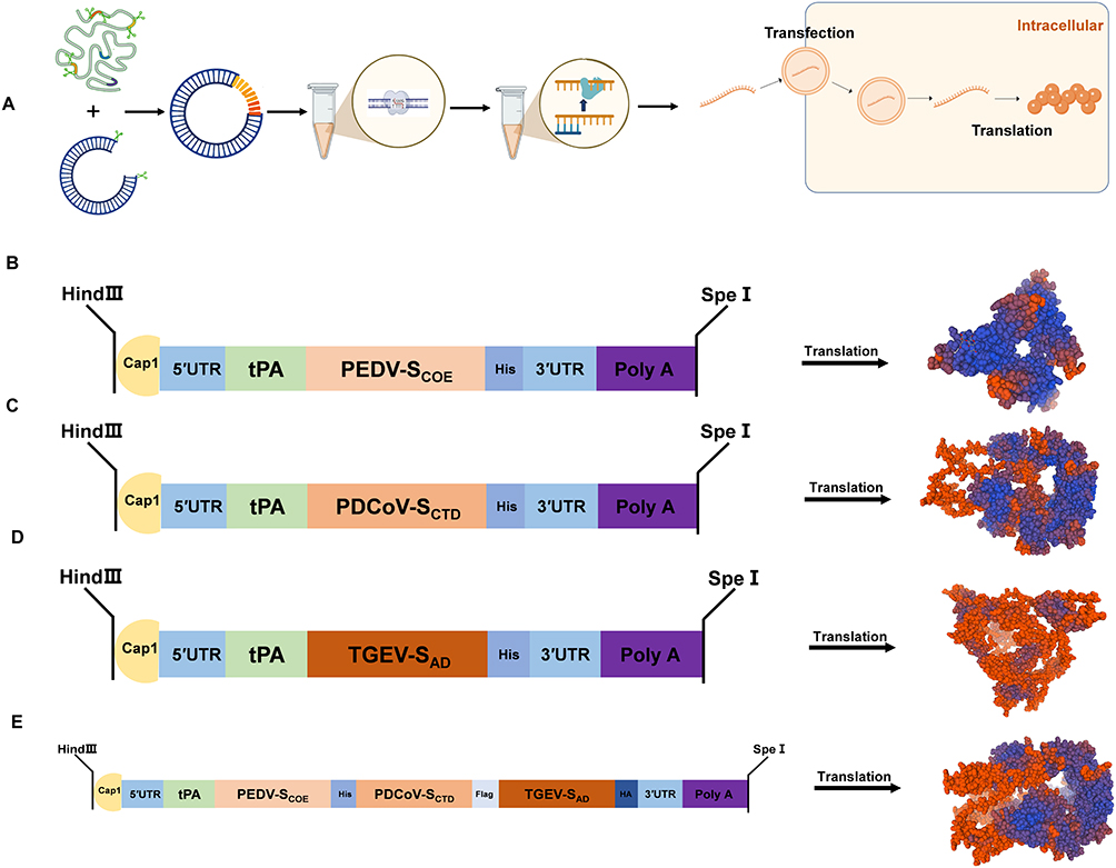 |
Figure 1 Schematic diagrams of the mRNA structure, predicted protein tertiary structures, and preparation workflow. (A) Preparation workflow and in vitro transcription of monovalent and trivalent mRNA vaccines. (B) Schematic of PEDV-SCOE mRNA and predicted tertiary protein structure. (C) Schematic of PDCoV-SCTD mRNA and predicted protein structure. (D) Schematic of TGEV-SAD mRNA and predicted protein structure. (E) Schematic of the trivalent PEDV-SCOE-PDCoV-SCTD-TGEV-SAD mRNA and predicted fusion protein structure. Polypeptide chains are colored from N-terminus (red) to C-terminus (blue) to visualize domain organization. Each model represents in silico structural predictions based on SWISS-MODEL. |
Favorable Physicochemical Characteristics of SM-102 LNPs Contributing to Efficient mRNA Delivery
The synthesized mRNAs (PEDV-SCOE-GS-CMV, PDCoV-SCTD-GS-CMV, TGEV-SAD-GS-CMV, and trivalent PEDV-SCOE-PDCoV-SCTD-TGEV-SAD-GS-CMV) were encapsulated in lipid nanoparticles (LNPs) to improve stability and protect against nuclease degradation, forming core-shell structures (Figure 2A). DLS analysis indicated particle sizes ranging from 91.4 to 102.6 nm, with polydispersity indices (PDI) of 0.099–0.148 across formulations, consistent with monodisperse populations. Zeta potentials were ±15 mV in 10% sucrose/Tris-HCl (pH 7.4 ± 0.5), suggesting favorable electrostatic interactions for cellular uptake (Figure 2B–E). TEM revealed spheroidal particles with uniform morphology and minimal aggregation for all four formulations (Figure 2F–I).
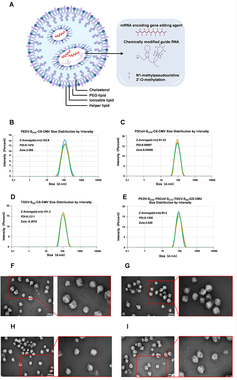 |
Figure 2 Characterization of lipid nanoparticles (LNPs) encapsulating mRNA and visualization by electron microscopy. (A) Schematic representation of LNP structure. (B–E) Particle size, polydispersity index (PDI), and zeta potential of LNP-encapsulated PEDV-SCOE-GS-CMV, PDCoV-SCTD-GS-CMV, TGEV-SAD-GS-CMV, and trivalent mRNA (PEDV-SCOE-PDCoV-SCTD-TGEV-SAD-GS-CMV) measured by dynamic light scattering (DLS). Three independent measurements are shown in different colored lines for each sample. (F–I) Representative transmission electron microscopy (TEM) images of LNPs loaded with PEDV-SCOE-GS-CMV, PDCoV-SCTD-GS-CMV, TGEV-SAD-GS-CMV, and trivalent PEDV-SCOE-PDCoV-SCTD-TGEV-SAD-GS-CMV mRNAs, showing particle size and morphology. Data are expressed as the mean ± SD of three independent preparations (n = 3). |
Robust Expression of mRNA-Encoded Vaccine Antigens in Eukaryotic Cells
Expression of both naked mRNA and LNP-encapsulated constructs was assessed by Western blot and immunofluorescence assay (IFA). HEK-293T cells were transfected with naked mRNA using Lipofectamine 3000 or treated directly with mRNA/LNPs. Protein expression was analyzed 24 h post-transfection. Western blot detected specific bands corresponding to His-, Flag-, and HA-tagged antigens in both trivalent mRNA-LNP and naked mRNA groups (PEDV-SCOE-PDCoV-SCTD-TGEV-SAD-GS-CMV), while no bands were observed in the control group (Figure 3A–D). Monovalent constructs showed expected bands using anti-His antibody (Figure 3E–G). IFA confirmed fluorescence signals and consistent subcellular localization for all constructs (Figure 3H), verifying correct translation and cytoplasmic targeting.
 |
Figure 3 mRNA in vitro protein expression validation. 1: Naked mRNA; 2: mRNA/LNP; 3: Blank control. (A–D) Western blot detection of His-, Flag-, and HA-tagged antigens. Comparable bands were detected with antibodies incubated either simultaneously or individually. (E–G) Western-blot results showed that the protein expressions of PEDV-SCOE-GS-CMV, PDCoV-SCTD-GS-CMV and TGEV-SAD-GS-CMV were detected using a monoclonal anti-His antibody. (H) Immunofluorescence assay (IFA) of cells transfected with the trivalent mRNA (PEDV-SCOE-PDCoV-SCTD-TGEV-SAD-GS-CMV); non-transfected cells served as negative control. Images captured at 200× magnification. Experiments were performed in triplicate. |
The Trivalent mRNA Vaccine Elicits Potent Systemic and Mucosal Immunity
BALB/c mice were immunized intramuscularly on days 0 and 14, with sera collected on days 14 and 28 (Figure 4A). Analysis of day-28 sera revealed that the 10 µg dose of the trivalent mRNA vaccine induced the highest IgG titers (28.80 mg/mL; Figure 4B), which were significantly greater than those in mice receiving PEDV/TGEV bivalent inactivated vaccine or PDCoV inactivated vaccine (20.35 mg/mL; Figure 4C and D). The 10 µg dose also elicited robust cellular responses, with IFN-γ and IL-4 concentrations peaking at 824.61 pg/mL and 61.12 pg/mL, respectively (Figure 4E and H). These values exceeded those in the commercial vaccine groups (Figure 4F, G, I and J), indicating concurrent activation of Th1 and Th2 pathways. Notably, the 15 µg dose group exhibited peak IgA production (84.46 µg/mL; Figure 4K), exceeding the levels induced by PEDV/TGEV bivalent and PDCoV inactivated vaccines (Figure 4L and M), suggesting effective induction of mucosal immunity.
 |
Figure 4 (A) Immunization schedule and sample collection timeline. (B–D) Antigen-specific IgG titers for: trivalent vaccine doses (B), comparison with commercial PEDV+TGEV vaccines (C), and comparison with commercial PDCoV vaccines (D). (E–G) IFN-γ secretion levels for: trivalent vaccine doses (E), PEDV+TGEV groups (F), and PDCoV groups (G). (H–J) IL-4 secretion levels for: trivalent vaccine doses (H), PEDV+TGEV groups (I), and PDCoV groups (J). (K–M) Antigen-specific IgA titers for: trivalent vaccine doses (K), PEDV+TGEV groups (L), and PDCoV groups (M). Data are presented as the mean ± SD of five mice per group, without standard deviation. Statistical analysis was performed using one-way ANOVA with multiple comparisons; horizontal lines indicate group comparisons; ns: no significant difference, * p < 0.05, ** p < 0.01, *** p < 0.001. |
Trivalent mRNA-LNP Vaccine Elicits Superior T-Cell and Neutralizing Antibody Responses with Favorable in-vivo Safety
Splenocyte proliferation assays on day 28 post-boost demonstrated significantly elevated stimulation indices (SI) across all vaccine groups when restimulated with inactivated PEDV, PDCoV, or TGEV. The 10 µg trivalent mRNA vaccine elicited markedly enhanced virus-specific T-cell responses compared to mice receiving PEDV/TGEV bivalent or PDCoV inactivated vaccines (Figure 5A–C), whereas the monovalent mRNA vaccines also showed robust proliferative activity. Concanavalin A (ConA) induced mitogenesis confirmed equivalent baseline T cell viability across all groups. Enhancements against PEDV and TGEV in the trivalent group were particularly notable, representing increases of 83.3% and 82.5%, respectively, relative to the commercial vaccines. The 15 µg trivalent regimen induced higher neutralizing antibody titers against all three viruses. The peak PEDV neutralization titer reached 1:153, substantially higher than the 1:78 titer observed in mice receiving PEDV/TGEV bivalent or PDCoV inactivated vaccines. Monovalent mRNA controls achieved neutralizing titers equivalent to 68%, 97.7%, and 95.02% of the respective commercial vaccines, while naïve animals remained seronegative throughout the study period (Figure 5D–F). These results indicate that the trivalent mRNA vaccine induces systemic, mucosal, cellular, and neutralizing antibody responses in mice, exceeding those elicited by commercial vaccines.
 |
Figure 5 Proliferative response of mouse splenic lymphocytes and serum neutralizing antibody titers. (A–C) Splenic lymphocyte proliferation assays specific for PEDV, PDCoV, and TGEV, respectively. (D–F) Serum neutralizing antibody titers against PEDV, PDCoV, and TGEV, respectively. Data are presented as the mean ± SD of five mice per group. Statistical analysis was performed using one-way ANOVA with multiple comparison tests; horizontal lines indicate significantly different groups, and asterisks indicate significance (ns: no significant difference, * p < 0.05, ** p < 0.01, *** p < 0.001). |
Discussion
The co-circulation of PEDV, PDCoV, and TGEV continues to threaten swine production globally.39 Conventional vaccines often fail to provide broad-spectrum protection against these diverse pathogens.1,40 This study introduces a trivalent mRNA-lipid nanoparticle (LNP) vaccine engineered to simultaneously target these three major swine coronaviruses.21,41 By leveraging the modularity of the mRNA platform, this approach enables a rapid and precise strategy to address mixed infections in field outbreaks.42
A two-dose prime-boost regimen induced both humoral and cellular immunity. The trivalent formulation maintained the immunogenicity of individual antigens, with antigen-sparing potential: doses of 10–15 μg elicited balanced Th1/Th2 and mucosal responses. Specifically, the 10 μg dose produced systemic immunity, whereas the 15 μg dose increased mucosal IgA and virus-neutralizing antibody titers, relevant for blocking viral entry at the intestinal epithelium.
Immune profiling showed that the 10 μg dose induced the highest levels of IgG, IFN-γ, IL-4, and splenocyte proliferation, consistent with a balanced Th1/Th2 response.43,44 In contrast, the 15 μg dose increased mucosal IgA secretion and neutralizing antibody titers.45,46 These findings suggest that while 10 μg is sufficient for systemic immunity, 15 μg may be more effective for stimulating local mucosal responses, important for preventing viral entry and fecal-oral transmission.47,48
Compared with conventional inactivated vaccines, the trivalent mRNA vaccine exhibited superior immunogenicity.21 Inactivated PEDV and TGEV vaccines typically induce weak, short-lived immunity with limited cross-protection.48,49 In contrast, the mRNA platform generated higher levels of neutralizing antibodies, robust IgA responses, and a pronounced Th1-biased profile characterized by IFN-γ production,45,50 which supports CD8⁺ T cell activation and long-term immune memory.51,52 The use of SM-102, a clinically validated ionizable lipid, enabled endosomal escape and antigen expression, contributing intrinsic adjuvanticity for potential translational application.53
Mechanistically, immune responses elicited by the trivalent mRNA-LNP vaccine may involve multiple regulatory pathways. JAK1/STAT3 signaling could modulate inflammatory injury.54 SIRT6-mediated regulation may contribute to systemic immune homeostasis,55 while AMPK signaling and metabolic reprogramming may support T and B cell activation.56 Microbiota-derived metabolites and macrophage polarization may influence mucosal immunity, highlighting the role of the gut-immune axis.36,57 Single-cell transcriptomics in IUGR piglets revealed ileal immune heterogeneity and intestinal immune impairment, emphasizing the need to evaluate vaccines in the natural host for balanced Th1/Th2 and mucosal responses.37 Microbiota-modulating interventions such as Litsea cubeba essential oil can enhance growth, immune function, and mucosal health, providing mechanistic support for enteric vaccine efficacy.35
Despite these observations, limitations exist that inform translational development. Murine models provide proof-of-concept but do not fully replicate porcine immunity and pathogenesis. Subsequent validation in porcine challenge models is required to assess protective efficacy and refine dosing strategies.58–60 Additionally, the small sample size (n = 5 per group) may limit statistical power, and the study duration (28 days) is relatively short for assessing long-term immune responses. Long-term durability of immune responses, including antibody persistence and T-cell memory, requires further investigation, particularly with emerging viral variants.61,62 Safety assessments should include local and systemic reactogenicity, off-target effects, and tolerability.63,64 No significant antigenic competition was observed in the trivalent formulation; however, future multivalent vaccines may require optimization of antigen ratios to avoid immune dominance or masking.65,66 Manufacturing scalability and LNP formulation, including potential cost-effective lipids such as DOTAP or Dlin-MC3, should be considered for commercial translation.67,68
In summary, the trivalent mRNA-LNP vaccine induces systemic and mucosal immunity with a balanced Th1/Th2 profile, exceeding responses induced by conventional inactivated vaccines. Mechanistic insights, microbiota interactions, and translational considerations support its potential as a multivalent vaccine platform in swine, providing preclinical proof-of-concept and laying the groundwork for next-generation enteric coronavirus vaccines.
Conclusions
In conclusion, this study demonstrates that the trivalent mRNA-LNP vaccine, designed to target PEDV, PDCoV, and TGEV, induces humoral, mucosal, and cellular immune responses in a murine model. Integration of mechanistic insights, microbiota interactions, and multivalent antigen presentation suggests that this platform may overcome some limitations of conventional vaccines and supports potential broad-spectrum protection against enteric coronaviruses. The efficacy of the vaccine against circulating and emerging viral variants remains to be determined, and validation in swine models is necessary to confirm protective efficacy, optimize dosing, and assess long-term immunity and safety. These results support future translational studies for next-generation veterinary vaccines capable of controlling complex, co-circulating enteric pathogens.
Ethics Approval and Informed Consent
All experimental animal procedures were performed in strict accordance with the guidelines of the Institutional Animal Care and Use Committee of Institute of Animal Science and Veterinary Medicine, Shandong Academy of Agricultural Sciences (No: IASVM-2025-006). The Institutional Animal Care and Use Committee were approved by Institute of Animal Science and Veterinary Medicine, Shandong Academy of Agricultural Sciences (IACC20060101,1 Jan, 2006). Mice were anesthetized by diethyl ether inhalation in a closed chamber containing ether-soaked cotton wool. Once deep anesthesia was confirmed by absence of reflex to toe pinch, the animals were euthanized by cervical dislocation.
Acknowledgments
We would like to thank the Shandong Academy of Agricultural Sciences for their fundamental support. The authors declare that they have not used AI-generated work in this manuscript.
Author Contributions
All authors made a significant contribution to the work reported, whether that is in the conception, study design, execution, acquisition of data, analysis and interpretation, or in all these areas; took part in drafting, revising or critically reviewing the article; gave final approval of the version to be published; have agreed on the journal to which the article has been submitted; and agree to be accountable for all aspects of the work.
Funding
This work was supported by the Agricultural Science and Technology Innovation Project of Shandong Academy of Agricultural Sciences (CXGC2026A09), the Major Scientific and Technological Innovation Project (MSTIP) (grant no. 2023CXGC010705), the Taishan Scholars Program (NO. tstp20250742), Shandong Province Pig Industry Technology System (grant no. SDAIT-08-06).
Disclosure
The authors declare that they have no known competing financial interests or personal relationships that could have appeared to influence the work reported in this paper.
References
1. Turlewicz-Podbielska H, Pomorska-Mól M. Porcine Coronaviruses: overview of the State of the Art. Virol Sin. 2021;36(5):833–14. doi:10.1007/s12250-021-00364-0
2. Jia X, Liu H, Sun Y, et al. Effective preparation and immunogenicity analysis of antigenic proteins for prevention of porcine enteropathogenic coronaviruses PEDV/TGEV/PDCoV. Int J Biol Macromol. 2025;308:142394. doi:10.1016/j.ijbiomac.2025.142394
3. Wood E. An apparently new syndrome of porcine epidemic diarrhoea. Vet Rec. 1977;100(12):243–244. doi:10.1136/vr.100.12.243
4. Doyle LP, Hutchings LM. A transmissible gastroenteritis in pigs. J Am Vet Med Assoc. 1946;108:257–259.
5. Woo PCY, Lau SKP, Lam CSF, et al. Discovery of a Novel Bottlenose Dolphin Coronavirus Reveals a Distinct Species of Marine Mammal Coronavirus in Gammacoronavirus. J Virol. 2014;88(2):1318–1331. doi:10.1128/JVI.02351-13
6. Guo J, Lai Y, Yang Z, et al. Coinfection and nonrandom recombination drive the evolution of swine enteric coronaviruses. Emerg Microbes Infect. 2024;13(1). doi:10.1080/22221751.2024.2332653
7. Li Y, Niu JW, Zhou X, et al. Development of a multiplex qRT-PCR assay for the detection of porcine epidemic diarrhea virus, porcine transmissible gastroenteritis virus and porcine Deltacoronavirus. Front Vet Sci. 2023;10:1158585. doi:10.3389/fvets.2023.1158585
8. Chang SH, Bae JL, Kang TJ, et al. Identification of the epitope region capable of inducing neutralizing antibodies against the porcine epidemic diarrhea virus. Mol Cells. 2002;14(2):295–299. doi:10.1016/S1016-8478(23)15106-5
9. Oh J, Lee KW, Choi HW, Lee C. Immunogenicity and protective efficacy of recombinant S1 domain of the porcine epidemic diarrhea virus spike protein. Arch Virol. 2014;159(11):2977–2987. doi:10.1007/s00705-014-2163-7
10. Okda FA, Lawson S, Singrey A, et al. The S2 glycoprotein subunit of porcine epidemic diarrhea virus contains immunodominant neutralizing epitopes. Virology. 2017;509:185–194. doi:10.1016/j.virol.2017.06.013
11. Su M, Zheng G, Xu X, Song H. Antigen epitopes of animal coronaviruses: a mini-review. Animal Diseases. 2023;3(1):14. doi:10.1186/s44149-023-00080-0
12. Bosch BJ, van der Zee R, de Haan CAM, Rottier PJM. The Coronavirus Spike Protein Is a Class I Virus Fusion Protein: structural and Functional Characterization of the Fusion Core Complex. J Virol. 2003;77(16):8801–8811. doi:10.1128/JVI.77.16.8801-8811.2003
13. Ho TT, Trinh VT, Tran HX, et al. The immunogenicity of plant-based COE-GCN4pII protein in pigs against the highly virulent porcine epidemic diarrhea virus strain from genotype 2. Front Vet Sci. 2022;9:940395. doi:10.3389/fvets.2022.940395
14. Yan S, Luo Y, Zhan N, et al. Intranasal delivery of a recombinant adenovirus vaccine encoding the PEDV COE elicits potent mucosal and systemic antibody responses in mice. Microbiol Spectr. 2024;12(10). doi:10.1128/spectrum.00692-24
15. Hou X, Jiang X, Jiang Y, et al. Oral Immunization against PEDV with Recombinant Lactobacillus casei Expressing Dendritic Cell-Targeting Peptide Fusing COE Protein of PEDV in Piglets. Viruses. 2018;10(3):106. doi:10.3390/v10030106
16. Gelhaus S, Thaa B, Eschke K, Veit M, Schwegmann-Weßels C. Palmitoylation of the Alphacoronavirus TGEV spike protein S is essential for incorporation into virus-like particles but dispensable for S–M interaction. Virology. 2014;464–465:397–405. doi:10.1016/j.virol.2014.07.035
17. Chen R, Fu J, Hu J, et al. Identification of the immunodominant neutralizing regions in the spike glycoprotein of porcine deltacoronavirus. Virus Res. 2020;276:197834. doi:10.1016/j.virusres.2019.197834
18. Gerdts V, Zakhartchouk A. Vaccines for porcine epidemic diarrhea virus and other swine coronaviruses. Vet Microbiol. 2017;206:45–51. doi:10.1016/j.vetmic.2016.11.029
19. Li Z, Fan B, Ouyang C, et al. Liposomal hydrogel-based oral vaccine delivery for targeted induction of intestinal mucosal immunity. Mater Today Bio. 2026;37:102975. doi:10.1016/j.mtbio.2026.102975
20. Luo Y, Feng Y, Ding S, et al. An adenovirus-vectored strategy expressing IFN-λ3 and IL-22 protects neonatal piglets from porcine epidemic diarrhea virus. Virology. 2026;618:110835. doi:10.1016/j.virol.2026.110835
21. Pardi N, Hogan MJ, Porter FW, Weissman D. mRNA vaccines — a new era in vaccinology. Nat Rev Drug Discov. 2018;17(4):261–279. doi:10.1038/nrd.2017.243
22. McMahon M, O’Dell G, Tan J, et al. Assessment of a quadrivalent nucleoside-modified mRNA vaccine that protects against group 2 influenza viruses. Proc Natl Acad Sci. 2022;119(45). doi:10.1073/pnas.2206333119
23. Fang E, Liu X, Li M, et al. Advances in COVID-19 mRNA vaccine development. Signal Transduct Target Ther. 2022;7(1):94. doi:10.1038/s41392-022-00950-y
24. Bollman B, Nunna N, Bahl K, et al. An optimized messenger RNA vaccine candidate protects non-human primates from Zika virus infection. NPJ Vaccines. 2023;8(1):58. doi:10.1038/s41541-023-00656-4
25. Wollner CJ, Richner M, Hassert MA, Pinto AK, Brien JD, Richner JM. A Dengue Virus Serotype 1 mRNA-LNP Vaccine Elicits Protective Immune Responses. J Virol. 2021;95(12). doi:10.1128/JVI.02482-20
26. Zhang P, Narayanan E, Liu Q, et al. A multiclade env–gag VLP mRNA vaccine elicits tier-2 HIV-1-neutralizing antibodies and reduces the risk of heterologous SHIV infection in macaques. Nat Med. 2021;27(12):2234–2245. doi:10.1038/s41591-021-01574-5
27. Patra T, Meyer K, Haga Y, Reagan EK, Weissman D, Ray R. Hepatitis C virus E1 and modified E2 delivered from an mRNA vaccine induces protective immunity. NPJ Vaccines. 2023;8(1):42. doi:10.1038/s41541-023-00635-9
28. Verbeke R, Hogan MJ, Loré K, Pardi N. Innate immune mechanisms of mRNA vaccines. Immunity. 2022;55(11):1993–2005. doi:10.1016/j.immuni.2022.10.014
29. Pardi N, Hogan MJ, Weissman D. Recent advances in mRNA vaccine technology. Curr Opin Immunol. 2020;65:14–20. doi:10.1016/j.coi.2020.01.008
30. Samaridou E, Heyes J, Lutwyche P. Lipid nanoparticles for nucleic acid delivery: current perspectives. Adv Drug Deliv Rev. 2020;154–155:37–63. doi:10.1016/j.addr.2020.06.002
31. Hassett KJ, Benenato KE, Jacquinet E, et al. Optimization of Lipid Nanoparticles for Intramuscular Administration of mRNA Vaccines. Mol Ther Nucleic Acids. 2019;15:1–11. doi:10.1016/j.omtn.2019.01.013
32. Sharma N, Sharma M, Sharma A, Anam, Bhardwaj S. Stability-centric Development of mRNA Vaccines: a Comprehensive Review of Design, Delivery, and Regulatory Considerations. Rev Recent Clin Trials. 2026;21:1. doi:10.2174/0115748871396452251203154434
33. Jung K, Saif LJ. Porcine epidemic diarrhea virus infection: etiology, epidemiology, pathogenesis and immunoprophylaxis. Vet J. 2015;204(2):134–143. doi:10.1016/j.tvjl.2015.02.017
34. Laczkó D, Hogan MJ, Toulmin SA, et al. A Single Immunization with Nucleoside-Modified mRNA Vaccines Elicits Strong Cellular and Humoral Immune Responses against SARS-CoV-2 in Mice. Immunity. 2020;53(4):724–732.e7. doi:10.1016/j.immuni.2020.07.019
35. Chen F, Wang Y, Wang K, et al. Effects of Litsea cubeba essential oil on growth performance, blood antioxidation, immune function, apparent digestibility of nutrients, and fecal microflora of pigs. Front Pharmacol. 2023;14:1166022. doi:10.3389/fphar.2023.1166022
36. Yan S, Xie Y, Xv J, et al. Shengjiang San attenuates sepsis-induced acute intestinal injury via Lactobacillus murinus derived IAAld-mediated macrophage polarization through NF-κB and TGF-β signaling. J Ethnopharmacol. 2026;357:120910. doi:10.1016/j.jep.2025.120910
37. He Y, Guo Y, Liang X, Hu H, Xiong X, Zhou X. Single-Cell Transcriptome and Microbiome Profiling Uncover Ileal Immune Impairment in Intrauterine Growth-Retarded Piglets. Curr Pharm Des. 2026;32(8):617–636. doi:10.2174/0113816128411269250707073647
38. Clegg B, Reddy GA, Velankar KY, et al. Zinc-Mediated Loading and Release of His-Tagged Recombinant Proteins in Self-Assembling Peptide Coacervates. ACS Appl Bio Mater. 2026;9(2):1064–1075. doi:10.1021/acsabm.5c02044
39. Liu Q, Wang HY. Porcine enteric coronaviruses: an updated overview of the pathogenesis, prevalence, and diagnosis. Vet Res Commun. 2021;45(2–3):75–86. doi:10.1007/s11259-021-09808-0
40. Song X, Li Y, Wang C, et al. Efficacy evaluation of a bivalent subunit vaccine against epidemic PEDV heterologous strains with low cross-protection. J Virol. 2024;98(10). doi:10.1128/jvi.01309-24
41. Chaudhary N, Weissman D, Whitehead KA. mRNA vaccines for infectious diseases: principles, delivery and clinical translation. Nat Rev Drug Discov. 2021;20(11):817–838. doi:10.1038/s41573-021-00283-5
42. Le T, Sun C, Chang J, Zhang G, Yin X. mRNA Vaccine Development for Emerging Animal and Zoonotic Diseases. Viruses. 2022;14(2):401. doi:10.3390/v14020401
43. Sallusto F, Geginat J, Lanzavecchia A. Central Memory and Effector Memory T Cell Subsets: function, Generation, and Maintenance. Annu Rev Immunol. 2004;22(1):745–763. doi:10.1146/annurev.immunol.22.012703.104702
44. Liu MA. Immunologic Basis of Vaccine Vectors. Immunity. 2010;33(4):504–515. doi:10.1016/j.immuni.2010.10.004
45. Holmgren J, Czerkinsky C, Lycke N, Svennerholm AM. Mucosal Immunity: implications for Vaccine Development. Immunobiology. 1992;184(2–3):157–179. doi:10.1016/S0171-2985(11)80473-0
46. Neutra MR, Kozlowski PA. Mucosal vaccines: the promise and the challenge. Nat Rev Immunol. 2006;6(2):148–158. doi:10.1038/nri1777
47. Al-Talib M, Dimonte S, Humphreys IR. Mucosal T-cell responses to chronic viral infections: implications for vaccine design. Cell Mol Immunol. 2024;21(9):982–998. doi:10.1038/s41423-024-01140-2
48. Fan L, Yi X, Zhong C, et al. A trivalent enteric coronaviruses inactivated vaccine provides effective protection against PEDV, TGEV, and PDCoV. Vet Microbiol. 2025;308:110630. doi:10.1016/j.vetmic.2025.110630
49. Jung K, Saif LJ, Wang Q. Porcine epidemic diarrhea virus (PEDV): an update on etiology, transmission, pathogenesis, and prevention and control. Virus Res. 2020;286:198045. doi:10.1016/j.virusres.2020.198045
50. Sahin U, Karikó K, Türeci Ö. mRNA-based therapeutics — developing a new class of drugs. Nat Rev Drug Discov. 2014;13(10):759–780. doi:10.1038/nrd4278
51. Leong KY, Tham SK, Poh CL. Revolutionizing immunization: a comprehensive review of mRNA vaccine technology and applications. Virol J. 2025;22(1):71. doi:10.1186/s12985-025-02645-6
52. Karl V, Hofmann M, Thimme R. Role of antiviral CD8+ T cell immunity to SARS-CoV-2 infection and vaccination. J Virol. 2025;99(4). doi:10.1128/jvi.01350-24
53. Nelson AL, Mancino C, Gao X, et al. β-catenin mRNA encapsulated in SM-102 lipid nanoparticles enhances bone formation in a murine tibia fracture repair model. Bioact Mater. 2024;39:273–286. doi:10.1016/j.bioactmat.2024.05.020
54. He J, Feng X, Liu Y, et al. Graveoline attenuates D-GalN/LPS-induced acute liver injury via inhibition of JAK1/STAT3 signaling pathway. Biomed. Pharmacother. 2024;177:117163. doi:10.1016/j.biopha.2024.117163
55. Wang J, Luo J, Rotili D, et al. SIRT6 Protects Against Lipopolysaccharide-Induced Inflammation in Human Pulmonary Lung Microvascular Endothelial Cells. Inflammation. 2024;47(1):323–332. doi:10.1007/s10753-023-01911-5
56. Luo J, Wang L, Zhang L, et al. Targeting adipocyte differentiation with CRT0066101: activation of AMPK signaling in 3T3-L1 cells. Front Pharmacol. 2025;16:5587. doi:10.3389/fphar.2025.1645587
57. Liu K, Kong L, Cui H, et al. Thymosin α1 reverses oncolytic adenovirus-induced M2 polarization of macrophages to improve antitumor immunity and therapeutic efficacy. Cell Rep Med. 2024;5(10):101751. doi:10.1016/j.xcrm.2024.101751
58. Meganck RM, Baric RS. Developing therapeutic approaches for twenty-first-century emerging infectious viral diseases. Nat Med. 2021;27(3):401–410. doi:10.1038/s41591-021-01282-0
59. Meurens F, Summerfield A, Nauwynck H, Saif L, Gerdts V. The pig: a model for human infectious diseases. Trends Microbiol. 2012;20(1):50–57. doi:10.1016/j.tim.2011.11.002
60. Lee CY, Lowen AC. Animal models for SARS-CoV-2. Curr Opin Virol. 2021;48:73–81. doi:10.1016/j.coviro.2021.03.009
61. Suthar MS. Durability of immune responses to SARS-CoV-2 infection and vaccination. Semin Immunol. 2024;73:101884. doi:10.1016/j.smim.2024.101884
62. Roe MD, Coggins SA, Darcey ES, et al. Estimating the decay of protective antibodies induced by SARS-CoV-2 mRNA vaccination and hybrid immunity. Npj Viruses. 2025;3(1):76. doi:10.1038/s44298-025-00156-3
63. Al-Azzani H, Arthur Vithran DT, Aliouat H, Zhou W, Mao X. Precision bacterial immunotherapy: an integrated mechanistic taxonomy and translational roadmap against antimicrobial resistance. Front Immunol. 2025;16. doi:10.3389/fimmu.2025.1675682.
64. Du Z, Zeng Y, Zhao Z, et al. Integrative Approaches to Uncover the Therapeutic Action of Huaiqihuang in Myocarditis: network Pharmacology, Molecular Docking, and Molecular Dynamics. Curr Pharm Des. 2026;32:1. doi:10.2174/0113816128393399251021101651
65. Pardi N, Krammer F. mRNA vaccines for infectious diseases — advances, challenges and opportunities. Nat Rev Drug Discov. 2024;23(11):838–861. doi:10.1038/s41573-024-01042-y
66. Lu Y, Qian C, Huang Y, et al. Advancing mRNA vaccines: a comprehensive review of design, delivery, and efficacy in infectious diseases. Int J Biol Macromol. 2025;319:145501. doi:10.1016/j.ijbiomac.2025.145501
67. Polidori I, Truszkowska M, Richter LM, Bernkop-Schnürch A. Design and mechanistic evaluation of charge-converting fusogenic liposomes for efficient nucleic acid delivery. Colloids Surf B Biointerfaces. 2026;265:115744. doi:10.1016/j.colsurfb.2026.115744
68. Truong LB, Li S, Domkofski C, et al. Messenger RNA and guide RNA distributions in lipid nanoparticles impact gene-editing efficiency in vivo. Mol Ther. 2026. doi:10.1016/j.ymthe.2026.04.028
 © 2026 The Author(s). This work is published and licensed by Dove Medical Press Limited. The
full terms of this license are available at https://www.dovepress.com/terms
and incorporate the Creative Commons Attribution
- Non Commercial (unported, 4.0) License.
By accessing the work you hereby accept the Terms. Non-commercial uses of the work are permitted
without any further permission from Dove Medical Press Limited, provided the work is properly
attributed. For permission for commercial use of this work, please see paragraphs 4.2 and 5 of our Terms.
© 2026 The Author(s). This work is published and licensed by Dove Medical Press Limited. The
full terms of this license are available at https://www.dovepress.com/terms
and incorporate the Creative Commons Attribution
- Non Commercial (unported, 4.0) License.
By accessing the work you hereby accept the Terms. Non-commercial uses of the work are permitted
without any further permission from Dove Medical Press Limited, provided the work is properly
attributed. For permission for commercial use of this work, please see paragraphs 4.2 and 5 of our Terms.