
Back to Journals » International Medical Case Reports Journal » Volume 17
Traumatic Posterior Fossa Acute Epidural Hematoma with Frontal Hemorrhagic Contusion in a 25-Year-Old Male: Immediate Craniotomy and Successful Recovery
Authors Ahmed LH  , Nadew TK, Ali AN , Mohamud EH , Nur MA
Received 20 June 2024
Accepted for publication 29 October 2024
Published 4 November 2024 Volume 2024:17 Pages 927—931
DOI https://doi.org/10.2147/IMCRJ.S478686
Checked for plagiarism Yes
Review by Single anonymous peer review
Peer reviewer comments 3
Editor who approved publication: Dr Xudong Zhu
Liban Hussein Ahmed,1 Tadese Kebede Nadew,1 Amal Naleye Ali,2 Esmail Husein Mohamud,2,3 Mohamed Abdikarim Nur2
1Department of Neurosurgery, Jazeera Specialist Hospital, Mogadishu, Somalia; 2Faculty of Medicine, Somali National University, Mogadishu, Somalia; 3Department of Internal Medicine, Jazeera Specialist Hospital, Mogadishu, Somalia
Correspondence: Liban Hussein Ahmed, Department of Neurosurgery, Jazeera Specialist Hospital, Mogadishu, Somalia, Email [email protected]
Abstract: Traumatic posterior fossa extradural hematoma (PFEDH) is a rare but potentially life-threatening condition. It is characterized by the accumulation of blood between the dura mater and occipital bone, leading to compression of the brainstem. We report the case of a 25-year-old male who presented to the emergency department following a fall, exhibiting confusion and a Glasgow Coma Scale (GCS) score of 14. Imaging revealed a significant acute epidural hematoma with associated mass effect, frontal hemorrhagic contusion, and right transverse sinus rupture. The initial hematoma volume was calculated to be 44.41 cm3 using the ellipsoid formula. Prompt neurosurgical intervention was performed, including craniotomy for hematoma evacuation and suction tube placement. Despite a decline in GCS score postoperatively, subsequent surgical management led to hematoma resolution and neurological improvement. The hematoma volume had decreased to 33.19 cm3 after the second intervention. After 15 days, the patient achieved a GCS score of 15. Our case highlights the importance of early recognition, emergent surgical intervention, and standardized management protocols for the treatment of PFEDH. In addition, it emphasizes the value of quantitative hematoma measurements in guiding treatment decisions. Prompt diagnosis and treatment are crucial to alleviate the potentially fatal consequences of this rare neurological condition.
Keywords: posterior fossa hematoma, traumatic epidural hematoma, craniotomy, neurosurgery, traumatic brain injury
Introduction
Traumatic posterior fossa extradural hematoma (PFEDH) is a rare neurological disorder. It is characterized by the accumulation of blood between the dura mater and occipital bone, resulting in compression of the brainstem.1 Despite accounting for only 4–7% of all extradural hematoma (EDH) cases, traumatic PFEDH can rapidly deteriorate clinically, potentially leading to fatal outcomes if not promptly diagnosed and treated.2 Patients with PFEDH may present with symptoms, such as headache, vomiting, altered consciousness, and neurological deficits, making early detection crucial.3 Computed tomography (CT) and magnetic resonance imaging (MRI) are essential diagnostic tools for identifying PFEDH and assessing the extent of hematoma and brainstem compression.4 Treatment often involves urgent neurosurgical intervention, such as craniotomy for hematoma evacuation, to relieve brainstem compression and prevent further neurological damage.5 In some cases, PFEDH may be accompanied by other traumatic brain injuries, such as frontal contusions or intraparenchymal hemorrhages, which can complicate the clinical presentation.6 In this case, the patient presented with an associated frontal hemorrhagic contusion, further complicating the clinical management. Early detection and intervention are essential to minimize the risks associated with this uncommon condition.7
Case Presentation
A 25-year-old male presented to our emergency department (ED) one hour after a road traffic accident (RTA). Upon initial assessment, the patient was awake and alert. The patient exhibited overlying occipital abrasion. Although no palpable skull fracture was identified, the patient experienced confusion and disorientation. His Glasgow Coma Scale (GCS) score was 14 (E4, V4, and M6). Physical examination revealed symmetric and reactive pupils, with no evidence of focal neurological deficits. The power motor was 5/5 bilaterally and the sensation and cranial nerves were intact. Further evaluation in the ED revealed normal vital signs. A body temperature was 36.2°C, respiratory rate was 20 beats/min, pulse rate was 79 bpm, blood pressure was 140/100 mmHg, and oxygen saturation was 97% on room air. Examination of the neck, chest, heart, and abdomen yielded unremarkable results. Blood results showed creatinine at 0.94 mg/dl, urea at 18 mg/dl, PT at 12 seconds, and platelet count at 3000/microliter. Given the acute nature of the trauma and the young age of the patient, it was determined that renal and liver function tests were not immediately essential for the management of this case. Computed tomography (CT) showed an acute epidural hematoma with air pockets along the right parieto-occipital convexity, indicating a right transverse sinus rupture and an associated frontal hemorrhagic contusion (Figure 1). Within one hour of admission, the patient’s neurological status deteriorated. With a GCS decrease to 13 (E3, V4, M6). Urgent consultation with neurosurgery was sought, and the patient underwent emergent craniotomy for evacuation of the extradural hematoma and placement of a suction tube in the intensive care unit (ICU). The patient was placed in a prone position. Unilateral paramedian suboccipital craniectomy was performed using a high-speed drill, and the hematoma was completely evacuated (Figure 2).
 |
Figure 1 Acute epidural hematoma with air pockets along the right parieto-occipital convexity-Likely right transverse sinus rupture. |
 |
Figure 2 Postoperative complete evacuation of hematoma. |
Thirty hours after the operation, the patient’s GCS score decreased to 12 (E3, V4, and M5). Due to this deterioration, a control CT scan was performed, revealing a hematoma. After neurosurgical consultation, urgent evacuation was performed. Subsequent CT imaging revealed a sizable acute epidural hematoma with a mass effect and minimal right acute subdural hematoma, prompting consideration for reoperation (Figure 3). Hematoma evacuation, bleeding control, and suction tube insertion were performed. The volume of the hematoma was calculated using the ellipsoid volume equation: A×B×C/2, where A, B, and C are the maximum diameter of the hemorrhage in three 3 dimensions. This gives the hematoma volume in cubic centimeters. As per the ellipsoid formula, the initial volume was 2.19×6.73×6/2 = 44.41 cm3, and the subsequent volume was 2.18×6.09×5/2 = 33.19 cm3. Following the second surgical intervention, the patient’s GCS score improved to 13 (E3, V4, and M6). Imaging indicated a minimal residual subdural hematoma and stable right parietal bone craniotomy. After 15 days, repeat CT imaging demonstrated resolution of the hematoma with intact craniotomy sites, and the patient’s GCS score improved to 15 (E4, V5, and M6) (Figure 4).
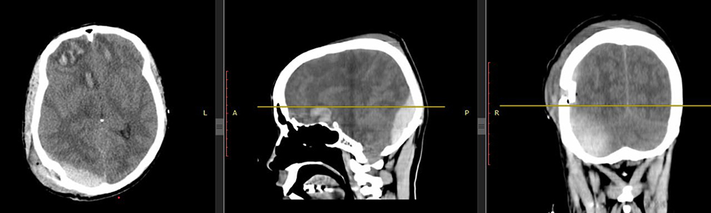 |
Figure 3 Right sizable acute epidural hematoma with mass effect as described, minimal negligible right acute subdural hematoma, and right parietal bone craniotomy. |
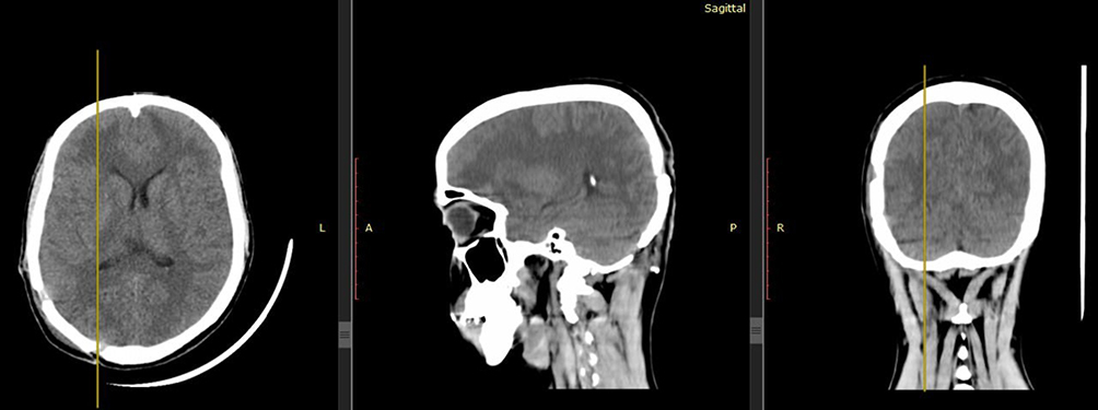 |
Figure 4 Complete evacuation of hematoma. |
Discussion
PFEDH is a rare condition that is often associated with skull fractures and occurs in approximately 0.3% of all craniocerebral injuries. However, it is noteworthy that in our case involving a 25-year-old adult, no palpable skull fracture was evident, despite the presence of PFEDH. This highlights the possibility of PFEDH occurring without associated fractures, particularly in patients with more elastic bones such as children. Despite the absence of a fracture, trauma-induced detachment of the periosteal dura mater and subsequent vascular rupture likely contributed to bleeding in our patient. This is consistent with the characteristic venous origin observed in most PFEDH cases.5 PFEDHs typically arise from injuries to the transverse or sigmoid sinuses following occipital bone fractures. The absence of a visible fracture in our patient shows the complexity of the etiology of PFEDH and the potential for hematoma formation, even without detectable skull fractures. This finding emphasizes the importance of considering PFEDH as a differential diagnosis, particularly in cases of head trauma, regardless of the presence of visible skull fractures.
The classical clinical presentation of extradural hematoma typically involves a brief period of posttraumatic unconsciousness followed by a lucid interval, during which patients may appear asymptomatic before neurological deterioration due to brainstem compression occurs. However, in the case of PFEDHs, this classical description may not apply. Instead, patients often present with a history of road traffic accidents (RTAs) and loss of consciousness (LOC) without experiencing a lucid interval. Despite this atypical presentation, timely diagnosis is crucial for optimal outcome. In this case, the patient presented without a lucid interval and remained conscious throughout the initial evaluation, deviating from the classical presentation. This finding adds to the understanding that PFEDH can present atypically. In addition to PFEDH, the patient also had an associated frontal hemorrhagic contusion, which further complicated the clinical management. In managing PFEDH, bilateral involvement of the infratentorial and supratentorial compartments is rare, but requires special consideration. Surgical intervention is typically required to evacuate hematomas from both compartments when there is a significant midline shift, basal cistern effacement, or brainstem compression.7 PFEDH accounts for approximately 4–7% of all extradural hematomas, a much lower incidence than the more common supratentorial extradural hematomas (EDHs), which are often associated with skull fractures and occur more frequently in the temporal and parietal regions.2,8 The unique anatomical challenges posed by PFEDH, particularly the potential for rapid brainstem compression, make early diagnosis and intervention even more critical than in other types of EDHs.7,9 Studies have suggested that early craniotomy is crucial for reducing intracranial pressure and preventing neurological deterioration.5,7 Compared to other types of extradural hematomas, posterior fossa EDHs present unique challenges because of their proximity to critical brainstem structures.10 This underscores the importance of early and aggressive management to prevent potentially life-threatening complications. The treatment strategies for PFEDHs may involve surgical or conservative approaches. Although a limited percentage of PFEDH cases can be managed nonoperatively, surgery is typically preferred. It is indicated for cases with specific criteria, including hematoma size, midline shift, effaced basal cisterns, compression of the fourth ventricle, and related supratentorial lesions.11 These parameters were carefully monitored during both surgeries to ensure optimal outcomes. Recent advances in diagnostic imaging, including high-resolution CT and MRI, have significantly improved the early detection of PFEDH, allowing for more precise assessment of hematoma size, location, and brainstem involvement.7 In terms of treatment, emerging protocols emphasize early craniotomy to prevent further neurological deterioration, even in cases where there are no overt signs of skull fractures.8 These advances, combined with tailored postoperative care, have improved survival rates and neurological outcomes. This report represents a single case of PFEDH, and, as such, the findings cannot be generalized to all PFEDH cases. Additionally, there are certain limitations to these imaging techniques. Although CT and MRI provided sufficient diagnostic clarity, a midline shift could have provided additional insights. Furthermore, the management of PFEDH lacks standardized decision-making criteria; in this case, clinical judgment plays a critical role in the timing of surgical interventions and postoperative care. Overall, the management of PFEDHs requires individualized treatment plans based on patient-specific characteristics and clinical presentation.12
Conclusion
Our case describes a patient who developed PFEDH and experienced rapid recovery following immediate surgical intervention. This case serves as an example of an uncommon presentation of PFEDH with a transverse sinus rupture. Successful outcomes depend on early diagnosis using cerebral computed tomography, prompt emergency evacuation, and bleeding control measures. This case highlights the importance of maintaining a high index of suspicion for PFEDH in patients with head trauma, even in the absence of typical symptoms such as skull fractures or a lucid interval. The presence of transverse sinus rupture further complicates management, necessitating careful evaluation of the venous structures. Additionally, this case underscores the need for standardized protocols and guidelines to improve the consistency and outcomes of PFEDH management.
Ethics Approval
Institutional approval was not required for the publication of this case report.
Consent
Written informed consent was obtained from the patient for the publication of this case report and any accompanying images. The patient was informed about the purpose of this publication, and consent included permission to share details of the case anonymously for educational and research purposes. Patient identity was protected to maintain confidentiality.
Funding
The authors have no funding to declare.
Disclosure
The authors report no conflicts of interest in this work.
References
1. Sharafat S, Azam F, Khan Z. Posterior fossa epidural hematoma: a 6-year management experience. Pak J Med Sci. 2022;39(1). doi:10.12669/pjms.39.1.6408
2. Verma S, Borkar S, Singh P, et al. Traumatic posterior fossa extradural hematoma: experience at level I trauma center. Asian J Neurosurg. 2018;13(2):227–232. doi:10.4103/1793-5482.228536
3. Prasad GL, Gupta DK, Sharma BS, Mahapatra AK. Traumatic pediatric posterior fossa extradural hematomas: a tertiary-care trauma center experience from India. Pediatr Neurosurg. 2015;50(5):250–256. doi:10.1159/000438488
4. Caruso G, Fiore P, Gorgoglione ML, Ricciardo G, Caffo M. Unusual case of posterior fossa extradural hematoma in a child: review of the literature. Pediatr Neurosurg. 2020;55(6):399–403. doi:10.1159/000511906
5. Siddique MS, Gregson BA, Fernandes HM, et al. Comparative study of traumatic and spontaneous intracerebral hemorrhage. J Neurosurg. 2002;96(1):86–89. doi:10.3171/jns.2002.96.1.0086
6. Kalhoro A, Panezai AS, Hassan S, Rehman L, Javed F. Assessment of modes of injury and outcome in patients with traumatic posterior fossa extradural hematomas: a study of a tertiary care hospital. Pak J Neurol Surg. 2021;25(2):149–155. doi:10.36552/pjns.v25i2.521
7. Parashar VS, Kankane VK, Jaiswal G, Gupta TK. Traumatic posterior fossa extradural hematoma. Roman Neurosurg. 2019;160–165. doi:10.33962/roneuro-2019-030
8. Anurag P, Rajeev B, Radheyshyam M. Factors affecting outcome in posterior fossa EDH: an analytical study at tertiary referral hospital. Roman Neurosurg. 2016;30(2):267–271. doi:10.1515/romneu-2016-0041
9. Finger G, Martins OG, Basso LS, et al. Acute spontaneous subdural hematoma in posterior fossa: great outcome. World Neurosurg. 2018;119:146–150. doi:10.1016/j.wneu.2018.07.220
10. Narasimhan V. Traumatic posterior fossa extradural hematoma–experience at a tertiary care centre. J Med Sci Clin Res. 2019;7(2). doi:10.18535/jmscr/v7i2.170
11. Bhardwaj S, Sharma V, Sharma S, Purohit D, Chopra S. Traumatic posterior fossa hematoma, a rare entity: study of 21 cases. J Neurosci Rural Pract. 2019;10(04):675–682. doi:10.1055/s-0039-1696610
12. Songara A, Patil H, Nayaran S. Traumatic posterior fossa extradural hematoma: case report and review of literature. Inter Surg J. 2016;369–371. doi:10.18203/2349-2902.isj20160261
 © 2024 The Author(s). This work is published and licensed by Dove Medical Press Limited. The
full terms of this license are available at https://www.dovepress.com/terms
and incorporate the Creative Commons Attribution
- Non Commercial (unported, 3.0) License.
By accessing the work you hereby accept the Terms. Non-commercial uses of the work are permitted
without any further permission from Dove Medical Press Limited, provided the work is properly
attributed. For permission for commercial use of this work, please see paragraphs 4.2 and 5 of our Terms.
© 2024 The Author(s). This work is published and licensed by Dove Medical Press Limited. The
full terms of this license are available at https://www.dovepress.com/terms
and incorporate the Creative Commons Attribution
- Non Commercial (unported, 3.0) License.
By accessing the work you hereby accept the Terms. Non-commercial uses of the work are permitted
without any further permission from Dove Medical Press Limited, provided the work is properly
attributed. For permission for commercial use of this work, please see paragraphs 4.2 and 5 of our Terms.