
Back to Journals » International Journal of Nanomedicine » Volume 21
Toward Safe and Effective Gene Therapy: Non-Viral Nanostructured Delivery Systems
Authors Madani F  , Izadi E, Motamedi M, Ganjalikhani Hakemi M, Saltanatpour Z , Webster TJ , Mohammadi SF
Received 25 January 2026
Accepted for publication 30 May 2026
Published 15 July 2026 Volume 2026:21 598766
DOI https://doi.org/10.2147/IJN.S598766
Checked for plagiarism Yes
Review by Single anonymous peer review
Peer reviewer comments 2
Editor who approved publication: Prof. Dr. RDK Misra
Fatemeh Madani,1 Elaheh Izadi,2 Maral Motamedi,1 Mazdak Ganjalikhani Hakemi,3 Zohreh Saltanatpour,2 Thomas J Webster,4– 7 Seyed Farzad Mohammadi2
1Department of Medical Nanotechnology, Tehran University of Medical Sciences, Tehran, Iran; 2Translational Ophthalmology Research Center, Tehran University of Medical Sciences, Tehran, Iran; 3Regenerative and Restorative Medicine Research Center (REMER), Research Institute for Health Sciences and Technologies (SABITA), Istanbul Medipol University, Istanbul, Turkey; 4Division of Pre-College and Undergraduate Studies, Brown University, Providence, RI, USA; 5School of Biomedical Engineering and Health Sciences, Hebei University of Technology, Tianjin, People’s Republic of China; 6School of Engineering, Saveetha University, Chennai, India; 7Department of Pharmacy, University of the Basque Country, Vitoria, Spain
Correspondence: Zohreh Saltanatpour, Email [email protected] Thomas J Webster, Email [email protected]
Abstract: Gene therapy has emerged as a transformative strategy for treating a wide range of genetic and acquired disorders. Despite its potential, clinical translation is hindered by the limitations of viral vectors, including immunogenicity, insertional mutagenesis, limited cargo capacity, and production challenges. Consequently, non-viral gene delivery systems have gained increasing attention as safer, more versatile alternatives. These platforms, including lipid-based nanoparticles, polymers, dendrimers, inorganic nanocarriers, and hybrid systems, offer customizable physicochemical properties, scalable manufacturing, and reduced risk of adverse immune responses. This review systematically expresses recent advances in non-viral vectors, focusing on the key parameters that influence cellular uptake, endosomal escape, nuclear localization, and overall transfection efficiency in various disease, especially cancers. Additionally, we evaluate recent preclinical and clinical studies, highlighting promising translational outcomes and therapeutic applications. By comparing different non-viral strategies and discussing their mechanistic underpinnings, this review underscores the potential of non-viral vectors to overcome the inherent limitations of viral delivery and to drive the development of next-generation gene therapy approaches that are safer, more adaptable, and clinically relevant.
Keywords: nanomedicine, gene therapy, gene delivery, non-viral vectors, nanoparticles
Graphical Abstract:

Introduction
Over the past three decades, significant technological advancements have transformed gene delivery systems from the direct delivery of naked nucleic acids to more sophisticated platforms employing biomaterials as non-viral vectors.1,2 Gene therapy offers great capability to address cancers, inherited disorders, and viral infections, by addressing their genetic origins through gene replacement, inactivation, or the introduction of therapeutic proteins at the target site.3,4 Multiple forms of genetic material such as plasmid DNA (pDNA), messenger RNA (mRNA), small interfering RNA (siRNA), microRNA, antisense oligonucleotides, and genome-editing complexes like CRISPR/Cas9 have been utilized in preclinical settings with considerable success.5,6 However, the clinical translation of these therapies remains limited, primarily due to delivery challenges. Naked nucleic acids are prone to enzymatic degradation and rapid systemic clearance, while their size and negative charge hinder efficient cellular uptake. Additional intracellular barriers, particularly the nuclear membrane, further restrict effective gene expression.7–9 These limitations highlight the need for delivery platforms that can protect genetic material and facilitate targeted intracellular transport.10
Non-viral vectors have emerged as attractive candidates due to their favorable safety profiles, high loading capacity, and design flexibility. These systems include physical methods and synthetic carriers such as polymers and lipid-based nanostructures, although many still face challenges related to delivery efficiency and biological barriers.5–7 Recent advances in nanotechnology have enabled the development of more effective and biocompatible delivery platforms. Despite encouraging preclinical outcomes, the clinical translation of non-viral systems remains challenging and, in some cases, controversial.11 This review focuses on recent advances in non-viral gene delivery systems for the treatment of various disease, especially cancers. Furthermore in vitro, in vivo, and clinical trials are highlighted, with particular emphasis on current clinical trial outcomes and the challenges limiting their successful translation.
Principles of Non-Viral Gene Delivery
Non-viral vectors have emerged as promising alternatives for gene delivery due to their lower immunogenicity, design flexibility, and capacity to carry large genetic payloads.1,12 Unlike viral systems, they lack intrinsic mechanisms for efficient cellular entry and intracellular trafficking; therefore, their successful application depends on rational design strategies that enable them to overcome multiple biological barriers, including cellular internalization, endosomal escape, and sustained gene expression. Advances in nanotechnology, polymer chemistry, and biomaterials science have significantly contributed to the development of non-viral platforms with improved targeting ability, controlled release, and biocompatibility.13–16
Non-viral gene delivery systems can be broadly categorized into physical, chemical, and hybrid approaches. Physical methods, such as electroporation, microinjection, and ultrasound-mediated delivery, rely on external forces to introduce genetic material into cells. In contrast, chemical systems including polymers, cationic lipids, and dendrimers form complexes with nucleic acids (polyplexes or lipoplexes), facilitating cellular uptake through endocytosis or membrane fusion. Hybrid systems combine these strategies to further enhance delivery efficiency.17–19
From a material perspective, non-viral vectors are commonly designed as nanoparticle (NP)-based systems. NPs, typically ranging from 1 to 1000 nm in size, exhibit unique physicochemical properties that distinguish them from bulk materials and make them suitable for gene delivery applications.18,20 These systems protect nucleic acids from enzymatic degradation, improve cellular uptake, and enable controlled intracellular release. Their performance is strongly influenced by key parameters such as size, zeta potential, and polydispersity index (PDI). For biomedical applications, NP size is generally maintained between 1 and 200 nm to enhance tissue penetration and cellular uptake. Surface charge plays a critical role in stability and membrane interaction, where moderate positive charge improves transfection efficiency, while excessive charge may induce cytotoxicity. A low PDI (<0.3) ensures uniformity and reproducibility of delivery behavior.21–25 Based on composition, NP-based non-viral vectors can be classified into several groups, including lipid-based, polymer-based, inorganic, dendrimer-based, carbon-based, and hybrid systems.14,26–28 Each category employs distinct mechanisms to encapsulate, protect, and deliver genetic material.
Lipid NPs (LNPs) are among the most widely used non-viral delivery systems. Due to their structural similarity to biological membranes, they exhibit excellent biocompatibility and can encapsulate both hydrophilic and hydrophobic molecules. This property enables efficient delivery of diverse nucleic acids, including DNA, RNA, and siRNA. Following cellular uptake, LNPs facilitate the release of genetic material into the cytoplasm. However, their stability in biological fluids remains a challenge, as interactions with serum proteins may lead to destabilization and reduced delivery efficiency.29,30
Polymer-based NPs (PNPs) represent another versatile class of non-viral vectors with high loading capacity and tunable properties. Cationic polymers interact electrostatically with negatively charged nucleic acids, forming compact structures that protect genetic cargo and promote cellular uptake via endocytosis.31 Their chemical structure can be modified to enhance biocompatibility and reduce toxicity.32 However, issues such as cytotoxicity at high concentrations and non-specific interactions remain concerns. Parameters such as the nitrogen-to-phosphate (N/P) ratio play a critical role in determining complex stability, transfection efficiency, and overall biological performance.33–35 Figure 1 provides a schematic view of how the N/P ratio affects the efficiency of gene delivery systems. Breakthroughs in polymer chemistry have contributed to the development of biodegradable and biocompatible polymers, which can enhance the safety profile of these vectors while preserving efficacy.33–35
 |
Figure 1 Effect of the nitrogen to phosphate ratio on the functionality of genetic material delivery platforms. |
Inorganic NPs, including gold NPs (Au NPs) and iron oxide NPs (IONPs), offer advantages such as large surface area, ease of synthesis, and high reproducibility.36 The surface functionalization of IONPs is another key feature, enabling the attachment of targeting ligands or other molecules that can enhance cellular uptake and specificity to the targeted tissue. The stability of these NPs facilitates their efficient accumulation within cells, where they can exert an influence on cellular behavior, such as promoting gene expression or altering cell growth.4 Additionally, their resistance under various environmental conditions further supports their potential for gene delivery applications; though challenges related to their potential toxicity and aggregation still need to be considered.37–39
To improve delivery performance, non-viral vectors are often functionalized with specific chemical groups. These modifications serve various purposes:
- Functional groups like peptides or antibodies can be attached to the vector backbone to confer specificity. This ensures that the gene delivery system selectively targets the intended cells, mitigating off-target risks and advancing the therapeutic outcome.40,41
- The backbone can also be modified to strengthen the complex’s integrity while circulating in vivo, such as by adding hydrophilic groups like polyethylene glycol (PEG), which prevents the aggregation of the vector and protects it from immune reactions.42
- pH-sensitive functional groups can be incorporated to make the vector responsive to environmental changes, helping the vector escape the endosome after cellular uptake. At acidic pH levels in the endosome, these groups can trigger a conformational change in the vector that destabilizes endosomes, releasing the pDNA into the cytoplasm.43,44
Another factor that should be considered is the interactions of the gene delivery system within biological compartments, particularly serum proteins, immune cells, and the extracellular matrix. Effective gene delivery depends on the vector’s ability to evade serum protein binding (which can prevent cellular uptake) and resist degradation by nucleases. Modifying the vector with specific functional groups, such as PEGylation, can enhance the vector’s ability to extend systemic circulation time by shielding it from serum proteins and immune surveillance. Additionally, these modifications can help the vector and circumvent quick elimination by the mononuclear phagocyte system (MPS), which is responsible for removing foreign particles from the body.45–47
After cellular uptake, the genetic materials must be transported within the cell to reach its target site, typically the nucleus, for gene expression. The process of intracellular trafficking involves several steps (Figure 2):
- Endocytosis: After binding to the cell membrane, the vector is internalized by the cell through endocytosis. However, once inside the cell, the genetic materials are trapped within an endosome, and the proton-rich environment inside endosomes causing the degradation of genetic substances if it is not released in time.
- Endosomal Escape: Ensuring successful endosomal escape is a critical challenge for non-viral gene delivery approaches. To impede this phenomenon, functional groups can be included to promote the rupture of the endosomal membrane resulting from the decrease in pH within the endosome, allowing the genetic materials to escape into the cytoplasm. Once in the cytoplasm, the genetic materials can be transported to the nucleus.
- Nuclear Localization: Genetic materials must reach the nucleus for gene expression. Some vectors incorporate nuclear localization signals (NLS) or other functional groups which assist in the penetration of the genetic materials into the nucleus, where it can be transcribed and translated into proteins. This step is crucial, in particular, when the aim is to modify the expression of specific genes within the target cells.48–51
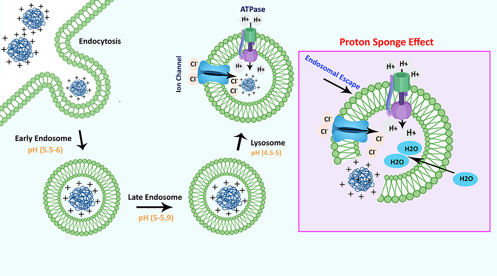 |
Figure 2 The process of successful intracellular trafficking of genetic materials. |
Non-viral platforms are missing the built-in capacity of viruses to effectively traverse cellular barriers. Therefore, during their engineering process, it is necessary to optimize them in order to promote endocytosis and endosomal escape, resulting in the cytosolic delivery of nucleic acids. The achievement of endosomal escape can be facilitated through the utilization of several techniques inherent in the vector, such as the proton-sponge effect aided by NPs.52
Non-Viral Gene Delivery Nanoparticles
In the context of gene delivery, NPs can encapsulate genetic material offering protection from enzymatic degradation while facilitating its delivery. The size, zeta potential and functional groups of NPs are critical factors influencing their cellular uptake, interaction with cellular membranes, and ability to escape endosomal compartments for efficient gene release. Furthermore, the ability of NPs to cross biological barriers, such as the blood-brain barrier (BBB) or tumor vasculature, enhances their potential for targeted gene therapy.18,20
The physicochemical properties of NPs, including size, zeta potential, and PDI, are essential to their performance in gene delivery. The size of NPs typically ranges from 1 to 200 nm for biomedical applications, with smaller NPs facilitating better cellular uptake and tissue penetration, which are crucial for effective gene delivery. Zeta potential, which reflects the surface charge of NPs, influences their stability in suspension and their interaction with cell membranes. A positively charged surface enhances binding to the negatively charged cell membrane, improving transfection efficiency, while excessively high charges may cause cytotoxicity. The PDI, which measures the uniformity of NP size distribution, is ideally kept below 0.3 for consistent and reliable gene delivery, as a narrow PDI ensures uniform behavior across the NP population. Together, these factors govern the effectiveness and safety of NPs as delivery vehicles for genetic materials.21–25 NPs can be categorized based on their composition into five primary groups: organic, inorganic, dendrimer, carbon-based and hybrids.26,27 Figure 3 depicts various types of NPs in gene delivery applications.
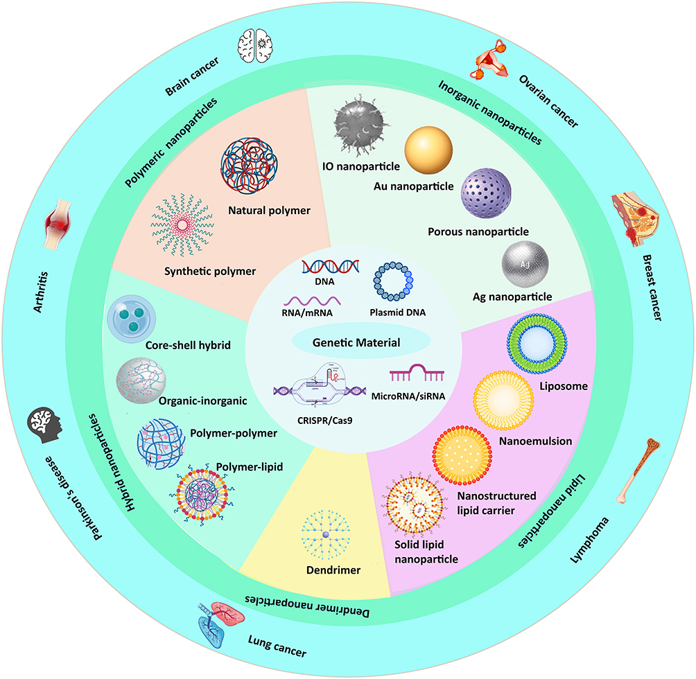 |
Figure 3 Various types of NPs used in gene delivery. |
Lipid NPs (LNPs)
LNPs are very effective delivery systems for various nucleic acids containing DNA, mRNA, siRNA and pDNA.53,54 Administering bare, chemically unaltered nucleic acids directly, is inefficient since they are easily broken down by nucleases and can be potentially harmful because of the immunological response of the host. The utilization of LNPs, which incorporate ionizable or cationic lipid molecules to promote nucleic acid condensation, presents an effective and straightforward approach to address these challenges and effectively transport nucleic acid to tissue.55
Cationic lipids possess a broad spectrum of sizes, a significant capacity for DNA loading, and excellent storage stability.56 The cationic lipids used in liposomes are amphiphilic and composed of a cationic amine group connected to a hydrocarbon chain. One crucial characteristic is their capacity for electrostatic interaction between their positively charged head group and the negatively charged nucleic acids enabling encapsulation of the nucleic acids.57 There is a wide range of commercially available LNPs and lipofectamine is a commonly referenced example.58 Nevertheless, the application of LNPs is limited due to their short-term expression levels and cytotoxicity.59 Various attempts have been made to improve LNPs formulations while improving various factors that can make them suitable for clinical use. These factors include: (i) being easy to replicate and scale up,60 (ii) effectively encapsulating genetic materials,60 (iii) controlling their physical and chemical characteristics such as size, charge and hydrophobicity,61 (iv) assessing their pharmacokinetic properties such as preventing fast clearance and accumulating in specific tissues,62 (v) studying the processes of internalization; and (vi) understanding the principles of nucleic acid release.63
Liposomes
Liposomes are a kind of LNP and as the oldest version of LNP, are structures consisting of either unilamellar or multilamellar phospholipid bilayers that surround an aqueous core, allowing for the encapsulation of genetic materials. The preparation of liposomes is induced by the interaction between polar head groups (hydrophilic) and nonpolar tail groups (hydrophobic) of phospholipids.54 Widely used in numerous therapeutic applications, liposomes serve as carriers for drugs and genes owing to intrinsic potential in biodegradation, effectiveness, low toxicity, and convenient preparation.57,64 One remarkable example of gene delivery via LNPs includes Onpattro®, an invaluable tool for gene regulation in the liver.65 Several other nucleic acid drugs based on LNPs have been subjected to clinical evaluation for the management of genetic diseases, cancers, or infectious disease.66
Micelles
Micelles are nanostructures formed through the self-assembly of amphiphilic structures (polymers or lipids) characterized by hydrophilic and hydrophobic parts, such as surfactants or block copolymers. These molecules spontaneously organize into a spherical or cylindrical shape in an aqueous environment (10 to 100 nm).67 Micelles are endowed with properties that enable them to be suitable options for gene delivery systems. Their structure enables the encapsulation of both aqueous and lipid-soluble drugs within the hydrophilic shell and hydrophobic core, respectively. This property enables the encapsulation of a broad range of genetic materials, such as pDNA, siRNA, and mRNA.68,69 Another advantage of using micelles in the gene delivery field involves enhancing the bioavailability of compounds with limited solubility, which is applicable for hydrophobic nucleic acids. The small size of micelles promotes efficient cellular uptake through endocytosis, a vital process in gene delivery. Following internalization, micelles can evade entrapment within endosomes, which is crucial for releasing the encapsulated genetic material into the cytoplasm for effective gene expression.68,70,71
Solid Lipid NPs (SLNs)
SLNs are a type of nanostructure constructed from solid lipids that remain solid at both room and body temperature which have received great recognition in the realm of advanced delivery platforms (50 to 1000 nm). Their solid lipid core enhances stability by protecting encapsulated compounds from degradation.72–74
Controlled release, along with surface modification using targeting ligands, enhance the efficiency of SLNs in gene delivery and reduce off-target effects. Moreover, SLNs have greater potential for gene therapy because of their capability to breach cellular and tissue barriers, such as the BBB, and their non-toxic profile. Additionally the concept of endosomal escape in gene delivery systems, such as SLNs, pertains to the ability of these nanostructures to release genetic material from the endosome into the cytoplasm.75
Nanostructured Lipid Carriers (NLCs)
NLCs (50 to 500 nm) are advanced nanostructures that have revolutionized delivery system technology by intelligently combining solid and liquid lipids. The hybrid structure of the NLC matrix provides a significant improvement in performance such as: stability, flexibility, and loading capacity compared to SLNs. High shear homogenization, microemulsion, or solvent evaporation are common preparation methods.76,77 The incorporation of a semi-crystalline matrix in the formulation enhances its colloidal stability, which is contingent upon the quantity of solid lipid employed. However, it is stated that NLCs are more readily synthesized and sterilized compared to other SLNs, and they exhibit less toxicity.78
A study by Erasmus et al successfully generated an NLC formulation and paired it with self-amplifying mRNA producing Zika virus antigens. Zika virus-rvRNA was combined with NLC at N:P ratios of 50 and 15, respectively. The NPs had mean diameter of 40–100 nm with zeta potential from 15 to 28 mV. This combination resulted in complete defense against the Zika virus in C57BL/6 mice, immunized with a single intramuscular (I.M.) dose (10 ng).79
Despite their potential, NLCs face key limitations in gene delivery applications, including suboptimal nucleic acid loading capacity, instability of lipid–nucleic acid complexes, potential cytotoxicity, and manufacturing complexities. These challenges have hindered their progression into clinical development.80 As of 2023, no NLC-based formulations for mRNA delivery or vaccines have received approval from the FDA.80,81 It is predicted that by optimizing formulation methods and conducting long term studies, clinical applications of these nanostructures will be improved over the next decade.82
Polymer NPs (PNPs) and Polymeric Micelles
In the last twenty years, there has been significant utilization of biodegradable polymers in biomedical applications.83,84 Biodegradable polymers may be broadly grouped into two distinct classes namely natural and synthetic polymers.32,85 Both of them have distinct merits and drawbacks. One notable characteristic of natural polymers is their exceptional biocompatibility, bioactivity, and ability to undergo cell-activated proteolytic breakdown.32 Nevertheless, the exact purification, characterization, and identification of the chemical composition and content of natural polymers pose significant challenges, leading to an issue of batch-to-batch variance.84,86
The formulation optimization of PNPs and micelles for gene delivery has attracted considerable interest recently because of the numerous advantages they offer. Micelles possess characteristics that make them well suited for use in gene delivery systems. Their architecture allows them to carry both water-soluble and lipid-soluble compounds, with hydrophilic substances accommodated in the outer shell and hydrophobic ones in the core. As a result, they can encapsulate a wide variety of genetic materials, including pDNA, siRNA, and mRNA.68,69
Despite their considerable therapeutic potential, several critical challenges must be addressed to fully realize their clinical utility. Among these are the requirements of biocompatibility, biodegradability, and effective encapsulation of nucleic acids, enabling controlled and targeted delivery, tailoring physicochemical properties, enhancing cellular uptake and endosomal escape, prolonging systemic circulation while minimizing clearance, and ensuring scalability and reproducibility of production processes.32,87 Collectively, these considerations underscore the necessity for the continued optimization of PNPs as non-viral gene delivery vectors to improve their safety, efficacy, and translational potential.32,88
Natural Polymers
Natural polymer NPs with a size range of about 50–200 nm89 have significant advantages including biocompatibility, biodegradability, and functional versatility. Alginate, gelatin, albumin, polysaccharides, chitosan and cyclodextrins, are widely used to fabricate natural PNPs for gene delivery. For example, chitosan is a positively charged polysaccharide, that is widely used for encapsulating nucleic acids and drugs because of its property of adhering to mucus and its ability to form NPs with controlled release profiles.90 Alginate is an important material in the development of biomedical technologies extracted from brown seaweed. It is mainly used to develop hydrogels and NP formulations and biological scaffolds. It should be mentioned that the excellent biocompatibility and gelation properties under physiological conditions make hydrogels a very appropriate option in biomedicine.91 Alginate NP sizes range from nanometer to sub-micron (~50–300 nm depending on their preparation method).92 Moreover, chemical modification of biopolymers has the potential to increase their stability and responsiveness to environmental stimuli and efficiency. Natural polymers with their inherent biodegradability and low toxicity result in the creation of safer and more effective therapeutic platforms.93
Gelatin and albumin are the predominant proteins utilized in gene-delivery vectors. Gelatin polymers are extensively applied for the fabrication of vectors because of their notable attributes of minimal antigenicity and excellent biocompatibility.94 Gelatin is derived from animal collagen. This polymer has amphiphilic properties, characterized by the presence of cationic and anionic charges, together with hydrophobic groups, at a predominant 1:1:1 ratio.95 It possesses several reactive groups on its surface, which serve to connect and alter various molecules, thus, endowing the polymer with a diverse range of functions. Gelatin possesses amino acid sequences in its structure, namely the cell adhesive Arg-Gly-Asp (RGD) peptide, which sets it apart from other polymers.96–99
Polysaccharide-based gene-delivery vectors have predominantly utilized chitosan, which is made up of irregularly scattered β-(1-4)-linked D-glucosamine and N-acetyl-D-glucosamine, owing to its inherent positive charge. Chitosan expresses meager solubility in physiological conditions and because of its low pKa, it has inefficient complexation potential.100 Chitosan is a polysaccharide with positive charges, which is derived from chitin (an abundant biopolymer found primarily in fungus and arthropods).101 Chitosan is biocompatible and biodegradable, making it an attractive material for gene delivery. It can form NPs by electrostatically binding, facilitating the intracellular transfection of genetic material, because of its cationic nature.101–105
Cyclodextrins (cyclic oligosaccharides) exist naturally and are classified as natural polymers106 linked by (α-1,4) bonds, arranged in a basket-like pattern exhibiting an amphipathic nature with an “endo-exo” structure. The stability of structures formed through the interaction of unmodified cyclodextrins with pDNA is limited, and their impact on transfection efficiency is relatively modest. The cavity size is precisely defined at the sub-nanometer scale and varies according to the type of cyclodextrin, with approximate internal diameters of ~0.5 nm for α-cyclodextrin, ~0.6–0.7 nm for β-cyclodextrin, and ~0.7–0.8 nm for γ-cyclodextrin.107
Synthetic Polymers
Synthetic polymers have been known as famous substances for the development of nanostructures and their size ranges can be tuned from nanometers to micrometers.32,108 In contrast to natural polymers, synthetic polymers offer the benefit of exact control over molecular weight, chemical composition, and structural characteristics, enabling the precise adjustment of their performance and functionality.109 PNPs expressed promising results for controlled release systems and, furthermore, they can be easily modified with targeting ligands or genetic materials; providing multiple functions simultaneously.110
Aliphatic polyesters are known for biodegradation84,111 and various forms of polyesters, which differ based on the monomer, have been widely utilized for gene-delivery in recent years. Polyester-based gene-delivery polymers that are frequently employed in various applications encompass polyethylenimine (PEI) poly(beta-amino ester) (PBAE), PEG, poly-L-lysine (PLL), polycaprolactone (PCL) and poly(lactic-co-glycolic acid) (PLGA) which have been chosen as potential vectors.112 In this review manuscript, we will describe the structures that have been studied more extensively than others.
PEI demonstrates the proton sponge effect, which is a phenomenon where positively charged polymers or NPs typically in the nanometer range (~50–300 nm), once inside acidic endosomal compartments accumulate protons, and this accumulation leads to an increase in osmotic pressure. The osmotic imbalance disrupts the integrity of the endosomal membrane allowing the encapsulated genetic material to escape into the cytoplasm.113,114 The buffering property of the substance induces swelling and rupture of endosomes, leading to polyplexes release. Endosome escape is believed to be caused by several causes, apart from the proton sponge effect. Protonation of PEI leads to the elongation of the polymer chain because of electrostatic repulsion, resulting in the swelling of the polymer.95,115–118 To achieve cellular absorption through ligand-mediated mechanisms, PEI can be combined with PEG to protect the surface charges of PEI before attaching a ligand like the anti-HER1 antibody for precise delivery.119,120
PBAE is a cationic polymer which demonstrates pH-responsive properties, providing useful properties for nucleic acid delivery.87,121–124 The ability to modify PBAE structures to reach sustained release makes them suitable for numerous gene delivery platforms.96
PEG is a hydrophilic, biocompatible polymer broadly exploited in gene delivery. PEG chains can be linked to NPs or biomolecules; this procedure increases the stability and pharmacokinetic properties of gene delivery systems by reducing opsonization, lowering renal clearance, and prolonging circulation time in the bloodstream. In gene delivery, PEGylated NPs, such as LNPs and PNPs (eg, PEG-PLGA), facilitate systemic administration, and promote targeted cellular uptake that improve transfection efficiency. Nevertheless, cellular internalization and endosomal escape are affected by extreme PEGylation.45
Biswal et al designed a pDNA expressing dihydrofolate reductase (DHFR) siRNA that was condensed with folate-PEG-PEI and introduced into KB cells and A549 cells. This complex formed self-assembled polyelectrolyte complexes with acidic genetic materials and cationic polymers. The study demonstrated that siRNA was successfully transported into KB cells, resulting in the suppression of DHFR. Conversely, A549 cells had an inability to internalize the siRNA nanostructure. It was determined that the complexation enabled targeted delivery by FR positive cells, thereby establishing them as a delivery system with an excellent level of specificity.125
Zhang et al designed a system focused on the controlled delivery of Pt (IV) for platinum-resistant ovarian cancer (PROC). The findings from an in vivo study on BALB/c nude mice showed that the NPs had an ability to selectively target tumor sites, resulting in enhanced gene silencing and antitumor effectiveness via subcutaneous (S.C.) administration.126
PLL is a positively charged polymer composed of repeating lysine units; this structure allows PLL to form electrostatic bonds with negatively charged nucleic acids, thereby, enhancing their encapsulation and facilitating their delivery. The biocompatibility and ability of PLL to facilitate cellular uptake make it a popular choice for gene delivery systems. This application enhances the efficiency of transfection preserving the genetic material from degradation.127
Li et al designed a PNP system that exhibited long-lasting gene transport vector. This system was biodegradable and demonstrated effective gene transfer when employing the pDNA expressing GFP (pGFP). The NPs consisted of three parts: (i) a PEG outer layer, (ii) a shell of PLGA, and (iii) and a core of PBAE/pGFP. The average particle diameter was about 165 nm with a poly dispersity index (PDI) of ~0.1 indicating that the particles were very homogeneous in size at about 130 nm. Furthermore, it was observed that adding the cationic polymer PBAE to PLGA-PEG NPs significantly increased pGFP entrapment efficiency (from 3% to 97%).6
Another investigation by Pretzer et al, was performed to synthesize a biotin-conjugated block copolymer micelle. The NPs were 50–60 nm and the zeta-potential was 4 mV. In this study at a N/P ratio= 20, the transfection efficiency for pDNA was variable in various formulations, however by enhancing the N/P ratio ≥ 100, polyplexes expressed similar transfection efficiencies about 72% and 85% for N/P =100 and 300, respectively.128
Inorganic NPs (NPs)
Controlling the size, morphology and surface charge of inorganic NPs allows for the optimization of cellular uptake and endosome escape: two key factors in efficient gene delivery. These features, combined with magnetic fields or light as external stimuli, make inorganic NPs a promising tool as further described below.129
AuNPs
AuNPs are considered ideal candidates in this realm because of biocompatibility, chemical stability and extensive surface modification capabilities. Through thiol bonds or electrostatic interactions, AuNPs can be modified. These NPs are considered promising alternatives to conventional gene delivery methods by providing a favorable balance between cytotoxicity and delivery efficiency.130
Silver NPs (AgNPs)
AgNPs are prominent candidates for targeted gene therapy for infection which results from intrinsic antimicrobial properties and surface modification capabilities. By surface modification with cationic agents, the efficiency of gene transfer will be improved. These NPs exert both antimicrobial effects and enhance genetic material delivery.131
Iron Oxide NPs (IONPs)
Among all inorganic NPs, IONPs have been studied more and in recent studies, various forms of IONPs have been designed specifically to diagnose and treat cancer.132 Coating these NPs with polymers or lipids improve their biocompatibility and facilitates the loading of nucleic acids, thereby increasing their potential as gene transfer vectors.131 The size of these entities renders them highly suitable for surface functionalization. These alterations in the diameter of NPs serve as the foundation for various pharmacological and biological uses, including diagnosis, drug and gene delivery.133 Two approaches for gene loading in IONPs are:
- The initial method involves the application of cationic chemicals on the surface of IONPs, followed by their binding to negatively charged genes via electrostatic absorption. PEI can be applied as a layer-by-layer coating on the surface of IONPs. The ultimate altered IONPs possess a positive charge to facilitate gene loading. In addition, PEI has the ability to directly alter the surface of IONPs, forming a core-shell platform.134 According to the results of a study by Bi et al, the platform can bind effectively with siRNA, leading to the formation of evenly distributed NPs.135 In addition to PEI, other polycations such as poly-arginine, PLL, and chitosan have the potential to be employed for creating surface modification on IONPs to enhance gene binding efficacy.136,137
- The second method is the utilization of gene/carrier complexes, wherein the surface charge does not play a decisive role.135,138
The prospective applications of IONPs in biological domains are attributed to their unique qualities, such as a greater surface-volume ratio, increased surface energy, and severe reactivity, which can be achieved by reducing their size to below 100 nm.139
Silica NPs
Silica-coated metal NPs significantly boost gene delivery system efficiency by creating a stable protective structure. These NPs consist of two main parts: a metal core (usually gold, silver or iron) and a porous silica shell that plays a key role in protecting and delivering genetic material. Surface functionalization of these NPs with amine groups, creates a positive charge that forms bonds with the negative charge of nucleic acids via electrostatic interactions which improves the stability of the gene-NPs complex.140
Dendrimers
Dendrimers are branched, tree-like macromolecules composed of a central core and repeating units. Their structure is defined at the nanoscale and their uniformity and their multiple end groups can be modified to bind to nucleic acids, which increases the loading capacity and stability of genetic material. Dendrimers also facilitate efficient cellular uptake and endosome escape, which are essential for successful gene delivery. In addition, their size and shape can be tuned to optimize biodistribution and reduce toxicity.141,142 Polyamidoamine (PAMAM) dendrimers are a class of three-dimensional branched platforms characterized by active amine groups.143
Upon endocytosis of the PAMAM/nucleic acids complex, the amino groups undergo protonation, leading to a substantial inflow of chloride ions. The osmotic pressure within the endosome subsequently increases and ultimately induces endosomal deterioration.52 The second generation of PAMAM dendrimers have been developed with specific modifications to improve gene delivery to the brain. These dendrimers contain arginine (R) and histidine (H) residues, which demonstrate a key role in increasing efficiency by attaching the oligopeptide RRHRH to their surface. Cytotoxicity research showed that PAMAM dendrimers were non-toxic to various cell lines. The results of in vivo investigations demonstrated dendrimers had superior gene delivery efficiency.144 In another study, the polyplex structure was formed by dendrimers via self-assembly at several N/P ratios including: 0.5, 1, 2, 5, 10 and 20. The results of that study demonstrated that this structure could be used as a new and environmentally friendly method for gene or drug delivery, while being less expensive and, unlike traditional carriers, biodegradable.145
Hybrid NPs
Hybrid NPs are composites made up of multiple organic and inorganic components. These NPs combine the beneficial properties of specific components to improve their performance in various applications. In this way, they can offer both types of materials in a single constituent unit. This combination helps improve the efficiency of NPs in gene delivery, making them suitable for medical and biological applications because it enhances properties such as stability, biocompatibility, drug loading, and release control. The organic components, like polymers and lipids, are biocompatible and biodegradable, due to their similarity to biological compounds. These structures allow for efficient gene loading and protection, but inorganic materials like metal or silica NPs improve stability, targeting, and controlled release. The key benefit of this combination is to create synergy between the properties of various organic or inorganic components. Functionalization with ligands (specific for cell targeting) and tuning surface features to improve cell entry and endosome escape are two key mechanisms which are implicated in the function of hybrid NPs for gene delivery. Hybrid NPs create a multifunctional platform for the delivery of various genetic materials such as pDNA, siRNA, and mRNA due to their combined features. This technology has been particularly successful for cancer treatment and genetic diseases.146–148
Hybrid NPs can solve the challenges associated with single substance NPs in gene delivery, such as the toxicity of NPs. By combining different properties of organic and inorganic materials, hybrid NPs can also offer additional advantages including: enhanced transfection efficiency, additional capabilities, and the capacity to modulate gene expression kinetics.149 In a study, novel biodegradable PNPs for gene delivery were developed, which are a combination of two polymers: PLGA and PBAE. While PLGA alone has low gene delivery efficiency, its combination with PBAE increases the strength, stability and persistence of the particles. This combination also facilitates fine-tuning of the particle dimensions, which helps in passive targeting to different tissues or phagocytic antigen-presenting cells (APC). Cellular and animal investigations with nanoplatforms of 75–85% PLGA and 15–25% PBAE are the most efficient for gene delivery to APCs, while being relatively safe. These particles have a stimulatory influence on APCs contributing to their value in genetic vaccination strategies.150
When lipid shells are paired with a polymer core, they facilitate improved targeting as well as binding of particles to specific cells. This is especially beneficial for hard-to-transfect cells, such as immune cells.151 Persano et al showed that PBAE/mRNA complexes (polyplexes) coated with a lipid layer with a diameter about 50 nm induced complete gene transfer in DC2.4 cells (similar to dendritic cells). In contrast, uncoated polyplexes showed no detectable gene transfer in DC2.4 cells.152 Subsequently, polyplexes (coated with lipids) were employed for the purpose of delivering mRNA encoding ovalbumin to immune cells through subcutaneous administration. This process resulted in the stimulation of immune cells, as evidenced by measuring IFN-β levels in blood, as well as the activation of T cells and lymph node cells.152
Moreover, lipid coatings have the ability to provide protection to the cationic polymer core, hence mitigating the issue of excessive positive charge-induced toxicity. This becomes highly pertinent within the framework of in vivo applications.153 Additionally, it has been reported that liposome-coated PBAE particles exhibit negligible toxicity in vivo.153,154
Achieving effective transfection in vivo is strongly governed by the balance between lipids and PEG-lipids. Therefore, it is of the utmost importance to optimize these ratios to achieve greater efficacy of transfection.153 Researchers have succeeded in developing efficient IONPs for DNA delivery using low molecular weight linear PEI (Mw 423 and 1800). This investigation expressed that a N/P ratio of 1 effectively enhanced the DNA binding capacity in comparison to PEI alone.155 Wang et al developed multifunctional NPs based on IONPs using PEI coatings which had the size range of 10–15 nm. These NPs were used for both MRI imaging and gene delivery to cancer cells. PEI as a cationic polymer, adsorbs siRNA through electrostatic interactions and prevents in vitro enzymatic degradation. This system effectively restrains cell growth and promotes both apoptotic and autophagic pathways in glioblastoma (U251) cells. This novel technology has great potential for personalized cancer medicine.156
In another study conducted by Du et al, a magnetic mesoporous silica loaded microbubble (M-MSN@MBs) was designed specifically for the purpose of ultrasound-mediated imaging and gene transfection. The pDNA incorporated in M-MSNs and the lipid microbubbles were loaded with pDNA-carrying M-MSNs. These NPs had a mean diameter of 1120 ± 345 nm with a zeta potential of −17.65 ± 2.8 mV. The findings of this study suggested that the designed vector had favorable biocompatibility, stability for DNA binding, performance in ultrasonic imaging, and response to magnetic stimuli. The M-MSNs that were modified with PEI showed significant efficacy in preserving the loaded pDNA from enzyme breakdown.157
In research by Lin et al, a nanostructure for pDNA delivery to MSCs was fabricated. The nanostructures that were smaller than 400 nm, targets the CD44 receptor on MSCs via hyaluronic acid (HA). The lipid-polymer nanostructures have two parts of DOTAP for DNA compaction and PLGA-PEG-HA for stability and controlled release. With its high transgene efficiency and meager cytotoxicity, this system has great potential for numerus gene therapy applications.158
Wang et al investigated the simultaneous treatment of high gene transfection and magnetic resonance imaging (MRI) contrast to enable MRI-guided visual gene transfection.159 In this study, polymers grafted onto the surface of IONPs@SiO2 NPs achieved both enhanced transfection efficiency and real-time MRI by utilizing an external magnetic field. In this investigation, the transfection efficiency on C6 and HepG2 cell lines using pDNA pRL-CMV as the reporter gene exhibited a threefold increase when subjected to a magnetic field compared to the control culture. These structures had a size between 500–600 nm and had a one dimensional peapod-like shape that improved penetration into cells as it was revealed that under the influence of a magnetic field, the transfection efficiency could be further enhanced.159 While there is a large number of hybrid NPs, only some of them are presented and summarized in Figure 4. The major classes of non-viral gene delivery systems, along with their mechanisms of nucleic acid encapsulation, in vivo distribution profiles, advantages, and safety considerations, are summarized in Table 1.
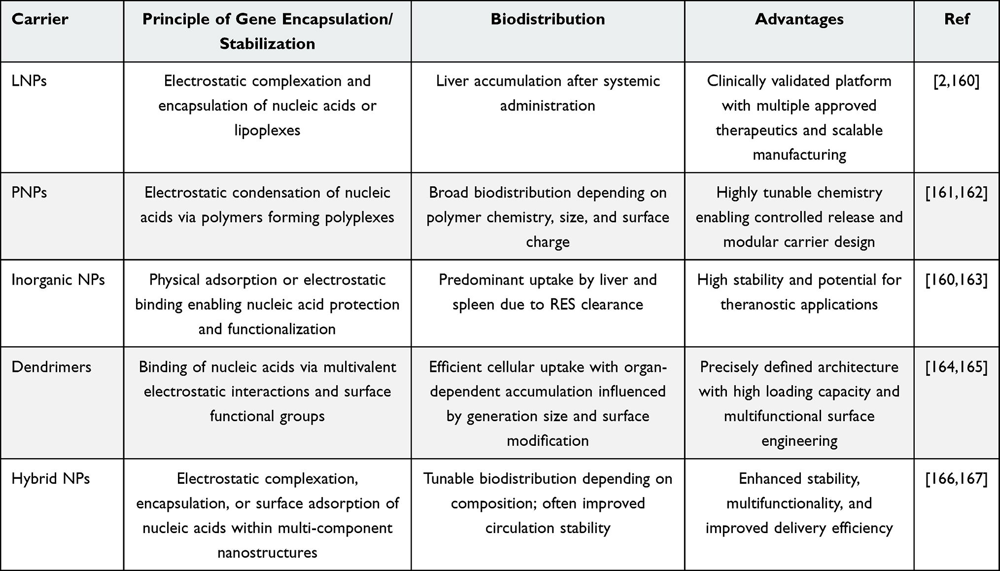 |
Table 1 Major Classes of Non-Viral Gene Delivery Systems and Their Characteristic Mechanisms of Nucleic Acid Encapsulation, Biodistribution Behavior, Advantages, and Safety Considerations |
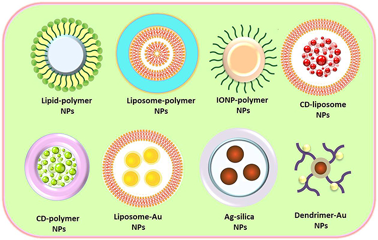 |
Figure 4 Schematic representation of some types of hybrid NPs formed from combinations of diverse material classes. These include organic–organic hybrids (lipid-polymer NPs and liposome-polymer NPs), organic–inorganic hybrids (IONP-polymer NPs, CD-liposome NPs, CD-polymer NPs and liposome-Au NPs), and inorganic–inorganic hybrids (Ag-silica NPs and dendrimer-Au NPs). The figure illustrates the structural diversity and enhanced multifunctional properties resulting from these hybrid configurations. |
Therapeutic Applications of Non-Viral Gene Delivery Systems
Non-viral gene delivery systems have been widely explored across a broad range of disease settings, owing to their favorable safety profile, tunability, and capacity to deliver diverse genetic cargos. Their applications span cancer therapy as well as genetic, infectious, and neurological disorders, in addition to vaccination strategies.168 However, a substantial proportion of studies remains limited to in vitro investigations. Notably, in vitro models often fail to accurately replicate the complexity of in vivo environments, leading to discrepancies in transfection efficiency, cellular uptake, and toxicity. Furthermore, variations in cell types, culture conditions, and analytical methods reduce reproducibility and hinder direct comparisons across studies.169,170 The following sections summarize the major disease areas in which non-viral gene delivery systems have been investigated, along with representative recent studies.
Cancer Therapy
Cancer represents one of the most extensively investigated applications of non-viral gene delivery systems. These platforms enable targeted modulation of gene expression within tumor cells and the tumor microenvironment.171 Common strategies include silencing oncogenes using siRNA,172 restoring tumor suppressor genes through pDNA, and delivering mRNA to induce the expression of therapeutic proteins.173 NPs have demonstrated the ability to preferentially accumulate in tumor tissues through the enhanced permeability and retention (EPR) effect. Surface functionalization with targeting ligands further improves selectivity toward cancer cells, minimizing off-target effects.8 In addition, NPs have been employed to co-deliver multiple therapeutic agents, such as chemotherapeutics and nucleic acids, resulting in synergistic antitumor effects.174 Beyond direct tumor targeting, non-viral systems are also being explored for cancer immunotherapy. For example, the delivery of mRNA encoding tumor-associated antigens can stimulate antigen presentation and activate cytotoxic T-cell responses.175 Despite these advances, challenges such as heterogeneous tumor environments, limited penetration into solid tumors, and variable transfection efficiency continue to hinder clinical translation.176
Genetic Disorders
Non-viral gene delivery offers a promising approach for the treatment of inherited genetic diseases by enabling gene replacement, correction, or silencing. Delivery systems have been developed to transport functional copies of defective genes or genome-editing tools such as CRISPR/Cas9 to target cells. However, achieving efficient and sustained gene expression in target tissues remains a major limitation.177 Recent advances in NPs engineering and targeted delivery strategies have improved the therapeutic potential of these systems. For example, lipid- and polymer-based carriers have demonstrated enhanced gene expression and partial functional recovery in preclinical models of cystic fibrosis178 and muscular dystrophies.179 Despite these encouraging results, challenges related to tissue-specific targeting and long-term expression continue to limit clinical translation.30
Infectious Diseases and Vaccination
Non-viral gene delivery systems have gained significant attention in the prevention and treatment of infectious diseases, particularly through nucleic acid-based vaccines. Recent advances have demonstrated that LNP-mediated delivery of mRNA enables rapid and efficient antigen expression, leading to robust humoral and cellular immune responses. Notably, the clinical success of mRNA vaccines against coronavirus disease 2019 (COVID-19) represents a major milestone, highlighting the translational potential of non-viral delivery platforms.180 These systems function by protecting nucleic acids from degradation, facilitating cellular uptake, and promoting antigen expression in host cells, which subsequently triggers immune activation. In addition to mRNA vaccines, non-viral vectors have been utilized to deliver DNA vaccines, siRNA, and antisense oligonucleotides for the inhibition of viral replication and modulation of host immune responses.181 Targeted delivery strategies have further enhanced vaccine efficacy. For example, NPs-based systems can be engineered to accumulate in lymphoid tissues, improving antigen presentation and immune activation. This lymph node-targeting capability is particularly important for inducing strong adaptive immune responses and has been widely explored in next-generation vaccine design.182,183
Neurological Disorders
The treatment of neurological diseases using gene therapy is particularly challenging due to the presence of the BBB, which restricts the delivery of therapeutic agents to the central nervous system.184 NPs have been engineered to overcome this barrier through size optimization, surface modification, and ligand-mediated targeting.184 These systems have shown potential in delivering therapeutic genes or RNA molecules for conditions such as Parkinson’s185 and multiple sclerosis.186
Cardiovascular and Inflammatory Diseases
Non-viral gene delivery has also been investigated for cardiovascular and inflammatory conditions. In cardiovascular diseases, gene delivery strategies aim to promote angiogenesis,187 tissue regeneration, and repair following injury.188 Similarly, in inflammatory and autoimmune disorders, nucleic acid-based approaches can modulate the expression of key cytokines and signaling pathways.189
Table 2 summarizes recent in vitro and in vivo studies on gene delivery nanostructures.
 |
Table 2 Recent Cellular/Animal Applications of Non-Viral Gene Delivery Systems |
Clinical Trials of Non-Viral Gene Delivery Systems
In recent years, non-viral nanostructures representing a hopeful alternative to viral-based gene delivery have been studied clinically. With the advancement of nanotechnology, it is expected that non-viral nanostructures will be increasingly used to treat various disease like cancer and genetic diseases.35,231 Figure 5 depicts clinical trials of gene-delivery structures, performed via the application of NPs. LNPs have been identified as an efficient delivery system for CRISPR/Cas9 components in gene editing therapy. They are used in the treatment of genetic disease such as hereditary transthyretin amyloidosis with polyneuropathy (ATTRv-PN) due to their bioavailability, safety, and immunogenicity. The first clinical trial using LNP for CRISPR/Cas9 delivery was performed by Intelaya Therapeutics. NTLA-2001 delivered via I.V. infusion for CRISPR/Cas9 gene therapy which targeted the TTR gene in hepatocytes for the treatment of ATTRv-PN. In that study by Intelaya Therapeutics, NTLA-2001 was given to patients at different doses, and initial results showed significant reductions in TTR protein levels.
 |
Figure 5 Clinical trials of nanostructures as gene delivery systems. |
In another clinical trial (NCT01621867), researchers investigated the potency and safety aspects of a non-viral gene therapy approach via cationic liposomes for treating cystic fibrosis (CF). This Phase II, randomized, double-blind, placebo-controlled trial involved repeated nebulization of a pDNA encoding the cystic fibrosis transmembrane conductance regulator (CFTR) gene. The goal was to restore functional CFTR protein expression in the airway epithelium, thereby improving lung function in CF patients. Unlike viral vectors, the non-viral delivery method used here aimed to minimize immune responses and allow for repeated dosing. The study concluded that the treatment showed a modest but beneficial effect on lung function, and no considerable treatment-associated side effects were reported, reflecting good patient tolerance. These findings support the potential of non-viral CFTR gene therapy as a promising strategy for cystic fibrosis.
In another clinical trial, the efficacy of a non-viral delivery system was evaluated through the use of an mRNA-based cancer vaccine known as Autogene Cevumeran (RO7198457), in combination with the immune checkpoint inhibitor atezolizumab (NCT03480152). This Phase 1/2 study focused on participants with locally advanced or metastatic tumors and aimed to determine the safety, tolerability, and immune activation caused by the treatment. Autogene Cevumeran utilizes a non-viral platform to deliver mRNA encoding patient-specific neoantigens, designed to trigger a focused immune reaction directed at cancer cells. The non-viral nature of the LNPs for mRNA delivery allowed for flexibility in design, rapid manufacturing, and a reduced risk of insertional mutagenesis. The study was completed and demonstrated that the mRNA-based vaccine, when used in combination with atezolizumab, could generate a neoantigen-specific T cell response, supporting the promise of non-viral gene delivery in personalized cancer immunotherapy. Table 3 summarizes representative FDA-approved nucleic acid therapeutics and gene therapy products, highlighting their therapeutic modality, delivery strategy, and major clinical applications.
 |
Table 3 Representative FDA-Approved Nucleic Acid Therapeutics and Gene Therapy Products, Including mRNA Vaccines, siRNA Therapeutics, Antisense Oligonucleotides, Viral Vector-Mediated Gene Therapies, and CRISPR-Based Genome Editing Platforms, Together with Their Corresponding Delivery Systems and Therapeutic Applications |
Future Directions of Non-Viral Gene Delivery Technologies Employing Nanostructures
The efficiency of gene therapy is highly contingent upon the vector’s effectiveness in ensuring both secure and specific gene delivery. Poorly designed vectors might trigger non-specific interactions, low transfection efficiency, or even adverse phenotypic outcomes.239 Although viral vectors such as retroviruses, lentiviruses, adenoviruses, and adeno-associated viruses (AAVs) exhibit high gene transfer efficiency, their broader application is constrained by several technical and clinical limitations. These include immunogenicity and the risk of insertional mutagenesis, limited cargo capacity (eg, AAV ≈ 4.7 kb), which restricts delivery of large genes or complex regulatory elements, and challenges in large-scale manufacturing, such as high production costs, scalability issues, and lot-to-lot variability.4,240,241 These limitations have driven increasing interest in non-viral vectors, including physical methods (eg, electroporation, microinjection, and ultrasound) and chemical carriers such as NPs.4
Nanotechnology has become an integral component of gene therapy, offering versatile platforms for non-viral gene delivery. However, clinical translation remains limited due to ongoing concerns regarding safety, efficacy, and specificity. To overcome these barriers, future research must focus on a deeper mechanistic understanding of NP–cell interactions, including cellular attachment, endocytosis, intracellular trafficking, and gene expression pathways. Elucidating these processes will allow for the rational design of nanocarriers that can efficiently navigate extracellular and intracellular barriers.242 A key challenge in non-viral delivery is non-specific biodistribution, which can lead to systemic toxicity. Therefore, developing specific targeting strategies, either through ligand-functionalized nanocarriers or localized delivery techniques, is essential. NPs functionalized with peptides, antibodies, or aptamers have shown promise in enhancing cellular specificity and reducing off-target effects, particularly for hard-to-reach tissues such as the brain.243 Equally important is the design of multifunctional and stimuli-responsive nanostructures, capable of releasing their cargo upon physiological cues such as pH, redox potential, or enzyme activity. These systems can enhance gene release in target cells while minimizing premature degradation.244,245
Besides NPs, alternative delivery platforms such as conjugation-based systems have also emerged as promising approaches for nucleic acid therapeutics. In particular, small-molecule conjugates including N-acetylgalactosamine (GalNAc), cholesterol, peptides, antibodies, and aptamer-mediated systems have demonstrated significant potential for improving cellular uptake, tissue specificity, pharmacokinetics, and therapeutic efficacy.246,247 Among these, GalNAc conjugation has achieved notable clinical success in liver-targeted siRNA delivery through receptor-mediated uptake via the asialoglycoprotein receptor, leading to the development of several approved RNA therapeutics.248,249 These conjugation strategies are structurally and mechanistically distinct from nanoparticle-based carriers; however, the integration of conjugation technologies with NP systems may further enhance targeting efficiency, stability, and controlled delivery, paving the way for the next generation of multifunctional nucleic acid delivery platforms.
Furthermore, the use of advanced in vitro models, including tissue-engineered constructs and organoids, as well as computational simulations and AI-based predictive modeling, can help in understanding NP biodistribution, kinetics, and optimization of design parameters. Such tools will be invaluable for accelerating the preclinical evaluation of gene delivery platforms.250–252 Ultimately, the goal is to engineer nanocarriers that are non-toxic, do not provoke immune responses, degrade naturally with high loading capacity for genetic material, extend circulation time in vivo, and effectively penetrate through barriers including the BBB. Achieving this will not only enable clinical breakthroughs in treating genetic disorders and cancers but will also solidify the convergence of nanotechnology and gene therapy as a cornerstone of next-generation medicine. This evolving field has already attracted significant interest from academia, industry, and biotechnology entrepreneurs, paving the way for impactful innovations and transformative patient outcomes.253,254
Conclusion
Non-viral gene delivery systems offer a safer and more versatile alternative to viral vectors, with advantages including reduced immunogenicity, greater cargo capacity, and scalable synthesis. Despite progress in vector design, challenges such as low transfection efficiency, poor endosomal escape, and limited in vivo targeting persist. Future efforts should focus on engineering stimuli-responsive, biodegradable carriers with enhanced targeting and intracellular delivery capabilities. Integration with gene-editing tools (eg, CRISPR/Cas), development of theranostic platforms, and incorporation of disease-specific ligands hold promise for improving therapeutic outcomes. Advancing in vivo validation, standardizing evaluation methods, and addressing translational barriers will be essential to realize the full clinical potential of non-viral gene delivery.
Acknowledgments
During the preparation of this work, the authors used AI (quillbot.com) in the writing process to improve the readability and language of the manuscript. After using this AI, the authors reviewed and edited the content as needed and take full responsibility for the content of the published article.
Disclosure
The authors report no conflicts of interest in this work.
References
1. Yin H, Kanasty RL, Eltoukhy AA, Vegas AJ, Dorkin JR, Anderson DG. Non-viral vectors for gene-based therapy. Nat Rev Genet. 2014;15(8):541–29. doi:10.1038/nrg3763
2. Francia V, Schiffelers RM, Cullis PR, Witzigmann D. The biomolecular Corona of lipid nanoparticles for gene therapy. Bioconjugate Chem. 2020;31(9):2046–2059. doi:10.1021/acs.bioconjchem.0c00366
3. Kareem MA, Aher A, Thitame S. Nanotechnology-enhanced CRISPR systems for gene therapy–A review. Multidiscipl Rev. 2025;8(7):2025225. doi:10.31893/multirev.2025225
4. Schambach A, Buchholz CJ, Torres-Ruiz R, et al. A new age of precision gene therapy. Lancet. 2024;403(10426):568–582. doi:10.1016/S0140-6736(23)01952-9
5. Dunbar CE, High KA, Joung JK, Kohn DB, Ozawa K, Sadelain M. Gene therapy comes of age. Science. 2018;359(6372):eaan4672. doi:10.1126/science.aan4672
6. Li Z, Ho W, Bai X, et al. Nanoparticle depots for controlled and sustained gene delivery. J Control Release. 2020;322:622–631. doi:10.1016/j.jconrel.2020.03.021
7. Torres-Vanegas JD, Cruz JC, Reyes LH. Delivery systems for nucleic acids and proteins: barriers, cell capture pathways and nanocarriers. Pharmaceutics. 2021;13(3):428. doi:10.3390/pharmaceutics13030428
8. Madani F, Morovvati H, Webster TJ, et al. Combination chemotherapy via poloxamer 188 surface-modified PLGA nanoparticles that traverse the blood–brain–barrier in a glioblastoma model. Sci Rep. 2024;14(1):19516. doi:10.1038/s41598-024-69888-1
9. Minasbekyan LA, Badalyan HG. Physical model of the nuclear membrane permeability mechanism. Biophys Rev. 2023;15(5):1195–1207. doi:10.1007/s12551-023-01136-8
10. Hosseinkhani H. Viral/non-viral vectors in DNA/RNA delivery technology. Rec Prog Mat. 2024;6(4):1–45. doi:10.21926/rpm.2404027
11. Chen Q, Yu T, Gong J, Shan H. Advanced nanomedicine delivery systems for cardiovascular diseases: viral and non-viral strategies in targeted therapy. Molecules. 2025;30(4):962. doi:10.3390/molecules30040962
12. Schlich M, Palomba R, Costabile G, et al. Cytosolic delivery of nucleic acids: the case of ionizable lipid nanoparticles. Bioeng Transl Med. 2021;6(2):e10213. doi:10.1002/btm2.10213
13. Patil S, Gao Y-G, Lin X, et al. The development of functional non-viral vectors for gene delivery. Int J Mol Sci. 2019;20(21):5491.
14. Khan M. Polymers as efficient non-viral gene delivery vectors: the role of the chemical and physical architecture of macromolecules. Polymers. 2024;16(18):2629.
15. Singh D. Beyond the membrane: exploring non-viral methods for mitochondrial gene delivery. Mitochondrion. 2024;78:101922.
16. Cucchiarini M. Human gene therapy: novel approaches to improve the current gene delivery systems. Discov Med. 2016;21(118):495–506.
17. Jiao L, Sun Z, Sun Z, Liu J, Deng G, Wang X. Nanotechnology-based non-viral vectors for gene delivery in cardiovascular diseases. Front Bioeng Biotechnol. 2024;12:1349077. doi:10.3389/fbioe.2024.1349077
18. Sharma D, Arora S, Singh J, Layek B. A review of the tortuous path of nonviral gene delivery and recent progress. Int J Biol Macromol. 2021;183:2055–2073. doi:10.1016/j.ijbiomac.2021.05.192
19. Zu H, Gao D. Non-viral vectors in gene therapy: recent development, challenges, and prospects. AAPS J. 2021;23(4):78. doi:10.1208/s12248-021-00608-7
20. Ashrafizadeh M, Zarrabi A, Bigham A, et al. (Nano) platforms in breast cancer therapy: drug/gene delivery, advanced nanocarriers and immunotherapy. Med Res Rev. 2023;43(6):2115–2176. doi:10.1002/med.21971
21. Labhasetwar V. Nanotechnology for drug and gene therapy: the importance of understanding molecular mechanisms of delivery. Curr Opin Biotechnol. 2005;16(6):674–680. doi:10.1016/j.copbio.2005.10.009
22. Nair A, Bu J, Bugno J, et al. Size-dependent drug loading, gene complexation, cell uptake, and transfection of a novel dendron-lipid nanoparticle for drug/gene co-delivery. Biomacromolecules. 2021;22(9):3746–3755. doi:10.1021/acs.biomac.1c00541
23. Alallam B, Oo MK, Nasir MHM, Taher M, Taher M. Influence of nanoparticles surface coating on physicochemical properties for CRISPR gene delivery. J Drug Delivery Sci Technol. 2021;66:102910. doi:10.1016/j.jddst.2021.102910
24. Rezić I. Nanoparticles for biomedical application and their synthesis. Polymers. 2022;14(22):4961. doi:10.3390/polym14224961
25. Yusuf A, Almotairy ARZ, Henidi H, Alshehri OY, Aldughaim MS. Nanoparticles as drug delivery systems: a review of the implication of nanoparticles’ physicochemical properties on responses in biological systems. Polymers. 2023;15(7):1596. doi:10.3390/polym15071596
26. Joudeh N, Linke D. Nanoparticle classification, physicochemical properties, characterization, and applications: a comprehensive review for biologists. J Nanobiotechnol. 2022;20(1):262. doi:10.1186/s12951-022-01477-8
27. Bhatia S, Bhatia S. Nanoparticles types, classification, characterization, fabrication methods and drug delivery applications. Nat Polymer Drug Del Syst. 2016;33–93.
28. Qadir A, Gao Y, Suryaji P, et al. Non-viral delivery system and targeted bone disease therapy. Int J Mol Sci. 2019;20(3):565. doi:10.3390/ijms20030565
29. Luiz MT, Tofani LB, Araújo VH, et al. Gene therapy based on lipid nanoparticles as non-viral vectors for Glioma treatment. Current Gene Therapy. 2021;21(5):452–463. doi:10.2174/1566523220999201230205126
30. Taghdiri M, Mussolino C. Viral and non-viral systems to deliver gene therapeutics to clinical targets. Int J Mol Sci. 2024;25(13):7333. doi:10.3390/ijms25137333
31. Madani F, Mujokoro B, Mohammadi S, Khosravani M, Adabi M. Chitosan/PVA nanofibers for implantable drug delivery systems. Nanomed Res J. 2022;7(2):150–155.
32. Madani F, Esnaashari SS, Webster TJ, Khosravani M, Adabi M. Polymeric nanoparticles for drug delivery in glioblastoma: state of the art and future perspectives. J Control Release. 2022;349:649–661. doi:10.1016/j.jconrel.2022.07.023
33. Sarvari R, Nouri M, Agbolaghi S, et al. A summary on non-viral systems for gene delivery based on natural and synthetic polymers. Int J Polym Mater Polym Biomater. 2022;71(4):246–265. doi:10.1080/00914037.2020.1825081
34. Patra D. Polymer-based nanoparticles as efficient non-viral vectors for gene delivery in CAR-T cell therapy. Exon. 2024;1(2):87–96. doi:10.69936/en15y0024
35. Faneca H. Non-viral gene delivery systems. MDPI. 2021;13:446.
36. Thomas CE, Ehrhardt A, Kay MA. Progress and problems with the use of viral vectors for gene therapy. Nat Rev Genet. 2003;4(5):346–358. doi:10.1038/nrg1066
37. Sung YK, Kim SW. Recent advances in polymeric drug delivery systems. Biomater Res. 2020;24(1):12. doi:10.1186/s40824-020-00190-7
38. Rani K. Biomedical applications of silver and gold nanoparticles: effective and safe non-viral delivery vehicles. J Appl Biotechnol Bioeng. 2017;3(2):
39. Valdés-Sánchez L, Borrego-González S, Montero-Sánchez A, et al. Mesoporous silica-based nanoparticles as non-viral gene delivery platform for treating retinitis pigmentosa. J Clin Med. 2022;11(8):2170. doi:10.3390/jcm11082170
40. Romani C, Gagni P, Sponchioni M, Volonterio A. Selectively fluorinated PAMAM–arginine conjugates as gene delivery vectors. Bioconjugate Chem. 2023;34(6):1084–1095. doi:10.1021/acs.bioconjchem.3c00139
41. Limpikirati PK, Rivai B, Ardiansah I, Sriwidodo S, Luckanagul JA, Luckanagul JA. Complexed hyaluronic acid-based nanoparticles in cancer therapy and diagnosis: research trends by natural language processing. Heliyon. 2025;11(1). doi:10.1016/j.heliyon.2024.e41246
42. Mahmoud LM, Kaur P, Stanton D, Grosser JW, Dutt M. A cationic lipid mediated CRISPR/Cas9 technique for the production of stable genome edited citrus plants. Plant Methods. 2022;18(1):33. doi:10.1186/s13007-022-00870-6
43. Hamimed S, Jabberi M, Chatti A. Nanotechnology in drug and gene delivery. Naunyn-Schmiedeberg’s Arch Pharmacol. 2022;395(7):769–787. doi:10.1007/s00210-022-02245-z
44. Bebbington C, Yarranton G. Antibodies for the treatment of bacterial infections: current experience and future prospects. Curr Opin Biotechnol. 2008;19(6):613–619. doi:10.1016/j.copbio.2008.10.002
45. Grun MK, Suberi A, Shin K, et al. PEGylation of poly (amine-co-ester) polyplexes for tunable gene delivery. Biomaterials. 2021;272:120780. doi:10.1016/j.biomaterials.2021.120780
46. Fu Y, Ding Y, Zhang L, Zhang Y, Liu J, Yu P. Poly ethylene glycol (PEG)-Related controllable and sustainable antidiabetic drug delivery systems. Eur J Med Chem. 2021;217:113372. doi:10.1016/j.ejmech.2021.113372
47. Blanco E, Shen H, Ferrari M. Principles of nanoparticle design for overcoming biological barriers to drug delivery. Nat Biotechnol. 2015;33(9):941–951. doi:10.1038/nbt.3330
48. Smith SA, Selby LI, Johnston AP, Such GK. The endosomal escape of nanoparticles: toward more efficient cellular delivery. Bioconjugate Chem. 2018;30(2):263–272. doi:10.1021/acs.bioconjchem.8b00732
49. Lam JK, Chow MY, Zhang Y, Leung SW. siRNA versus miRNA as therapeutics for gene silencing. Mol Ther Nucleic Acids. 2015;4:e252. doi:10.1038/mtna.2015.23
50. Tasset A, Bellamkonda A, Wang W, et al. Overcoming barriers in non-viral gene delivery for neurological applications. Nanoscale. 2022;14(10):3698–3719. doi:10.1039/D1NR06939J
51. Ma Z, Zheng Y, Chao Z, et al. Visualization of the process of a nanocarrier-mediated gene delivery: stabilization, endocytosis and endosomal escape of genes for intracellular spreading. J Nanobiotechnol. 2022;20(1):124. doi:10.1186/s12951-022-01336-6
52. Vermeulen LM, De Smedt SC, Remaut K, Braeckmans K. The proton sponge hypothesis: fable or fact? Eur J Pharm Biopharm. 2018;129:184–190. doi:10.1016/j.ejpb.2018.05.034
53. Kulkarni JA, Cullis PR, Van Der Meel R. Lipid nanoparticles enabling gene therapies: from concepts to clinical utility. Nucleic Acid Therapeut. 2018;28(3):146–157. doi:10.1089/nat.2018.0721
54. Hajj KA, Whitehead KA. Tools for translation: non-viral materials for therapeutic mRNA delivery. Nature Rev Mater. 2017;2(10):1–17. doi:10.1038/natrevmats.2017.56
55. Whitehead KA, Langer R, Anderson DG. Knocking down barriers: advances in siRNA delivery. Nat Rev Drug Discov. 2009;8(2):129–138. doi:10.1038/nrd2742
56. Madry H, Trippel S. Efficient lipid-mediated gene transfer to articular chondrocytes. Gene Ther. 2000;7(4):286–291. doi:10.1038/sj.gt.3301086
57. Granot Y, Peer D. Delivering the right message: challenges and opportunities in lipid nanoparticles-mediated modified mRNA therapeutics—An innate immune system standpoint.
58. Li J, Zhao X, Ye L, Coates P, Caton-Rose F. Multiple shape memory behavior of highly oriented long-chain-branched poly (lactic acid) and its recovery mechanism. J Biomed Mater Res Part A. 2019;107(4):872–883. doi:10.1002/jbm.a.36604
59. Cucchiarini M, Rey-Rico A. Controlled gene delivery systems for articular cartilage repair. Adv Biomat Biomed Appl. 2017;261–300.
60. Belliveau NM, Huft J, Lin PJ, et al. Microfluidic synthesis of highly potent limit-size lipid nanoparticles for in vivo delivery of siRNA. Mol Ther Nucleic Acids. 2012;1:e37. doi:10.1038/mtna.2012.28
61. Chen S, Tam YYC, Lin PJ, Sung MM, Tam YK, Cullis PR. Influence of particle size on the in vivo potency of lipid nanoparticle formulations of siRNA. J Control Release. 2016;235:236–244. doi:10.1016/j.jconrel.2016.05.059
62. Akinc A, Querbes W, De S, et al. Targeted delivery of RNAi therapeutics with endogenous and exogenous ligand-based mechanisms. Mol Ther. 2010;18(7):1357–1364. doi:10.1038/mt.2010.85
63. Basha G, Novobrantseva TI, Rosin N, et al. Influence of cationic lipid composition on gene silencing properties of lipid nanoparticle formulations of siRNA in antigen-presenting cells. Mol Ther. 2011;19(12):2186–2200. doi:10.1038/mt.2011.190
64. Kranz LM, Diken M, Haas H, et al. Systemic RNA delivery to dendritic cells exploits antiviral defence for cancer immunotherapy. Nature. 2016;534(7607):396–401. doi:10.1038/nature18300
65. Akinc A, Maier MA, Manoharan M, et al. The Onpattro story and the clinical translation of nanomedicines containing nucleic acid-based drugs. Nat Nanotechnol. 2019;14(12):1084–1087. doi:10.1038/s41565-019-0591-y
66. Witzigmann D, Kulkarni JA, Leung J, Chen S, Cullis PR, van der Meel R. Lipid nanoparticle technology for therapeutic gene regulation in the liver. Adv Drug Del Rev. 2020;159:344–363.
67. Movassaghian S, Merkel OM, Torchilin VP. Applications of polymer micelles for imaging and drug delivery. Wiley Interdiscip Rev Nanomed Nanobiotechnol. 2015;7(5):691–707. doi:10.1002/wnan.1332
68. Atanase LI. Micellar drug delivery systems based on natural biopolymers. Polymers. 2021;13(3):477. doi:10.3390/polym13030477
69. Yousefpour Marzbali M, Yari khosroushahi A. Polymeric micelles as mighty nanocarriers for cancer gene therapy: a review. Cancer Chemother Pharmacol. 2017;79(4):637–649. doi:10.1007/s00280-017-3273-1
70. Pereira-Silva M, Jarak I, Alvarez-Lorenzo C, et al. Micelleplexes as nucleic acid delivery systems for cancer-targeted therapies. J Control Release. 2020;323:442–462. doi:10.1016/j.jconrel.2020.04.041
71. Gothwal A, Khan I, Kesharwani P, Chourasia MK, Gupta U. Micelle-based drug delivery for brain tumors. In: Nanotechnology-Based Targeted Drug Delivery Systems for Brain Tumors. 2018:307–326.
72. J END, Omidi Y. Solid lipid-based nanocarriers as efficient targeted drug and gene delivery systems. TrAC Trends Anal Chem. 2016;77:100–108. doi:10.1016/j.trac.2015.12.016
73. Akanda M, Getti G, Nandi U, Mithu MS, Douroumis D. Bioconjugated solid lipid nanoparticles (SLNs) for targeted prostate cancer therapy. Int J Pharm. 2021;599:120416. doi:10.1016/j.ijpharm.2021.120416
74. Mohammed HA, Khan RA, Singh V, et al. Solid lipid nanoparticles for targeted natural and synthetic drugs delivery in high-incidence cancers, and other diseases: roles of preparation methods, lipid composition, transitional stability, and release profiles in nanocarriers’ development. Nanotechnol Rev. 2023;12(1). doi:10.1515/ntrev-2022-0517.
75. Bukke SPN, Venkatesh C, Bandenahalli Rajanna S, et al. Solid lipid nanocarriers for drug delivery: design innovations and characterization strategies—a comprehensive review. Discover Appl Sci. 2024;6(6). doi:10.1007/s42452-024-05897-z.
76. Wang H, Liu S, Jia L, et al. Nanostructured lipid carriers for MicroRNA delivery in tumor gene therapy. Cancer Cell Int. 2018;18:101. doi:10.1186/s12935-018-0596-x
77. Iqbal MA, Md S, Sahni JK, Baboota S, Dang S, Ali J. Nanostructured lipid carriers system: recent advances in drug delivery. J Drug Target. 2012;20(10):813–830. doi:10.3109/1061186X.2012.716845
78. Gómez-Aguado I, Rodríguez-Castejón J, Vicente-Pascual M, Rodríguez-Gascón A, Solinís MÁ, Del Pozo-Rodríguez A. Nanomedicines to deliver mRNA: state of the art and future perspectives. Nanomaterials. 2020;10(2):364. doi:10.3390/nano10020364
79. Erasmus JH, Khandhar AP, Guderian J, et al. A nanostructured lipid carrier for delivery of a replicating viral RNA provides single, low-dose protection against Zika. Mol Ther. 2018;26(10):2507–2522. doi:10.1016/j.ymthe.2018.07.010
80. Del Pozo-Rodriguez A, MÁ S, Rodríguez-Gascón A. Applications of lipid nanoparticles in gene therapy. Eur J Pharm Biopharm. 2016;109:184–193. doi:10.1016/j.ejpb.2016.10.016
81. Fatima M, An T, Hong KJ. Revolutionizing mRNA vaccines through innovative formulation and delivery strategies. Biomolecules. 2025;15(3). doi:10.3390/biom15030359
82. Beloqui A, Solinis MA, Rodriguez-Gascon A, Almeida AJ, Preat V. Nanostructured lipid carriers: promising drug delivery systems for future clinics. Nanomedicine. 2016;12(1):143–161. doi:10.1016/j.nano.2015.09.004
83. Chen CK, Lin WJ, Hsia Y, Lo LW. Synthesis of polylactide-based core–shell interface cross-linked micelles for anticancer drug delivery. Macromol Biosci. 2017;17(3):1600191. doi:10.1002/mabi.201600191
84. Chen C-K, Huang P-K, Law W-C, Chu C-H, Chen N-T, Lo L-W. Biodegradable polymers for gene-delivery applications. Int J Nanomed. 2020;2131–2150.
85. Mitchell MJ, Billingsley MM, Haley RM, Wechsler ME, Peppas NA, Langer R. Engineering precision nanoparticles for drug delivery. Nat Rev Drug Discov. 2021;20(2):101–124. doi:10.1038/s41573-020-0090-8
86. Doppalapudi S, Jain A, Khan W, Domb AJ. Biodegradable polymers—an overview. Polym Adv Technol. 2014;25(5):427–435. doi:10.1002/pat.3305
87. Li Y, Tian R, Xu J, Zou Y, Wang T, Liu J. Recent developments of polymeric delivery systems in gene therapeutics. Polym Chem. 2024;15(19):1908–1931. doi:10.1039/D4PY00124A
88. Moreno-Lanceta A, Medrano-Bosch M, Edelman ER, Melgar-Lesmes P. Polymeric nanoparticles for targeted drug and gene delivery systems. In: Pharmaceutical Nanobiotechnology for Targeted Therapy. Springer; 2022:561–608.
89. Thomas T, Tajmir-Riahi H-A, Pillai C. Biodegradable polymers for gene delivery. Molecules. 2019;24(20):3744. doi:10.3390/molecules24203744
90. Figueiró F, Bernardi A, Frozza RL, et al. Resveratrol-loaded lipid-core nanocapsules treatment reduces in vitro and in vivo glioma growth. J biomed nanotechnol. 2013;9(3):516–526. doi:10.1166/jbn.2013.1547
91. Suruchi S, Harsh R, Varsha S, Raghav D, Taniya G, Meenakshi T. Alginate based nanoparticles and its application in drug delivery systems. J Pharmaceut Negative Res. 2022;13:1463–1469.
92. Augst AD, Kong HJ, Mooney DJ. Alginate hydrogels as biomaterials. Macromol Biosci. 2006;6(8):623–633. doi:10.1002/mabi.200600069
93. Idrees H, Zaidi SZJ, Sabir A, Khan RU, Zhang X, Hassan SU. A review of biodegradable natural polymer-based nanoparticles for drug delivery applications. Nanomaterials. 2020;10(10):1970. doi:10.3390/nano10101970
94. Zhao H, Lin ZY, Yildirimer L, Dhinakar A, Zhao X, Wu J. Polymer-based nanoparticles for protein delivery: design, strategies and applications. J Mat Chem B. 2016;4(23):4060–4071. doi:10.1039/C6TB00308G
95. Mendes BB, Conniot J, Avital A, et al. Nanodelivery of nucleic acids. Nat Rev Meth Prim. 2022;2(1):24. doi:10.1038/s43586-022-00104-y
96. Cai X, Dou R, Guo C, et al. Cationic polymers as transfection reagents for nucleic acid delivery. Pharmaceutics. 2023;15(5):1502. doi:10.3390/pharmaceutics15051502
97. Liu D, Nikoo M, Boran G, Zhou P, Regenstein JM. Collagen and gelatin. Annual Rev Food Sci Technol. 2015;6:527–557. doi:10.1146/annurev-food-031414-111800
98. Alipal J, Pu’Ad NM, Lee T, et al. A review of gelatin: properties, sources, process, applications, and commercialisation.
99. Elzoghby AO. Gelatin-based nanoparticles as drug and gene delivery systems: reviewing three decades of research. J Control Release. 2013;172(3):1075–1091. doi:10.1016/j.jconrel.2013.09.019
100. Chen C-K, Huang S-C. Preparation of reductant–responsive N-maleoyl-functional chitosan/poly (vinyl alcohol) nanofibers for drug delivery. Mol Pharm. 2016;13(12):4152–4167. doi:10.1021/acs.molpharmaceut.6b00758
101. Elieh-Ali-Komi D, Hamblin MR. Chitin and chitosan: production and application of versatile biomedical nanomaterials. Int J Adv Res. 2016;4(3):411.
102. Wu Y, Rashidpour A, Almajano MP, Metón I. Chitosan-based drug delivery system: applications in fish biotechnology. Polymers. 2020;12(5):1177. doi:10.3390/polym12051177
103. Desai N, Rana D, Salave S, et al. Chitosan: a potential biopolymer in drug delivery and biomedical applications. Pharmaceutics. 2023;15(4):1313. doi:10.3390/pharmaceutics15041313
104. Aranda-Barradas ME, Trejo-López SE, Del Real A, et al. Effect of molecular weight of chitosan on the physicochemical, morphological, and biological properties of polyplex nanoparticles intended for gene delivery. Carbohydr Polym Technol Appl. 2022;4:100228. doi:10.1016/j.carpta.2022.100228
105. Anraku M, Hiraga A, Iohara D, et al. Preparation and antioxidant activity of PEGylated chitosans with different particle sizes. Int J Biol Macromol. 2014;70:64–69. doi:10.1016/j.ijbiomac.2014.06.026
106. Crini G, Fourmentin S, Fenyvesi É, Torri G, Fourmentin M, Morin-Crini N. Cyclodextrins, from molecules to applications. Environ Chem Lett. 2018;16:1361–1375. doi:10.1007/s10311-018-0763-2
107. Zhang J, Ma PX. Cyclodextrin-based supramolecular systems for drug delivery: recent progress and future perspective. Adv Drug Delivery Rev. 2013;65(9):1215–1233. doi:10.1016/j.addr.2013.05.001
108. Perumal S. Polymer nanoparticles: synthesis and applications. MDPI. 2022;14:5449.
109. Wei Q, Deng NN, Guo J, Deng J. Synthetic polymers for biomedical applications. Int J Biomater. 2018;2018:7158621. doi:10.1155/2018/7158621
110. Begines B, Ortiz T, Perez-Aranda M, et al. Polymeric nanoparticles for drug delivery: recent developments and future prospects. Nanomaterials. 2020;10(7):1403. doi:10.3390/nano10071403
111. Ivanova T, Golubeva E. Aliphatic polyesters for biomedical purposes: design and kinetic regularities of degradation in vitro. Russian J Phys Chem B. 2022;16(3):426–444. doi:10.1134/S1990793122030162
112. Yilmaz ZE, Jérôme C. Polyphosphoesters: new trends in synthesis and drug delivery applications. Macromol Biosci. 2016;16(12):1745–1761. doi:10.1002/mabi.201600269
113. Benjaminsen RV, Mattebjerg MA, Henriksen JR, Moghimi SM, Andresen TL. The possible “proton sponge” effect of polyethylenimine (PEI) does not include change in lysosomal pH. Mol Ther. 2013;21(1):149–157. doi:10.1038/mt.2012.185
114. Valente J, Pereira P, Sousa A, Queiroz J, Sousa F. Effect of plasmid DNA size on chitosan or polyethyleneimine polyplexes formulation. Polymers. 2021;13(5):793. doi:10.3390/polym13050793
115. Sabín Fernández JD, Alatorre Meda M, Miñones Conde J, Domínguez Arca V, Prieto G. New insights on the mechanism of polyethylenimine transfection and their implications on gene therapy and DNA vaccines. Colloids Surf B. 2022;210:11219.
116. Yoshitomi T, Ozaki Y, Thangavel S, Nagasaki Y. Redox nanoparticle therapeutics to cancer—increase in therapeutic effect of doxorubicin, suppressing its adverse effect. J Control Release. 2013;172(1):137–143. doi:10.1016/j.jconrel.2013.08.011
117. Chatterjee S, Kon E, Sharma P, Peer D. Endosomal escape: a bottleneck for LNP-mediated therapeutics. Proc Natl Acad Sci. 2024;121(11):e2307800120. doi:10.1073/pnas.2307800120
118. Cho SK, Lee RT, Hwang YH, Kwon YJ. Chemically tuned intracellular gene delivery by core-shell nanoparticles: effects of proton buffering, acid degradability, and membrane disruption. ChemMedChem. 2022;17(7):e202100718. doi:10.1002/cmdc.202100718
119. Ewe A, Przybylski S, Burkhardt J, Janke A, Appelhans D, Aigner A. A novel tyrosine-modified low molecular weight polyethylenimine (P10Y) for efficient siRNA delivery in vitro and in vivo. J Control Release. 2016;230:13–25. doi:10.1016/j.jconrel.2016.03.034
120. Ewe A, Höbel S, Heine C, et al. Optimized polyethylenimine (PEI)-based nanoparticles for siRNA delivery, analyzed in vitro and in an ex vivo tumor tissue slice culture model. Drug Delivery Transl Res. 2017;7:206–216. doi:10.1007/s13346-016-0306-y
121. Gong J-H, Wang Y, Xing L, et al. Biocompatible fluorinated poly (β-amino ester) s for safe and efficient gene therapy. Int J Pharm. 2018;535(1–2):180–193. doi:10.1016/j.ijpharm.2017.11.015
122. Cutlar L, Zhou D, Gao Y, et al. Highly branched poly (β-amino esters): synthesis and application in gene delivery. Biomacromolecules. 2015;16(9):2609–2617. doi:10.1021/acs.biomac.5b00966
123. Shi J, Zhang Y, Ma B, et al. Enhancing the gene transfection of poly (β-amino ester)/DNA polyplexes by modular manipulation of Amphiphilicity. ACS Appl Mater Interfaces. 2023;15(36):42130–42138. doi:10.1021/acsami.3c03802
124. Qin Y, Ou L, Zha L, Zeng Y, Li L. Delivery of nucleic acids using nanomaterials. Mol Biomed. 2023;4(1):48.
125. Biswal BK, Debata NB, Verma RS. Development of a targeted siRNA delivery system using FOL-PEG-PEI conjugate. Mol Biol Rep. 2010;37:2919–2926.
126. Zhang Q, Kuang G, He S, et al. Photoactivatable prodrug-backboned polymeric nanoparticles for efficient light-controlled gene delivery and synergistic treatment of platinum-resistant ovarian cancer. Nano Lett. 2020;20(5):3039–3049.
127. Zhang X, Oulad-Abdelghani M, Zelkin AN, et al. Poly(L-lysine) nanostructured particles for gene delivery and hormone stimulation. Biomaterials. 2010;31(7):1699–1706.
128. Pretzer I, Bushiri D, Weberskirch R. Biotin-functionalized block catiomers as an active targeting approach in gene delivery. Macromol Mater Eng. 2023;308(6):2200627.
129. Shahalaei M, Azad AK, Sulaiman W, et al. A review of metallic nanoparticles: present issues and prospects focused on the preparation methods, characterization techniques, and their theranostic applications. Front Chem. 2024;12:1398979.
130. Kanu GA, Parambath JBM, Abu Odeh RO, Mohamed AA. Gold nanoparticle-mediated gene therapy. Cancers. 2022;14(21):5366.
131. Sharma AR, Lee YH, Bat-Ulzii A, Bhattacharya M, Chakraborty C, Lee SS. Recent advances of metal-based nanoparticles in nucleic acid delivery for therapeutic applications. J Nanobiotechnol. 2022;20(1):501.
132. Zhang S, Wu L, Cao J, et al. Effect of magnetic nanoparticles size on rheumatoid arthritis targeting and photothermal therapy. Colloids Surf B Biointerfaces. 2018;170:224–232.
133. Nedyalkova M, Donkova B, Romanova J, Tzvetkov G, Madurga S, Simeonov V. Iron oxide nanoparticles–in vivo/in vitro biomedical applications and in silico studies. Adv Colloid Interface Sci. 2017;249:192–212.
134. Pandey AP, Sawant KK. Polyethylenimine: a versatile, multifunctional non-viral vector for nucleic acid delivery. Mater Sci Eng C. 2016;68:904–918.
135. Bi Q, Song X, Hu A, et al. Magnetofection: magic magnetic nanoparticles for efficient gene delivery. Chin Chem Lett. 2020;31(12):3041–3046.
136. Veiseh O, Kievit FM, Mok H, et al. Cell transcytosing poly-arginine coated magnetic nanovector for safe and effective siRNA delivery. Biomaterials. 2011;32(24):5717–5725.
137. Arsianti M, Lim M, Marquis CP, Amal R. Polyethylenimine based magnetic iron-oxide vector: the effect of vector component assembly on cellular entry mechanism, intracellular localization, and cellular viability. Biomacromolecules. 2010;11(9):2521–2531.
138. Ma Y, Wang X, Gu H. Enhancement of the efficiency of PEI/liposome transfection by magnetofectins formed via electrostatic self-assembly. Chin Sci Bull. 2012;57:4005–4011.
139. Xiong F, Huang S, Gu N. Magnetic nanoparticles: recent developments in drug delivery system. Drug Dev Ind Pharm. 2018;44(5):697–706.
140. Liu B, Duan H, Liu Z, Liu Y, Chu H. DNA-functionalized metal or metal-containing nanoparticles for biological applications. Dalton Trans. 2024;53(3):839–850.
141. Apartsin EK. Dendrimers for drug delivery: where do we stand in 2023? Pharmaceutics. 2023;15(12):2740.
142. Wang J, Li B, Qiu L, Qiao X, Yang H. Dendrimer-based drug delivery systems: history, challenges, and latest developments. J Biol Eng. 2022;16(1):18.
143. Li J, Liang H, Liu J, Wang Z. Poly (amidoamine)(PAMAM) dendrimer mediated delivery of drug and pDNA/siRNA for cancer therapy. Int J Pharm. 2018;546(1–2):215–225.
144. Lee Y, Lee J, Kim M, Kim G, Choi JS, Lee M. Brain gene delivery using histidine and arginine-modified dendrimers for ischemic stroke therapy. J Control Release. 2021;330:907–919.
145. Stefanovic S, McCormick K, Fattah S, Brannigan R, Cryan S-A, Heise A. Star-shaped poly (l-lysine) with polyester bis-MPA dendritic core as potential degradable nano vectors for gene delivery. Polym Chem. 2023;14(27):3151–3159.
146. Mukherjee A, Waters AK, Kalyan P, Achrol AS, Kesari S, Yenugonda VM. Lipid-polymer hybrid nanoparticles as a next-generation drug delivery platform: state of the art, emerging technologies, and perspectives. Int J Nanomed. 2019;14:1937–1952.
147. Gajbhiye KR, Salve R, Narwade M, Sheikh A, Kesharwani P, Gajbhiye V. Lipid polymer hybrid nanoparticles: a custom-tailored next-generation approach for cancer therapeutics. Mol Cancer. 2023;22(1):160.
148. Wang C, Pan C, Yong H, et al. Emerging non-viral vectors for gene delivery. J Nanobiotechnol. 2023;21(1):272.
149. Karlsson J, Rhodes KR, Green JJ, Tzeng SY. Poly (beta-amino ester) s as gene delivery vehicles: challenges and opportunities. Expert Opin Drug Delivery. 2020;17(10):1395–1410.
150. Fields RJ, Quijano E, McNeer NA, et al. Modified poly (lactic-co-glycolic acid) nanoparticles for enhanced cellular uptake and gene editing in the lung. Adv Healthcare Mater. 2014;4(3):361.
151. Vandenbroucke RE, De Geest BG, Bonné S, et al. Prolonged gene silencing in hepatoma cells and primary hepatocytes after small interfering RNA delivery with biodegradable poly (β-amino esters). J Gene Med. 2008;10(7):783–794.
152. Persano S, Guevara ML, Li Z, et al. Lipopolyplex potentiates anti-tumor immunity of mRNA-based vaccination. Biomaterials. 2017;125:81–89.
153. Kaczmarek JC, Kauffman KJ, Fenton OS, et al. Optimization of a degradable polymer–lipid nanoparticle for potent systemic delivery of mRNA to the lung endothelium and immune cells. Nano Lett. 2018;18(10):6449–6454.
154. Brito LA, Chandrasekhar S, Little SR, Amiji MM. In vitro and In vivo studies of local arterial gene delivery and transfection using lipopolyplexes-embedded stents. J Biomed Mater Res Part A. 2010;93(1):325–336.
155. McBain SC, Yiu HHP, El Haj A, Dobson J. Polyethyleneimine functionalized iron oxide nanoparticles as agents for DNA delivery and transfection. J Mater Chem. 2007;17(24):2561–2565.
156. Wang X, Zhu L, Hou X, Wang L, Yin S. Polyethylenimine mediated magnetic nanoparticles for combined intracellular imaging, siRNA delivery and anti-tumor therapy. RSC Adv. 2015;5(123):101569–101581. doi:10.1039/C5RA18464A
157. Du M, Chen Y, Tu J, et al. Ultrasound responsive magnetic mesoporous silica nanoparticle-loaded microbubbles for efficient gene delivery. ACS Biomater Sci Eng. 2020;6(5):2904–2912. doi:10.1021/acsbiomaterials.0c00014
158. Lin WJ, Lee W-C, Shieh M-J. Hyaluronic acid conjugated micelles possessing CD44 targeting potential for gene delivery. Carbohydr Polym. 2017;155:101–108. doi:10.1016/j.carbpol.2016.08.021
159. Wang R, Hu Y, Zhao N, Xu F-J. Well-defined peapod-like magnetic nanoparticles and their controlled modification for effective imaging guided gene therapy. ACS Appl Mater Interfaces. 2016;8(18):11298–11308. doi:10.1021/acsami.6b01697
160. Kulkarni JA, Witzigmann D, Thomson SB, et al. The current landscape of nucleic acid therapeutics. Nat Nanotechnol. 2021;16(6):630–643. doi:10.1038/s41565-021-00898-0
161. Duncan R, Ringsdorf H, Satchi-Fainaro R. Polymer therapeutics: polymers as drugs, drug and protein conjugates and gene delivery systems: past, present and future opportunities. Polymer Therapeutics I. 2005;1–8.
162. Rai R, Alwani S, Badea I. Polymeric nanoparticles in gene therapy: new avenues of design and optimization for delivery applications. Polymers. 2019;11(4):745. doi:10.3390/polym11040745
163. Loh XJ, Lee T-C, Dou Q, Deen GR. Utilising inorganic nanocarriers for gene delivery. Biomater Sci. 2016;4(1):70–86. doi:10.1039/C5BM00277J
164. Esfand R, Tomalia DA. Poly (amidoamine)(PAMAM) dendrimers: from biomimicry to drug delivery and biomedical applications. Drug Discovery Today. 2001;6(8):427–436. doi:10.1016/S1359-6446(01)01757-3
165. Torabi Fard N, Ahmad panahi H, Moniri E, Reza Soltani E, Mahdavijalal M. Stimuli-responsive dendrimers as nanoscale vectors in drug and gene delivery systems: a review study. J Polym Environ. 2024;32(10):4959–4985. doi:10.1007/s10924-024-03280-y
166. Almatroudi A. Advances in mesoporous silica and hybrid nanoparticles for drug delivery: synthesis, functionalization, and biomedical applications. Pharmaceutics. 2025;17(12):1602. doi:10.3390/pharmaceutics17121602
167. He C, Lu J, Lin W. Hybrid nanoparticles for combination therapy of cancer. J Control Release. 2015;219:224–236. doi:10.1016/j.jconrel.2015.09.029
168. Geng G, Xu Y, Hu Z, et al. Viral and non-viral vectors in gene therapy: current state and clinical perspectives. EBioMedicine. 2025;118.
169. Wilhelm S, Tavares AJ, Dai Q, et al. Analysis of nanoparticle delivery to tumours. Nat Rev Mater. 2016;1(5):16014. doi:10.1038/natrevmats.2016.14
170. Faria M, Björnmalm M, Thurecht KJ, et al. Minimum information reporting in bio–nano experimental literature. Nat Nanotechnol. 2018;13(9):777–785. doi:10.1038/s41565-018-0246-4
171. Kanvinde S, Kulkarni T, Deodhar S, Bhattacharya D, Dasgupta A. Non-viral vectors for delivery of nucleic acid therapies for cancer. BioTech. 2022;11(1):6. doi:10.3390/biotech11010006
172. Subhan MA, Filipczak N, Torchilin VP. Advances with lipid-based nanosystems for siRNA delivery to breast cancers. Pharmaceuticals. 2023;16(7):970. doi:10.3390/ph16070970
173. Balaraman AK, Babu MA, Moglad E, et al. Exosome-mediated delivery of CRISPR-Cas9: a revolutionary approach to cancer gene editing. Pathol Res Pract. 2025;266:155785. doi:10.1016/j.prp.2024.155785
174. Madani F, Esnaashari SS, Bergonzi MC, et al. Paclitaxel/methotrexate co-loaded PLGA nanoparticles in glioblastoma treatment: formulation development and in vitro antitumor activity evaluation. Life Sci. 2020;256:117943. doi:10.1016/j.lfs.2020.117943
175. Magoola M, Niazi SK. Engineering universal cancer immunity: non-tumor-specific mRNA vaccines trigger epitope spreading in cold tumors. Vaccines. 2025;13(9):970. doi:10.3390/vaccines13090970
176. Shamaeizadeh A, Beigi A, Naghib SM, Tajabadi M, Rahmanian M, Mozafari M. Smart nanobiomaterials for gene delivery in localized cancer therapy: an overview from emerging materials and devices to clinical applications. Curr Cancer Drug Targets. 2025;25(8):892–922. doi:10.2174/0115680096288917240404060506
177. Matuszek Z, Brown BL, Yrigollen CM, Keiser MS, Davidson BL. Current trends in gene therapy to treat inherited disorders of the brain. Mol Ther. 2025;33(5):1988–2014. doi:10.1016/j.ymthe.2025.03.057
178. Liu X, Campos-Gomez J, Luo M, et al. LUNAR LNP delivery of CFTR mRNA restores channel function and improves mucociliary clearance in ferret cystic fibrosis airways. Mol Ther. 2026;34(4):2044–2062. doi:10.1016/j.ymthe.2025.12.040
179. Andreana I, Ghosh A, Repellin M, et al. Nanoparticle delivery of AMPK activator 991 prevents its toxicity and improves muscle homeostasis in Duchenne muscular dystrophy. Mol Ther Meth Clin Develop. 2025;33(3):101564. doi:10.1016/j.omtm.2025.101564
180. Hou X, Zaks T, Langer R, Dong Y. Lipid nanoparticles for mRNA delivery. Nature Rev Mater. 2021;6(12):1078–1094. doi:10.1038/s41578-021-00358-0
181. Singh D. mRNA-Encoded antibodies as a next-generation therapeutic paradigm: a rapid and adaptive platform for the prevention and treatment of emerging and re-emerging infectious diseases—A critical review. Immunol Res. 2026;74(1):7. doi:10.1007/s12026-025-09737-z
182. Castelli JM, Poljakov K, Jwa Y, et al. In vivo production of an anti-HIV antibody in mice by non-viral gene knockin in primate hematopoietic stem and progenitor cells. Mol Ther. 2026;34:2754–2769. doi:10.1016/j.ymthe.2026.01.038
183. Thron LK, Pumtang-on P, Chang JW, et al. NK cell immunotherapy administered at the time of HIV recrudescence is associated with viral control. bioRxiv. 2025. doi:10.1101/2025.11.19.688897
184. Madani F, Morovvati H, Khosravani M, Adabi M. Progress in Nanotechnology-Enabled Drug Delivery for Glioblastoma: reports Since 2020. Nanomed Res J. 2025;10(2):139–147.
185. Kwatra M, Kwak G, Li H, Suk JS, Ko HS. Polymeric nanoparticle-mediated GBA1 gene therapy is neuroprotective in a preclinical model of Parkinson’s disease. Drug Delivery Transl Res. 2026;16(3):894–910. doi:10.1007/s13346-025-01944-3
186. Shamaeizadeh N, Varshosaz J, Mirian M, Aliomrani M. Glutathione targeted tragacanthic acid-chitosan as a non-viral vector for brain delivery of miRNA-219a-5P: an in vitro/in vivo study. Int J Biol Macromol. 2022;200:543–556. doi:10.1016/j.ijbiomac.2022.01.100
187. Schiffer M, Wagner K, Carls E, et al. Nanoparticle-assisted targeting of heart lesions with cardiac myofibroblasts: combined gene and cell therapy. Theranostics. 2025;15(10):4287. doi:10.7150/thno.103816
188. Scalzo S, Santos AK, Ferreira HA, et al. Ionizable lipid nanoparticle-mediated delivery of plasmid DNA in cardiomyocytes. Int J Nanomed. 2022;2865–2881.
189. Gao M, Yin L, Zhang B, et al. Targeting ischemic myocardium: nanoparticles loaded with long noncoding RNA AK156373 siRNA alleviate myocardial infarction. ACS nano. 2025;19(19):18475–18491. doi:10.1021/acsnano.5c01641
190. Zhupanyn P, Ewe A, Büch T, et al. Extracellular vesicle (ECV)-modified polyethylenimine (PEI) complexes for enhanced siRNA delivery in vitro and in vivo. J Control Release. 2020;319:63–76. doi:10.1016/j.jconrel.2019.12.032
191. Zhang W, Xu W, Lan Y, He X, Liu K, Liang Y. Antitumor effect of hyaluronic-acid-modified chitosan nanoparticles loaded with siRNA for targeted therapy for non-small cell lung cancer. Int J Nanomed. 2019;5287–5301. doi:10.2147/IJN.S203113
192. Peng L-H, Huang Y-F, Zhang C-Z, et al. Integration of antimicrobial peptides with gold nanoparticles as unique non-viral vectors for gene delivery to mesenchymal stem cells with antibacterial activity. Biomaterials. 2016;103:137–149. doi:10.1016/j.biomaterials.2016.06.057
193. Vinhas R, Mendes R, Fernandes AR, Baptista PV. Nanoparticles—emerging potential for managing leukemia and lymphoma. Front Bioeng Biotechnol. 2017;5:79. doi:10.3389/fbioe.2017.00079
194. Zou Y, Zheng M, Yang W, et al. Virus-mimicking chimaeric polymersomes boost targeted cancer siRNA therapy in vivo. Adv Mater. 2017;29(42):1703285. doi:10.1002/adma.201703285
195. Palombarini F, Masciarelli S, Incocciati A, et al. Self-assembling ferritin-dendrimer nanoparticles for targeted delivery of nucleic acids to myeloid leukemia cells. J Nanobiotechnol. 2021;19(1):172. doi:10.1186/s12951-021-00921-5
196. Jain M, Est-Witte SE, Shannon SR, et al. Biodegradable targeted polymeric mRNA nanoparticles enable in vivo CD19 CAR T cell generation and lead to B cell depletion. Sci Adv. 2026;12(11):eadz1722. doi:10.1126/sciadv.adz1722
197. Eksi OB, Guler A, Akdeniz M, Atalay P, Hamurcu Z, Aydin O. Development of silver-based hybrid nanoparticles loaded with eEF2 K-siRNA and quercetin against triple-negative breast cancer. Drug Delivery Transl Res. 2026;16(1):268–290. doi:10.1007/s13346-025-01860-6
198. Zou Y, Sun X, Wang Y, et al. Single siRNA nanocapsules for effective siRNA brain delivery and glioblastoma treatment. Adv Mater. 2020;32(24):2000416. doi:10.1002/adma.202000416
199. Chiang C-Y, Hsieh M-S, Chen M-Y, et al. Lipid nanoparticle-formulated DNA acts as a potent immune modulator for cancer immunotherapy through interferon signaling pathways. Theranostics. 2026;16(5):2342. doi:10.7150/thno.121364
200. Panda P, Mohapatra R. DNA-functionalized superparamagnetic Fe3O4 nanoparticles: in Vitro antioxidant and anticancer assessment in MCF-7 cancer cells. Next Nanotechnol. 2026;9:100370. doi:10.1016/j.nxnano.2026.100370
201. Fang Y, Shen Q, Lin Y, et al. PARPi combining nanoparticle LIN28B siRNA for the management of malignant ascites. Adv Sci. 2026;13(16):e10547. doi:10.1002/advs.202510547
202. Wei W, Sun J, Guo X-Y, et al. Microfluidic-based holonomic constraints of siRNA in the kernel of lipid/polymer hybrid nanoassemblies for improving stable and safe in vivo delivery. ACS Appl Mater Interfaces. 2020;12(13):14839–14854. doi:10.1021/acsami.9b22781
203. Li -P-P, Yan Y, Zhang H-T, et al. Biological activities of siRNA-loaded lanthanum phosphate nanoparticles on colorectal cancer. J Control Release. 2020;328:45–58. doi:10.1016/j.jconrel.2020.08.027
204. Sharifiaghdam M, Shaabani E, Sharifiaghdam Z, et al. Enhanced siRNA delivery and selective apoptosis induction in H1299 cancer cells by layer-by-layer-assembled Se nanocomplexes: toward more efficient cancer therapy. Front Mol Biosci. 2021;8:639184. doi:10.3389/fmolb.2021.639184
205. Chen H, Fan X, Zhao Y, et al. Stimuli-responsive polysaccharide enveloped liposome for targeting and penetrating delivery of survivin-shRNA into breast tumor. ACS Appl Mater Interfaces. 2020;12(19):22074–22087. doi:10.1021/acsami.9b22440
206. Choi KY, Correa S, Min J, et al. Binary targeting of siRNA to hematologic cancer cells in vivo using layer-by-layer nanoparticles. Adv Funct Mater. 2019;29(20):1900018. doi:10.1002/adfm.201900018
207. Shaabani E, Sharifiaghdam M, De Keersmaecker H, et al. Layer by layer assembled chitosan-coated gold nanoparticles for enhanced siRNA delivery and silencing. Int J Mol Sci. 2021;22(2):831. doi:10.3390/ijms22020831
208. Kapadia CH, Ioele SA, Day ES. Layer-by-layer assembled PLGA nanoparticles carrying miR-34a cargo inhibit the proliferation and cell cycle progression of triple-negative breast cancer cells. J Biomed Mater Res Part A. 2020;108(3):601–613. doi:10.1002/jbm.a.36840
209. Fang T, Deng Y, Chen M, Luo T, Ning T, Chen G. Nanoparticles-mediated intratumoral gene editing targeting PD-L1 and Galectin-9 for improved cancer immunotherapy. Biomaterials. 2026;324:123511. doi:10.1016/j.biomaterials.2025.123511
210. Zhou J, Che J, Xu L, Yang W, Zhou W, Zhou C. Tumor-derived extracellular vesicles containing long noncoding RNA PART1 exert oncogenic effect in hepatocellular carcinoma by polarizing macrophages into M2. Digestive Liver Dis. 2022;54(4):543–553. doi:10.1016/j.dld.2021.07.005
211. da Silva NJA, Alves MTR, França FA, Guimarães PPG. Gold nanoparticle-based delivery of Cas13d for targeted RNA virus defense. Mol Ther Nucleic Acids. 2025;36(3).
212. Darji S, Qu F, Henager M, et al. Optimizing mRNA delivery with targeted elastin-like polypeptide–based LENN formulations: insights into the endocytosis mechanism. Proc Natl Acad Sci. 2026;123(2):e2502486122. doi:10.1073/pnas.2502486122
213. O’Shaughnessy L, Khosravi R, Robins J, et al. Dual end-functionalisation of poly (beta-amino ester) gene delivery vectors using multicomponent chemistry. RSC Appl Polymers. 2026;4:432–450. doi:10.1039/D5LP00251F
214. Li Y, Yao L, Liu J, et al. EDAC-mediated O-acylisourea rearrangement for tertiary amine cationization of hyaluronic acid (HA) and its application as structural backbones in virus-inspired polyplexes. Bioact Mater. 2026;58:274–282. doi:10.1016/j.bioactmat.2025.12.012
215. Zhu H, Yu H, Huang M, et al. Feasibility of combining JAK1 gene editing via CRISPR-CasRx with EGCG–lactoferrin nanoparticle therapy in a microneedle-based platform for atopic dermatitis. Mater Today Bio. 2026;37:102884. doi:10.1016/j.mtbio.2026.102884
216. Gottimukkala KS, Lane DD, Cunningham R, et al. CRISPR-AuNP: physicochemical optimization of a gold nanoparticle platform for cost-effective and modular non-viral gene editing in HSPCs. Genet Ther. 2026;1–15.
217. Apaydin DC, Sadhnani G, Carlaw T, et al. Lipid nanoparticle-based non-viral in situ gene editing of congenital ichthyosis-causing mutations in human skin models. Cell Stem Cell. 2026;33(2):233–252.e12. doi:10.1016/j.stem.2026.01.001
218. Lee S, Lee J, Jeon J, et al. PLGA Nanoparticle-based Anti-TLR2 scFv Gene Delivery for the Treatment of Alzheimer’s Disease. Exp Neurobiol. 2026;35(2):81–95. doi:10.5607/en25047
219. Graham F, Hutchinson DW, Moon TJ, et al. Lipid Nanoparticle-Mediated Cd14 siRNA Delivery Ameliorates the Acute Inflammatory Response to Intracortical Microelectrode Implantation. Acta Biomater. 2026;213:691–709. doi:10.1016/j.actbio.2026.01.055
220. Narcisse D, Benkowski R, Dwyer M, Mohanty S. Nano-Enhanced Optical Delivery of Multi-Characteristic Opsin Gene for Spinal Optogenetic Modulation of Pain. Bioengineering. 2026;13(4):479. doi:10.3390/bioengineering13040479
221. Wu JR, Hernandez Y, Canjels A, Bailey KL, Kwon EJ. Enhanced neuronal transfection in the injured brain following systemic delivery of peptide-targeted lipid nanoparticles. Mol Ther Nucleic Acids. 2026;37(1):102817. doi:10.1016/j.omtn.2025.102817
222. Wang X, Liang Q, Mao Y, et al. Bioreducible, branched poly (β-amino ester) s mediate anti-inflammatory ICAM-1 siRNA delivery against myocardial ischemia reperfusion (IR) injury. Biomater Sci. 2020;8(14):3856–3870. doi:10.1039/D0BM00631A
223. C LCM, Patil R, Refaat A, Lal S, Wang X, Gentile C. Acetylcholine-loaded nanoparticles protect against doxorubicin-induced toxicity in in vitro cardiac spheroids. Biofabrication. 2025;17(2):025023. doi:10.1088/1758-5090/adb7c2
224. Mohr T, Schiffer M, Ramanujam D, et al. Efficient in vivo targeting of the myocardial scar using Moloney murine leukaemia virus complexed with nanoparticles. J Physiol. 2026;604(4):1708–1735. doi:10.1113/JP288020
225. Zhang J, Chen C, Fu H, et al. MicroRNA-125a-loaded polymeric nanoparticles alleviate systemic lupus erythematosus by restoring effector/regulatory T cells balance. ACS nano. 2020;14(4):4414–4429. doi:10.1021/acsnano.9b09998
226. Zhang J, Zhang Y, Ma Y, Luo L, Chu M, Zhang Z. Therapeutic potential of exosomal circRNA derived from synovial mesenchymal cells via targeting circEDIL3/miR-485-3p/PIAS3/STAT3/VEGF functional module in rheumatoid arthritis. Int J Nanomed. 2021:7977–7994.
227. Cai X, Chen M, Cao G, et al. Polymer-lipid hybrid nanoparticle enhances mRNA delivery and T cell-mediated immunity. bioRxiv. 2026:
228. Zhang S, Zhou Y, Zhang Q, et al. Fine-tuning positive-surface-charge carbon dots for high-efficiency and low-cytotoxicity gene delivery. Nanomaterials. 2026;16(3):169. doi:10.3390/nano16030169
229. Sikhosana N, du Toit LC, Fru PN, Walvekar P, Choonara YE. In vitro evaluation of a cationic polymer-lipid conjugate as a potential nanosystem for gene delivery. Int J Pharm. 2026;692:126641. doi:10.1016/j.ijpharm.2026.126641
230. Yuan W, Huang M, Chen L, et al. Nano MiRNA-functionating tetrahedral framework nucleic acid for cartilage-targeted ferritinophagy modulation to attenuate temporomandibular joint osteoarthritis. Small Sci. 2026;6(1):e2500267. doi:10.1002/smsc.202500267
231. Jiang T, Gonzalez KM, Cordova LE, Lu J. Nanotechnology-enabled gene delivery for cancer and other genetic diseases. Expert Opin Drug Deliv. 2023;20(4):523–540. doi:10.1080/17425247.2023.2200246
232. Pantelić I, Ilić T, Nikolić I, Savić S. Lipid nanoparticles employed in mRNA-based COVID-19 vaccines: an overview of materials and processes used for development and production. Arhiv za farmaciju. 2022;72(1):20–35. doi:10.5937/arhfarm72-33660
233. McDougall R, Ramsden D, Agarwal S, et al. The nonclinical disposition and pharmacokinetic/pharmacodynamic properties of N-Acetylgalactosamine–conjugated small interfering RNA are highly predictable and build confidence in translation to human. Drug Metab Dispos. 2022;50(6):781–797. doi:10.1124/dmd.121.000428
234. Zhang L, Liang Y, Liang G, et al. The therapeutic prospects of N-acetylgalactosamine-siRNA conjugates. Front Pharmacol. 2022;13:1090237. doi:10.3389/fphar.2022.1090237
235. Pacione M, Siskind CE, Day JW, Tabor HK. Perspectives on Spinraza (Nusinersen) treatment study: views of individuals and parents of children diagnosed with spinal muscular atrophy. J Neuromusc Dis. 2019;6(1):119–131. doi:10.3233/JND-180330
236. Darrow JJ. Luxturna: FDA documents reveal the value of a costly gene therapy. Drug Discovery Today. 2019;24(4):949–954. doi:10.1016/j.drudis.2019.01.019
237. Ogbonmide T, Rathore R, Rangrej SB, et al. Gene therapy for spinal muscular atrophy (SMA): a review of current challenges and safety considerations for onasemnogene abeparvovec (Zolgensma). Cureus. 2023;15(3). doi:10.7759/cureus.36197.
238. Baylot V, Le TK, Taïeb D, Rocchi P, Colleaux L. Between hope and reality: treatment of genetic diseases through nucleic acid-based drugs. Commun Biol. 2024;7(1):489. doi:10.1038/s42003-024-06121-9
239. Raisin S, Belamie E, Morille M. Non-viral gene activated matrices for mesenchymal stem cells based tissue engineering of bone and cartilage. Biomaterials. 2016;104:223–237. doi:10.1016/j.biomaterials.2016.07.017
240. Kuzmin DA, Shutova MV, Johnston NR; Phase I, IIPhase II IP, Phase III I. The clinical landscape for AAV gene therapies. Nat Rev. 2021;20:173. doi:10.1038/d41573-021-00017-7
241. Blahetek G, Lindner B, Oti M, Schön C, Strobel B. AAV yield, bioactivity, and particle heterogeneity are impacted by genome size and non-coding DNA elements. Mol Ther Meth Clin Develop. 2025;33(3):101499. doi:10.1016/j.omtm.2025.101499
242. Wong JK, Mohseni R, Hamidieh AA, MacLaren RE, Habib N, Seifalian AM. Will nanotechnology bring new hope for gene delivery? Trends Biotechnol. 2017;35(5):434–451. doi:10.1016/j.tibtech.2016.12.009
243. Pavani G, Amendola M. Targeted gene delivery: where to land. Front Genome Editing. 2021;2:609650. doi:10.3389/fgeed.2020.609650
244. Majumder J, Minko T. Multifunctional and stimuli-responsive nanocarriers for targeted therapeutic delivery. Expert Opin Drug Delivery. 2021;18(2):205–227. doi:10.1080/17425247.2021.1828339
245. Yang C, Wu X, Liu J, Ding B. Stimuli-responsive nucleic acid nanostructures for efficient drug delivery. Nanoscale. 2022;14(48):17862–17870. doi:10.1039/D2NR05316K
246. Lin X, Chen L, Jia K, et al. Targeted delivery of nucleic acid therapeutics: emerging carriers and applications in common metabolic and inflammatory diseases. Int J Nanomed. 2026:1–34.
247. Gong Y, Jia H, Dang W, et al. Enhancing cell-mediated immunity through dendritic cell activation: the role of Tri-GalNAc-modified PLGA-PEG nanoparticles encapsulating SR717. Front Immunol. 2024;15:1490003. doi:10.3389/fimmu.2024.1490003
248. Scholten GJ, Grundmann C, Nordling Å, et al. A long-term human liver spheroid model for assessing silencing and durability of GalNAc-conjugated siRNAs. Clin Transl Sci. 2026;19(4):e70536. doi:10.1111/cts.70536
249. Springer AD, Dowdy SF. GalNAc-siRNA conjugates: leading the way for delivery of RNAi therapeutics. Nucleic Acid Therapeut. 2018;28(3):109–118. doi:10.1089/nat.2018.0736
250. Abolhassani H, Eskandari A, Poor AS, et al. Nanobiotechnological approaches for breast cancer management: drug delivery systems and 3D in-vitro models. Coord Chem Rev. 2024;508:215754. doi:10.1016/j.ccr.2024.215754
251. Aundhia C, Parmar G, Talele C, Shah N, Talele D. Impact of artificial intelligence on drug development and delivery. Curr Top Med Chem. 2024.
252. Harkos C, Hadjigeorgiou AG, Voutouri C, Kumar AS, Stylianopoulos T, Jain RK. Using mathematical modelling and AI to improve delivery and efficacy of therapies in cancer. Nat Rev Cancer. 2025:1–17.
253. Wang Y, Yao Y, Zhang Y, et al. Rational design of advanced gene delivery carriers: macrophage phenotype matters. Adv Mater. 2025;37(3):2401504. doi:10.1002/adma.202401504
254. Butt MH, Zaman M, Ahmad A, et al. Appraisal for the potential of viral and nonviral vectors in gene therapy: a review. Genes. 2022;13(8):1370. doi:10.3390/genes13081370
 © 2026 The Author(s). This work is published and licensed by Dove Medical Press Limited. The
full terms of this license are available at https://www.dovepress.com/terms
and incorporate the Creative Commons Attribution
- Non Commercial (unported, 4.0) License.
By accessing the work you hereby accept the Terms. Non-commercial uses of the work are permitted
without any further permission from Dove Medical Press Limited, provided the work is properly
attributed. For permission for commercial use of this work, please see paragraphs 4.2 and 5 of our Terms.
© 2026 The Author(s). This work is published and licensed by Dove Medical Press Limited. The
full terms of this license are available at https://www.dovepress.com/terms
and incorporate the Creative Commons Attribution
- Non Commercial (unported, 4.0) License.
By accessing the work you hereby accept the Terms. Non-commercial uses of the work are permitted
without any further permission from Dove Medical Press Limited, provided the work is properly
attributed. For permission for commercial use of this work, please see paragraphs 4.2 and 5 of our Terms.