Back to Journals » International Journal of Nanomedicine » Volume 21
Therapeutic Metal Ions: Engineering Biomaterials for Multimodal Disease Treatment
Authors Liu Y, Wu X, Su X, Li X ![]()
Received 12 February 2026
Accepted for publication 6 May 2026
Published 13 May 2026 Volume 2026:21 603122
DOI https://doi.org/10.2147/IJN.S603122
Checked for plagiarism Yes
Review by Single anonymous peer review
Peer reviewer comments 3
Editor who approved publication: Dr Sachin Mali
Yan Liu,1,* Xinlin Wu,2,* Xiulan Su,3 Xian Li3
1Research Department,The Affiliated Hospital of Inner Mongolia Medical University, Hohhot, People’s Republic of China; 2Department of Gastrointestinal Surgery, The Affiliated Hospital of Inner Mongolia Medical University, Hohhot, 010050, People’s Republic of China; 3Clinical Medical Research Center, Inner Mongolia Bioactive Peptide Engineering Laboratory, The Affiliated Hospital of Inner Mongolia Medical University, Hohhot, People’s Republic of China
*These authors contributed equally to this work
Correspondence: Xiulan Su; Xian Li, Clinical Medical Research Center, Inner Mongolia Bioactive Peptide Engineering Laboratory, The Affiliated Hospital, Inner Mongolia Medical University, Hohhot, 010050, People’s Republic of China, Tel/Fax +860471-3451716, Email [email protected]; [email protected]
Abstract: Metal ions possess unique catalytic, immunomodulatory, and antimicrobial properties, demonstrating multimodal therapeutic potential in chemodynamic therapy, tumor immunotherapy, tissue regeneration, and anti-infective applications. This review systematically outlines metal-based biomaterials, including metal-organic frameworks, nanoparticles, mixed matrix membranes, and protein-inspired metal polymers, as well as design strategies such as multi-metal synergy, dynamic responsiveness, biomimetic structures, and three-dimensional printing. Drawing on the principles of coordination chemistry, electron transfer, and signaling pathway interference, we elucidate the core mechanisms by which metal ions regulate cell death, immune responses, and tissue regeneration. Nevertheless, approximately 85% of current research remains at or below the cellular level, with in vivo studies accounting for less than 85%, and human clinical studies are completely lacking. Most metal ions have a narrow therapeutic window; non-specific release may induce oxidative stress and organ toxicity, and the long-term in vivo behavior of engineered carriers remains poorly understood. Key translational challenges include the absence of standardized guidelines for evaluating release kinetics and toxicology, difficulties in scalable manufacturing and batch-to-batch consistency, and unpredictable interactions with the host’s endogenous metal pool and immune system. In summary, metal-based biomaterials exhibit broad application value and unique therapeutic potential in the fields of antitumor therapy, anti-infection, tissue repair, and theranostics. Although current readiness for clinical translation remains at an early stage, the translational potential of these materials warrants further investigation. Future efforts should focus on systematic in vivo validation and optimization of material design to facilitate progression toward clinical application. Infographic on materials, coordination chemistry and applications in therapy and regeneration.The infographic presents three main sections. On the left, ’Materials’ include MOFs, nanoparticles, mixed matrix membranes and protein-inspired metal polymers, highlighting multimetal synergy, dynamic response and biomimetic structure. The center focuses on ’Coordination Chemistry, Electron Transfer, Signaling Interference’ and ’Cell Death, Immune, Tissue Regeneration’. On the right, ’Applications’ cover chemodynamic therapy, cancer immunotherapy, tissue regeneration and anti-infection. Below, key challenges are listed: narrow therapeutic window, non-specific release, lack of standards, scalability issues and immune interference. Future directions include systematic in vivo validation, material optimization and clinical translation.
Keywords: metal ions-biomaterials, material design, antitumor, anti-infection, tissue repair
Introduction
The design of biomaterials has undergone a transition from inert bulk implants to bioactive systems capable of actively modulating host responses. Among these, metal-based biomaterials have garnered considerable interest owing to their distinctive physicochemical and biological properties. Over the past decade, numerous reviews have summarized the synthesis and applications of metal-containing nanomaterials from a materials science perspective, often emphasizing nanoscale size effects. However, a critical paradigm shift is currently underway: researchers are increasingly recognizing that many remarkable biological effects, such as antibacterial, immunomodulatory, and tissue-regenerative functions, which do not originate from the collective physical properties of the nanoparticles themselves, but are actively mediated by metal ions released from or exposed on their surface. This transition from particles to ions, and the metal ion-mediated mechanism it entails, represents an emerging conceptual framework.
Metal ions play a pivotal role in the development and application of biomaterials, particularly in the context of disease treatment. These ions significantly influence the biological behavior of materials used in various medical applications, including cancer therapy, infection control, and tissue regeneration. The unique properties of metal ions, such as their ability to participate in redox reactions, facilitate enzymatic processes, and modulate cellular functions, render them valuable components in the design of advanced biomaterials.1,2 Zinc, iron, copper, magnesium, and strontium, for instance, have been demonstrated to promote osteogenesis, angiogenesis, and antibacterial activity, thereby improving outcomes in bone defect repair and beyond.3–5
However, existing accounts often rely on broad, generic statements regarding the anti-tumor, antimicrobial, and tissue-repair functions of metal ions, without critically examining the underlying mechanisms or the strength of the evidence. For example, although it is frequently stated that copper or zinc ions induce reactive oxygen species (ROS)-mediated cell death,6,7 the concentration dependence, cell-type specificity, and off-target effects are rarely systematically compared across studies. Similarly, the antimicrobial effects of silver or copper ions are often presented as a given, yet the quantitative relationship between ion release kinetics, bacterial resistance development, and host cell toxicity remains underexplored.8,9 Polymer-based hydrogels, for instance, can also deliver bioactive molecules, support cell infiltration, and modulate inflammation without the risk of metal toxicity.10 Small-molecule drugs achieve targeted anti-tumor or antimicrobial effects with well-defined pharmacokinetics.11 Biologics enable potent, specific modulation of signaling pathways.12 Given these alternatives, the mere demonstration that metal ions can inhibit tumors or kill bacteria does not constitute a knowledge gap; it merely restates the obvious. The true gap lies in the absence of a critical, comparative framework that identifies when and why metal ion-mediated strategies are superior, equivalent, or inferior to non-metallic ones, and under what conditions the trade-off between bioactivity and toxicity is justified.
Numerous reviews have focused on metal-containing nanomaterials, such as gold nanoparticles and magnetic iron oxides, with their core paradigm being the nanoscale size effect, wherein the metal elements are often regarded as static components that endow the nanostructures.13 Instead of descriptively cataloging functions, we explicitly compare metal-based biomaterials with non-metallic platforms across key parameters: mechanism of action, multi-targeting capacity, spatiotemporal controllability, immunogenicity, pharmacokinetics, dose control, biodegradability, regulatory considerations, and clinical translational hurdles. We argue that the unique value of metal ions lies in their ability to simultaneously engage multiple biological targets through coordination, redox, and ion-signaling pathways, which as a polypharmacology that is difficult to achieve with conventional small molecules or biologics. Conversely, we acknowledge their limitations: potential systemic toxicity, complex pharmacokinetics, and challenges in achieving sustained, tunable release. Furthermore, a key question is how to rationally design metal-based biomaterials that outperform or complement non-metallic alternatives in specific therapeutic contexts, while managing toxicity. Recent studies have shown that many biological effects (eg, antibacterial, immunomodulatory, pro-angiogenic) do not depend on the entirety of the nanoparticles but are dominated by the dynamic coordination, electron transfer, or signaling pathway interference of the released or exposed metal ions.14 Therefore, a three-level conceptual mechanism based on coordination chemistry, electron transfer, and ion signaling has been proposed, that has not been systematically articulated in prior reviews.
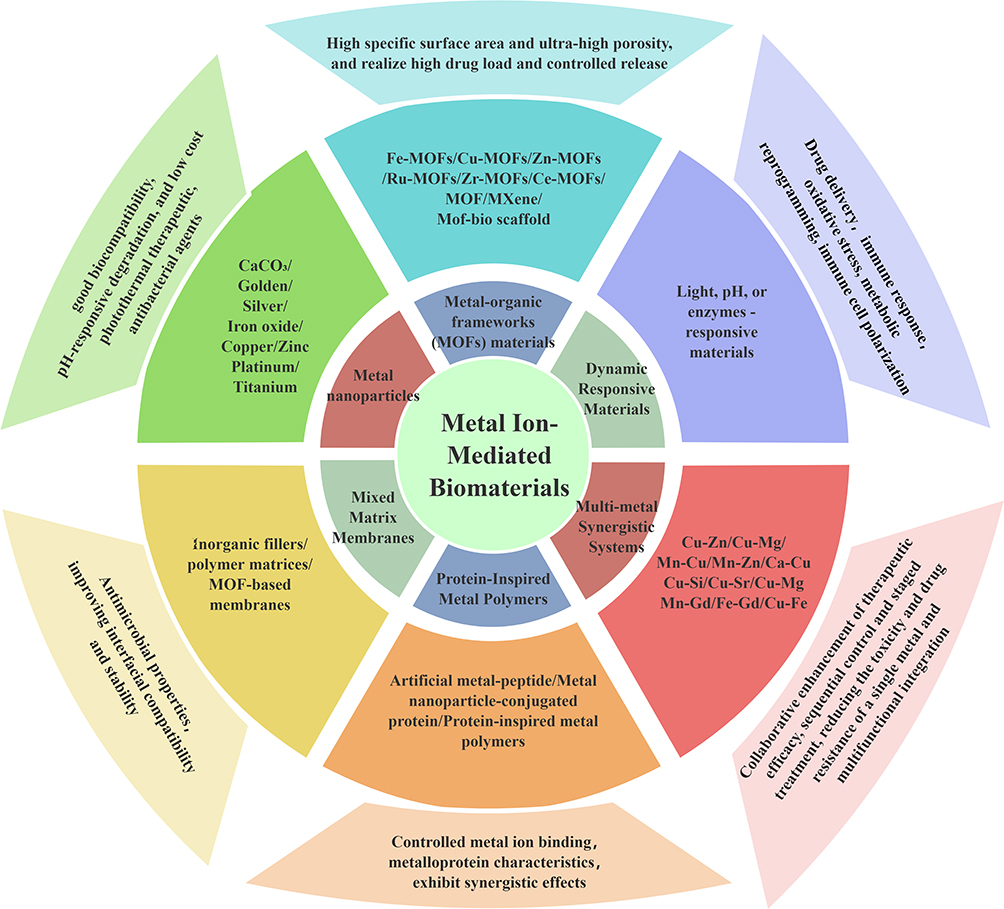 |
Figure 1 The classification, structural advantages, and functional directions of metal-based biomaterials are outlined as follows. Centered on the regulatory role of metal ions, these materials are divided into six major branches, including metal–organic frameworks (MOFs), metal nanoparticles, multi-metal synergistic systems, protein-inspired metallopolymers, and hybrid membrane materials, among others. Each branch offers unique physicochemical advantages, such as high porosity for drug loading, environmental responsiveness, antibacterial theranostics, and multi-component synergistic therapy. Collectively, these features enable a wide range of multifunctional biomedical applications, including drug delivery, immune regulation, oxidative stress intervention, and stepwise precision theranostics. |
This review aims to provide a comprehensive overview of the current state of research on metal-based biomaterials (Figure 1), focusing on their design, functional innovations, and clinical applications. It synthesizes existing knowledge on the applications of these biomaterials in oncology, infection management, and regenerative medicine, but does so within the proposed three-level framework and with the explicit boundary conditions defined above. The review is structured as follows: we first discuss the types of metal materials and the fundamental roles of metal ions in biomaterials; we then explore advanced material design strategies, followed by a systematic comparison with non-metallic platforms; subsequently, we discuss comprehensive considerations of drug delivery systems, mechanisms of metal ion-mediated action and biological effects, and disease treatment applications; finally, we outline clinical translational hurdles and future directions. By highlighting recent advancements and emerging trends, we underscore the significance of metal ions in developing innovative therapeutic strategies to address complex disease challenges. Understanding the mechanisms by which metal ions exert their effects will not only enhance biomaterial design but also pave the way for more effective and personalized treatment options.15
Types of Metal-Based Biomaterials
Metal-based biomaterials have garnered significant attention owing to their distinctive properties and potential biomedical applications. Based on composition and functionality, these materials can be classified into several categories. They offer numerous advantages, including specific responsiveness,16 multifunctional integration,17 biocompatibility,18 controlled release characteristics19 and synergistic enhancement,20 thereby serving as powerful tools for precision tumor therapy. For example, to counteract cancer cell evasion of programmed cell death (PCD) via immunosuppressive pathways, FeMn@R@H, which possesses acid-responsive properties, and disintegrates in the acidic tumor microenvironment (TME), releasing Fe3⁺ and Mn2⁺ ions to initiate Fenton-like reactions for ROS-mediated pyroptosis.16 Multifunctional integration addresses challenges such as orchestrating osteogenesis and modulating inflammatory responses with bioactive scaffolds. An example is the integration of poly(lactic-co-glycolic acid) (PLGA), type I collagen, zinc-imbued metal-organic frameworks (Zn-MOFs), and macrophage chemotactic factor (MCF) via extrusion-based 3D printing, concurrently enabling biological regeneration and inflammatory control.17 To address insufficient interfacial bone union, a strategy involving mussel adhesion-mediated ion coordination and molecular clicking applied to titanium substrates (DPA-Co/GFO) exhibits excellent biocompatibility and effectively promotes angiogenesis and osteogenesis.18 Numerous nanomedicine platforms have been developed to tackle low survival rates. Two-dimensional Ti3C2 MXene nanosheets (NSs) have emerged as a versatile platform that enhances drug release and biocompatibility while minimizing toxicity.19 Furthermore, to overcome challenges in immunotherapy such as insufficient T cell activation and infiltration, carrier-free small molecular self-assembly strategies have been employed. For instance, a Cel hydrogel composed of glycyrrhizic acid (GA), copper ions (Cu2⁺), and celastrol (Cel) increases reactive oxygen species (ROS) in conjunction with GA and Cel, synergistically expediting cellular apoptosis.20
Metal-Organic Frameworks (MOFs) Materials
Metal-organic frameworks (MOFs) have garnered considerable attention in biomedical applications due to their tunable pore structures, high drug-carrying capacity, and diverse metal ion sources. Based on metal sources and functionalities, MOFs can be classified into two main categories: those containing endogenous metals (essential bioelements such as Fe, Cu, and Zn) and those containing exogenous metals (including Ru, Zr, and Ce). MOFs are crystalline materials composed of metal ions coordinated with organic ligands, forming porous structures. Their high surface area, tunable pore sizes, and chemical versatility make them ideal for drug delivery systems, catalysis, and gas storage.21 In biomedicine, MOFs facilitate controlled release of therapeutic agents, enhance imaging capabilities, and improve cancer treatment efficacy by providing targeted drug delivery platforms. Recent advancements indicate that MOFs can be engineered to respond to specific stimuli such as pH or temperature, allowing precise control over drug release in tumor environments.22
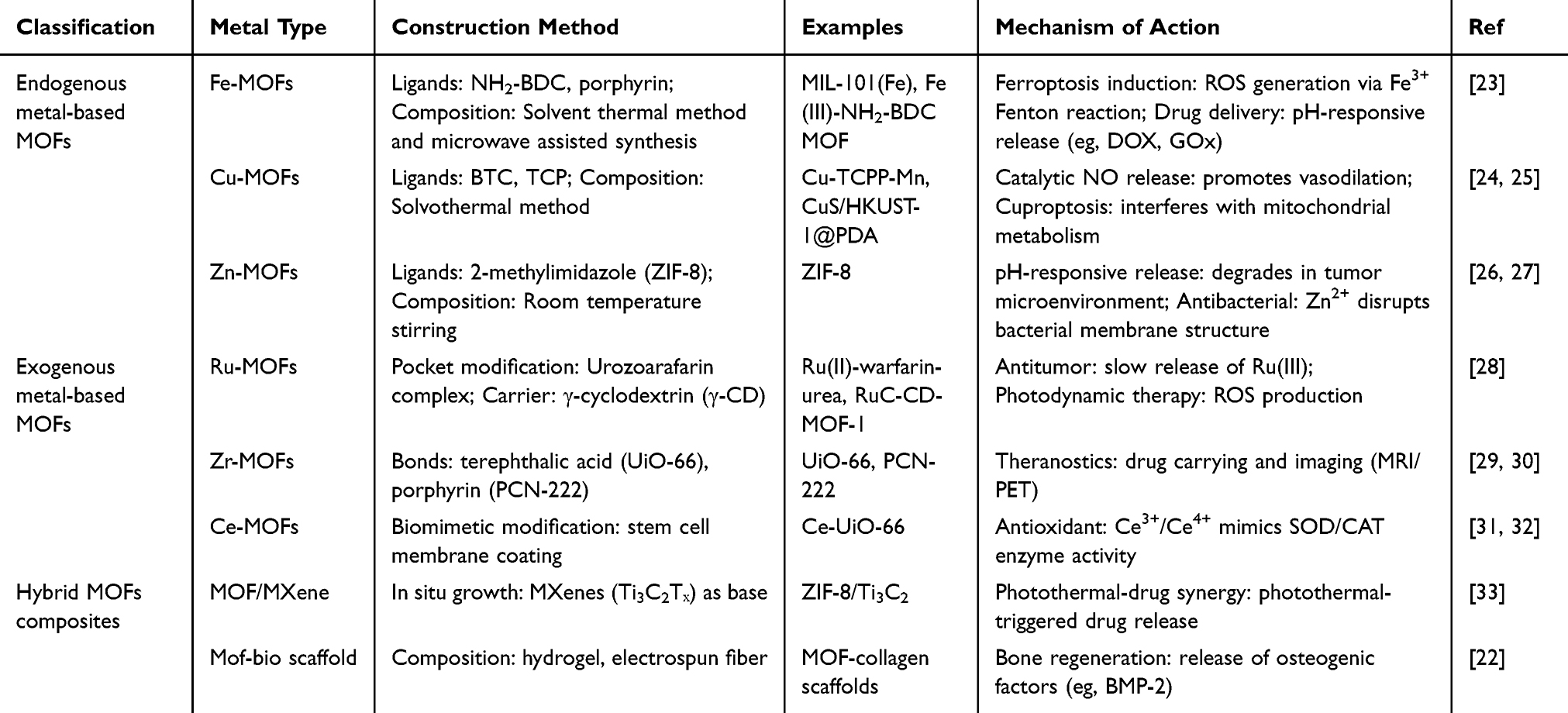 |
Table 1 Classification of Metal-Organic Frameworks (MOFs) in Biomedical Applications |
Table 1 summarizes the classification of MOF applications in biomedical contexts. Metal nanoparticles, including gold, silver, and platinum, exhibit unique physical and chemical properties that differ significantly from their bulk counterparts. These nanoparticles can enhance the efficacy of therapeutic agents through mechanisms such as photothermal therapy, wherein they absorb light and convert it into heat, selectively destroying cancer cells. Moreover, metal nanoparticles can serve as drug carriers, improving solubility and bioavailability. Their small size enables enhanced cellular uptake, making them effective in targeting tumor tissues while minimizing side effects on healthy cells.34 Metal-polyphenol coordination materials leverage interactions between metal ions and polyphenolic compounds to form stable complexes. These materials exhibit antioxidant properties and can be utilized in drug delivery applications. The coordination between metal ions and polyphenols can enhance the stability and solubility of therapeutic agents, rendering them suitable for various biomedical applications, including cancer therapy. The ability of these materials to respond to environmental changes, such as pH, further augments their potential as targeted drug delivery systems.35
However, classifying MOFs by metal source into endogenous versus exogenous is misleading, because the biological relevance of metal ions depends not on their “endogenous” label but on local concentration, coordination environment, and release kinetics. For example, while copper is an essential trace element, Cu-based MOFs can induce profound cytotoxicity at concentrations far below those of “exogenous” zirconium-based MOFs, depending on ligand design and degradation rate.23,24 The endogenous/exogenous binary is therefore a poor predictor of biocompatibility or therapeutic efficacy. A more meaningful axis is metal-ligand bond lability: MOFs with labile coordination bonds (eg, Cu-carboxylate) tend to release ions rapidly, favoring acute antimicrobial or antitumor effects but risking systemic toxicity.25
Stimulus-responsive design is often treated as a checklist rather than a trade-off analysis. Numerous studies demonstrate that MOFs can be engineered to respond to pH, temperature, glutathione, or enzymes.26,28 However, the literature rarely compares the clinical practicality of different triggers.
There is also a neglect of non-MOF alternatives in comparative evaluation. A MOF is rarely the only platform for a given application. Consider drug delivery: mesoporous silica nanoparticles offer comparable pore structures and surface functionalization with well-established safety profiles; polymer nanoparticles (PLGA, PEG-PLGA) provide simpler regulatory paths and decades of clinical data; liposomes achieve higher drug loading for hydrophobic agents. Where, then, do MOFs offer unique advantages? Three candidate domains emerge from critical reading: (i) multi-metal synergistic therapy, which the ability to incorporate two or more metal ions (eg, Fe3⁺/Cu2⁺) into the same framework to achieve combined ferroptosis/chemodynamic therapy;36 (ii) catalytic activity beyond drug delivery, MOFs as enzyme-mimicking nanozymes (eg, Ce-UiO-66 with phosphatase-like activity);37 (iii) cargo protection in harsh biological environments, the crystalline framework can shield labile biologics from proteases or low pH better than amorphous polymers.38 Conversely, MOFs are less advantageous when simple sustained release is needed or when metal toxicity is a primary concern.
Metal-Based Nanoparticles
Calcium Carbonate (CaCO3) Nanocarriers
In a CO2 atmosphere, Ca2⁺ and CO32− react to form nano-sized CaCO3 while simultaneously enabling hydrophobic drug loading. In the acidic tumor microenvironment (pH 6.5–6.8), CaCO3 decomposes into Ca2⁺ and CO2, releasing the loaded drug (eg, aminoglycosides, shenazone). Excess Ca2⁺ enters tumor cells, causing mitochondrial damage and a ROS burst, thereby inducing apoptosis. Degradation of CaCO3 also provides Ca2⁺ for bone formation, promoting bone regeneration. CaCO3 nanocarriers offer significant advantages in biomedical applications, including good biocompatibility, pH-responsive degradation, and low cost; however, key limitations remain in their construction. CaCO3 readily transitions from metastable vaterite to stable calcite in solution, leading to pore structure collapse and reduced drug-loading capacity. To enhance stability, CaCO3 microspheres can be coated with sodium polystyrene sulfonate (PSS) to stabilize the vaterite phase, delay phase transition, and improve drug-loading stability.39 Additionally, CaCO3 microspheres often suffer from insufficient loading efficiency and targeting, particularly for hygroscopic drugs, and lack active targeting. Biomimetic modification and composite carrier design can address these issues. For instance, coating CaCO3 nanoparticles with tumor cell membranes enables homologous targeting and immune evasion.40 While porous CaCO3 microspheres serve as drug reservoir systems, conventional preparation methods struggle to precisely control drug loading and release kinetics, especially for macromolecular drugs such as proteins and nucleic acids, due to the absence of specific binding sites on CaCO3 surfaces, resulting in low loading efficiency.
Naked CaCO3 nanoparticles lack inherent targeting capability, leading to poor accumulation at lesion sites and increased off-target toxicity. Surface modification of CaCO3 with proteins (eg, human serum albumin, HSA) or specific peptides can enhance stability and introduce targeting functions. Nanocomposite technology combining CaCO3 with other materials creates hybrid systems with synergistic stabilization effects. For example, manganese-doped CaCO3 nanoparticles (Mn-doped CaCO3 NPs) exhibit enhanced stability and magnetic responsiveness.41 pH-triggered release is the most prominent controlled-release mechanism for CaCO3 carriers, dissolving rapidly in acidic environments (pH < 6.5) to release loaded drugs and Ca2⁺ ions. Additionally, pH-responsive Fe2⁺ delivery nanocarriers coated with CaCO3 nanoparticles enable intracellular Fe2⁺ delivery to enhance tumor chemotherapy.42 Here, a pertinent question arises: for a given therapeutic window (eg, 4–6 h of drug release in a tumor), does a simple pH-responsive CaCO3 system with optimized particle size perform as well as a multi-enzyme cascade system?
Furthermore, wrapping CaCO3 nanoparticles with tumor cell membranes achieves homologous targeting and immune evasion. Cell membrane coating offers multi-antigen targeting and prolonged circulation, but it is technically complex, batch-dependent, and raises regulatory hurdles regarding source characterization and immunogenicity.43 Moreover, the “homologous targeting” claimed for cancer cell membranes has been challenged by recent studies demonstrating that the preferential uptake in homologous cells arises largely from qualitative variations in the protein corona, not from inherent membrane specificity.44
Enzyme-responsive release adds another dimension of controlled release to CaCO3 carriers. Newly designed CaCO3 composite carriers incorporate enzyme-sensitive linkers or coatings triggered by specific enzymes. For instance, a CaCO3 system containing glucose oxidase (GOD) and tirapazamine (TPZ) achieves a unique starvation-hypoxia activated synergistic therapy, enabling efficient, low-toxicity targeted therapy based on tumor microenvironment characteristics. As a non-inhibitor involvement strategy, calcium-based nanocatalysts (G/A@CaCO3-PEG) exert synergistic antineoplastic effects via low-temperature photothermal therapy.45 Moreover, the in vivo targeting ability of CaCO3 nanocarriers directly affects therapeutic efficacy and side effects. Lack of effective targeting leads to nanocarrier accumulation in non-target tissues, reducing therapeutic effects and increasing systemic toxicity. Modifying CaCO3 nanocarrier surfaces with specific ligands that recognize and bind to overexpressed receptors on pathological cells can significantly improve enrichment at target sites. Both ligand-receptor-mediated and antibody-mediated targeting enhance CaCO3 nanocarrier accumulation in pathological areas. Our study demonstrated that the combination of calcium carbonate (CaCO3), which adsorbs iron ions, and selenium-peptide nanoparticles incorporated into collagen scaffolds effectively suppressed gastric cancer by inducing ferroptosis and promoting apoptosis.46 Thus, by integrating chemotherapy, photodynamic/photothermal/acoustic therapy, and immunotherapy into a single multimetallic platform, this convergent strategy attacks diseases through multiple mechanisms, thereby addressing drug resistance and minimizing the risk of recurrence.
However, the distinct value proposition of CaCO3 is twofold: (i) therapeutic ion release, as Ca2⁺ itself contributes to mitochondrial damage and bone regeneration, which is not true for polymers or silica;3 (ii) like CO2 generation, gas production may enhance drug penetration in dense tumor stroma, an underexplored but potentially unique advantage.47 The limitations are equally clear: poor phase stability, lack of active targeting, and difficulty in loading macromolecular drugs due to the absence of specific binding sites.
Golden Nanoparticles
Gold nanoparticles (AuNPs) exhibit size- and shape-dependent optical properties due to surface plasmon resonance (SPR), making them attractive for photothermal therapy (PTT), radiotherapy sensitization, drug delivery, and immunotherapy.48 Gold star nanoparticles (AuSNs) serve as adjuvants to enhance the immune effect of virus-like particles, offering safety, facile production, and favorable immune properties.49 Consequently, it remains unknown whether the reported “adjuvant effect” of AuSNs is a shape effect or an artifact of different purification protocols. Gold nanorods (AuNRs) act as pro-angiogenic, anti-inflammatory, photothermal therapeutic, and antibacterial agents for synchronized delivery to full-thickness wounds in vivo.50 Examples include AuNRs@TF,51 Bi2MoO6/MoS2/AuNRs (BMO-MSA),52 hollow CuS@gold nanorods/polydopamine (HCuS@AuNRs/PDA) nanohybrids,53 AuNRs@MnO2@SiO2 nanoparticles,54 and hierarchical-structured AuNRs@MnO2@SiO2 (AMS) nanocarrier.55 Thus, AuNPs and AuNRs, with their surface plasmon resonance (SPR) effect, high photothermal conversion efficiency, ease of functionalization, and strong X-ray absorption capacity, are employed in photothermal therapy (PTT), radiotherapy sensitization, drug delivery, and immunotherapy. Crucially, none of these studies directly compared the multi-layered or hybrid systems against PEGylated AuNRs alone or a simple physical mixture of components, leaving the incremental therapeutic benefit of added architectural complexity unproven. However, it remains unclear whether different geometries confer genuine biological advantages or merely reflect differences in synthesis protocols and characterization methods.
Silver Nanoparticles
Silver nanoparticles (AgNPs) with broad-spectrum antimicrobial properties are increasingly studied for combating multidrug-resistant bacteria.56,57 AgNPs act as sources of released silver ions, enhancing the efficacy of common antibiotics.58 AgNPs synthesized using plant extracts, wherein different plant-derived phytochemical coatings confer distinct biological advantages, and merely reflect differences in synthesis conditions and nanoparticle physicochemical properties. Examples include colloidal AgNP solutions produced from aqueous extracts of Picea abies and Pinus nigra bark,59 AgNPs using Withania coagulans leaf extracts,60 Ag-Cu bimetallic nanoparticles using Peganum harmala leaf extract,61 AgNPs using bee bread extracts62 and bioactive Ag/Cu NCs using Sargassum latifolium extract.63 Notably, the plant extract itself introduces additional variability: the composition of bioactive phytochemicals varies with plant species, growth conditions, extraction protocols, and seasonal factors, leading to batch-to-batch inconsistencies in nanoparticle size, shape, and surface chemistry that are seldom quantified. Thus, through silver ion (Ag⁺) release, ROS production, and disruption of microbial membrane structure, AgNPs are used for antibacterial treatment, wound dressings, and combating biofilms.
Iron Oxide Nanoparticles
Magnetic iron oxide nanoparticles (FeₓOγ NPs, primarily Fe3O4 and γ-Fe2O3) are prominent nanomaterials due to their potential in magnetic separation, hyperthermia, targeted drug delivery, and catalysis.64 Owing to their unique magnetic properties, good biocompatibility, excellent superparamagnetism, and reliable traceability,65 iron oxide nanoparticles are used for brain imaging and drug delivery,66,67 bone remodeling,68 tumor therapy,69 atherosclerosis,70 diagnostic,71 immune cell labeling and cancer immunotherapy,72 antibiofilm,73 and chronic inflammatory diseases.74 Environmental sources of magnetic FeₓOγ NPs are complex, and they are regarded as significant risk factors for human health.75 Upon entering and penetrating deeply in vivo, these NPs persistently create interfaces for chemical reactions, generating reactive oxygen species (ROS) and causing chronic toxic effects.76 Iron oxide nanoparticles conjugated to anticancer drugs, targeting agents, or genetic vectors, or used in combination with bioactive molecules, represent promising systems for assisting osteosarcoma therapy.77 Due to their unique physicochemical properties and sizes, iron oxide nanoparticles (IONPs) possess magnetic resonance (MR) T1/T2 imaging capabilities.78 Research on IONPs to improve chemotherapeutic efficacy has been highlighted in brain tumors,79 ovarian cancer,80 osteosarcoma treatment,77 endometrial cancer,81 breast cancer,82 gastric cancer,83 and bone tissue engineering.84 For instance, Fe3O4@Glu-EA nanoparticles show considerable toxicity against gastric cancer cells and serve as an efficient platform for ellagic acid (EA) delivery, promising improved gastric cancer chemotherapy outcomes.85 Therefore, iron oxide nanoparticles, with their superparamagnetism, catalytic Fenton/Fenton-like reactions,86 ROS generation,87 and magnetic targeting,88 are applied in magnetic hyperthermia therapy (MHT),89 chemodynamic therapy (CDT),90 and as MRI contrast agents.
However, this enumeration of IONP-based applications, spanning imaging, drug delivery, hyperthermia, and tissue engineering, conceals several critical unresolved issues that a truly critical review must address. First, despite decades of research and initial FDA approval of ferumoxytol for anemia treatment, the clinical translation of SPIONs for imaging and therapeutic applications has been unexpectedly slow. As a recent clinical landscape review noted, SPIONs were initially approved as liver imaging agents nearly three decades ago, yet their broader adoption as universal nanodiagnostic and theranostic agents remains constrained by intrinsic physicochemical properties and in vivo behaviors.91 Second, the field has yet to resolve a fundamental tension regarding the source of therapeutic efficacy: is it the nanoparticles themselves or the iron ions they release? Third, the immunomodulatory roles of IONPs, ranging from M1 macrophage polarization to ferroptosis induction and STING pathway activation, remain mechanistically ambiguous.92 Finally, the long-term fate and toxicity of IONPs remain inadequately characterized. Although IONPs are often regarded as biodegradable because iron is an essential element, prolonged nanoparticle accumulation in the liver, spleen, and other organs may induce cell stress and toxicity, often mediated through oxidative stress and inflammation.93
Copper Based Nanoparticles
Among nanomaterials, copper and copper oxide nanoparticles stand out as promising candidates for numerous medical applications. However, achieving controlled release of therapeutic agents from copper nanoparticles remains a complex challenge that requires meticulous engineering and precise design. Examples include copper-based composite nanoparticles,94 ultra-small copper-based multienzyme-like nanoparticles,95 self-assembled copper-based nanoparticles,96 tragacanth gum-based copper oxide nanoparticles,97 spark discharge aerosol-generated copper-based nanoparticles,98 all of which aim to address current challenges such as biocompatibility and controlled release.
Copper-based nanoparticles (eg, copper oxide, copper sulfide, copper ions, copper alloys) are incorporated into biopolymer-based films; their effectiveness depends on filler concentration, dispersion state, and nanoparticle-biopolymer matrix interaction.99 They are also used to tackle drug-resistant bacteria and highly infectious viruses.100 Additionally, copper and copper oxide nanoparticles (CuNPs) possess unique physicochemical properties that render them highly promising for biomedical applications such as diagnosis, therapy, and theranostics;101 cancer imaging and therapy;102 control of hepatitis A virus vaccines;103 burn injuries;104 treatment of hepatocellular carcinoma;105 tumor immunotherapy;106 pancreatic cancer treatment;107 breast cancer immunotherapy;108 and lung cancer.109
However, the mechanistic foundation of cuproptosis, while elegantly described, has yet to be rigorously established in nanomedicine contexts. The precise relationship between nanoparticle physicochemical properties (size, surface charge, coating, dissolution kinetics) and the induction efficiency of cuproptosis remains largely uncharted. Moreover, the interplay between cuproptosis and other cell death pathways (eg, apoptosis, ferroptosis) is rarely quantified, leaving combination therapy design without rational guidance.110 Furthermore, the path to clinical translation remains obstructed by persistent challenges in biocompatibility and controlled release. As noted in multiple contemporary analyses, copper-based nanomaterials face formidable hurdles before clinical adoption: biocompatibility remains a significant concern, and achieving controlled release of therapeutic agents from copper nanoparticles poses a complex challenge that demands meticulous engineering and precise design.111
Thus, copper nanoparticles are used in chemodynamic therapy (CDT), photothermal therapy (PTT), and anti-angiogenesis by catalyzing Fenton/Fenton-like reactions, exhibiting photothermal effects, and inducing cuproptosis.67 A fundamental ambiguity persists: is the therapeutic effect attributable to the copper nanoparticles themselves or to the copper ions they release? The mechanism of cuproptosis is driven by the controlled or excessive release of copper ions from the material. Upon entering cells, these ions bind directly to enzymes in the mitochondrial tricarboxylic acid cycle, leading to abnormal aggregation and functional impairment of the enzymes, which disrupts cellular energy metabolism. Concurrently, copper ions promote excessive generation of reactive oxygen species, inducing oxidative stress. This dual action ultimately triggers a unique form of programmed cell death that is dependent on mitochondrial respiration and protein lipoylation.
Zinc Based Nanoparticles
Zinc oxide nanoparticles (ZnO NPs) have attracted significant attention due to their potent antimicrobial properties. Plant-based biosynthesis of ZnO NPs offers a cost-effective and sustainable alternative to chemical and physical methods.112 Synthesis methods include using glucose and sucrose (constituting over 70% of dried extract);113 Cassia fistula and Melia azadarach leaf extracts;114 Thryallis glauca leaf extract;115 Butea monosperma flowers and Glycyrrhiza glabra roots;116 Momordica charantia and Curcuma zedoaria plant extracts;117 and Mallotus philippinensis leaf extract.118
Interestingly, some researchers have pointed out that the toxic effects of ZnO NPs are caused by Zn2⁺ release;119 yet another in vivo study found that ZnO NPs are significantly more effective than ionic zinc in reducing hyperglycemia.120 One possibility is that the insulin-mimetic activity of ZnO NPs is not solely determined by the free Zn2⁺ concentration. For instance, ZnO NPs synthesized with plant extracts may have surface-attached phytocompounds (such as catechin derivatives and polyphenols) that synergistically enhance hypoglycemic activity through other mechanisms (eg, DPP-4 enzyme inhibition), rather than merely affecting Zn2⁺ release. Existing studies indicate that ZnO NPs may be more effective than conventional zinc salts in ameliorating diabetes, but this advantage is accompanied by a significant risk of oxidative stress. In a 56-day rat study comparing ZnO NPs (1, 3, 10 mg/kg) with zinc sulfate (ZnSO4, 30 mg/kg), ZnO NPs exhibited stronger antidiabetic activity, as reflected by better glucose metabolism, higher insulin levels, and improved zinc status.121 In the same study, ZnO NPs induced severe oxidative stress, particularly at higher doses, manifested by altered antioxidant enzyme activities, elevated lipid peroxidation levels, and a marked decrease in total antioxidant capacity. This suggests that ZnO NPs may represent a double-edged sword: greater efficacy but also greater toxicity. However, the therapeutic window between their superior efficacy and increased toxicity risk has not been systematically defined. Currently, risk–benefit assessment data necessary for clinical translation are lacking.
Therefore, zinc nanoparticles, through zinc ion (Zn2⁺) release, ROS production, insulin activity simulation, and enzyme inhibition, are used for antibacterial treatment, diabetes management (insulin mimicry), and wound healing.
Platinum Nanoparticles
Platinum nanoparticles (PtNPs) show significant promise in cancer therapy by enhancing the effects of platinum-based chemotherapies such as cisplatin,122 applications include tumor photothermal therapy,123 neuroblastoma treatment,124 ovarian cancer immunotherapy,125 chemo-photodynamic bladder cancer therapy126 and self-reinforcing hypoxic oncotherapy.127 Other applications involve adjuvant therapy in silicosis management,128 anti-infective therapy,129 acne vulgaris treatment,130 and virus detection.131 PtNPs exhibit various enzyme-mimetic activities (eg, peroxidase (POD), catalase (CAT)), high catalytic efficiency, and enhanced ROS generation, making them suitable for catalytic therapy, sonodynamic therapy (SDT), and antioxidant scavenging.
PtNPs can be prepared via various methods: chemical reduction,132 electrochemical processes,133 plasma methods,134 microemulsion methods,135 template methods (using soft structures such as micelles or vesicles from surfactants, block copolymers, or biomolecules to guide nucleation and growth),136 and biological synthesis (using plant extracts, microorganisms, or biomolecules to reduce platinum ions and stabilize nanoparticles).137 However, the synthesis reproducibility and scalability of PtNPs present a critical, often overlooked challenge.
Despite decades of research and the well-established clinical use of platinum-based small-molecule drugs, the clinical translation of PtNPs has been unexpectedly slow and fraught with fundamental challenges. A recent systematic review of 108 studies on PtNP toxicological effects revealed a stark reality: while PtNPs generally show good biocompatibility in vitro, significant toxicity, highly dependent on size, concentration/dose, coating, and biological system, has been consistently reported. Most critically, no data from human epidemiological studies have been published so far. In addition, a fundamental ambiguity persists regarding whether the anticancer activity of PtNPs represents a true nano-specific effect or merely a sustained-release mechanism for ionic platinum species. A comparative study on ultrasmall 2 nm PtNPs versus core-shell 30 nm Au@PtNPs against hepatocellular carcinoma revealed that both nanoparticle types induced selective cytotoxicity in cells with high oxidative status while remaining inactive in cells with lower oxidative status.138
Thus, constructing PtNPs involves fine-tuning processes in which size, morphology, structure, and dispersion depend strongly on the selected materials based on the precursors, reducing agents, stabilizers, templates, carriers and preparation methods.
Titanium Based Nanoparticles
Titanium-based nanoparticles are nanoscale materials with titanium as the core element, typically ranging from 1 to 100 nanometers, and include titanium metal, titanium dioxide (TiO2), or other titanates. To meet specific requirements, they can be enhanced by doping with other metal elements (eg, silver, iron) or by surface modifications (eg, PEG, antibodies). Titanium-based nanoparticles are notable for their unique properties: photocatalytic activity,139 high biocompatibility,140 surface and interfacial effects,141 and designable morphology and structure.142,143 These properties render them useful in photodynamic therapy,144 drug delivery,145 biological imaging146 and antimicrobial materials.147 Here, TiO2 nanoparticles can be enhanced by doping with other metal elements or surface modifications. However, do these modifications reduce TiO2’s intrinsic toxicity, or do they simply introduce new safety concerns? PEGylation, for instance, has been widely adopted for prolonging circulation and reducing opsonization.148 Nevertheless, a growing body of evidence has identified significant drawbacks of PEGylation, including steric hindrance, diminished cellular uptake, and immunogenicity (anti-PEG antibodies, accelerated blood clearance).149
While TiO2 is often positioned as biocompatible and less toxic than other nanomaterials (ie, copper oxide, zinc oxide, and manganese oxide), this characterization obscures a more nuanced and less reassuring reality. Although TiO2 nanoparticles (TiO2-NPs) were once considered inert, a large number of in vitro studies have confirmed their cytotoxicity and genotoxicity, with mechanisms primarily involving the generation of reactive oxygen species (ROS) and the activation of inflammatory and cell death signaling pathways. The International Agency for Research on Cancer (IARC) has classified TiO2 as a Group 2B carcinogen when inhaled. However, TiO2 also exhibits fundamental limitations in certain therapeutic strategies. The photocatalytic activity that makes TiO2 attractive for photodynamic therapy also imposes a fundamental practical limitation: its dependence on UV light. Pristine TiO2 has a wide bandgap of approximately 3.2 eV, meaning it can only be activated by ultraviolet (UV) light, a wavelength range with limited tissue penetration depth and known phototoxic effects on healthy tissue.150
There is a significant gap between animal experiments and human clinical settings. For instance, when used for drug delivery, nanomaterials rapidly adsorb proteins in the bloodstream to form a protein corona, which completely alters their surface properties and biological identity. As a result, their targeting ability and in vivo distribution behavior deviate substantially from the intended design. This explains why many targeting strategies that perform excellently in simple models often suffer a sharp decline in therapeutic efficacy in complex in vivo environments.
Mixed Matrix Membranes (MMMs)
Mixed matrix membranes (MMMs) incorporate fillers such as metal-organic frameworks (MOFs) and covalent-organic frameworks (COFs) into polymers to enhance gas separation performance. Molecular simulations of gas adsorption and diffusion in MOFs and COFs, combined with theoretical permeation models, allow calculation of H2, N2, CH4, and CO2 permeabilities for nearly one million MOF/COF/polymer MMM combinations.151 These membranes can be engineered for improved gas separation, water purification, or drug delivery systems.152 In biomedicine, MMMs facilitate controlled drug release while providing mechanical stability and biocompatibility. Incorporating metal ions can further enhance their antimicrobial properties, making them suitable for various healthcare applications.34 MMMs combine the advantages of inorganic fillers (eg, MOFs, zeolites, two-dimensional materials) dispersed within a polymer matrix, showing significant potential in gas separation and water treatment. However, MMM research faces multiple limitations in materials, methodologies, and practical implementation.
Inorganic fillers and polymer matrices possess vastly different physical and chemical properties, leading to interfacial defects that reduce separation performance. For instance, MOFs often disperse poorly in organic solvents and tend to agglomerate, affecting film uniformity.153 MOF-based membranes face issues such as framework flexibility, defects, and grain orientation. MMMs encounter bottlenecks including MOF aggregation, polymer matrix plasticization and aging, and poor interfacial compatibility.154 Preparing high-quality MMMs remains challenging due to insufficient interfacial interaction. Strategies such as coating modification and priming techniques, for example, incorporating ionic liquid (IL)-modified UiO-66-NH2 filler into microporous organic polymers (PIM-1), can produce dense, defect-free MMMs, thereby improving interfacial compatibility and stability.155 Developing defect-free MMMs is challenging due to poor MOF-polymer matrix compatibility; surface-modification strategies using polymers with intrinsic microporosity grafted onto UiO-66-NH2 within Pebax-supported MMMs show promising separation performance.156 To address fatal MMM defects such as non-selective pores arising from poor phase compatibility, amidoxime-modified UiO-66@PIM-1 MMMs enhance CO2 separation and anti-aging performance.157
Protein-Inspired Metal Polymers
Protein-inspired metal polymers are innovative functional materials formed by combining proteins or protein analogs with metal elements via coordination bonds, covalent bonds, or non-covalent interactions. These materials integrate the structural precision and functional diversity of biological macromolecules with the catalytic, optical, and magnetic properties of metal ions or nanoclusters, demonstrating broad application potential in biomedical engineering, catalysis, sensing, and flexible electronics. Traditional metalloprotein preparation often relies on complex biosynthetic pathways, which are costly and difficult to scale up. Furthermore, high concentrations of heavy metal ions can cause uncontrollable protein denaturation, limiting practical applications.158 Researchers have therefore developed biomimetic strategies: engineering natural proteins via genetic or chemical modification for controlled metal ion binding, and designing polymers with protein-like structures that mimic key metalloprotein characteristics. Composites of metal nanoclusters and proteins retain the characteristics of each component and may exhibit synergistic effects, showing unique advantages in bioimaging, catalysis, and sensing.159
New strategies involve mediating secondary structure transitions in polyamino acids using metal ions.160 From the perspective of artificial metal-peptide assemblies (MPAs), adaptive materials with protein-like nanocavities can be created through metal coordination and non-covalent interactions.161 Using clinically available deferoxamine (DFO) as an exogenous ligand template to modify polyamino acid side chains enables controlled secondary conformation transition from α-helix to β-sheet by adjusting ligand amount and chelating metal ions. Divalent tin ions (Sn2⁺) effectively reduce disulfide bonds in proteins, forming tin-sulfur bonds (-C-S-Sn-S-C-), which promotes rapid, controllable protein assembly applicable to the preparation of protein amyloid aggregates in metal composites.162 Two primary strategies are used to construct metal nanoparticle-conjugated protein systems: direct synthesis and post-modification. Direct synthesis involves in situ formation of metal nanoparticles within protein templates by reducing metal precursors using reductants or the proteins’ own redox groups.159
Material Design and Functional Innovation
Multi-Metal Synergistic Systems
Integrating multi-metal synergistic systems shows significant promise in enhancing the biological functions of biomaterials. Examples include: antimicrobial and osteogenesis-promoting synergistic systems (Cu-Zn,163 Cu-Mg164), immune regulation and anti-tumor synergistic systems (Mn-Cu,165 Mn-Zn,166 Ca-Cu167), angiogenesis-promoting and osteogenesis-promoting synergistic systems (Cu-Si,168 Cu-Sr,169 Cu-Mg164), and integrated diagnosis and treatment systems (Mn-Gd,170 Fe-Gd,171 Cu-Fe.172) Or instance, synergistic effects between Fe3⁺ and Cu2⁺ have been extensively studied for their roles in promoting angiogenesis and osteogenesis, which are vital for bone regeneration.173 These metal ions can influence exosome secretion from mesenchymal stem cells (MSCs) and macrophages, thereby modulating inflammation and promoting tissue healing. The application of Mg2⁺ and Sr2⁺ in bone repair illustrates another facet; these ions enhance osteogenic differentiation and improve the mechanical properties of biomaterials, making them suitable for orthopedic and dental applications.174 Designing such systems requires careful consideration of optimal concentrations and interactions to achieve desired therapeutic outcomes while minimizing potential cytotoxicity.
Achieving the fine balance between release rates is critical, as overly rapid release can lead to toxicity while excessively slow release may compromise efficacy, posing a significant challenge in materials science. The Jahn-Teller effect of Cu2⁺ effectively modulates and optimizes the electronic structure and spin state of Fe3⁺. Under Cu2⁺ regulation, NiFe-LDHs transform from ferrimagnetic to ferromagnetic, and CuNiFe-LDHs exhibit significantly enhanced oxygen evolution reaction (OER) performance under magnetic fields compared to NiFe-LDHs.175 While Mg2⁺ and Sr2⁺ are well known for promoting osteogenesis and modulating immune responses in bone repair, their interplay with other metal ions within the complex in vivo microenvironment poses significant challenges. Unpredictable inter-ion reactions, such as precipitation and redox processes, can alter the intended therapeutic efficacy. Moreover, the systemic and long-term biosafety of these multi-metal interactions, including their potential for cumulative toxicity and sensitization, remains largely elusive.
Dynamic Responsive Materials
Dynamic responsive materials represent a significant advancement, enabling controlled release triggered by external stimuli such as light, pH, or enzymes. For example, light-controlled release mechanisms using hydrogels responsive to specific wavelengths allow precise drug delivery in targeted therapies.176 Enzyme-responsive materials release therapeutic agents in response to pathological enzymatic activities, such as those observed in cancer.177 Designing these materials often involves incorporating dynamic bonds that enable reversible structural changes, thereby enhancing real-time functionality, improving therapeutic efficacy, and reducing side effects associated with conventional drug delivery.
Traditional delivery systems often lack dynamic responsiveness to the inflammatory microenvironment and struggle to match temporal changes in inflammation. For instance, the immune response after spinal cord injury is time-dependent, yet traditional systems find precise, synchronized intervention challenging. Activating the TRPM7 channel in dendritic cells (DCs) induces the HIF-1α-TGF-β axis, suppressing effector T cell activation while promoting regulatory T cell (Treg) formation, thereby alleviating adaptive immune inflammation. In chitosan-hyaluronic acid-magnesium (CSHA-Mg) gel, sustained Mg2⁺ release reduces pro-inflammatory macrophages (M1) and fosters an anti-inflammatory microenvironment.178 However, targeting deficiencies lead to drug accumulation in non-target tissues, increasing the risk of side effects. Biomimetic self-assembled nanoparticles with lipopolysaccharide-free EC-K1 outer membranes prolong circulation time, improve distribution in the intracranial microenvironment, and exhibit high biocompatibility.179 Self-contained injectable hydrogels that store Mg2⁺ while carrying nucleus pulposus (NP) cells aim to inhibit intervertebral disc degeneration (IVDD) through immunoregulation.178
Metal ion-based dynamic responsive materials show great potential in smart materials such as shape memory polymers, MOFs, and ionic dynamic cross-linked networks, but they face key limitations. Many require specific triggering conditions such as temperature, pH, light, redox environment, which limits their application in complex settings. Thermo-responsive shape memory polymers (SMPs) need precise temperature control, while photo-responsive MOFs require specific light wavelengths.180 Near-infrared (NIR)-responsive gold nanorods promote thermo-responsive targeted delivery of the heme oxygenase 1 (HO-1) gene, overcoming the challenge of non-specific gene expression in healthy tissues.181 Most materials rely on single stimuli such as pH or ROS, struggling to adapt to dynamic multiple signals (cytokine gradients, mechanical changes) in inflammatory microenvironments.182 Redox state changes of certain metal ions (Cu2⁺/Cu⁺, Fe3⁺/Fe2⁺) can affect material dynamics but may cause irreversible structural damage. However, some coordination systems are highly reversible and stable For example, the stiffness of metal–terpyridine crosslinked hydrogels can be switched via redox reactions,183 and the Zn2⁺-induced densified network has exhibited excellent oxidative stability and ultrafast self-healing behavior.184
Most studies report in vitro release kinetics in idealized buffer solutions, which bear little resemblance to the dynamic, heterogeneous, and mechanically active inflammatory microenvironment. Parameters such as cytokine gradients, intermittent mechanical loading, fluctuating oxygen tension, and protein corona formation are rarely incorporated into the design or evaluation pipeline. Consequently, the claimed real-time functionality and reduced side effects often fail to materialize in preclinical animal models, and rarely discussed in the primary literature. Studies on the organ distribution and excretion of inhaled Ag, Au, CuO, and ZnO NPs have provided critical data on the long-term fate of metal-based nanomaterials. Hadrup et al reported that silver was retained in the lung for >2,000 hours and remained detectable in the liver and spleen 2,000 hours post-exposure, while gold persisted in multiple organs for >600 hours185 These findings underscore the need for systematic evaluation of the long-term stability and fate of metal ion-mediated dynamic systems. For MOFs, the systemic toxicity of degradation products depends on multiple factors, including degradation kinetics, biodistribution, tissue accumulation, and excretion.186
Notably, a fundamental ambiguity remains: does dynamic responsiveness truly offer any genuine advantage over a well-designed sustained-release system? Taking the CSHA-Mg gel, sustained Mg2⁺ release is presented as a feature. Yet a simple, non-responsive Mg2⁺-eluting polymer might achieve comparable immunomodulation with far less design complexity.187,188
Metal ions themselves possess unique physicochemical properties that enable them to serve as contrast agents for imaging modalities such as magnetic resonance imaging (MRI), positron emission tomography (PET), photoacoustic (PA) imaging, or computed tomography (CT). By integrating diagnostic and therapeutic functions, real-time monitoring of patient diseases and individualized adjustment of treatment dosages can be achieved. Individualized therapy places extremely high demands on the scalable manufacturing and quality control of metal-based biomaterials. Therefore, integrating multiple triggers (eg, photothermal, enzyme-responsive) is being explored to achieve deep coupling between multi-modal responsive material design and pathological mechanisms.
Biomimetic Structural Design
Biomimetic structural design draws inspiration from natural systems to create materials that replicate complex biological functionalities. For instance, ferritin nanocages are promising for drug delivery due to their inherent stability, biocompatibility, and ability to encapsulate therapeutic agents.177 These nanocages can be engineered for enhanced targeting, enabling specific delivery to tumor sites via receptor-mediated endocytosis. Incorporating nacre-like structures improves the mechanical properties of composite materials, making them suitable for load-bearing bone repair applications.189 Integrating such designs enhances material performance and opens new avenues for advanced therapeutic systems that mimic natural biological functions.
Metal ions (eg, Fe3⁺, Cu2⁺, Zn2⁺) form dynamic crosslinking networks with polymer chain coordination groups (eg, carboxyl, oxime, amino), endowing materials with self-healing capabilities, adjustable mechanical properties, and stimulus responsiveness. Using biomolecules (proteins, polysaccharides) as templates guides the biomimetic mineralization of MOFs or inorganic materials, forming biocompatible nanostructures. Zinc-based MOF mineralization regulated by bovine serum albumin (BSA) forms uniform, stable nanocarriers for targeted tumor drug delivery.190 Modifying porous materials (COFs, MOFs) with metal ion-responsive molecules (crown ethers, carboxyl groups) enables controllable ion transport switching, simulating cell membrane ion channels.191 Reconstructing the ionic solvation sheath using metal ion-solvent molecule coordination reduces side reactions and optimizes electrochemical performance. Biomimetic interface layers constructed on metal surfaces mimic the selective transport and protection mechanisms of biofilms. Bimetallic or polymetallic ion synergy (Fe/Cu, Zn/Mg) enhances the catalytic, mechanical, or therapeutic properties of materials. Metal imbalance can trigger synergistic cuproptosis and ferroptosis, offering innovative cancer treatment solutions. Thus, metal ion-mediated biomimetic materials, through combined approaches, are expected to find wider applications in tissue engineering.
Green Synthesis Methods
Green synthesis, particularly microbial production, is advancing biomaterial manufacturing for regenerative medicine. This approach offers sustainable, cost-effective routes to complex biomolecules under mild conditions, thereby reducing the environmental impact of traditional methods. Microbial systems enable precise DNA-template assembly, ensuring high reproducibility for tissue scaffolding and drug delivery.192 Incorporating therapeutic metal ions (eg, Zn, Mg, Sr) further enhances material properties, promoting osteogenesis and angiogenesis.193 Through tailored scaffold design that controls ion release, this strategy improves biological efficacy while adhering to green chemistry principles.
3D Printing and Personalization
Integrating 3D printing technology into personalized medical device manufacturing represents a significant advancement in regenerative medicine, enabling the production of customized bone scaffolds that precisely match patient anatomical requirements. For example, an electron/ion-flux dual-gradient 3D porous Zn anode achieves a hierarchical porous structure and continuous conducting network via 3D printing.194 Combining facile metal-coordination self-assembly with 3D printing enables the engineering of bioactive scaffolds for osteosarcoma-related bone defects, minimizing microstructural heterogeneity.195 Proof-of-concept micro-extrusion-based 3D coaxial printing technology addresses challenges such as low exposed adsorption sites and slow ion diffusion rates in heavy metal removal.196 Direct ink writing using polymer precursor inks based on metal-polymer coordination effects opens pathways toward 3D-printed optoelectronic devices derived from functional oxides.197 3D metal/alloy nanoarchitectures, leveraging metal ion-carboxyl group complexation, incorporate various metal ions into acrylic acid-based polymer scaffolds, enabling advanced nanoengineering applications.198 Allium-based liquid metals (LM) and polymer binders serve as multifunctional inks for 3D-printing self-standing scaffolds or for Zn2⁺ plating/stripping, a challenging task for energy storage devices.199
However, highly efficient cross-linking ions (Cu2⁺, Fe3⁺, Sr2⁺) are cytotoxic above certain concentrations. Even physiological ions such as Ca2⁺ can disrupt cellular signaling at high local concentrations. Ionically cross-linked hydrogel networks are more dynamic and reversible than covalently cross-linked ones, resulting in lower mechanical strength, which makes them prone to deformation and rupture, thereby limiting their use in load-bearing tissue engineering. Cross-linking depends on metal ion diffusion, typically proceeding in an outside-in manner, creating a cross-linking gradient characterized by highly cross-linked, hard outer layers and poorly cross-linked, soft inner layers, which may lead to incomplete gelling.
Therefore, metal ion-mediated 3D printed biomaterials remain a topic of active research due to their mild cross-linking conditions (room temperature, aqueous environment) and biomimetic characteristics (simulating the ionic environment of the extracellular matrix).
Comprehensive Considerations of Drug Delivery Systems
Precise Targeting Strategy
Metal ion-mediated precision targeting strategies show great potential in cancer treatment, which can be achieved through metal ion catalytic activity, redox regulation, immune activation, or synergy with other therapies. In the context of precision medicine, innovative targeting strategies are paramount for enhancing therapeutic efficacy and minimizing off-target effects. Promising approaches combine antibodies and aptamers (short, single-stranded nucleic acids that bind specific targets with high affinity). This dual targeting enables more precise delivery of therapeutic agents to diseased cells while sparing healthy tissues. Recent studies have demonstrated that aptamer-antibody bioconjugates enhance the specificity and effectiveness of drug delivery systems. For instance, aptamers engineered to recognize specific cancer cell markers can direct cytotoxic agents precisely to tumor sites.200 Another innovative strategy employs bacterial membrane camouflage technology, wherein therapeutic nanoparticles are disguised with bacterial membranes to evade the immune system and improve circulation time in the bloodstream. By mimicking the natural properties of bacteria, these membrane-coated nanoparticles can effectively target and penetrate tumor tissues, thereby enhancing the delivery of chemotherapeutic agents. This method improves drug bioavailability and reduces systemic toxicity, holding promise for various malignancies. Table 2 summarizes the methods and materials based on metal ion-mediated precision targeting strategies.
 |
Table 2 Metal Ion-Mediated Precision Targeting Strategies |
Integrating stimuli-responsive elements further advances these targeting strategies. Nanoparticles can be designed to release their payloads in response to specific environmental triggers, such as pH changes or enzymes characteristic of the tumor microenvironment, thereby ensuring site-specific release to maximize therapeutic potential and minimize side effects.
Spatiotemporal Controlled Release
Metal ion-mediated spatiotemporal controlled release is a prominent research focus in materials science, biomedical engineering, and environmental engineering. By triggering metal ion release via external stimuli (light, sound, electric fields) or endogenous changes (pH, ROS), this approach enables precise drug delivery, environmental remediation, and energy conversion applications. Spatiotemporal controlled release is increasingly important in drug delivery systems for enhancing therapeutic efficacy and minimizing side effects. Effective mechanisms include dual-responsive systems triggered by tumor microenvironment stimuli such as glutathione (GSH) and pH. These systems exploit the elevated GSH levels and acidic microenvironment characteristic of cancer cells to facilitate controlled drug release.207 Another innovative approach involves designing hydrogels that respond to external stimuli such as light or ultrasound. These hydrogels undergo physical changes upon triggering, enabling controlled release of encapsulated drugs. For example, hydrogels exposed to near-infrared (NIR) light enhance localized chemotherapeutic agent delivery, improving treatment outcomes and reducing systemic toxicity.208
Developing oxygen self-supplying systems enhances the efficacy of photodynamic therapy (PDT). Integrating oxygen-generating components into drug delivery systems overcomes the hypoxia that limits PDT effectiveness. These systems release oxygen in response to specific stimuli, improving reactive oxygen species (ROS) generation, which is crucial for tumor ablation.209 Table 3 outlines spatiotemporal controlled release strategies based on metal ion mediation.
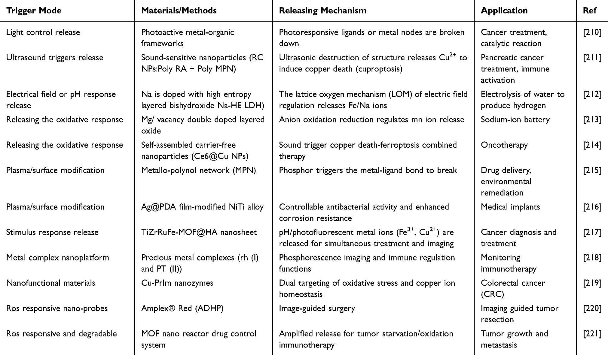 |
Table 3 Metal Ion-Mediated Spatiotemporal Controlled Release Strategies |
Gd3⁺-MOFs exhibit dual functionality that is promising for biomedical applications, integrating diagnosis and treatment. They serve as both imaging agents and therapeutic delivery systems. Gd3⁺-MOFs demonstrate excellent MRI properties, enabling precise tumor imaging alongside targeted drug delivery. Incorporating therapeutic agents within MOFs allows localized treatment, minimizing systemic side effects and enhancing efficacy.222
64Cu-labeled nanoparticles represent another innovative theranostic approach. These nanoparticles enable both imaging and therapy, particularly in cancer treatment. 64Cu labeling facilitates in vivo tracking, allowing real-time monitoring of drug delivery and therapeutic response. This dual functionality enhances the precision of cancer therapy and provides insights into the pharmacokinetics and biodistribution of therapeutic agents.200 Integrating diagnostics and therapeutics via such multifunctional platforms significantly advances personalized medicine, with the potential to improve patient outcomes and reduce complications.
The collaboration of zinc ions (Zn2⁺) with PD-L1 inhibitors exemplifies synergistic immunotherapy strategies. Zn2⁺ enhances the efficacy of PD-L1 inhibitors, promoting robust anti-tumor immune responses. This collaborative approach improves therapeutic outcomes and provides a framework for novel combination therapies targeting multiple pathways involved in tumor progression.223 Integrating various modalities within collaborative platforms highlights the importance of interdisciplinary approaches in advancing treatment strategies, ultimately improving the management of complex diseases and patient care.
Pharmacokinetic Behavior
AuNPs have long been regarded as biologically inert and difficult to degrade in the body, making their long-term pharmacokinetic (PK) behavior a key focus of research in this field. Recent studies, however, have challenged this notion: the PK behavior of AuNPs is determined not only by size and surface modification but also dynamically regulated by the formation of a protein corona. In vivo, AuNPs initially undergo extensive distribution via the mononuclear phagocyte system (MPS), including the liver and spleen, and a fraction is subsequently excreted through the kidneys, provided the particles are sufficiently small (<5–8 nm) and possess an appropriate surface charge.224 In terms of PK modeling for metal nanoparticles, physiologically based pharmacokinetic (PBPK) models have begun to be used to simulate the distribution of gold nanoparticles in animals,225 and are expected to be extended to predicting the PK of other metal-based nanomedicines. Regarding toxicokinetics, metal nanoparticles face challenges such as dose-dependent cytotoxicity, hepatic retention (30–40%), and oxidative stress (2- to 10-fold increase in ROS). PEGylation can reduce macrophage uptake by 60–75%, while biodegradable hybrid materials can lower long-term accumulation by 70–80%.226
Mixed matrix membranes (MMMs) are primarily used for transdermal drug delivery and implantable local controlled release systems. Their PK characteristics are fundamentally different from those of metal-based nanoparticles: the drug is slowly released from the membrane matrix via a diffusion mechanism and, after absorption through the skin or local tissues, either enters the systemic circulation or acts only locally. The most notable PK advantage of protein-inspired metallopolymers lies in their oral delivery potential. Orthogonally shaped protein–metal hybrid nanocrystals, inspired by the insulin–Zn complex, have demonstrated the ability to control insulin release and maintain its stability in vitro, achieving sustained glucose lowering in rats, as well as an absolute bioavailability exceeding 20% after oral capsule administration in conscious pigs.227
Dose Control
Dose control of metal nanoparticles faces two levels of challenges: the administered dose of the particles themselves and the delivered dose of their loaded therapeutic agents. AuNPs can achieve a drug loading efficiency exceeding 90% and increase tumor targeting efficiency by 3- to 5-fold. However, the safety window of metal-based nanoparticles is narrow: the cytotoxic IC50 falls within the range of 10–40 μg/mL, and cationic surface modification can enhance cytotoxicity by 2- to 3-fold compared to anionic surfaces.226 Another challenge in dose control lies in batch-to-batch variability; the primary cause of clinical translation failure is manufacturing irreproducibility, rather than insufficient biological efficacy.
The core advantage of MMMs in dose control lies in their ability to achieve zero-order release kinetics, meaning the drug is released at a constant rate, thereby avoiding peak-and-trough fluctuations in plasma drug concentration. Current research on MMMs can achieve precise control over release kinetics at the laboratory scale; however, the variability introduced by industrial-scale membrane fabrication processes represents a key bottleneck limiting clinical translation.
The reversibility of metal coordination endows protein-inspired metallopolymers with unique advantages in dose control: by varying metal ion concentration, ligand type, or pH conditions, drug loading and release rates can be modulated over a wide range. The modular design of metal-coordination-driven protein assembly, ranging from oligomers to nanocages to higher-order protein structures, and provides multi-level regulatory windows for dose control.
Immune Compatibility
Regardless of the type of nanomaterial, once it enters a biological fluid, its surface rapidly adsorbs biomolecules such as proteins to form a protein corona. The protein corona not only determines the biological identity of the nanomaterial but also directly influences immune recognition and clearance. Furthermore, the surface modification of metal nanoparticles has a decisive impact on their immunocompatibility. For instance, a study on the immune effects of iron oxide nanoparticles (IONPs) with different surface coatings in a mouse model of acute myeloid leukemia revealed that surface chemistry can determine whether nanoparticles act as immune activators or immune depleters.228 The immunocompatibility risk of MMMs is relatively low for two main reasons: first, MMMs are typically used for transdermal or local implantation and do not directly enter the bloodstream; second, the polymer matrix of MMMs has well-established biocompatibility and has been approved by the FDA for various medical devices. However, for biodegradable MMMs, like degradable scaffolds used in tissue engineering, degradation products may release immunologically active fragments, necessitating the inclusion of immunocompatibility assessment. Research in this dimension within the field remains relatively limited. The immunocompatibility of protein-inspired metallopolymers can be both an advantage and a challenge. The advantage lies in the fact that their metal-coordination design is derived from physiological metal–protein interactions, theoretically conferring low foreign immunogenicity. The challenge is that the protein component itself may elicit an immune response, especially when non-human proteins are used. Additionally, metal ions released from the material may trigger metal-sensitive immune reactions.
Biodegradation
Metal nanoparticles have evolved from being traditionally considered non-degradable in vivo to achieving controlled degradation via glomerular filtration, active proximal tubular reabsorption and clearance, or acidic lysosomal transformation. They can now be designed as biodegradable hybrid materials or small-sized, kidney-clearable particles. Mixed matrix membranes, in contrast, are classified into non-degradable types for long-term implantation and degradable types for temporary support. Protein-inspired metallopolymers exploit the reversibility of metal coordination and enzymatic degradation by proteases to achieve on-demand clearance; however, the metabolic fate and long-term safety of the metal ions still require systematic evaluation.
Regulatory Considerations
The FDA has not yet established a legal definition of nanomaterial. However, for materials designed to exhibit specific size-dependent properties, if one or more dimensions are up to 1000 nm, the FDA may also consider them as nanomaterials. The biggest challenge in nanomedicine regulation is the lack of a unified regulatory framework globally. Moreover, challenges in biocompatibility, nanotoxicology, and regulatory oversight continue to limit the clinical translation of nanomedicines.
Mechanisms of Metal Ion-Mediated Action
Chemical Dynamics Therapy (CDT)
Chemodynamic therapy (CDT) utilizes metal ions to catalyze reactions that generate ROS in the presence of hydrogen peroxide, thereby enhancing oxidative stress within tumor cells and leading to cell death. Metal ions such as iron and manganese are pivotal in this process, rendering CDT a promising cancer treatment strategy. The selective targeting of tumor cells while minimizing damage to healthy tissues represents a significant advantage.86 However, CDT is constrained by overexpressed glutathione (GSH) in tumors and intrinsic tumor resistance to conventional organic photosensitizers. Lanthanide-doped nanoparticles (LDNPs) coated with inorganic bimetallic copper/manganese silicate nanospheres (CMSNs) and modified with sodium alginate (SA) enable second near-infrared (NIR-II, 1000–1700 nm) imaging-guided CDT and photodynamic therapy (PDT), modulating the tumor microenvironment for cancer theranostics.229 A biomimetic copper-ion-mediated GSH-responsive nanomedicine achieves cascade anticancer effects by combining CDT with chemotherapy.230 Traditional nanomedicine for solid tumors suffers from inefficient penetration and multiple resistance effects; chemo-dynamic immunotherapy (CDIT) based on a transcytosis tumor oxygenator (MnPO2/MC3) with effective chemodynamic effects suppresses tumor growth while enhancing adaptive anti-tumor immunity.231
Metal Ion Interference with Cellular Homeostasis
Metal ions disrupt cellular homeostasis by altering essential nutrient balances and signaling pathways. Excessive levels induce oxidative stress, inflammation, and apoptosis in cancer cells. This interference can be harnessed therapeutically to induce tumor cell death while sparing normal cells.34 However, ion interference therapy (MIIT) outcomes are often compromised by the intrinsic ion homeostasis maintenance systems of cancer cells (Figure 2). An ion homeostasis perturbator (CTC) co-encapsulates carvacrol (CAR) and meso-tetra-(4-carboxyphenyl)porphine (TCPP) into pH-sensitive nano-CaCO3, thereby disrupting self-defense mechanisms during ion imbalance.232 The diversity of metal ions and the intricacies of cellular metabolism challenge full understanding, impeding progress. Various amplification strategies focusing on ionic homeostasis and cancer cell metabolism have been developed to enhance MIIT efficacy.233 An in situ mineralization-synthesized ion homeostasis disruptor (PCCa) targets cellular calcium buffering for calcium ion therapy.234 To overcome metastasis, recurrence, and poor lethality against bulk tumor cells, coordination nanomedicine engineered from 2,5-dihydroxyterephthalic acid (DHT) complexed with zinc ions (Zn2⁺) acts as a double-effect nanodisrupter of tumor iron (Fe) and redox homeostasis for catalysis-boosted therapy.235 Inhibiting the copper transporter ATP7A enhances cuproptosis, thereby inhibiting tumor invasion and metastasis.236 Moreover, zinc ions are cytocidal to mutp53-carrying cells by restoring p53 function and abrogating mutp53. Zinc-doped Prussian blue (ZP) nanoparticles combine Zn2⁺-based and photothermal therapeutic effects, revealing the interplay between hyperthermia and mutp53 degradation in cancer treatment.237
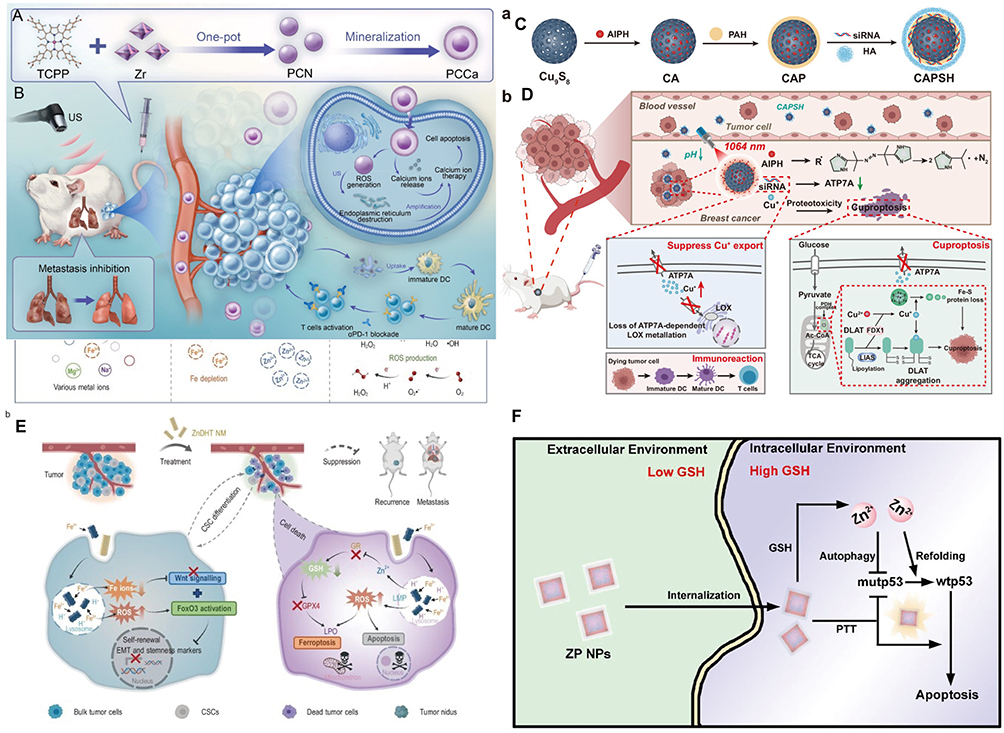 |
Figure 2 Schematic illustration of metal ion interference with cellular homeostasis. (A) Preparation of PCCa. (B) Tumor cell death process via calcium ion therapy and PCCa-mediated cancer immunotherapy.234 (C) Synthesis of CAPSH. (D) Copper sulfide-based nanocarriers regulating copper homeostasis for synergistic breast cancer treatment.236(E) Fe3⁺-triggered cascade functioning of ZnDHT and its anti-cancer mechanisms.235 (F) Combinational effects of ion interference and photothermal treatment using ZP NPs.237 |
Thus, metal ions can catalyze oxygen generation in hypoxic tumor microenvironments, alleviating oxygen deficiency and enhancing the effectiveness of radiotherapy and chemotherapy. Improved oxygen availability increases the treatment sensitivity of tumor cells, leading to better therapeutic outcomes.
Immune Modulation
Metal ions crucially modulate immune responses, influencing the activation and function of immune cells such as T cells and macrophages. Zinc ions enhance dendritic cell activation, promoting robust anti-tumor immune responses. This immunomodulatory effect can improve the efficacy of cancer immunotherapy by enhancing the body’s natural cancer-fighting ability.35,238 Nutritional metal ions in metalloimmunotherapy, such as cyclic dinucleotide (CDN) STING agonists and Mn2⁺, offer a new platform for local and systemic cancer treatments, initiating robust anti-tumor immunity and achieving remarkable therapeutic efficacy.239 Achieving high-quality bone-implant integration requires a balance between immune defense and anti-inflammation. Abaloparatide (ABL) integrated into a zinc-phenolic network constructs a multifunctional nanointerface (ABL@ZnTA) that enhances implant osseointegration, balancing infection defense and osteogenesis promotion in orthopedic and dental implants (Figure 3A–D).240 Long-term implant stability depends on perfect integration with surrounding tissues; metal ion-regulated immune responses promote bone tissue formation, thereby enhancing integration effectiveness. A multifunctional coating with biomimetic micro-/nano-structures on titanium surfaces achieves sequential Cu2⁺ and Zn2⁺ release, addressing the challenges of titanium implant failure (Figure 3E).241 An ATP-responsive Mg/Zn-MOF used as an ion-interference strategy in periodontitis immunotherapy effectively diminishes inflammatory cell infiltration.242
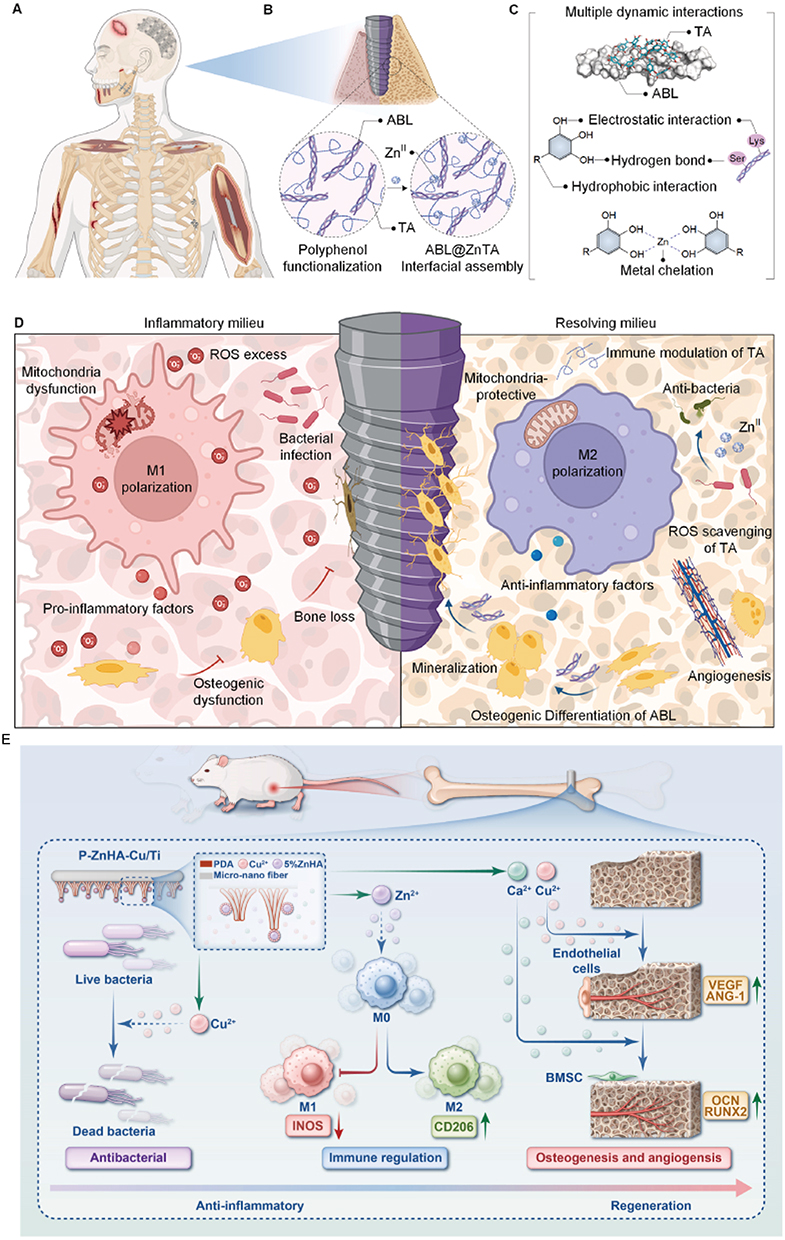 |
Figure 3 (A) Clinical scenarios for functional coatings: traumatic fractures, immediate dental implantation in bacterial infection microenvironments. (B) TA-ABL and ABL@ZnTA nanointerface preparation on Ti implants. (C) Multiple TA-ABL interactions; TA-ZnII chelation. (D) Osseointegration on clinical Ti implants via anti-inflammation and immunomodulation against bacterial infection.240 (E) Ti with sequential release function promoting angiogenesis and osteogenic differentiation.241 |
Structural Regulation and Intelligent Response
Significant progress has been made in Metal-based biomaterials for structural regulation and intelligent response, with applications in antimicrobial properties,243 tissue repair,244,245 cancer therapy,246 and biosensing.247 Thus, the development of novel non-antibiotic strategies for treating infections and accelerating wound healing represents a key application for these materials, which leverage the unique chemical characteristics of metal ions, including coordination, redox activity, and photothermal/catalytic effects, for dynamic structural modulation and environmental responsiveness. Co-assembling glycyrrhizic acid (GA) and rhein (Rh) via Zn2⁺ modulation improves antibacterial efficacy and anti-inflammatory capacity.245 An ion-mediated immunotherapy agent (IMIA) engineered through ion-immunotherapy reshapes the immunosuppressive tumor microenvironment (TME), awakening systemic immune responses and long-term immune memory.246 Surfaces designed for specific biomolecule recognition are essential for cancer theranostics, biosensing, and tissue engineering. New grafting methods, such as polydopamine (PDA)-assisted Ca2⁺-mediated grafting using hyaluronic acid (HA), increase grafting density, enhance interactions, enable selective recognition, and offer simplicity, versatility, and substrate flexibility for biomedical applications.247 Another conjugation strategy, cytomembrane-mediated biospecific transport of metal ions without other materials, also offers versatile biofunctions for effective chemotherapy.248
Limitations in structural regulation and intelligent response include difficulty in precisely controlling metal ion release rate, spatial distribution, and concentration gradients, which may cause localized toxicity or insufficient efficacy. Long-term degradation products of certain materials may trigger immune responses. Photothermal/photodynamic materials such as InNNi3249 and Ti-PMOF-DMA250 are constrained by tissue penetration depth and hypoxic microenvironments. Future trends include combining multiple mechanisms, such as photothermal/sonodynamic/ion release (eg, Type I/II SDT synergistic starvation therapy using Au/Yb-TCPP@GOx).251 Optimizing cell membrane modification strategies improves targeting efficiency (eg, macrophage membrane for inflammation targeting).252 Developing pH/enzyme/ROS multi-responsive metal ion carriers enables on-demand release.253
Mechanisms of Metal Ion-Mediated Biological Effects
Coordination Chemistry: Binding of Metal Ions to Biomacromolecules
Metal ions can be stably delivered through coordination interactions.14 Using a metal-phenolic network, the researchers co-immobilized Al3⁺ and Mn2⁺ with ovalbumin into a biocompatible coordination network. At a more fundamental level, the coordination function of metal ions as enzyme cofactors in immune signal transduction is critical, especially the activation of the cGAS-STING pathway by Mn2⁺.254 Moreover, a high-copper environment promotes tumor angiogenesis by upregulating VEGF expression, while copper chelators are used to inhibit angiogenesis and delay tumor progression.255 This direct coordination serves as the key molecular basis for copper-mediated regulation of angiogenesis. Whether in immunomodulation or angiogenesis, coordination chemistry represents the initial step through which metal ions initiate biological effects.
Although the binding affinity and stoichiometry of the Cu2⁺-angiogenin interaction are well characterized, most evidence comes from in vitro binding assays. In the complex in vivo milieu, competing metal ions (eg, Zn2⁺) and chelating proteins (eg, albumin, metallothionein) can dramatically alter the effective free Cu2⁺ concentration. Thus, the quantitative significance of this direct coordination under physiological or pathological copper overload conditions remains uncertain.
Electron Transfer: Redox Regulation and Reactive Oxygen Species Generation
Electron transfer represents another core mechanism by which metal ions exert biological effects, and it is particularly prominent in antibacterial activity and cell death regulation. Generalova et al256 systematically summarized three major antibacterial mechanisms of metal/metal oxide nanoparticles: physical cell-wall interaction, release of metal ions, and reactive oxygen species (ROS) generation. Unlike the single mode of action of conventional antibiotics, nanoparticles can exhibit multiple mechanisms simultaneously, producing synergistic effects that include membrane disruption, DNA and protein damage, enzyme inactivation, and oxidation of cellular components, ultimately leading to cell death. The review classified and discussed the specific antibacterial mechanisms of various metal nanoparticles, including Ag, Au, Cu/CuO, TiO2, ZnO, and Fe2O3/Fe3O4. The intrinsic link between ion release and ROS generation lies in the fact that metal ions (especially transition metal ions such as Fe2⁺/Fe3⁺ and Cu2⁺) can catalyze the Fenton reaction and Fenton-like reactions, converting relatively inert H2O2 into highly reactive hydroxyl radicals (·OH), thereby causing irreparable oxidative damage to bacteria. While Fenton chemistry is well established in cell-free systems, its actual contribution within bacterial cells is often inferred from indirect evidence (eg, ROS scavengers or iron chelators). Direct quantification of hydroxyl radicals in live bacteria during nanoparticle exposure remains technically challenging. Wang et al254 took a more systematic view, noting that transition metals (Fe, Zn, Mn, Cu, Co) play central roles in metabolism and immunity due to their unique redox properties, performing three major functions within metalloproteins: providing structural support, acting as enzyme cofactors, and mediating electron transfer Cheng et al257 extended this principle to the field of tumor immunotherapy, elaborating in detail how multiple metal ions, regulate the tumor microenvironment and enhance immunotherapy efficacy by inducing programmed cell death (PCD), such as ferroptosis (driven by iron-dependent lipid peroxidation) and cuproptosis (copper-induced proteotoxic stress through direct binding to tricarboxylic acid (TCA) cycle enzymes). These newly discovered PCD paradigms all depend on the unique redox chemistry of metal ions, reaffirming the key role of the ion-centric paradigm in cell fate determination.
The discovery of cuproptosis and ferroptosis has undoubtedly advanced the field. However, most mechanistic studies rely on genetic or pharmacological manipulations that drastically alter intracellular metal pools, conditions rarely encountered in clinical metal-based nanomedicine. Moreover, distinguishing between direct ion-mediated PCD and secondary effects arising from general oxidative stress remains a significant challenge.
Signaling Pathway Interference: Multi-Target Regulation by Ion Signal Transduction
The most distinctive capability of metal ions lies in their ability to simultaneously regulate multiple signaling pathways, a form of polypharmacology that is difficult to achieve with conventional small molecules or biologics. Among the pathways uncovered so far, the cGAS-STING pathway stands as the most representative example.
Li et al systematically explained how manganese ions modulate the activity of CD8⁺ T cells and NK cells by regulating the cGAS-STING pathway.258 Mn2⁺ directly binds to cGAS, enhances its sensitivity to double-stranded DNA, promotes cGAMP production, and thereby activates the STING-dependent type I interferon response. In terms of the breadth of signaling pathway regulation, biometallic ions not only affect a single immune checkpoint pathway but also synergistically enhance immunotherapeutic effects through multiple steps, such as activating immune cells, enhancing tumor antigen presentation, and remodeling the tumor microenvironment.255 The immunomodulatory function of metal ions is multi-layered: by inducing distinct forms of ferroptosis or cuproptosis, metal ions not only directly kill tumor cells but also release damage-associated molecular patterns (DAMPs) that activate antigen-presenting cells, thereby bridging innate and adaptive immune responses.257 In the field of angiogenesis, metal ions such as magnesium, zinc, copper, strontium, and cobalt have been shown to synergistically regulate the process of new blood vessel formation by activating multiple signaling pathways, including Wnt, PI3K/Akt, MAPK, and HIF-1α/VEGF. This ability to achieve multi-pathway synergistic regulation is precisely the core advantage that distinguishes metal ions from conventional drugs.
While polypharmacology is touted as an advantage, it is also a double-edged sword. The same multi-target engagement that produces therapeutic synergy can lead to unpredictable off-target effects, narrow therapeutic windows, and complex toxicity profiles that are difficult to model preclinically. For example, activation of the HIF-1α/VEGF axis by cobalt or copper ions may promote angiogenesis in ischemic tissue but could also accelerate tumor progression or induce aberrant vascular leakage.259,260 Copper chelation therapies such as penicillamine and tetrathiomolybdate inhibit tumor microvascular supply, reduce tumor volume, and decrease vascular permeability in animal models by lowering plasma copper levels; reduced plasma copper levels suppress the formation of breast cancer micrometastases.261 Multiple clinical trials have been conducted to investigate copper chelation as an adjuvant or primary therapy. Thus, a more balanced view would acknowledge that metal ions clinical success depends on exquisite spatial and temporal control, which a challenge that remains far from solved.
Disease Treatment Applications of Metal Ion-Mediated Action
Metal ion-mediated therapies have vast applications in disease treatment, particularly in oncology, where they offer innovative strategies for combating cancer. Representative paradigms include metal peroxides in chemoreactive nanomedicine: copper peroxide (CuO2), calcium peroxide (CaO2), magnesium peroxide (MgO2), zinc peroxide (ZnO2), barium peroxide (BaO2), and titanium peroxide (TiOₓ) nanosystems. These have been broadly explored in catalytic nanotherapeutics, photodynamic therapy, radiation therapy, antibacterial infection control, tissue regeneration, and synergistic applications.262 As shown in Figure 4, metal-based materials, such as hydrogels, microspheres, nanofiber scaffolds, and coatings, which deliver anti-tumor therapies through mechanisms including chemical kinetics, glutathione depletion, mitochondrial dysfunction, and immune activation. They combat infections by disrupting biofilm formation and membrane structures. Furthermore, their antibacterial properties help prevent infections, while their antioxidant capabilities neutralize excessive ROS and mitigate inflammatory damage.
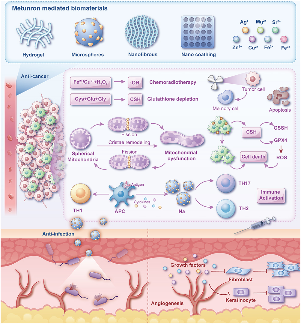 |
Figure 4 Multi-scenario biological functions of metal-ion-loaded biomaterials. These biomaterials can be fabricated into various morphologies such as hydrogels, microspheres, and nanofibers, and loaded with a range of metal ions including silver, magnesium, copper, and iron. They function in three major directions: Antitumor: they kill tumor cells through multiple pathways, including chemodynamic therapy, mitochondrial dysfunction, glutathione depletion, ferroptosis, and immune activation. Anti-infection: the released metal ions achieve broad-spectrum antibacterial and antimicrobial effects. Tissue repair: they induce angiogenesis and regulate the proliferation of fibroblasts and keratinocytes, thereby accomplishing wound tissue regeneration. |
Antitumor Therapy
Antitumor therapy using metal ions encompasses various strategies that enhance treatment efficacy while minimizing side effects. Metal phosphate nanomaterials (Metal-BP) and their nanocomposites, as drug delivery systems, have achieved breakthroughs in tumor diagnosis (MRI, fluorescence, photoacoustic, and nuclear imaging) and therapies (chemotherapy, gene therapy, PTT, PDT, and radiotherapy).263 Porphyrin-based MOFs, as multifunctional nanomaterials, offer high porosity, tunable structures, excellent stability, and versatile applications, leading to better therapeutic outcomes, reduced side effects, and improved selectivity for cancer therapy.264 Copper-based MOFs (Cu-MOFs) excel in catalysis, biocompatibility, photothermal conversion, and metabolism regulation, attracting increasing attention for antitumor applications.265 DNAzyme-loaded MOFs, such as the chlorin e6-modified DNAzyme (Ce6-DNAzyme) therapeutic nanosystem, produce ROS and provide fluorescence signals for combined gene therapy and PDT.266 Other MOF-based approaches include nanozyme-engineered MOFs for catalytic cascade-enhanced synergistic cancer therapy.267 Different MOF structures have also been applied in cancer therapy, including heterostructures of Ag-doped MOFs,268 petaloid R848@petaloid MOFs,269 aperture-modulated isoreticular MOFs,270 nanoscale MOFs,271 polydopamine-coated MOFs,272 multifaceted carbonized Cu2-ₓSe@cMOF nanoplatforms,273 dual-functional bimetallic (GOx@MnCoMOF) nanoplatforms,274 and tailored core-shell dual MOFs (MIL-88-ICG@ZIF-8-DOX nanoparticles) as versatile nanomotors for effective synergistic antitumor therapy.275
 |
Figure 5 The anti-tumor mechanisms of these metal-based biomaterials are distinct: Iron-based biomaterials function by potentiating oxidative stress to induce ferroptosis. Copper-based biomaterials mediate cell death via Cu2⁺ binding to lipidated proteins, which induces proteotoxic stress through abnormal protein aggregation and FDX1-dependent impairment of iron-sulfur cluster proteins. Manganese-based biomaterials act by competitive inhibition of Golgi Mn2⁺/Fe2⁺ transporters (eg, ATP2C1/SPCA1) by Mn2⁺. The resultant intra-Golgi Fe2⁺ deficiency disrupts glycosylation, causing proteostatic stress and caspase-independent cell death (By FigDraw). |
Catalytic therapy uses metal ions to facilitate the generation of cytotoxic agents within tumor cells. As shown in Figure 5, iron-, copper-, and manganese-based biomaterials induce apoptosis through distinct ion-mediated pathways. Iron-based materials release iron ions, triggering lipid peroxidation and accumulation of reactive oxygen species (ROS), thereby initiating ferroptosis. Copper-based materials regulate the valence state of copper ions, interfere with protein maturation and assembly, and induce proteotoxic stress leading to cell death. Manganese-based materials cause intracellular manganese overload, disrupt endoplasmic reticulum protein folding, and trigger cell death through non-apoptotic pathways. Thus, these three types of biomaterials achieve differential cell regulation via completely independent programmed cell death pathways. Copper ions (Cu⁺/Cu2⁺) generate ROS, enhancing oxidative stress and promoting apoptosis.276 Manganese ions (Mn2⁺) alleviate tumor hypoxia, thereby improving the effectiveness of other therapies.86 Synergistic therapy has prompted extensive research into new treatments that overcome the limitations of traditional therapies. Metal catalysts with enzyme-like functions serve as carriers and are combined with glucose oxidase (GOx), ensuring preservation of enzyme activity in normal tissue and specific activation within tumors.277
Metal ions enhance anti-tumor immune responses, making them valuable in immunotherapy. Zinc ions (Zn2⁺) activate dendritic cells, promoting T cell activation and enhancing the overall immune response.278 Magnesium ions (Mg2⁺) modulate antitumor immunity, highlighting the potential of metal ions in enhancing immunotherapy efficacy.279 A novel multifunctional copper-piceatannol/HA nanopill (Cu-Pic/HA NPs) enhances pyroptosis and cuproptosis, remodeling the immunosuppressive tumor microenvironment (TME) for cancer immunotherapy.280 Most chemotherapy drugs lack tumor specificity, have low bioavailability, and face clinical obstacles. Metal-phenolic network nanoparticles self-assembled from copper-coordinated chlorogenic acid (ChA) (ChA-Cu NPs) overcome the tumor immunosuppressive microenvironment (TIME) and low immunogenicity in solid tumors.281 Cell pyroptosis and cuproptosis represent promising cancer therapy strategies. Tumor microenvironment-responsive Cu/CaO2 nanocomposites serve as a multimodal platform that facilitates significant ROS generation,87 leading to pyroptosis, immunogenic cell death (ICD), and T cell infiltration via antitumor immune response.282
Therefore, in the field of anticancer therapy, metal ions exert potent antitumor effects by inducing novel programmed cell death modalities such as ferroptosis, cuproptosis, and calcicoptosis, and through activation of immune pathways such as the cGAS–STING pathway.257 In regenerative medicine and tissue engineering, ions including Sr2⁺, Mg2⁺, Zn2⁺, and Cu2⁺ have been systematically integrated into scaffold designs to harness their osteoinductive and angiogenic functions, effectively promoting multi-tissue synergistic repair.283 In the area of anti-infection, strategies based on metal ions (eg, Ag⁺, Cu2⁺, Ga3⁺) provide new avenues for overcoming antibiotic resistance. Furthermore, metal ion–driven delivery systems, such as metal–organic frameworks (MOFs), metal hydrogels, and metalloprotein nanomedicines, have enabled major breakthroughs including oral delivery of macromolecular drugs, crossing the blood–brain barrier, and theranostic capabilities that integrate imaging-guided therapy.284–286
Antimicrobial Treatment
Antimicrobial treatment is increasingly important due to the rise of antibiotic resistance and the need for effective strategies to combat infections. This section explores various aspects of antimicrobial treatment, focusing on the mechanisms of action of silver, copper, and gallium ions, as well as the role of biofilm disruption in enhancing therapeutic efficacy. Examples include integrated metal complexes for diagnosis and treatment,287 metal-drug coordination complexes,288 bimetallic nanocatalyst/nanoreactors,289 and bioceramics/coatings with metal ion doping.290
Tuberculosis, a globally significant infectious disease primarily caused by Mycobacterium tuberculosis, has seen its treatment efficacy drastically reduced owing to drug resistance issues associated with conventional medications. This urgent need for multi-target therapeutic approaches has driven the development of innovative solutions. Emerging antibacterial therapies, such as light-activated therapy and gas therapy, demonstrate unique advantages. However, current multi-component drugs based on nanocarriers still face challenges including complex preparation processes, poor stability, and unclear pharmacokinetic profiles. Thus, a novel integrated photoactive manganese-based carbon monoxide (CO) release molecule has been developed for the synergistic treatment of mycobacterial infections, based on small molecular metal complexes with photoactivity and carbon monoxide release function.288
Silver ions (Ag⁺) possess potent antibacterial properties, interacting with bacterial cell membranes and intracellular components. The mechanism involves ROS release that induces oxidative stress, causing cellular damage and bacterial death. Ag⁺ penetrates membranes, disrupts metabolism, and inhibits DNA replication. Silver nanoparticles that controllably release Ag⁺ significantly reduce bacterial viability while minimizing resistance development, making silver a promising candidate for various medical antimicrobial applications.291,292 Osteomyelitis remains a formidable challenge; multidrug-resistant bacteria have renewed interest in antimicrobial biomaterials that use antiseptic silver ions.293 In situ silver nanoparticle coatings built on bacterial biofilms offer antimicrobial activities for implant-surrounding tissue cells.294 A Sr@Ag-based scaffold fabricated by coaxial 3D printing provides anti-inflammatory and pro-osteogenic effects, holding promise for infectious bone defect diseases.295
Copper ions (Cu2⁺) are potent antimicrobials against multidrug-resistant bacteria. Their antibacterial action primarily involves ROS generation and bacterial membrane disruption, leading to increased permeability, leakage of cellular contents, and cell death. Copper effectively inhibits resistant strains such as MRSA. Its dual mechanism, direct membrane disruption combined with oxidative stress induction, and makes it valuable against bacterial infections, especially in the context of rising antibiotic resistance.296,297 In vital pulp therapy (VPT), a SrCuSi4O10/gelatin methacrylate (SC/Gel) composite hydrogel improves dentine-pulp complex repair through multiple bioactivities via sustained release of Sr2⁺, Cu2⁺, and SiO32− ions (Figure 6A).298
 |
Figure 6 (A) SC/Gel composite hydrogel application with NIR irradiation for infected dental pulp treatment, releasing Sr2⁺, Cu2⁺, SiO32− ions promoting antibacterial effects, angiogenesis, odontogenesis.298 (B) BP-Ga3⁺ combination increases BP stability; NIR irradiation releases Ga3⁺ exerting antibacterial effects.299 |
Gallium ions (Ga3⁺) exhibit unique antibacterial properties by mimicking iron, which is essential for bacterial growth. Ga3⁺ interferes with iron uptake, causing nutritional deprivation and growth inhibition. This mechanism is particularly effective against iron-dependent pathogens, disrupting their metabolism. Recent research highlights gallium’s potential against resistant bacteria, making it a promising candidate for novel antimicrobial therapies. Targeting bacterial iron metabolism provides a strategic advantage against infections that are increasingly difficult to treat with conventional antibiotics.300,301 In wound healing, an innovative antibacterial technology uses a two-dimensional nanomaterial that anchors Ga3⁺ onto the surface of black phosphorus (BP); Ga3⁺ employs a “Trojan horse strategy” to disrupt iron metabolism while enhancing BP stability (Figure 6B).299
Iron chelators disrupt biofilms by sequestering iron, which is essential for bacterial growth and biofilm formation. Limiting iron availability weakens biofilm structural integrity and enhances antibiotic susceptibility. Iron chelation inhibits biofilm formation and promotes the dispersal of established biofilms, making bacteria more vulnerable. This dual action positions iron chelators as valuable adjuncts to antimicrobial therapy, particularly against biofilm-associated infections.302,303
Bone Repair
In the field of orthopedics, metal ion-functionalized biomaterials have made significant progress in bone defect repair. Studies have shown that applying layered double hydroxide (LDH)-functionalized bone cement to critical-size bone defects not only enhances the mechanical properties of the material but also promotes bone tissue regeneration through the sustained release of functional metal ions.304 Specifically, CuMgFe-LDH-loaded bone cement demonstrates superior performance in multiple aspects: the material achieves an inhibition rate of up to 99.99% against Staphylococcus aureus, significantly enhances cell proliferation and differentiation, increases calcium nodule deposition by 1.29-fold, and doubles the expression of osteogenesis-related genes.304
Strontium ions (Sr2⁺) enhance osteoblast differentiation and activity. Sr2⁺ stimulates the proliferation of mesenchymal stem cells (MSCs) and their differentiation into osteoblasts, a process crucial for bone formation.305 The mechanism involves activation of signaling pathways such as Wnt/β-catenin, which are essential for osteogenic differentiation.306,307 Sr2⁺ enhances the expression of osteogenic markers, including alkaline phosphatase (ALP),308 osteocalcin (OCN),308,309 and runt-related transcription factor 2 (Runx2),310 thereby promoting mineralization and bone matrix formation. Incorporating Sr2⁺ into bone graft materials improves their osteoconductive properties, enhancing bone healing in preclinical models.311 The release of Sr2⁺ from biomaterials creates a localized environment conducive to bone regeneration, making it a valuable strategy for bone repair.305
Magnesium ions (Mg2⁺) are vital for bone growth and regeneration, playing essential roles in osteoblast function and the regulation of bone mineralization. Mg2⁺ enhances osteoblast proliferation and differentiation, thereby increasing bone formation.312 The incorporation of Mg2⁺ into biomaterials improves their mechanical properties and biocompatibility, making them attractive for bone repair.313 Mg2⁺ in amorphous calcium magnesium phosphate (ACMP) bioceramics incorporated into methacrylated gelatin (GelMA) bioink triggers osteogenic differentiation of encapsulated pre-osteoblasts and stimulates bone regeneration.314 Mg2⁺ modulates osteogenic gene expression and promotes the synthesis of bone matrix proteins, facilitating the healing of bone defects.315 Additionally, Mg2⁺ exerts anti-inflammatory effects, supporting a favorable environment for bone regeneration. Using magnesium nanoparticles (MgNPs) as templates, magnesium-enriched graphene oxide nanoscrolls (MgNPs@GNSs) have been designed as immunomodulatory agents that combinationally modulate inflammatory responses for bone defect repair.316 Incorporating Mg2⁺ into bone grafts and scaffolds significantly improves healing outcomes, highlighting its therapeutic potential. Injectable biomimetic porous hydrogels (MgO/MgCO3@PLGA (PMM) hydrogels) serve as in situ repair systems for irregular bone defects and accelerate bone regeneration.317
In the field of orthopedic implants, novel medium-entropy alloys (MEAs) such as TiMoHfCux (x = 0, 5, 10) have simultaneously achieved excellent mechanical properties, antibacterial characteristics, and osteogenic activity through the introduction of hafnium and copper elements. The TiMoHfCu10 alloy demonstrates an antibacterial rate of up to 97% against Escherichia coli and exhibits outstanding osteogenic performance in a rat femoral condyle model. This is primarily attributed to the nanoscale needle-like (Ti, Hf)2Cu precipitates and the micro-area potential difference (MAPD) effect formed between these precipitates and the matrix.318
Nerve Regeneration
Zinc ions (Zn2⁺) play a critical role in nerve injury repair, as they are involved in cell proliferation, differentiation, and regeneration. A chimeric hydrogel microsphere has been designed, featuring an Mg2⁺-crosslinked methacrylate gelatin “shell” and a Zn2⁺-loaded PLGA “core”.319 Zn2⁺ promotes the survival of neurons and glial cells, which is essential for nerve regeneration. In the context of nerve injury, Zn2⁺ enhances repair by modulating inflammatory responses and promoting angiogenesis, a process crucial for supplying nutrients and oxygen to regenerating nerves.320 Zn2⁺ stimulates neurotrophic factor secretion (nerve growth factor NGF,321 brain-derived neurotrophic factor BDNF322), thereby supporting neuronal survival and growth. Furthermore, Zn2⁺ influences macrophage polarization, promoting the pro-regenerative M2 phenotype that facilitates tissue repair. KZIF@HA spontaneously forms nanogels for sustained drug release; the release of Zn2⁺ reverses osteoarthritis (OA) progression by promoting M2 macrophage polarization and establishing an anti-inflammatory microenvironment.323 The application of Zn2⁺ in nerve repair strategies holds promise for improved functional recovery after peripheral nerve injuries.324
Angiogenesis
Copper ions (Cu2⁺) are essential for angiogenesis, the process of new blood vessel formation that is critical for tissue repair and regeneration. Cu2⁺ promotes angiogenesis by enhancing endothelial cell proliferation, migration, and tube formation. It stabilizes hypoxia-inducible factor (HIF),325 a key regulator of angiogenic factors such as vascular endothelial growth factor (VEGF).326 As shown in Figure 7, the release of metal ions from porous scaffolds, biomimetic nanoparticles, and metal-coated micro/nano-particles can promote endothelial cell migration and proliferation, regulate remodeling of the immune microenvironment, alleviate oxidative stress, and ultimately facilitate vascular regeneration as well as tissue repair and reconstruction through pro-angiogenic growth factors such as VEGF. TiCu/TiCuN multilayer coatings offer wear resistance and antibacterial effects; however, their broad application remains unrealized.327 The documented role of Cu2⁺ in promoting angiogenesis and tissue repair,328,329 presents a significant opportunity to develop enhanced multifunctional coatings, although a deeper understanding of their properties is still required. Moreover, the development of porous tantalum (Ta) scaffolds is hindered by the difficulty in balancing porosity against mechanical strength, and degradation rate against tissue ingrowth. The synthesis of in situ Ta-doped β-tricalcium phosphate (β-TCP) nanopowders via a microwave-ultrasound-assisted hydrothermal method aims to overcome these trade-offs by improving mechanical integrity, biodegradation kinetics, and osteoinductivity.330
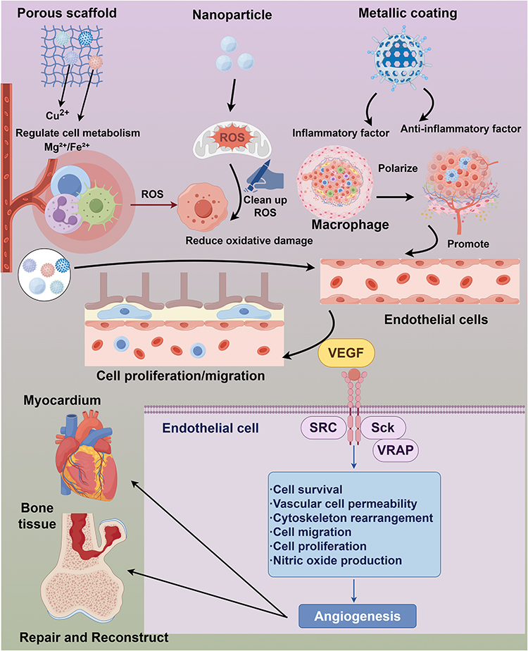 |
Figure 7 This schematic diagram, created with FigDraw. At the material level, metal-based biomaterials sustain the release of metal ions, which regulate cellular metabolism, scavenge reactive oxygen species (ROS) to alleviate oxidative damage, bidirectionally modulate inflammation, and induce reparative polarization of macrophages. At the cellular/molecular level, these actions activate vascular endothelial cells and drive angiogenesis via the VEGF signaling pathway. Ultimately, at the tissue/organ level, this results in defect regeneration and structural reconstruction of myocardial and bone tissues. |
To achieve sustained release, prolonged efficacy, reduced dosage, improved efficiency, and potentially lower side effects, dexketoprofen trometamol (DT)-loaded chitosan nanoparticles (CS-NPs) were designed and produced as an oral sustained-release delivery system using the spray drying method.331 Moreover, by utilizing an interpenetrating polymer network (IPN) structure and a nanocomposite strategy, the scaffold’s mechanical strength and degradation properties are simultaneously improved, thereby addressing the common performance drawbacks of single-component hydrogel materials. The innovative incorporation of CuO nanoparticles (CuONPs) enables the scaffold not only to passively support tissue growth but also to actively release biological signals that promote angiogenesis, overcoming the core challenge of nutrient delivery and metabolic waste removal in tissue engineering.332
Antioxidant and Anti-Inflammatory
The interplay between oxidative stress and inflammation is critical in the pathogenesis of chronic diseases such as diabetes, cardiovascular disorders, and neurodegenerative diseases. Antioxidants mitigate oxidative stress, while anti-inflammatory agents reduce inflammation. There is growing interest in using metal ions for their antioxidant and anti-inflammatory properties. These ions exhibit therapeutic potential and contribute to novel management strategies for diseases related to oxidative stress and inflammation.
Cerium ions (Ce3⁺, Ce4⁺) attract attention for their antioxidant properties. Their unique redox behavior enables participation in redox cycling, allowing them to effectively scavenge free radicals and ROS. The mechanism involves the conversion between Ce3⁺ and Ce4⁺: Ce3⁺ donates electrons to neutralize free radicals, while Ce4⁺ accepts electrons to stabilize reactive species. This dual functionality makes cerium oxide nanoparticles (nanoceria) particularly effective in biological systems, as they mimic natural antioxidant enzymes such as superoxide dismutase and catalase.333 Cerium ions also enhance the expression of endogenous antioxidant enzymes, thereby boosting cellular antioxidant capacity.334 The application of cerium in wound healing and tissue regeneration highlights its potential to reduce oxidative damage and promote healing through its antioxidant properties.335
Vanadium ions (V5+) are promising for diabetes treatment due to their insulin-mimetic properties. Vanadium enhances cellular glucose uptake, improves insulin sensitivity, and modulates signaling pathways involved in glucose metabolism.336 The mechanism involves the activation of insulin receptor substrates, which in turn stimulate glucose transporter proteins to facilitate cellular glucose entry. Okra extract-capped V2O5 nanoparticles (ONPs) exhibit significant absorptivity, mucoadhesion, and controlled release of vanadate ions as intermediates.337 Vanadium also possesses antioxidant properties that help mitigate oxidative stress associated with diabetes. By reducing oxidative damage, it protects pancreatic β-cells from apoptosis, thereby preserving insulin production.338 Vanadium’s dual role with enhancing insulin sensitivity while providing antioxidant benefits, positions it as a valuable therapeutic agent for the management of diabetes and its complications.339 Ongoing studies are exploring optimal delivery methods and formulations to maximize vanadium’s clinical therapeutic potential.340
Integrated Diagnosis and Treatment
Integrating diagnostic imaging with therapeutic strategies represents a significant advancement in modern medicine, particularly in oncology and cardiology. This approach combines imaging modalities such as MRI and PET/SPECT with targeted therapies, enabling personalized treatment based on individual patient profiles. Melanin nanoparticles, especially when combined with paramagnetic ions, have sparked interest as MRI contrast agents.341 The use of magnetic (Fe3⁺, Gd3⁺) and radioactive (64Cu, 99mTc) metal ions further enhances the efficacy of this integrated approach. Paramagnetic ions improve MRI contrast and visualization of pathological tissues, while radioactive metals enable PET/SPECT imaging, facilitating the detection of malignancies and guiding therapeutic interventions. This synergy optimizes patient management and enhances treatment precision, thereby improving clinical outcomes. Copper-64 (T1/2 = 12.7 h) emits positrons and beta particles, making it suitable for PET imaging and cancer radiotherapy.342 Labeling with 64Cu during the synthesis of melanin-like polymers (MPNs) directly into the polymeric matrix creates multimodal preclinical imaging agents.343 Ligand anchoring group-mediated radiolabeling (LAGMERAL), based on diphosphonate-polyethylene glycol (DP-PEG) decorated on inorganic nanoparticle surfaces, enables sensitive tumor diagnosis.344
The development of metal-ion theranostic agents faces key challenges. In particular, it is difficult to simultaneously optimize both imaging properties and therapeutic functions. Although smart probes responsive to pathological microenvironments are emerging, their activation sensitivity, specificity, and reliability still require improvement. Furthermore, integrating multimodal imaging and combination therapies into single platforms often leads to structural complexity, reduced reproducibility, higher costs, and potential interference between functional components.
Vanadium ions (V5+) are promising for diabetes management. Research indicates that vanadium mimics insulin action, enhancing cellular glucose uptake and lowering blood sugar levels. Silver vanadate nanoparticles (AgVO3) confer antimicrobial activity at the molecular level.345 Selecting suitable nanozymes that are easy to synthesize, tumor-specific, multifunctional, and therapeutically effective is meaningful for tumor therapy. Natural polyphenol tannic acid (TA) hybridized with mixed-valence vanadium oxide nanosheets (TA@VOₓ NSs) effectively inhibits tumor growth through synergistic chemodynamic therapy (CDT) and photothermal therapy (PTT).346 Titanium (Ti)-based metallic biomaterials (MBs) serve as mechanical supports and constraints in clinical settings due to their superb mechanical properties, great corrosion resistance, and good biocompatibility.347 Transition metal oxide (TMO) nanomaterials can initiate the tumor immunity cycle.348 pH-sensitive, tumor-tropism hybrid membrane-coated manganese oxide (MnO2) nanoparticles act as glutamine metabolism inhibitors, delivering small molecule drugs for chemo- and immunotherapy.349 The key challenge remains the precise delivery of metal ions or nanocarriers to target lesions with effective enrichment while reducing off-target effects on normal tissues.
The clinical application of conventional metal-ion agents is limited by poor targeting, low bioavailability, and short half-life. Although nanotechnology can partially address these issues, many nanocarriers suffer from instability, rapid clearance, and insufficient tumor accumulation due to the weak enhanced permeability and retention (EPR) effect in humans. Active targeting strategies are further hindered by complex modification processes and target heterogeneity. Thus, despite the promise of metal ions in theranostics, key challenges related to safety, targeting, functional integration, and clinical translation remain to be overcome.
In summary, the field of metal ion-based theranostics stands at a crossroads. The challenges outlined above are substantial but not insurmountable Such as conflicts in functional integration, inefficient targeting, and translational hurdles. Only through such a paradigm shift will metal ion-based theranostics fulfill their long-standing promise of personalized, precise, and safe disease management.
Wound Healing and Skin Regeneration
In the field of skin wound repair, metal ion-mediated biomaterials demonstrate significant therapeutic potential. In recent years, various innovative material systems have been developed to address different types of wound healing challenges. Metal-alginate (M-Alg) hydrogels,350 provide a moist wound environment through their three-dimensional network structure while enabling controlled release of metal ions for antibacterial effects. Gold nanoparticles (E-Au NPs),351 utilize their surface plasmon resonance characteristics to generate localized thermal effects under near-infrared light irradiation, effectively eliminating drug-resistant bacteria.
Nanosized zinc-based MOFs,352 leverage their high specific surface area and tunable pore sizes to achieve sustained release of zinc ions, inhibiting bacterial growth while promoting fibroblast migration. The nIon2⁺-COS/SA gel film, provides both a physical barrier and bioactive therapy for wounds through the synergistic effect of the natural antibacterial properties of chitosan and metal ions.353 Copper-zinc bimetallic organic frameworks (Cu/Zn-rich BMOFs), employ cascaded release of two metal ions, achieving rapid antibacterial effects followed by long-term healing promotion, thereby meeting the requirements of different stages of wound repair.354 Particularly noteworthy, tannic acid-europium metal-phenolic network nanoparticles (TA-Eu MPN), not only exert antibacterial functions but also enable real-time monitoring of the wound healing process through fluorescence imaging.355 Metal-doped cryogels combine temperature-sensitive properties with metal ion activity, allowing adaptive filling of irregular wounds.356 Cu-NP-embedded hydrogels effectively block antibiotic penetration barriers through sustained release of copper ions and inactivate bacteria by generating ROS.357 These materials have shown remarkable effects in preclinical studies: in antibiotic-resistant bacterial infections,358 they can disrupt biofilm structures; in diabetic wound models,359 they can stimulate fibroblast proliferation and angiogenesis; during infected wound healing,360 they can coordinately regulate inflammatory responses. It is noteworthy that recent studies have found that copper ions (Cu2⁺) at specific concentrations361 may instead promote bacterial proliferation, and induce intracellular acidification, highlighting the need for precise control of metal ion release kinetics.
These innovative material systems provide multifunctional solutions for skin tissue regeneration, demonstrating unique advantages in promoting collagen deposition, epithelialization, and angiogenesis, and showing broad clinical translation prospects. Future research should focus on optimizing the synergistic ratios of metal ions, developing intelligent responsive release systems, and establishing individualized precision treatment strategies.
Safety Analysis and Translational Challenges
Despite the significant potential of metal ions (eg, Fe3⁺/Fe2⁺, Cu2⁺, Zn2⁺, Mn2⁺, Co2⁺) in multimodal synergistic therapies, such as chemodynamic therapy, immunotherapy, and tissue regeneration, which owing to their unique catalytic, immunomodulatory, and antimicrobial activities, their clinical application faces severe safety bottlenecks. Most metal ions have a narrow therapeutic window between therapeutic and toxic concentrations, and non-specific release can induce oxidative stress, mitochondrial damage, neurotoxicity, or liver and kidney dysfunction. Moreover, the long-term stability, degradation product profiles, and in vivo distribution and clearance patterns of engineered carriers in physiological environments remain unclear.
Currently, approximately 85% of research in this field remains at the cellular level or below, with in vivo model studies accounting for less than 85%, and there is a complete absence of human clinical studies. Therefore, this review concludes that although basic mechanistic research is relatively sufficient, the overall translational readiness remains at an early stage (Technology Readiness Level 3–4), with a significant gap to reach TRL 6 or above for clinical application.
Key translational challenges include: the lack of standardized guidelines for evaluating metal ion release kinetics and toxicology; the difficulty in ensuring scalable manufacturing and batch-to-batch consistency of multifunctional synergistic systems; and the unpredictability of complex interactions between metal ions and the host’s endogenous metal pool, protein corona, and immune system.
Conclusions and Perspectives
In conclusion, metal-based biomaterials show promising potential in disease treatment, as demonstrated by their preliminary efficacy in cancer therapy, orthopedic applications, and antimicrobial treatments in vitro and in early animal models. However, further in vivo validation and clinical translation are needed to establish their definitive role. Nevertheless, the burgeoning interest in this field necessitates a balanced consideration of the diverse research perspectives and findings that underpin it.
Future metal-based biomaterials will place greater emphasis on multifunctional integration, achieving the combination of diagnosis, treatment, and monitoring. The application of diverse metal ions offers innovative therapeutic strategies, leveraging their distinct biochemical properties for specific clinical scenarios. While silver ions provide antimicrobial effects and iron ions aid oxygen transport, their toxicity requires careful evaluation. Future research must prioritize targeted delivery systems to enhance efficacy and minimize systemic exposure. Combining metal ions with conventional therapies (chemotherapy/immunotherapy) could yield synergistic effects. Critical challenges include establishing safe dosage thresholds, understanding long-term accumulation, and developing biodegradable carriers to ensure post-treatment clearance. A multidisciplinary approach is essential to balance therapeutic potential with biosafety.
To accelerate the clinical translation of this field, it is necessary to establish standardized in vivo tracing methods and long-term toxicological evaluation systems, and to clarify the accumulation organs, clearance pathways, and dose–toxicity relationships of different metal ions. This is a prerequisite for clinical translation. Achieving precise control over the release rate, action range, and duration of metal ions in complex physiological environments is the key to broadening their therapeutic window. Moreover, to bridge the gap from positive results in cell experiments to predictable efficacy in humans, two critical aspects must be addressed for successful clinical translation: long-term safety in large animals and biomarkers associated with clinical endpoints.
In summary, therapeutic metal ions are undergoing a profound paradigm shift from traditional auxiliary roles to multifunctional core therapeutic platforms. The field has achieved breakthrough progress in areas such as ion-induced regulation of cell death, remodeling of the tumor immune microenvironment, and functionalization of regenerative medicine scaffolds. However, clinical translation remains constrained by the irreproducibility of large-scale manufacturing, knowledge gaps in long-term biosafety, and fragmentation of global regulatory frameworks. In the future, only through the synergistic advancement of precision in materials design, deep understanding of biological mechanisms, and AI-driven predictive engineering can the clinical potential of therapeutic metal ions be translated into real-world therapies that benefit patients.
Data Sharing Statement
All data generated or analysed during this study areincluded in this published article.
Acknowledgments
The authors would like to thank all the participants of this study. They would like to acknowledge Editage for translating and editing this manuscript.
Funding
This work was supported by the Natural Science Foundation of China (No. 22568034, 22168028) and Key research and transformation technology projects of Inner Mongolia Autonomous Region (No. 2025YFSH0058) and Key project of Inner Mongolia Medical University (YKD2025ZD002). The Talent support project of Outstanding Young Talents “Yingcai Xingmeng” project of Inner Mongolia Autonomous Region and Inner Mongolia Medical University “Good at learning talent” (ZY20242128).
Disclosure
The authors declare that they have no known competing financial interests or personal relationships that could have appeared to influence the work reported in this paper.
References
1. Hong X, Tian G, Zhu Y, Ren T. Exogeneous metal ions as therapeutic agents in cardiovascular disease and their delivery strategies. Regen Biomater. 2024;11:rbad103. doi:10.1093/rb/rbad103
2. Wang H, Lin F, Zhang Y, Lin Y, Gao B, Kang D. Biomaterial-based vascularization strategies for enhanced treatment of peripheral arterial disease. J Nanobiotechnology. 2025;23(1):103. doi:10.1186/s12951-025-03140-4
3. Tao Y, Ayre WN, Jiang L, et al. Enhanced functionalities of biomaterials through metal ion surface modification. Front Bioeng Biotechnol. 2025;13:1522442. doi:10.3389/fbioe.2025.1522442
4. Luo Y, Liu H, Zhang Y, et al. Metal ions: the unfading stars of bone regeneration-from bone metabolism regulation to biomaterial applications. Biomater Sci. 2023;11(22):7268–45. doi:10.1039/D3BM01146A
5. Stadter J, Hoess A, Leemhuis H, et al. Incorporation of metal-doped silicate microparticles into collagen scaffolds combines chemical and architectural cues for endochondral bone healing. Acta Biomater. 2025;192:260–278. doi:10.1016/j.actbio.2024.12.029
6. Navratilova P, Emmer J, Tomas T, et al. Plastic response of macrophages to metal ions and nanoparticles in time mimicking metal implant body environment. Environ Sci Pollut Res Int. 2024;31(3):4111–4129. doi:10.1007/s11356-023-31430-7
7. Ying D, Zhang T, Qi M, Han B, Dong B. Artificial Bone Materials for Infected Bone Defects: advances in Antimicrobial Functions. ACS Biomater Sci Eng. 2025;11(4):2008–2036. doi:10.1021/acsbiomaterials.4c01940
8. Di Giacomo V, Cataldi A, Sancilio S. Biological Factors, Metals, and Biomaterials Regulating Osteogenesis through Autophagy. Int J Mol Sci. 2020;21(8):2789. doi:10.3390/ijms21082789
9. Bian Y, Cai X, Lv Z, et al. Layered Double Hydroxides: a Novel Promising 2D Nanomaterial for Bone Diseases Treatment. Adv Sci. 2023;10(24):e2301806. doi:10.1002/advs.202301806
10. Chakraborty A, Pacelli S, Alexander S, et al. Nanoparticle-Reinforced Tough Hydrogel as a Versatile Platform for Pharmaceutical Drug Delivery: preparation and in Vitro Characterization. Mol Pharm. 2023;20(1):767–774. doi:10.1021/acs.molpharmaceut.2c00564
11. Lucaciu LA, Seicean R, Seicean A. Small molecule drugs in the treatment of inflammatory bowel diseases: which one, when and why? - a systematic review. Eur J Gastroenterol Hepatol. 2020;32(6):669–677. doi:10.1097/MEG.0000000000001730
12. Hong Q, Kim H, Cai GY, Chen XM, He JC, Lee K. Modulation of TGF-β signaling new approaches toward kidney disease and fibrosis therapy. Int J Biol Sci. 2025;21(4):1649–1665. doi:10.7150/ijbs.101548
13. Nedylakova M, Medinger J, Mirabello G, Lattuada M. Iron oxide magnetic aggregates: aspects of synthesis, computational approaches and applications. Adv Colloid Interface Sci. 2024;323:103056. doi:10.1016/j.cis.2023.103056
14. Wang Z, Chen J, Wang T, De Rose R, Cortez-Jugo C, Caruso F. Amplifying the Immune Activity of Metal Ions through Metal-Phenolic Complexation. ACS Nano. 2025;19(15):15055–15068. doi:10.1021/acsnano.5c01842
15. de Souza JR, Anselmi C, Cardoso LM, et al. Electrospun SilkMA/silicate-chlorinated cobalt-doped bioactive glass composite for bone regeneration. J Mech Behav Biomed Mater. 2025;164:106929. doi:10.1016/j.jmbbm.2025.106929
16. Feng Z, Chen G, Zhong M, et al. An acid-responsive MOF nanomedicine for augmented anti-tumor immunotherapy via a metal ion interference-mediated pyroptotic pathway. Biomaterials. 2023;302:122333. doi:10.1016/j.biomaterials.2023.122333
17. Han Y, Jia X, Yang Y, et al. Study of bioactive 3D-printed scaffolds incorporating zinc-based MOF for bone defect repair and anti-inflammatory applications. Mater Today Bio. 2025;32:101884. doi:10.1016/j.mtbio.2025.101884
18. Zhou W, Liu Y, Dong J, et al. Mussel-Derived and Bioclickable Peptide Mimic for Enhanced Interfacial Osseointegration via Synergistic Immunomodulation and Vascularized Bone Regeneration. Adv Sci. 2024;11(32):e2401833. doi:10.1002/advs.202401833
19. Kumar R, Nag I, Mallick S, Vankayala R. NIR Light-Responsive Ti(3)C(2) MXenes Mediate Controlled Aqueous Degradation and Highly Efficient Photothermal Conversion for Targeted Cancer Phototherapy. ACS Appl Bio Mater. 2025;8(7):5998–6012. doi:10.1021/acsabm.5c00616
20. Wu L, Pi W, Huang X, et al. Orchestrated metal-coordinated carrier-free celastrol hydrogel intensifies T cell activation and regulates response to immune checkpoint blockade for synergistic chemo-immunotherapy. Biomaterials. 2025;312:122723. doi:10.1016/j.biomaterials.2024.122723
21. Lu K, Aung T, Guo N, Weichselbaum R, Lin W. Nanoscale Metal-Organic Frameworks for Therapeutic, Imaging, and Sensing Applications. Adv Mater. 2018;30(37):e1707634. doi:10.1002/adma.201707634
22. He X, Han Q, Zhang Y, Zhang H, Liu J, Zhou X. Effect of collagen-based scaffolds with hydroxyapatite on the repair of cartilage defects in the rabbit knee joint. J Orthop Surg Res. 2024;19(1):818. doi:10.1186/s13018-024-05323-5
23. Xu R, Yang J, Qian Y, et al. Ferroptosis/pyroptosis dual-inductive combinational anti-cancer therapy achieved by transferrin decorated nanoMOF. Nanoscale Horiz. 2021;6(4):348–356. doi:10.1039/D0NH00674B
24. Xiang K, Wu H, Liu Y, et al. MOF-derived bimetallic nanozyme to catalyze ROS scavenging for protection of myocardial injury. Theranostics. 2023;13(8):2721–2733. doi:10.7150/thno.83543
25. Zou P, Pan Y, Luo W, Huang R, Lin X, Xie Y. Construction of CuS/HKUST-1@PDA drug carrier for enhanced chemo-photothermal synergistic therapy triggered by near-infrared light in tumor treatment. Colloids Surf B Biointerfaces. 2025;254:114784. doi:10.1016/j.colsurfb.2025.114784
26. Li Z, Wan M, Cui D, et al. DNMTi@ZIF-8 Enhances Biomimetic Pulp Regeneration via Epigenetic Regulation. J Dent Res. 2025;104(7):743–752. doi:10.1177/00220345251315468
27. Saffar A, Bahrami AR, Saljooghi AS, Matin MM. ZIF-8/doxorubicin nanoparticles camouflaged with Cucurbita-derived exosomes for targeted prostate cancer therapy. J Mater Chem B. 2025;13(19):5705–5722. doi:10.1039/D5TB00086F
28. Marzabad MA, Kollárová S, Pan F, et al. Cyclodextrin-Based Metal-Organic Framework as an Application Platform for Bioactive Ruthenium(III) Complexes. Inorg Chem. 2025;64(22):10870–10878. doi:10.1021/acs.inorgchem.5c00813
29. Iqbal A, Ullah H, Iqbal M, et al. MOF UiO-66 and its composites: design strategies and applications in drug and antibiotic removal. Environ Sci Pollut Res Int. 2025;33(13):5767–5794. doi:10.1007/s11356-025-35922-6
30. Xia Z, Liu B, Xiao Y, Hu W, Deng M, Lü C. Integrating Hybrid Perovskite Nanocrystals into Metal-Organic Framework as Efficient S-Scheme Heterojunction Photocatalyst for Synergistically Boosting Controlled Radical Photopolymerization under 980 nm NIR Light. ACS Appl Mater Interfaces. 2023;15(49):57119–57133. doi:10.1021/acsami.3c13496
31. Chu L, Wang Z, Zhao T, Yang M, Wang G. Defective hierarchical porous Ce-UiO-66 excites phosphotungstic acid for superior low-temperature oxidative desulfurization. J Hazard Mater. 2025;494:138762. doi:10.1016/j.jhazmat.2025.138762
32. Chakraborty S, Sahoo KR, Bera D, Ghosh CK, Roy L. Mechanistic insights of free radical scavenging-driven stabilization of edible oils and their shelf life extension using CeO(2) nanoparticles. Food Chem. 2025;472:142834. doi:10.1016/j.foodchem.2025.142834
33. Xiong F, Hu H, Xue X, et al. Sandwich-structured continuous ZIF-8/Ti(3)C(2) MXene/ZIF-8 for efficient sterilization: enhanced photocatalytic activity, photothermal effect, and environmental safety. Water Res. 2024;259:121888. doi:10.1016/j.watres.2024.121888
34. Barkett NL, Weiss G, High B, Kurtin SE. Implementation of an Oral Antineoplastic Therapy Program: results From a Pilot Project. Clin J Oncol Nurs. 2022;26(1):61–70. doi:10.1188/22.CJON.61-70
35. Selvaraj C, Vijayalakshmi P, Alex AM, Alothaim AS, Vijayakumar R, Umapathy VR. Metalloproteins structural and functional insights into immunological patterns. Adv Protein Chem Struct Biol. 2024;141:67–86.
36. Chen K, Zhou A, Zhou X, He J, Xu Y, Ning X. Cellular Trojan Horse initiates bimetallic Fe-Cu MOF-mediated synergistic cuproptosis and ferroptosis against malignancies. Sci Adv. 2024;10(15):eadk3201. doi:10.1126/sciadv.adk3201
37. Li G, Cheng X, Zhu M, Tian Y, Chen L. Self-cascade colorimetric sensing of profenofos synergistically driven by phosphatase- and laccase-like activities of Ce-MOF nanozymes. Mikrochim Acta. 2026;193(4). doi:10.1007/s00604-026-07979-8
38. Yang L, Guo J, Wang L, et al. Transformation of the shape and shrinking the size of acid-resistant metal-organic frameworks (MOFs) for use as the vehicle of oral proteins. Biomater Sci. 2023;11(10):3726–3736. doi:10.1039/D3BM00191A
39. Yu W, Zhang W, Chen Y, et al. Cellular uptake of poly(allylamine hydrochloride) microcapsules with different deformability and its influence on cell functions. J Colloid Interface Sci. 2016;465:149–157. doi:10.1016/j.jcis.2015.11.065
40. Patil G, Khan MI, Patel DK, Sultana S, Prasad R, Ahmad I. Evaluation of cytotoxic, oxidative stress, proinflammatory and genotoxic responses of micro- and nano-particles of dolomite on human lung epithelial cells A(549). Environ Toxicol Pharmacol. 2012;34(2):436–445. doi:10.1016/j.etap.2012.05.014
41. Zheng C, Song Q, Zhao H, et al. A nanoplatform to boost multi-phases of cancer-immunity-cycle for enhancing immunotherapy. J Control Release. 2021;339:403–415. doi:10.1016/j.jconrel.2021.10.011
42. Han Y, Dong Z, Wang C, et al. Ferrous ions doped calcium carbonate nanoparticles potentiate chemotherapy by inducing ferroptosis. J Control Release. 2022;348:346–356. doi:10.1016/j.jconrel.2022.06.002
43. An Y, Ji C, Zhang H, et al. Engineered Cell Membrane Coating Technologies for Biomedical Applications: from Nanoscale to Macroscale. ACS Nano. 2025;19(12):11517–11546. doi:10.1021/acsnano.4c16280
44. Fontana F, Lindstedt H, Correia A, et al. Influence of Cell Membrane Wrapping on the Cell-Porous Silicon Nanoparticle Interactions. Adv Healthc Mater. 2020;9(17):e2000529. doi:10.1002/adhm.202000529
45. Wu J, Cai X, Williams GR, et al. 2D antimonene-integrated composite nanomedicine for augmented low-temperature photonic tumor hyperthermia by reversing cell thermoresistance. Bioact Mater. 2022;10:295–305. doi:10.1016/j.bioactmat.2021.08.018
46. Li X, Cao H, Fan D, Wu X, Su X. Calcium Carbonate Microspheres Loaded the Iron and Selenopeptide Conjugate with Human-Like Collagen Scaffold for Skin Filling and Gastric Cancer Therapy. ACS Applied Polymer Materials. 2024;6(21):13015–13033. doi:10.1021/acsapm.4c01861
47. Fu J, Yu W, Qian X, Wang Y, Ji J. A photocatalytic carbon monoxide-generating effervescent microneedle patch for improved transdermal chemotherapy. J Mater Chem B. 2023;11(24):5406–5415. doi:10.1039/D2TB02613A
48. Ko WC, Wang SJ, Hsiao CY, et al. Pharmacological Role of Functionalized Gold Nanoparticles in Disease Applications. Molecules. 2022;27(5):1551. doi:10.3390/molecules27051551
49. Teng Z, Sun S, Chen H, et al. Golden-star nanoparticles as adjuvant effectively promotes immune response to foot-and-mouth disease virus-like particles vaccine. Vaccine. 2018;36(45):6752–6760. doi:10.1016/j.vaccine.2018.09.030
50. Ahmed W, Li S, Liang M, Kang Y, Liu X, Gao C. Multifunctional Drug- and AuNRs-Loaded ROS-Responsive Selenium-Containing Polyurethane Nanofibers for Smart Wound Healing. ACS Biomater Sci Eng. 2024;10(6):3946–3957. doi:10.1021/acsbiomaterials.4c00363
51. Leng N, Zhou Y, Wen C, et al. GSH responsive AuNRs@TFF nanotheranostic for NIR-II photoacoustic imaging-guided CDT/PTT synergistic cancer therapy. Biomater Adv. 2025;166:214056. doi:10.1016/j.bioadv.2024.214056
52. Elsherbiny SM, Khalifa MA, Acheampong A, et al. Effective Nanocomposite Based on Bi(2)MoO(6)/MoS(2)/AuNRs for NIR-II Light-Boosted Photodynamic/Chemodynamic Therapy. Langmuir. 2023;39(24):8414–8423. doi:10.1021/acs.langmuir.3c00372
53. Zhang C, Yang P, Li J, Cao S, Liu Y, Shi J. Self-assembled hollow CuS@AuNRs/PDA nanohybrids with synergistically enhanced photothermal efficiency. Dalton Trans. 2024;53(34):14315–14324. doi:10.1039/D4DT02039A
54. Zhang WX, Zhou ZL, Lv QY, et al. O(2)-Generation-Enhanced Responsive Starvation/Photothermal Synergistic Tumor Therapy Based on the AuNRs@MnO(2)@SiO(2) Nanocarrier and Thermosensitive Biomimetic Camouflaging. ACS Appl Bio Mater. 2023;6(11):4775–4790. doi:10.1021/acsabm.3c00544
55. Zhang WX, Chen J, Guo Q, Lv QY, Song X, Cui HF. Reversal of doxorubicin-resistance of MCF-7/Adr cells via multiple regulations by glucose oxidase loaded AuNRs@MnO(2)@SiO(2) nanocarriers. Colloids Surf B Biointerfaces. 2025;253:114748. doi:10.1016/j.colsurfb.2025.114748
56. Menéndez Miranda M, Liu W, Godinez-Leon JA, et al. Colloidal Silver Nanoparticles Obtained via Radiolysis: synthesis Optimization and Antibacterial Properties. Pharmaceutics. 2023;15(7):1787. doi:10.3390/pharmaceutics15071787
57. Khalifa HO, Oreiby A, Mohammed T, et al. Silver nanoparticles as next-generation antimicrobial agents: mechanisms, challenges, and innovations against multidrug-resistant bacteria. Front Cell Infect Microbiol. 2025;15:1599113. doi:10.3389/fcimb.2025.1599113
58. Casals E, Gusta MF, Bastus N, Rello J, Puntes V. Silver Nanoparticles and Antibiotics: a Promising Synergistic Approach to Multidrug-Resistant Infections. Microorganisms. 2025;13(4):952. doi:10.3390/microorganisms13040952
59. Macovei I, Luca SV, Skalicka-Woźniak K, et al. Phyto-Functionalized Silver Nanoparticles Derived from Conifer Bark Extracts and Evaluation of Their Antimicrobial and Cytogenotoxic Effects. Molecules. 2021;27(1):217. doi:10.3390/molecules27010217
60. Khan A, Younis T, Anas M, et al. Withania coagulans-mediated green synthesis of silver nanoparticles: characterization and assessment of their phytochemical, antioxidant, toxicity, and antimicrobial activities. BMC Plant Biol. 2025;25(1):574. doi:10.1186/s12870-025-06533-7
61. Khan A, Anas M, Bibi F, et al. Phytochemical-Mediated Green Synthesis of Silver, Copper, and Ag-Cu Bimetallic Nanoparticles Using Peganum harmala Demonstrating Advanced Catalytic, Antioxidant, and Biomedical Applications. Appl Biochem Biotechnol. 2025;197(6):3630–3667. doi:10.1007/s12010-025-05186-4
62. Urcan AC, Criste AD, Szanto KI, et al. Antimicrobial and Antiproliferative Activity of Green Synthesized Silver Nanoparticles Using Bee Bread Extracts. Pharmaceutics. 2023;15(7):1797. doi:10.3390/pharmaceutics15071797
63. Elattar KM, Elmetwalli A, Alzahrani OR, et al. Green-synthesized silver-copper nanocomposites from Sargassum latifolium: antibacterial, anticancer, and in silico pharmacokinetic evaluation. Med Oncol. 2025;42(8):339. doi:10.1007/s12032-025-02899-8
64. Jacinto MJ, Silva VC, Valladão DMS, Souto RS. Biosynthesis of magnetic iron oxide nanoparticles: a review. Biotechnol Lett. 2021;43(1):1–12. doi:10.1007/s10529-020-03047-0
65. Wei H, Hu Y, Wang J, Gao X, Qian X, Tang M. Superparamagnetic Iron Oxide Nanoparticles: cytotoxicity, Metabolism, and Cellular Behavior in Biomedicine Applications. Int J Nanomed. 2021;16:6097–6113. doi:10.2147/IJN.S321984
66. Qiao R, Fu C, Forgham H, et al. Magnetic iron oxide nanoparticles for brain imaging and drug delivery. Adv Drug Deliv Rev. 2023;197:114822. doi:10.1016/j.addr.2023.114822
67. Gonzalez CC, Valverde PO. Polysaccharide-Based Magnetic Nanoparticles in Brain Cancer: a Review on the Diagnostic and Therapeutic Potential of Ferumoxytol. Mini Rev Med Chem. 2025;25(22):1685–1695. doi:10.2174/0113895575400653251008064030
68. Yang J, Wu J, Guo Z, Zhang G, Zhang H. Iron Oxide Nanoparticles Combined with Static Magnetic Fields in Bone Remodeling. Cells. 2022;11(20):3298. doi:10.3390/cells11203298
69. Geppert M, Himly M. Iron Oxide Nanoparticles in Bioimaging - An Immune Perspective. Front Immunol. 2021;12:688927. doi:10.3389/fimmu.2021.688927
70. Talev J, Kanwar JR. Iron Oxide Nanoparticles as Imaging and Therapeutic Agents for Atherosclerosis. Semin Thromb Hemost. 2020;46(5):553–562. doi:10.1055/s-0039-3400247
71. Sharma S, Lamichhane N, Parul TS, Roy I, Roy I. Iron Oxide Nanoparticles Conjugated with Organic Optical Probes for In Vivo Diagnostic and Therapeutic Applications. Nanomedicine. 2021;16(11):943–962. doi:10.2217/nnm-2020-0442
72. Chung S, Revia RA, Zhang M. Iron oxide nanoparticles for immune cell labeling and cancer immunotherapy. Nanoscale Horiz. 2021;6(9):696–717. doi:10.1039/D1NH00179E
73. Velusamy P, Su CH, Kannan K, Kumar GV, Anbu P, Gopinath SCB. Surface engineered iron oxide nanoparticles as efficient materials for antibiofilm application. Biotechnol Appl Biochem. 2022;69(2):714–725. doi:10.1002/bab.2146
74. Ansari SR, Mahajan J, Teleki A. Iron oxide nanoparticles for treatment and diagnosis of chronic inflammatory diseases: a systematic review. Wiley Interdiscip Rev Nanomed Nanobiotechnol. 2024;16(3):e1963. doi:10.1002/wnan.1963
75. Yang H, Liu L, Shu Z, et al. Magnetic iron oxide nanoparticles: an emerging threat for the environment and human health. J Environ Sci. 2025;152:188–202. doi:10.1016/j.jes.2024.04.045
76. Miri A, Najafzadeh H, Darroudi M, Miri MJ, Kouhbanani MAJ, Sarani M. Iron Oxide Nanoparticles: biosynthesis, Magnetic Behavior, Cytotoxic Effect. ChemistryOpen. 2021;10(3):327–333. doi:10.1002/open.202000186
77. Benjamin AS, Nayak S. Iron oxide nanoparticles coated with bioactive materials: a viable theragnostic strategy to improve osteosarcoma treatment. Discov Nano. 2025;20(1):18. doi:10.1186/s11671-024-04163-w
78. Liu H, Zhen Z, Chen F, Chen J, Chen W. Advancements in Iron Oxide Nanoparticles for Multimodal Imaging and Tumor Theranostics. Curr Med Chem. 2025;32(2):301–321. doi:10.2174/0109298673301359240705063544
79. Varalli L, Berlet R, Abenojar EC, et al. Applications and Efficacy of Iron Oxide Nanoparticles in the Treatment of Brain Tumors. Pharmaceutics. 2025;17(4):499. doi:10.3390/pharmaceutics17040499
80. Poonia N, Kumar V, Subudhi RN, et al. Iron oxide nanoparticles: a versatile nanoplatform for the treatment and diagnosis of ovarian cancer. Ther Deliv. 2025;16(4):379–392. doi:10.1080/20415990.2024.2442301
81. Jedryka MA, Klimczak P, Kryszpin M, et al. Superparamagnetic Iron Oxide Nanoparticles as a Tracer for Sentinel Lymph Node Mapping in Endometrial Cancer. Int J Mol Sci. 2025;26(2):781. doi:10.3390/ijms26020781
82. Ferreira VR, Ventura A, Cândido M, Ferreira-Strixino J, Raniero L. Curcumin-coated iron oxide nanoparticles for photodynamic therapy of breast cancer. Photochem Photobiol Sci. 2025;24(1):181–190. doi:10.1007/s43630-025-00682-2
83. Mohamad EA, Fikry EM, Rageh MM. Antitumor activity of functionalized iron oxide nanoparticles in Ehrlich tumor carcinoma-bearing mice enhanced by magnetic field. Naunyn Schmiedebergs Arch Pharmacol. 2025;398(9):12551–12570. doi:10.1007/s00210-025-04063-5
84. Wang P, Wang H, Wang X, Gu J, Huang C, Sun J. Preparing the functional biomaterial with osteogenic bioactivities by incorporating annealing pretreated silk fiber and iron oxide nanoparticles. Front Bioeng Biotechnol. 2025;13:1584081. doi:10.3389/fbioe.2025.1584081
85. Mashkoor NR, Heydarnezhad M, Aghabali T, et al. Ellagic Acid-Conjugated Iron Oxide Nanoparticles Inhibit the Cell Cycle in the G0/G1 Phase and Induce Extrinsic Apoptosis in Gastric Cancer Cell Line. Chem Biodivers. 2025;22(6):e202402294. doi:10.1002/cbdv.202402294
86. Cao W, Jin M, Yang K, et al. Fenton/Fenton-like metal-based nanomaterials combine with oxidase for synergistic tumor therapy. J Nanobiotechnology. 2021;19(1):325. doi:10.1186/s12951-021-01074-1
87. Liu J, Yang W, Huang Y, et al. Oxygen and hydrogen peroxide self-supplying magnetic nanoenzymes for cancer therapy through magneto-mechanical force, force-induced reactive oxygen species, chemodynamic effects, and cytotoxicity of Ca2+ ions. Nano Res. 2023;16(5):7134–7147.
88. Haider AJ, Al-Musawi S, Alnayli RS, et al. Superparamagnetic Iron Oxide Nanoparticles: multifunctional Targeted Platforms for Cancer Detection and Combination Therapy. BioNanoScience. 2025;15(4). doi:10.1007/s12668-025-02144-6
89. Xiong L, Liang B, Yu K. Magnetic hyperthermia in oncology: nanomaterials-driven combinatorial strategies for synergistic therapeutic gains. Mater Today Bio. 2025;33:102070. doi:10.1016/j.mtbio.2025.102070
90. Chauhan A, Saini A, Sharma D. The evolution of integrated magnetic hyperthermia and chemodynamic therapy for combating cancer: a comprehensive viewpoint. Nanoscale Adv. 2025;7(16):4820–4836. doi:10.1039/D4NA01004C
91. Li Y, Barmin RA, Zhang R, Kiessling F, Lammers T, Pallares RM. Clinical translation and landscape of superparamagnetic iron oxide nanoparticles. Adv Drug Deliv Rev. 2026;229:115756. doi:10.1016/j.addr.2025.115756
92. Yang C, Li S, Wang L. Engineered iron oxide nanoplatforms: reprogramming immunosuppressive niches for precision cancer theranostics. Mol Cancer. 2025;24(1):225. doi:10.1186/s12943-025-02443-2
93. Jacinto C, Javed Y, Lavorato G, et al. Biotransformation and biological fate of magnetic iron oxide nanoparticles for biomedical research and clinical applications. Nanoscale Adv. 2025;7(10):2818–2886. doi:10.1039/d5na00195a
94. Wang N, Liu Y, Peng D, et al. Copper-Based Composites Nanoparticles Improve Triple-Negative Breast Cancer Treatment with Induction of Apoptosis-Cuproptosis and Immune Activation. Adv Healthc Mater. 2024;13(28):e2401646. doi:10.1002/adhm.202401646
95. Lin CS, He MQ, An MY, et al. Ultra-Small Copper-Based Multienzyme-Like Nanoparticles Protect Against Hepatic Ischemia-Reperfusion Injury Through Scavenging Reactive Oxygen Species in Mice. Small. 2024;20(51):e2403313. doi:10.1002/smll.202403313
96. Wang Y, Zhang X, Ma Y, et al. Self-assembled copper-based nanoparticles for enzyme catalysis-enhanced chemodynamic/photodynamic/antiangiogenic tritherapy against hepatocellular carcinoma. J Nanobiotechnology. 2024;22(1):375. doi:10.1186/s12951-024-02626-x
97. Ihsan S, Munir H, Meng Z, et al. Tragacanth gum-based copper oxide nanoparticles: comprehensive characterization, antibiofilm, antimicrobial and photocatalytic potentials. Int J Biol Macromol. 2024;268(Pt 1):131600. doi:10.1016/j.ijbiomac.2024.131600
98. Giannakopoulos K, Lasithiotakis M, Karakasis C, et al. Spark Discharge Aerosol-Generated Copper-Based Nanoparticles: structural & Optical Properties; Application on the Antiviral (SARS-CoV-2) and Antibacterial Improvement of Face Masks. Chempluschem. 2024;89(8):e202400194. doi:10.1002/cplu.202400194
99. Zhang W, Roy S, Rhim JW. Copper-based nanoparticles for biopolymer-based functional films in food packaging applications. Compr Rev Food Sci Food Saf. 2023;22(3):1933–1952. doi:10.1111/1541-4337.13136
100. Li X, Cong Y, Ovais M, et al. Copper-based nanoparticles against microbial infections. Wiley Interdiscip Rev Nanomed Nanobiotechnol. 2023;15(4):e1888. doi:10.1002/wnan.1888
101. Vodyashkin A, Stoinova A, Kezimana P. Promising biomedical systems based on copper nanoparticles: synthesis, characterization, and applications. Colloids Surf B Biointerfaces. 2024;237:113861. doi:10.1016/j.colsurfb.2024.113861
102. Zhou M, Tian M, Li C. Copper-Based Nanomaterials for Cancer Imaging and Therapy. Bioconjug Chem. 2016;27(5):1188–1199. doi:10.1021/acs.bioconjchem.6b00156
103. Dorozhko EV, Solomonenko AN, Erkovich AV, et al. Copper-enhanced electrochemical immunosensor based on gold nanoparticles for the quality control of hepatitis A virus vaccines. Talanta. 2025;297(Pt A):128579. doi:10.1016/j.talanta.2025.128579
104. Rezvan H, Zolhavarieh SM, Nourian A, et al. Therapeutic Effects of Nanochelating-Based Copper Nanoparticles on Burn Wound Healing in Mouse Model. Avicenna J Med Biotechnol. 2025;17(1):2–13. doi:10.18502/ajmb.v17i1.17672
105. Chen S, Huang H, Li Q, et al. Carrier-free nanoparticles based on self-assembly of 5-FU and copper-genistein complexes for the combined treatment of hepatocellular carcinoma. Drug Deliv Transl Res. 2025;15(4):1299–1316. doi:10.1007/s13346-024-01676-w
106. Jin XK, Zhang SK, Zhang SM, et al. Disrupting Intracellular Homeostasis by Copper-Based Nanoinducer with Multiple Enzyme-Mimicking Activities to Induce Disulfidptosis-Enhanced Pyroptosis for Tumor Immunotherapy. Adv Mater. 2025;37(1):e2410957. doi:10.1002/adma.202410957
107. Gao S, Ge H, Gao L, et al. Silk Fibroin Nanoparticles for Enhanced Cuproptosis and Immunotherapy in Pancreatic Cancer Treatment. Adv Sci. 2025;12(18):e2417676. doi:10.1002/advs.202417676
108. Jing Q, Zhang J, Yuan L, et al. Copper-based hollow mesoporous nanogenerator with reactive oxygen species and reactive nitrogen species storm generation for self-augmented immunogenic cell death-mediated triple-negative breast cancer immunotherapy. J Colloid Interface Sci. 2025;688:688–702. doi:10.1016/j.jcis.2025.02.186
109. Luo C, Wu X, Zhang S, et al. Cuproptosis: a novel therapeutic mechanism in lung cancer. Cancer Cell Int. 2025;25(1):231. doi:10.1186/s12935-025-03864-1
110. Yang Z, Qingquan S, Baoyuan S, et al. Emerging nanodelivery systems for cuproptosis induction: design strategies and multidimensional regulatory mechanisms. Int J Pharm. 2026;690:126547. doi:10.1016/j.ijpharm.2025.126547
111. Woźniak-Budych MJ, Staszak K, Staszak M. Copper and Copper-Based Nanoparticles in Medicine-Perspectives and Challenges. Molecules. 2023;28(18):6687. doi:10.3390/molecules28186687
112. Khalif OI, Alkalifawi EJ. Green-Synthesized Zinc Oxide Nanoparticles for Acinetobacter baumannii Control: a Review of Plant-Based Approaches. Cureus. 2025;17(5):e83506. doi:10.7759/cureus.83506
113. Mohamed H, Maaza M, Krief A, Gibaud A. Decoding Plant-Based Green Synthesis of Zinc Oxide Nanoparticles. Chem Biodivers. 2025;22(6):e202402412. doi:10.1002/cbdv.202402412
114. Naseer M, Aslam U, Khalid B, Chen B. Green route to synthesize Zinc Oxide Nanoparticles using leaf extracts of Cassia fistula and Melia azadarach and their antibacterial potential. Sci Rep. 2020;10(1):9055. doi:10.1038/s41598-020-65949-3
115. Dey A, Somaiah S. Green synthesis and characterization of zinc oxide nanoparticles using leaf extract of Thryallis glauca (Cav.) Kuntze and their role as antioxidant and antibacterial. Microsc Res Tech. 2022;85(8):2835–2847. doi:10.1002/jemt.24132
116. Alam K, Din IU, Tariq S, et al. Green Synthesis and Characterization of Zinc Oxide Nanoparticles Biosynthesized from Butea monosperma Flowers and Glycyrrhiza glabra Roots and their Antioxidant and Antibacterial Properties. Appl Biochem Biotechnol. 2025;197(3):1630–1649. doi:10.1007/s12010-024-05102-2
117. Ihsan M, Din IU, Alam K, Munir I, Mohamed HI, Khan F. Green Fabrication, Characterization of Zinc Oxide Nanoparticles Using Plant Extract of Momordica charantia and Curcuma zedoaria and Their Antibacterial and Antioxidant Activities. Appl Biochem Biotechnol. 2023;195(6):3546–3565. doi:10.1007/s12010-022-04309-5
118. Khajuria AK, Kandwal A, Sharma RK, Bachheti RK, Worku LA, Bachheti A. In vitro antioxidant and antibacterial activities of biogenic synthesized zinc oxide nanoparticles using leaf extract of Mallotus philippinensis Mull Arg. Sci Rep. 2025;15(1):6541. doi:10.1038/s41598-025-85264-z
119. Yeh TK, Chen JK, Lin CH, et al. Kinetics and tissue distribution of neutron-activated zinc oxide nanoparticles and zinc nitrate in mice: effects of size and particulate nature. Nanotechnology. 2012;23(8):085102. doi:10.1088/0957-4484/23/8/085102
120. Siddiqui SA, Rashid MMO, Uddin MG, et al. Biological efficacy of zinc oxide nanoparticles against diabetes: a preliminary study conducted in mice. Biosci Rep. 2020;40(4). doi:10.1042/BSR20193972
121. Nazarizadeh A, Asri-Rezaie S. Comparative Study of Antidiabetic Activity and Oxidative Stress Induced by Zinc Oxide Nanoparticles and Zinc Sulfate in Diabetic Rats. AAPS Pharm Sci Tech. 2016;17(4):834–843. doi:10.1208/s12249-015-0405-y
122. Faderin E, Iorkula TH, Aworinde OR, et al. Platinum nanoparticles in cancer therapy: chemotherapeutic enhancement and ROS generation. Med Oncol. 2025;42(2):42. doi:10.1007/s12032-024-02598-w
123. Liu S, Tian L, Mu M, et al. Platinum Nanoparticles-Enhanced Ferritin-Mn(2+) Interaction for Magnetic Resonance Contrast Enhancement and Efficient Tumor Photothermal Therapy. Adv Healthc Mater. 2024;13(16):e2303939. doi:10.1002/adhm.202303939
124. Zhang X, Zhao Y, Teng Z, et al. Combination of Losartan and Platinum Nanoparticles with Photothermal Therapy Induces Immunogenic Cell Death Effective Against Neuroblastoma. Int J Nanomed. 2024;19:10213–10226. doi:10.2147/IJN.S467968
125. Zhang X, Tang D, Xiao H, Li B, Shang K, Zhao D. Activating the cGAS-STING Pathway by Manganese-Based Nanoparticles Combined with Platinum-Based Nanoparticles for Enhanced Ovarian Cancer Immunotherapy. ACS Nano. 2025;19(4):4346–4365. doi:10.1021/acsnano.4c12237
126. Hao Y, Chen Y, He X, et al. RGD peptide modified platinum nanozyme Co-loaded glutathione-responsive prodrug nanoparticles for enhanced chemo-photodynamic bladder cancer therapy. Biomaterials. 2023;293:121975. doi:10.1016/j.biomaterials.2022.121975
127. Wang Q, Meng J, Huang L, et al. Platinum-Coordinated Engineered Nanoreactors with O(2) Self-Amplificationand On-Demand Cascade Chemo-Drug Synthesis for Self-Reinforcing Hypoxic Oncotherapy. ACS Appl Mater Interfaces. 2023;15(14):17495–17506. doi:10.1021/acsami.2c22153
128. Ban G, Chen Y, Liang Y, et al. Exploring the efficacy and constraints of platinum nanoparticles as adjuvant therapy in silicosis management. Drug Deliv. 2025;32(1):2445257. doi:10.1080/10717544.2024.2445257
129. He X, Lv Y, Lin Y, et al. Platinum Nanoparticles Regulated V(2)C MXene Nanoplatforms with NIR-II Enhanced Nanozyme Effect for Photothermal and Chemodynamic Anti-Infective Therapy. Adv Mater. 2024;36(25):e2400366. doi:10.1002/adma.202400366
130. Pulumati A, Jaalouk D, Algarin YA, Kasheri E, Issa NT, Nouri K. Targeting sebaceous glands: a review of selective photothermolysis for Acne Vulgaris treatment. Arch Dermatol Res. 2024;316(7):356. doi:10.1007/s00403-024-02979-1
131. Baskar A, Madhivanan K, Atchudan R, Arya S, Sundramoorthy AK. Nanoparticle electrochemical biosensors for virus detection. Clin Chim Acta. 2025;566:120054. doi:10.1016/j.cca.2024.120054
132. Yen CH, Cui X, Pan HB, Wang S, Lin Y, Wai CM. Deposition of platinum nanoparticles on carbon nanotubes by supercritical fluid method. J Nanosci Nanotechnol. 2005;5(11):1852–1857. doi:10.1166/jnn.2005.421
133. Wang C, Xin D, Yue Q, et al. A novel electrochemical IL-6 sensor based on Au nanoparticles-modified platinum carbon electrode. Front Bioeng Biotechnol. 2023;11:1128934. doi:10.3389/fbioe.2023.1128934
134. Sánchez-Cachero A, López-Sanz S, Fariñas NR, Ríos Á, Martín-Doimeadios R. A method based on asymmetric flow field flow fractionation hyphenated to inductively coupled plasma mass spectrometry for the monitoring of platinum nanoparticles in water samples. Talanta. 2021;222:121513. doi:10.1016/j.talanta.2020.121513
135. Soleimani S, Salabat A, Tabor RF. Effect of surfactant type on platinum nanoparticle size of composite Pt/α-Al2O3 catalysts synthesized by a microemulsion method. J Colloid Interface Sci. 2014;426:287–292. doi:10.1016/j.jcis.2014.03.033
136. Pham XH, Tran VK, Hahm E, et al. Synthesis of Gold-Platinum Core-Shell Nanoparticles Assembled on a Silica Template and Their Peroxidase Nanozyme Properties. Int J Mol Sci. 2022;23(12):6424. doi:10.3390/ijms23126424
137. Fahmy SA, Fawzy IM, Saleh BM, Issa MY, Bakowsky U, Azzazy HME. Green Synthesis of Platinum and Palladium Nanoparticles Using Peganum harmala L. Seed Alkaloids: biological and Computational Studies. Nanomaterials. 2021;11(4):965. doi:10.3390/nano11040965
138. Wawrowicz K, Majkowska-Pilip A, Szwed M, Żelechowska-Matysiak K, Chajduk E, Bilewicz A. Oxidative Status as an Attribute for Selective Antitumor Activity of Platinum-Containing Nanoparticles against Hepatocellular Carcinoma. Int J Mol Sci. 2022;23(23):14773. doi:10.3390/ijms232314773
139. Cosma D, Urda A, Radu T, Rosu MC, Mihet M, Socaci C. Evaluation of the Photocatalytic Properties of Copper Oxides/Graphene/TiO(2) Nanoparticles Composites. Molecules. 2022;27(18):5803. doi:10.3390/molecules27185803
140. Rathore C, Yadav VK, Gacem A, et al. Microbial synthesis of titanium dioxide nanoparticles and their importance in wastewater treatment and antimicrobial activities: a review. Front Microbiol. 2023;14:1270245. doi:10.3389/fmicb.2023.1270245
141. Lin C, Song Y, Cao L, Chen S. Effective photocatalysis of functional nanocomposites based on carbon and TiO2 nanoparticles. Nanoscale. 2013;5(11):4986–4992. doi:10.1039/c3nr01033c
142. Babić Radić MM, Vukomanović M, Nikodinović-Runić J, Tomić S. Gelatin-/Alginate-Based Hydrogel Scaffolds Reinforced with TiO(2) Nanoparticles for Simultaneous Release of Allantoin, Caffeic Acid, and Quercetin as Multi-Target Wound Therapy Platform. Pharmaceutics. 2024;16(3):372. doi:10.3390/pharmaceutics16030372
143. Pansani TN, Phan TH, Lei Q, Kondyurin A, Kalionis B, Chrzanowski W. Extracellular-Vesicle-Based Coatings Enhance Bioactivity of Titanium Implants-SurfEV. Nanomaterials. 2021;11(6):1445. doi:10.3390/nano11061445
144. Shi Z, Zhang K, Zada S, et al. Upconversion Nanoparticle-Induced Multimode Photodynamic Therapy Based on a Metal-Organic Framework/Titanium Dioxide Nanocomposite. ACS Appl Mater Interfaces. 2020;12(11):12600–12608. doi:10.1021/acsami.0c01467
145. Castillejo A, Martínez G, Delgado-Pujol EJ, et al. Enhanced porous titanium biofunctionalization based on novel silver nanoparticles and nanohydroxyapatite chitosan coatings. Int J Biol Macromol. 2025;299:139846. doi:10.1016/j.ijbiomac.2025.139846
146. Wang XN, Song Y, Tang W, Li P, Li B. Integration of fluorescence and MALDI imaging for microfluidic chip-based screening of potential thrombin inhibitors from natural products. Biosens Bioelectron. 2023;237:115527. doi:10.1016/j.bios.2023.115527
147. Zong E, Zhang C, Wu S, et al. Titanium dioxide nanoparticles functionalized chitosan toward bio-based antibacterial adsorbent for enhanced phosphate capture. Int J Biol Macromol. 2023;241:124511. doi:10.1016/j.ijbiomac.2023.124511
148. Taghavimandi F, Kim MG, Lee M, Shin K. Beyond PEGylation: nanoparticle surface modulation for enhanced cancer therapy. Health Nanotechnology. 2025;1(1). doi:10.1186/s44301-025-00014-4
149. Hatakeyama H, Akita H, Harashima H. The polyethyleneglycol dilemma: advantage and disadvantage of PEGylation of liposomes for systemic genes and nucleic acids delivery to tumors. Biol Pharm Bull. 2013;36(6):892–899. doi:10.1248/bpb.b13-00059
150. Hou Z, Zhang Y, Deng K, et al. UV-emitting upconversion-based TiO2 photosensitizing nanoplatform: near-infrared light mediated in vivo photodynamic therapy via mitochondria-involved apoptosis pathway. ACS Nano. 2015;9(3):2584–2599. doi:10.1021/nn506107c
151. Aydin S, Altintas C, Erucar I, Keskin S. Computational Investigation of Dual Filler-Incorporated Polymer Membranes for Efficient CO(2) and H(2) Separation: MOF/COF/Polymer Mixed Matrix Membranes. Ind Eng Chem Res. 2023;62(6):2924–2936. doi:10.1021/acs.iecr.2c04500
152. Arundhathi B, Pabba M, Raj SS, Sahu N, Sridhar S. Advancements in Mixed-Matrix Membranes for Various Separation Applications: state of the Art and Future Prospects. Membranes. 2024;14(11):224.
153. Gao GK, Wang YR, Zhu HJ, et al. Rapid Production of Metal-Organic Frameworks Based Separators in Industrial-Level Efficiency. Adv Sci. 2020;7(24):2002190. doi:10.1002/advs.202002190
154. Duan Y, Li L, Shen Z, Cheng J, He K. Engineering Metal-Organic-Framework (MOF)-Based Membranes for Gas and Liquid Separation. Membranes. 2023;13(5):480.
155. Lu J, Zhang X, Xu L, et al. Preparation of Amino-Functional UiO-66/PIMs Mixed Matrix Membranes with [bmim][Tf(2)N] as Regulator for Enhanced Gas Separation. Membranes. 2021;11(1):781.
156. Husna A, Hossain I, Jeong I, Kim TH. Mixed Matrix Membranes for Efficient CO(2) Separation Using an Engineered UiO-66 MOF in a Pebax Polymer. Polymers. 2022;14(4):655. doi:10.3390/polym14040655
157. Gao J, Sun Y, Kang F, et al. Amidoxime Modified UiO-66@PIM-1 Mixed-Matrix Membranes to Enhance CO(2) Separation and Anti-Aging Performance. Membranes. 2023;13(9):781.
158. Saif B, Zhang W, Zhang X, Gu Q, Yang P. Sn-Triggered Two-Dimensional Fast Protein Assembly with Emergent Functions. ACS Nano. 2019;13(7):7736–7749. doi:10.1021/acsnano.9b01392
159. Zhang B, Liu Z, Zhang R, Yao Q, Xie J. Precision Metal Nanoclusters Meet Proteins: crafting Next-Gen Hybrid Materials. ACS Nano. 2025;19(4):3997–4010. doi:10.1021/acsnano.4c15366
160. Liu H, Zhou Y, Liu Y, et al. Protein-Inspired Polymers with Metal-Site-Regulated Ordered Conformations. Angew Chem Int Ed Engl. 2023;62(6):e202213000. doi:10.1002/anie.202213000
161. Dong J, Liu Y, Cui Y. Artificial Metal-Peptide Assemblies: bioinspired Assembly of Peptides and Metals through Space and across Length Scales. J Am Chem Soc. 2021;143(42):17316–17336. doi:10.1021/jacs.1c08487
162. Qin R, Liu Y, Tao F, Li C, Cao W, Yang P. Protein-Bound Freestanding 2D Metal Film for Stealth Information Transmission. Adv Mater. 2019;31(5):e1803377. doi:10.1002/adma.201803377
163. Lee YH, Kang YM, Heo MJ, et al. The survival role of peroxisome proliferator-activated receptor gamma induces odontoblast differentiation against oxidative stress in human dental pulp cells. J Endod. 2013;39(2):236–241. doi:10.1016/j.joen.2012.11.006
164. Wang B, Wu Z, Wang S, et al. Mg/Cu-doped TiO(2) nanotube array: a novel dual-function system with self-antibacterial activity and excellent cell compatibility. Mater Sci Eng C Mater Biol Appl. 2021;128:112322. doi:10.1016/j.msec.2021.112322
165. Zhang M, Wang W, Wu F, et al. Biodegradable Poly(γ-glutamic acid)@glucose oxidase@carbon dot nanoparticles for simultaneous multimodal imaging and synergetic cancer therapy. Biomaterials. 2020;252:120106. doi:10.1016/j.biomaterials.2020.120106
166. Ahmadi F, Saeedi M, Akbari J, et al. Nanohybrid Based on (Mn, Zn) Ferrite Nanoparticles Functionalized With Chitosan and Sodium Alginate for Loading of Curcumin Against Human Breast Cancer Cells. AAPS Pharm Sci Tech. 2023;24(8):222. doi:10.1208/s12249-023-02683-9
167. Lu H, Wang Q, Qin T, et al. Disruption of mitochondrial homeostasis by bimetallic ca/cu nanocomposites against hepatocellular carcinoma. J Control Release. 2025;387:114244. doi:10.1016/j.jconrel.2025.114244
168. Shi M, Chen Z, Farnaghi S, et al. Copper-doped mesoporous silica nanospheres, a promising immunomodulatory agent for inducing osteogenesis. Acta Biomater. 2016;30:334–344. doi:10.1016/j.actbio.2015.11.033
169. Wu Y, Huo S, Liu S, Hong Q, Wang Y, Lyu Z. Cu-Sr Bilayer Bioactive Glass Nanoparticles/Polydopamine Functionalized Polyetheretherketone Enhances Osteogenic Activity and Prevents Implant-Associated Infections through Spatiotemporal Immunomodulation. Adv Healthc Mater. 2023;12(32):e2301772. doi:10.1002/adhm.202301772
170. Su Y, Guo D, Liang Y, et al. Tumor-microenvironment triggered Mn-Gd based nanosystem for breast carcinoma suppression via synergistic radiotherapy and glutathione-depleting along with glucose oxidase combination enhanced Ros storm. J Nanobiotechnology. 2025;23(1):557. doi:10.1186/s12951-025-03636-z
171. Zhang M, Wu T, Zhang H, et al. Mesoporous carbon hemispheres integrated with Fe-Gd nanoparticles for potential MR/PA imaging-guided photothermal therapy. J Mater Chem B. 2024;12(3):658–666. doi:10.1039/D3TB02073H
172. Zhang Y, Zhang N, Xing J, et al. In situ hydrogel based on Cu-Fe(3)O(4) nanoclusters exploits oxidative stress and the ferroptosis/cuproptosis pathway for chemodynamic therapy. Biomaterials. 2024;311:122675. doi:10.1016/j.biomaterials.2024.122675
173. Luo X, Xiao D, Zhang C, Wang G. The Roles of Exosomes upon Metallic Ions Stimulation in Bone Regeneration. J Funct Biomater. 2022;13(3):126. doi:10.3390/jfb13030126
174. Qian J, Qin H, Zeng P, et al. Metal-organic Zn-zoledronic acid and 1-hydroxyethylidene-1,1-diphosphonic acid nanostick-mediated zinc phosphate hybrid coating on biodegradable Zn for osteoporotic fracture healing implants. Acta Biomater. 2023;166:685–704. doi:10.1016/j.actbio.2023.05.020
175. Sun Z, Lin L, He J, et al. Regulating the Spin State of Fe(III) Enhances the Magnetic Effect of the Molecular Catalysis Mechanism. J Am Chem Soc. 2022;144(18):8204–8213. doi:10.1021/jacs.2c01153
176. Liu H, Zhao J, Xue Y, et al. X-Ray-Induced Drug Release for Cancer Therapy. Angew Chem Int Ed Engl. 2023;62(39):e202306100. doi:10.1002/anie.202306100
177. Cao S, Ma D, Ji S, Zhou M, Zhu S. Self-Assembled Ferritin Nanoparticles for Delivery of Antigens and Development of Vaccines: from Structure and Property to Applications. Molecules. 2024;29(17):4221. doi:10.3390/molecules29174221
178. Jiang Y, Wang J, Sun D, et al. A hydrogel reservoir as a self-contained nucleus pulposus cell delivery vehicle for immunoregulation and repair of degenerated intervertebral disc. Acta Biomater. 2023;170:303–317. doi:10.1016/j.actbio.2023.08.023
179. Chen H, Zhou M, Zeng Y, et al. Biomimetic Lipopolysaccharide-Free Bacterial Outer Membrane-Functionalized Nanoparticles for Brain-Targeted Drug Delivery. Adv Sci. 2022;9(16):e2105854. doi:10.1002/advs.202105854
180. Ni C, Chen D, Yin Y, et al. Shape memory polymer with programmable recovery onset. Nature. 2023;622(7984):748–753. doi:10.1038/s41586-023-06520-8
181. Zhang H, Jia J, Liu H, Han H, Li Q. Near-infrared light-driven metabolic reprogramming of synoviocytes for the treatment of rheumatoid arthritis. Nat Commun. 2025;16(1):6590. doi:10.1038/s41467-025-61923-7
182. Jain I, Chan AHP, Yang G, et al. Combinatorial extracellular matrix tissue chips for optimizing mesenchymal stromal cell microenvironment and manufacturing. NPJ Regen Med. 2025;10(1):21. doi:10.1038/s41536-025-00408-z
183. Fadeev M, Davidson-Rozenfeld G, Biniuri Y, Yakobi R, Cazelles R. Redox-triggered hydrogels revealing switchable stiffness properties and shape-memory functions. Polym Chem. 2018;9(21):2905–2912. doi:10.1039/C8PY00515J
184. Lee J, Moon HH, Paeng K, Song C. Reversible Assembly of Terpyridine Incorporated Norbornene-Based Polymer via a Ring-Opening Metathesis Polymerization and Its Self-Healing Property. Polymers. 2018;10(10):1173. doi:10.3390/polym10101173
185. Hadrup N, Vogel U, Jacobsen NR. Biokinetics of inhaled silver, gold, copper oxide, and zinc oxide nanoparticles: a review. Nanotoxicology. 2025;19(3):259–289. doi:10.1080/17435390.2025.2476994
186. Liu Z, Deng H, Zheng Y, et al. Synthesis, Characterization, and Toxicity Evaluation of Size-Dependent Iron-Based Metal-Organic Frameworks. Nanomaterials. 2025;15(12):927.
187. Dai Y, Wu J, Wang J, et al. Magnesium Ions Promote the Induction of Immunosuppressive Bone Microenvironment and Bone Repair through HIF‐1α‐TGF‐β Axis in Dendritic Cells. Small. 2024;20(33). doi:10.1002/smll.202311344
188. Zhou H, He Z, Cao Y, et al. An injectable magnesium-loaded hydrogel releases hydrogen to promote osteoporotic bone repair via ROS scavenging and immunomodulation. Theranostics. 2024;14(9):3739–3759. doi:10.7150/thno.97412
189. Kong Y, Ma S, Zhou F. Bioinspired Interfacial Friction Control: from Chemistry to Structures to Mechanics. Biomimetics. 2024;9(4):200.
190. Rehman SU, Kong S, Zhang J, et al. Hydrophilic Metal-Organic Frameworks Regulated by Biomineralized Protein for Enhanced Stability and Drug Delivery. Nano Lett. 2024;24(49):15652–15661. doi:10.1021/acs.nanolett.4c04147
191. Lu J, Xu H, Yu H, et al. Ultrafast rectifying counter-directional transport of proton and metal ions in metal-organic framework-based nanochannels. Sci Adv. 2022;8(14):eabl5070. doi:10.1126/sciadv.abl5070
192. Kannappan S, Jo K, Kim KK, Lee JH. Utilizing peptide-anchored DNA templates for novel programmable nanoparticle assemblies in biological macromolecules: a review. Int J Biol Macromol. 2024;256(Pt 2):128427. doi:10.1016/j.ijbiomac.2023.128427
193. Šalandová M, van Hengel IAJ, Apachitei I, Zadpoor AA, van der Eerden BCJ, Fratila-Apachitei LE. Inorganic Agents for Enhanced Angiogenesis of Orthopedic Biomaterials. Adv Healthc Mater. 2021;10(12):e2002254. doi:10.1002/adhm.202002254
194. He H, Zeng L, Luo D, et al. 3D Printing of Electron/Ion-Flux Dual-Gradient Anodes for Dendrite-Free Zinc Batteries. Adv Mater. 2023;35(17):e2211498. doi:10.1002/adma.202211498
195. Huang B, Li G, Cao L, et al. Nanoengineered 3D-printing scaffolds prepared by metal-coordination self-assembly for hyperthermia-catalytic osteosarcoma therapy and bone regeneration. J Colloid Interface Sci. 2024;672:724–735. doi:10.1016/j.jcis.2024.06.055
196. Wu Y, Tang C, Si P, Zhang D. 3D coaxial printing of porous construct stacked with hollow filaments for heavy metal removal. Sci Total Environ. 2023;887:164120. doi:10.1016/j.scitotenv.2023.164120
197. Chen H, Wang J, Peng S, et al. A Generalized Polymer Precursor Ink Design for 3D Printing of Functional Metal Oxides. Nanomicro Lett. 2023;15(1):180. doi:10.1007/s40820-023-01147-w
198. Han C, Li R, Jiang M, et al. 3D Printing of Metal and Alloy Nanoarchitectures through a Complexation-Driven Strategy. Nano Lett. 2025;25(20):8294–8302. doi:10.1021/acs.nanolett.5c01420
199. Shi G, Peng X, Zeng J, et al. A Liquid Metal Microdroplets Initialized Hemicellulose Composite for 3D Printing Anode Host in Zn-Ion Battery. Adv Mater. 2023;35(25):e2300109. doi:10.1002/adma.202300109
200. Peters K, Staehlke S, Rebl H, Jonitz-Heincke A, Hahn O. Impact of Metal Ions on Cellular Functions: a Focus on Mesenchymal Stem/Stromal Cell Differentiation. Int J Mol Sci. 2024;25(18):10127. doi:10.3390/ijms251810127
201. Zheng SJ, Yang M, Luo JQ, et al. Manganese-Based Immunostimulatory Metal-Organic Framework Activates the cGAS-STING Pathway for Cancer Metalloimmunotherapy. ACS Nano. 2023;17(16):15905–15917. doi:10.1021/acsnano.3c03962
202. Bao J, Wang J, Chen S, et al. Coordination Self-Assembled AuTPyP-Cu Metal-Organic Framework Nanosheets with pH/Ultrasound Dual-Responsiveness for Synergistically Triggering Cuproptosis-Augmented Chemotherapy. ACS Nano. 2024;18(12):9100–9113. doi:10.1021/acsnano.3c13225
203. Zhao C, Pan Y, Cao L, et al. Metal-Phenolic Outer Membrane Vesicles for Cancer Radioimmunotherapy. J Am Chem Soc. 2025;147(30):26195–26207.
204. Ding J, Zhang W, Xue F, et al. Highly dispersive AuNCs/ChOx@ZIF-8/PEI nanocomplexes for fluorescent detection of cholesterol in human serum. Mikrochim Acta. 2022;189(5):203. doi:10.1007/s00604-022-05306-5
205. Vashitha A, Khan SS. Recent advances in the development of chitosan based nanocarriers for drug delivery application: critical challenges, outlooks and promises in cancer therapy. Int J Biol Macromol. 2025;321:146184. doi:10.1016/j.ijbiomac.2025.146184
206. Liu C, Guo L, Cheng Y, et al. A Mitochondria-Targeted Nanozyme Platform for Multi-Pathway Tumor Therapy via Ferroptosis and Cuproptosis Regulation. Adv Sci. 2025;12(36):e17616. doi:10.1002/advs.202417616
207. Taghipour YD, Zarebkohan A, Salehi R, et al. Enhanced docetaxel therapeutic effect using dual targeted SRL-2 and TA1 aptamer conjugated micelles in inhibition Balb/c mice breast cancer model. Sci Rep. 2024;14(1):24603. doi:10.1038/s41598-024-75042-8
208. Zhang H, Wang J, Wu R, et al. Self-Supplied Reactive Oxygen Species-Responsive Mitoxantrone Polyprodrug for Chemosensitization-Enhanced Chemotherapy under Moderate Hyperthermia. Adv Healthc Mater. 2024;13(12):e2303631. doi:10.1002/adhm.202303631
209. Wang L, Fang C, Xu B, et al. A ZnO-based Catalytic System for the Synthesis of Hydrogen Peroxide from Air. Angew Chem Int Ed Engl. 2025;64(15):e202424984. doi:10.1002/anie.202424984
210. Zeng L, Huang L, Han G. Dye Doped Metal-Organic Frameworks for Enhanced Phototherapy. Adv Drug Deliv Rev. 2022;189:114479. doi:10.1016/j.addr.2022.114479
211. Huang J, Hu F, Zhang H, et al. Ultrasound-Triggered Nanoparticles Induce Cuproptosis for Enhancing Immunogenic Sonodynamic Therapy. Adv Mater. 2025;37(29):e2504228. doi:10.1002/adma.202504228
212. Wang F, Feng L, Zhang M, Cong H. Engineering oxygen nonbonding states in high entropy hydroxides for scalable water oxidation. Nat Commun. 2025;16(1):6624. doi:10.1038/s41467-025-61766-2
213. Cai C, Li X, Li J, et al. Transition metal vacancy and position engineering enables reversible anionic redox reaction for sodium storage. Nat Commun. 2025;16(1):100. doi:10.1038/s41467-024-54998-1
214. Zhu Y, Niu X, Ding C, et al. Carrier-Free Self-Assembly Nano-Sonosensitizers for Sonodynamic-Amplified Cuproptosis-Ferroptosis in Glioblastoma Therapy. Adv Sci. 2024;11(23):e2402516. doi:10.1002/advs.202402516
215. Xu W, Lin Z, Pan S, et al. Direct Assembly of Metal-Phenolic Network Nanoparticles for Biomedical Applications. Angew Chem Int Ed Engl. 2023;62(45):e202312925. doi:10.1002/anie.202312925
216. Li Y, Yin Y, Li L. Conferring NiTi alloy with controllable antibacterial activity and enhanced corrosion resistance by exploiting Ag@PDA films as a platform through a one-pot construction route. Heliyon. 2024;10(14):e34154. doi:10.1016/j.heliyon.2024.e34154
217. Jiang T, Tang Z, Tian S, et al. Metal-organic nanostructures based on sono/chemo-nanodynamic synergy of Ti(x)O(y)/Ru reaction units: for ultrasound-induced dynamic cancer therapy. J Nanobiotechnology. 2025;23(1):533. doi:10.1186/s12951-025-03599-1
218. Xiang W, He S, Zheng Z, et al. A Self-Adaptive Rhodium(I) Complex-Based Nanoplatform with Type II Immunogenic Cell Death for Near-Infrared Phosphorescence Imaging and Cancer Immunotherapy. Adv Healthc Mater. 2025;14(27):e01871. doi:10.1002/adhm.202501871
219. Dong S, Cao H, Yuan Y, et al. A Novel “Three-in-One” Copper-Based Metal-Organic Framework Nanozyme Eradicates Colorectal Cancer and Overcomes Chemoresistance for Tumor Therapy. Adv Sci. 2025;12(6):e2413422. doi:10.1002/advs.202413422
220. Sun K, Xu R, Xue B, et al. ROS-responsive ADPH nanoparticles for image-guided surgery. Front Chem. 2023;11:1121957. doi:10.3389/fchem.2023.1121957
221. Dai L, Yao M, Fu Z, et al. Multifunctional metal-organic framework-based nanoreactor for starvation/oxidation improved indoleamine 2,3-dioxygenase-blockade tumor immunotherapy. Nat Commun. 2022;13(1):2688. doi:10.1038/s41467-022-30436-y
222. Wong HL, Tsang CY, Beyer S. Metal-Organic Frameworks (MOFs) and Their Composites as Emerging Biomaterials for Osteoarthritis Treatment. Biomimetics. 2023;8(1):97.
223. Xu H, Ge L, Zhou S, et al. Enzyme-Mimetic, Cascade Catalysis-Based Triblock Polypeptide-Assembled Micelles for Enhanced Chemodynamic Therapy. Biomacromolecules. 2024;25(11):7349–7360. doi:10.1021/acs.biomac.4c01027
224. Huang Y, Yu M, Zheng J. Proximal tubules eliminate endocytosed gold nanoparticles through an organelle-extrusion-mediated self-renewal mechanism. Nat Nanotechnol. 2023;18(6):637–646. doi:10.1038/s41565-023-01366-7
225. Singh P, Joshi AS, Singh H, Mijakovic I. Medical importance and pharmacokinetics of gold nanoparticles in the human body. Mol Cancer. 2025;24(1):252. doi:10.1186/s12943-025-02418-3
226. Mehmood S, Iraqui S, Ojha RK, Sharma N, Marlinda AR. Therapeutic potential and toxicological challenges of metal nanoparticles in drug delivery: a comprehensive review. Nanomedicine. 2025;70:102862. doi:10.1016/j.nano.2025.102862
227. Durán-Lobato M, Tovar S, Cuñarro J, et al. Bioinspired orthogonal-shaped protein-biometal nanocrystals enable oral protein absorption. J Control Release. 2025;377:17–36. doi:10.1016/j.jconrel.2024.11.016
228. Li Y, Wu W, Liu Q, et al. Specific surface-modified iron oxide nanoparticles trigger complement-dependent innate and adaptive antileukaemia immunity. Nat Commun. 2024;15(1):10400. doi:10.1038/s41467-024-54810-0
229. Wang Q, Ye J, Wang J, et al. Tumor-responsive nanomedicine based on Ce(3+)-modulated up-/downconversion dual-mode emission for NIR-II imaging-guided dynamic therapy. J Mater Chem B. 2022;10(20):3824–3833. doi:10.1039/D2TB00626J
230. Wang Q, Li X, Mao J, et al. Biomimic Binding Affinity Gradients Triggered GSH-Response of Core-Shell Nanoparticles for Cascade Chemo/Chemodynamic Therapy. Adv Healthc Mater. 2022;11(2):e2101634. doi:10.1002/adhm.202101634
231. Zhong K, Song W, Li Z, et al. Cationizable transcytosis manganese nano-oxygenator for enhanced chemo-dynamic immunotherapy in deep tumour tissue. J Mater Chem B. 2025;13(6):2091–2099. doi:10.1039/D4TB02303J
232. Huang J, He J, Wang J, et al. Calcium carbonate-actuated ion homeostasis perturbator for oxidative damage-augmented Ca(2+)/Mg(2+) interference therapy. Biomaterials. 2023;302:122340. doi:10.1016/j.biomaterials.2023.122340
233. Li Y, Wang Y, Zhao L, Stenzel MH, Jiang Y. Metal ion interference therapy: metal-based nanomaterial-mediated mechanisms and strategies to boost intracellular “ion overload” for cancer treatment. Mater Horiz. 2024;11(18):4275–4310. doi:10.1039/D4MH00470A
234. Yang M, Hu Y, Hao X, et al. Ultrasound-actuated ion homeostasis perturbator for oxidative damage-augmented Ca2+ interference therapy and combined immunotherapy. Mater Today Bio. 2025;32:101666. doi:10.1016/j.mtbio.2025.101666
235. Chen J, Wang Y, Huang J, et al. Cascade specific endogenous Fe(3+) interference and in situ catalysis for tumor therapy with stemness suppression. Natl Sci Rev. 2025;12(2):nwae434. doi:10.1093/nsr/nwae434
236. Guan M, Cheng K, Xie XT, et al. Regulating copper homeostasis of tumor cells to promote cuproptosis for enhancing breast cancer immunotherapy. Nat Commun. 2024;15(1):10060. doi:10.1038/s41467-024-54469-7
237. Jin Q, Zuo W, Lin Q, et al. Zinc-doped Prussian blue nanoparticles for mutp53-carrying tumor ion interference and photothermal therapy. Asian J Pharm Sci. 2022;17(5):767–777. doi:10.1016/j.ajps.2022.07.003
238. Muñoz SM, Vallejos-Baccelliere G, Manubens A, et al. Structural insights into a functional unit from an immunogenic mollusk hemocyanin. Structure. 2024;32(6):812–823.e4. doi:10.1016/j.str.2024.02.018
239. Sun X, Zhang Y, Li J, et al. Amplifying STING activation by cyclic dinucleotide-manganese particles for local and systemic cancer metalloimmunotherapy. Nat Nanotechnol. 2021;16(11):1260–1270. doi:10.1038/s41565-021-00962-9
240. Xu L, Fang J, Pan J, et al. Zinc finger-inspired peptide-metal-phenolic nanointerface enhances bone-implant integration under bacterial infection microenvironment through immune modulation and osteogenesis promotion. Bioact Mater. 2024;41:564–576. doi:10.1016/j.bioactmat.2024.08.009
241. Chen L, Guo Z, Duan G, et al. The multifaceted biomimetic titanium implant promotes bone integration with sequential antibacterial and immune modulation properties. Bioact Mater. 2025;51:494–511. doi:10.1016/j.bioactmat.2025.05.015
242. Yang Q, Sun X, Ding Q, et al. An ATP-responsive metal-organic framework against periodontitis via synergistic ion-interference-mediated pyroptosis. Natl Sci Rev. 2024;11(8):nwae225. doi:10.1093/nsr/nwae225
243. Wang X, He L, Cai Q, et al. Photothermal therapy for bacteria-infected wound healing via a cation-anion inverted antiperovskite with full-spectrum solar absorption. Chem Sci. 2025;16(34):15638–15651.
244. Zhang H, Zhao Y, Zhang Y, Hang R, Yao X, Hang R. Exosomes derived from macrophages upon cobalt ion stimulation promote angiogenesis. Colloids Surf B Biointerfaces. 2021;203:111742. doi:10.1016/j.colsurfb.2021.111742
245. Yu X, Liu S, Wan Z, Yang X. Bioactive herbal supramolecular hydrogels with a hierarchical nanofibrillar structure via metal ion mediated co-assembly. Nanoscale. 2025;17(33):19169–19181. doi:10.1039/D5NR02063H
246. Liang JL, Huang QX, Chen QW, et al. Perturbing Organelle-Level K(+)/Ca(2+) Homeostasis by Nanotherapeutics for Enhancing Ion-Mediated Cancer Immunotherapy. Adv Mater. 2025;37(12):e2416574. doi:10.1002/adma.202416574
247. Xu T, Tai L, Zhang Y, et al. Polydopamine-assisted ion-mediated hyaluronic acid grafting for effective construction of hemocompatible platform with cancer cell recognition. Int J Biol Macromol. 2025;294:139827. doi:10.1016/j.ijbiomac.2025.139827
248. Zhang MK, Ye JJ, Li CX, et al. Cytomembrane-Mediated Transport of Metal Ions with Biological Specificity. Adv Sci. 2019;6(17):1900835. doi:10.1002/advs.201900835
249. Zhang J, Zhang L, Du L, Xin HL, Goodenough JB, Cui Z. Composition-Tunable Antiperovskite Cu(x) In(1-x) NNi(3) as Superior Electrocatalysts for the Hydrogen Evolution Reaction. Angew Chem Int Ed Engl. 2020;59(40):17488–17493. doi:10.1002/anie.202007883
250. Sheng W, Huang F, Dong X, Lang X. Solvent-controlled synthesis of Ti-based porphyrinic metal-organic frameworks for the selective photocatalytic oxidation of amines. J Colloid Interface Sci. 2022;628(Pt A):784–793. doi:10.1016/j.jcis.2022.07.185
251. Jiang X, Sun L, Zhao Y, et al. Using the Electronic Grab-Transport Mechanism to Construct Metal-Organic Frameworks with Type I/II Dual Sonodynamic Therapy Reinforcement. ACS Nano. 2025;19(30):27351–27369. doi:10.1021/acsnano.5c05276
252. Geng C, Ren X, Cao P, et al. Macrophage membrane‒biomimetic nanoparticles target inflammatory microenvironment for epilepsy treatment. Theranostics. 2024;14(17):6652–6670. doi:10.7150/thno.99260
253. Ge S, Wang X, Zhao X, et al. Responsive Multi-Arm PEG-Modified COF Nanocomposites: dynamic Photothermal, pH/ROS Dual-Responsive, Targeted Carriers for Rheumatoid Arthritis Treatment. Adv Healthc Mater. 2024;13(28):e2401744. doi:10.1002/adhm.202401744
254. Wang C, Zhang R, Wei X, Lv M, Jiang Z. Metalloimmunology: the metal ion-controlled immunity. Adv Immunol. 2020;145:187–241. doi:10.1016/bs.ai.2019.11.007
255. Zhao L, Gui Y, Cai J, Deng X. Biometallic ions and derivatives: a new direction for cancer immunotherapy. Mol Cancer. 2025;24(1):17. doi:10.1186/s12943-025-02225-w
256. Generalova AN, Dushina AO. Metal/metal oxide nanoparticles with antibacterial activity and their potential to disrupt bacterial biofilms: recent advances with emphasis on the underlying mechanisms. Adv Colloid Interface Sci. 2025;345:103626. doi:10.1016/j.cis.2025.103626
257. Cheng X, Park N, Lee Y. Metal-Based Nanomedicines for Inducing Programmed Cell Death to Enhance the Efficacy of Cancer Immunotherapy. Int J Nanomed. 2025;20:16055–16092. doi:10.2147/IJN.S558247
258. Lyu X, Li B, Lin Z. Targeting immune checkpoint therapy: the role of manganese in tumor immunotherapy. Cancer Pathog Ther. 2026;4(3):165–173. doi:10.1016/j.cpt.2025.10.001
259. Zhu T, Peng X, Cheng Z, et al. COMMD3 Expression Affects Angiogenesis through the HIF1 α/VEGF/NF-κB Signaling Pathway in Hepatocellular Carcinoma In Vitro and In Vivo. Oxid Med Cell Longev. 2022;2022(1):1655502. doi:10.1155/2022/1655502
260. Marzo T, La Mendola D. The Effects on Angiogenesis of Relevant Inorganic Chemotherapeutics. Curr Top Med Chem. 2021;21(1):73–86. doi:10.2174/1568026620666201126163436
261. Antoniades V, Sioga A, Dietrich EM, Meditskou S, Ekonomou L, Antoniades K. Is copper chelation an effective anti-angiogenic strategy for cancer treatment? Med Hypotheses. 2013;81(6):1159–1163. doi:10.1016/j.mehy.2013.09.035
262. Hu H, Yu L, Qian X, Chen Y, Chen B, Li Y. Chemoreactive Nanotherapeutics by Metal Peroxide Based Nanomedicine. Adv Sci. 2020;8(1):2000494. doi:10.1002/advs.202000494
263. Liu Y, Wang Y, Song S, Zhang H. Tumor Diagnosis and Therapy Mediated by Metal Phosphorus-Based Nanomaterials. Adv Mater. 2021;33(49):e2103936. doi:10.1002/adma.202103936
264. Oryani MA, Tarin M, Araghi LR, et al. Synergistic cancer treatment using porphyrin-based metal-organic Frameworks for photodynamic and photothermal therapy. J Drug Target. 2025;33(4):473–491. doi:10.1080/1061186X.2024.2433551
265. Qian Y, Wang C, Xu R, et al. Copper-based metal-organic frameworks for antitumor application. J Nanobiotechnology. 2025;23(1):135. doi:10.1186/s12951-025-03220-5
266. Wang H, Chen Y, Wang H, Liu X, Zhou X, Wang F. DNAzyme-Loaded Metal-Organic Frameworks (MOFs) for Self-Sufficient Gene Therapy. Angew Chem Int Ed Engl. 2019;58(22):7380–7384. doi:10.1002/anie.201902714
267. Liu C, Xing J, Akakuru OU, et al. Nanozymes-Engineered Metal-Organic Frameworks for Catalytic Cascades-Enhanced Synergistic Cancer Therapy. Nano Lett. 2019;19(8):5674–5682. doi:10.1021/acs.nanolett.9b02253
268. Meng X, Sun S, Gong C, et al. Ag-Doped Metal-Organic Frameworks’ Heterostructure for Sonodynamic Therapy of Deep-Seated Cancer and Bacterial Infection. ACS Nano. 2022;17(2):1174–1186.
269. Zhang C, Deng Z, Sun X, et al. Petaloid Metal-Organic Frameworks for Resiquimod Delivery To Potentiate Antitumor Immunity. ACS Appl Mater Interfaces. 2024;16(26):33093–33105. doi:10.1021/acsami.4c05290
270. Cai M, Liang W, Wang K, et al. Aperture Modulation of Isoreticular Metal Organic Frameworks for Targeted Antitumor Drug Delivery. ACS Appl Mater Interfaces. 2022;14(32):36366–36378. doi:10.1021/acsami.2c07450
271. Ni K, Lan G, Veroneau SS, Duan X, Song Y, Lin W. Nanoscale metal-organic frameworks for mitochondria-targeted radiotherapy-radiodynamic therapy. Nat Commun. 2018;9(1):4321. doi:10.1038/s41467-018-06655-7
272. He J, Wang G, Zhou Y, Li B, Shang P. Recent advances in polydopamine-coated metal-organic frameworks for cancer therapy. Front Bioeng Biotechnol. 2025;13:1553653. doi:10.3389/fbioe.2025.1553653
273. Zhao C, Tang X, Chen X, Jiang Z. Multifaceted Carbonized Metal-Organic Frameworks Synergize with Immune Checkpoint Inhibitors for Precision and Augmented Cuproptosis Cancer Therapy. ACS Nano. 2024;18(27):17852–17868. doi:10.1021/acsnano.4c04022
274. Xiao Y, Lai F, Xu M, et al. Dual-Functional Nanoplatform Based on Bimetallic Metal-Organic Frameworks for Synergistic Starvation and Chemodynamic Therapy. ACS Biomater Sci Eng. 2023;9(4):1991–2000. doi:10.1021/acsbiomaterials.2c01476
275. Wu B, Fu J, Zhou Y, et al. Tailored core‒shell dual metal-organic frameworks as a versatile nanomotor for effective synergistic antitumor therapy. Acta Pharm Sin B. 2020;10(11):2198–2211. doi:10.1016/j.apsb.2020.07.025
276. Guo Z, Gao X, Lu J, et al. Apoptosis and Paraptosis Induced by Disulfiram-Loaded Ca(2+)/Cu(2+) Dual-Ions Nano Trap for Breast Cancer Treatment. ACS Nano. 2024;18(9):6975–6989. doi:10.1021/acsnano.3c10173
277. Fu Y, Sun J, Wang Y, Li W. Glucose oxidase and metal catalysts combined tumor synergistic therapy: mechanism, advance and nanodelivery system. J Nanobiotechnology. 2023;21(1):400. doi:10.1186/s12951-023-02158-w
278. Hou G, Chen Y, Lei H, et al. Bimetallic peroxide nanoparticles induce PANoptosis by disrupting ion homeostasis for enhanced immunotherapy. Sci Adv. 2024;10(45):eadp7160. doi:10.1126/sciadv.adp7160
279. Dai Y, Wu J, Wang J, et al. Magnesium Ions Promote the Induction of Immunosuppressive Bone Microenvironment and Bone Repair through HIF-1α-TGF-β Axis in Dendritic Cells. Small. 2024;20(33):e2311344.
280. Zhu G, Xie Y, Wang J, et al. Multifunctional Copper-Phenolic Nanopills Achieve Comprehensive Polyamines Depletion to Provoke Enhanced Pyroptosis and Cuproptosis for Cancer Immunotherapy. Adv Mater. 2024;36(45):e2409066. doi:10.1002/adma.202409066
281. Zhou Y, Wang Q, Wang Q, et al. Self-assembled copper chlorogenic acid nanoparticles: inducing pyroptosis and cuproptosis to activate antitumor immunity. J Control Release. 2025;384:113941. doi:10.1016/j.jconrel.2025.113941
282. Li H, Wang Y, Liu Y, Jiang P. Tumor microenvironment-responsive Cu/CaO(2) nanocomposites for amplified pyroptosis and cuproptosis. J Colloid Interface Sci. 2025;698:138044. doi:10.1016/j.jcis.2025.138044
283. Yu P, Guo J, Nie G, et al. Advances Focusing on the Application of Various Ions in Tendon-Bone Healing. Tissue Eng Part B Rev. 2025. doi:10.1177/19373368251388823
284. Jiang A, Ma Y, Bao S, et al. Metal-directed nanomedicines for imaging-guided disease treatment. Acta Biomater. 2026;212:42–66. doi:10.1016/j.actbio.2026.01.032
285. Ma T, Mao Z, Xue B, Cao Y, Sun W. Metalloprotein-Based Nanomedicines: design Strategies, Functional Mechanisms, and Biomedical Applications. Int J Mol Sci. 2026;27(2):1076. doi:10.3390/ijms27021076
286. Jang Y, Bellier N, Canlas KKV, et al. Simulation-guided design of peptide-metal coordination interfaces for next-generation metallo-immunotherapy. Nano Converg. 2026;13(1). doi:10.1186/s40580-026-00545-1
287. Cui H, Zhao YY, Wu Q, et al. Microwave-responsive gadolinium metal-organic frameworks nanosystem for MRI-guided cancer thermotherapy and synergistic immunotherapy. Bioact Mater. 2024;33:532–544. doi:10.1016/j.bioactmat.2023.11.010
288. Lai Y, Wang L, Ma H, et al. One-for-All Photoactivatable Manganese-Based Carbon Monoxide-Releasing Molecules (CORMs) for Synergistic Therapy of Mycobacterial Infection. J Am Chem Soc. 2025;147(22):19353–19363. doi:10.1021/jacs.5c05490
289. Zhou H, Bao P, Lin YT, et al. Bimetallic nanoreactor mediates cascade amplification of oxidative stress for complementary chemodynamic-immunotherapy of tumor. Biomaterials. 2025;317:123075. doi:10.1016/j.biomaterials.2024.123075
290. Zhou X, Xie J, Zhou X, et al. Single-atom Zr doped heterojunction enhanced piezocatalysis for implant infection therapy through synergistic metal immunotherapy with sonodynamic and physical puncture. J Nanobiotechnology. 2025;23(1):243. doi:10.1186/s12951-025-03309-x
291. Pareek V, Gupta R, Devineau S, et al. Does Silver in Different Forms Affect Bacterial Susceptibility and Resistance? A Mechanistic Perspective. ACS Appl Bio Mater. 2022;5(2):801–817. doi:10.1021/acsabm.1c01179
292. Zhu Y, Lu F, Zhang G, Liu Z. Overview of signal transduction between LL37 and bone marrow-derived MSCs. J Mol Histol. 2022;53(2):149–157. doi:10.1007/s10735-021-10048-4
293. Lu M, Liao J, Dong J, et al. An effective treatment of experimental osteomyelitis using the antimicrobial titanium/silver-containing nHP66 (nano-hydroxyapatite/polyamide-66) nanoscaffold biomaterials. Sci Rep. 2016;6(1):39174. doi:10.1038/srep39174
294. Tran HA, Tran PA. In Situ Coatings of Silver Nanoparticles for Biofilm Treatment in Implant-Retention Surgeries: antimicrobial Activities in Monoculture and Coculture. ACS Appl Mater Interfaces. 2021;13(35):41435–41444. doi:10.1021/acsami.1c08239
295. Wang J, Zhang Q, Wang H, et al. A Sr@Ag-based spatiotemporal and step-release scaffold against chronic osteomyelitis, fabricated by coaxial 3D-printing. Biomaterials. 2025;314:122899. doi:10.1016/j.biomaterials.2024.122899
296. Chen K, Zhang J, Beeraka NM, et al. Novel Perspectives on Nanotechnological and Biomedical Implications of Monotherapy or Combination Regimen of Lactoferrin. Curr Pharm Des. 2023;29(20):1579–1591. doi:10.2174/1381612829666230622140926
297. Xu Z, Zhang C, Wang X, Liu D. Release Strategies of Silver Ions from Materials for Bacterial Killing. ACS Appl Bio Mater. 2021;4(5):3985–3999. doi:10.1021/acsabm.0c01485
298. Qiu Y, Tian J, Kong S, et al. SrCuSi(4) O(10) /GelMA Composite Hydrogel-Mediated Vital Pulp Therapy: integrating Antibacterial Property and Enhanced Pulp Regeneration Activity. Adv Healthc Mater. 2023;12(24):e2300546. doi:10.1002/adhm.202300546
299. Li Z, Ren J, Sui X, et al. A win-win platform: stabilized black phosphorous nanosheets loading gallium ions for enhancing the healing of bacterial-infected wounds through synergistic antibacterial approaches. Int Wound J. 2024;21(6):e14940. doi:10.1111/iwj.14940
300. Zhang Y, Li L, Jiang X, et al. Injectable dual-network hyaluronic acid nanocomposite hydrogel for prevention of postoperative breast cancer recurrence and wound healing. Int J Biol Macromol. 2025;291:139125. doi:10.1016/j.ijbiomac.2024.139125
301. Zou M, Chen C, Wang M, et al. Facile Fabrication of Injectable Multifunctional Hydrogels Based on Gallium-Polyphenol Networks with Superior Antibacterial Activity for Promoting Infected Wound Healing. Adv Healthc Mater. 2025;14(9):e2404283. doi:10.1002/adhm.202404283
302. Gómez SG, Ginebra MP, Gil FJ, Barraquer RI, Manero JM. Antibacterial and cytocompatible silver coating for titanium Boston Keratoprosthesis. Front Bioeng Biotechnol. 2024;12:1421706. doi:10.3389/fbioe.2024.1421706
303. Jing X, Wang Q, Du T, et al. Calcium chelator BAPTA‑AM protects against iron overload‑induced chondrocyte mitochondrial dysfunction and cartilage degeneration. Int J Mol Med. 2021;48(4). doi:10.3892/ijmm.2021.5029
304. Yang X, Peng S, Chen X, et al. Multi-metallic layered double hydroxides functionalized degradable bone cement: modulation of degradation, sustained ion release, antimicrobial, and osteogenic properties. Appl Clay Sci. 2025;265(000):107652. doi:10.1016/j.clay.2024.107652
305. Wu C, Zhou Y, Lin C, Chang J, Xiao Y. Strontium-containing mesoporous bioactive glass scaffolds with improved osteogenic/cementogenic differentiation of periodontal ligament cells for periodontal tissue engineering. Acta Biomater. 2012;8(10):3805–3815. doi:10.1016/j.actbio.2012.06.023
306. Looney M, O’Shea H, Boyd D. Preliminary evaluation of therapeutic ion release from Sr-doped zinc-silicate glass ceramics. J Biomater Appl. 2013;27(5):511–524. doi:10.1177/0885328211413621
307. Sun T, Ma D, Song Y, et al. Effects of 0.01 mM strontium on human periodontal ligament stem cell osteogenic differentiation via the Wnt/β-catenin signaling pathway. J Int Med Res. 2025;53(2):3000605251315024. doi:10.1177/03000605251315024
308. Pang B, Xian J, Chen J, et al. Cuttlefish Bone-Derived Calcium Phosphate Bioceramics Have Enhanced Osteogenic Properties. J Funct Biomater. 2024;15(8):212. doi:10.3390/jfb15080212
309. Wirsig K, Kilian D, von Witzleben M, Gelinsky M, Bernhardt A. Impact of Sr(2+) and hypoxia on 3D triple cultures of primary human osteoblasts, osteocytes and osteoclasts. Eur J Cell Biol. 2022;101(3):151256. doi:10.1016/j.ejcb.2022.151256
310. Chen S, Wang Y, Ma J. A facile way to construct Sr-doped apatite coating on the surface of 3D printed scaffolds to improve osteogenic effect. J Biomater Appl. 2022;37(2):344–354. doi:10.1177/08853282221087107
311. Murphy S, Boyd D, Moane S, Bennett M. The effect of composition on ion release from Ca-Sr-Na-Zn-Si glass bone grafts. J Mater Sci Mater Med. 2009;20(11):2207–2214. doi:10.1007/s10856-009-3789-y
312. Zhao Z, Li G, Ruan H, et al. Capturing Magnesium Ions via Microfluidic Hydrogel Microspheres for Promoting Cancellous Bone Regeneration. ACS Nano. 2021;15(8):13041–13054. doi:10.1021/acsnano.1c02147
313. Huang W, Wang X, Zhao Z. Ultrasound-Triggered Mg(2+) Blasting Release Hydrogel Microspheres for Promoting Bone Reconstruction. Adv Healthc Mater. 2025;14(3):e2402935. doi:10.1002/adhm.202402935
314. Kim JY, Kumar SB, Park CH, Kim CS. Development of cell-laden photopolymerized constructs with bioactive amorphous calcium magnesium phosphate for bone tissue regeneration via 3D bioprinting. Int J Biol Macromol. 2024;267(Pt 2):131412. doi:10.1016/j.ijbiomac.2024.131412
315. Wang L, Wang X, Wu J, et al. Magnesium Ions Induce Endothelial Cell Differentiation into Tip Cell and Enhance Vascularized Bone Regeneration. Adv Healthc Mater. 2025;14(19):e2500274. doi:10.1002/adhm.202500274
316. Zheng Z, Chen Y, Hong H, et al. The “Yin and Yang” of Immunomodulatory Magnesium-Enriched Graphene Oxide Nanoscrolls Decorated Biomimetic Scaffolds in Promoting Bone Regeneration. Adv Healthc Mater. 2021;10(2):e2000631. doi:10.1002/adhm.202000631
317. Zhou H, Yu K, Jiang H, et al. A Three-in-One Strategy: injectable Biomimetic Porous Hydrogels for Accelerating Bone Regeneration via Shape-Adaptable Scaffolds, Controllable Magnesium Ion Release, and Enhanced Osteogenic Differentiation. Biomacromolecules. 2021;22(11):4552–4568. doi:10.1021/acs.biomac.1c00842
318. Yu H, Li J, Ran T, et al. Enhancement of Antibacterial and Osteogenic Properties in Novel Ti-Mo-Hf-Cu Medium Entropy Alloys. Small. 2025;22(5):e05780. doi:10.1002/smll.202505780
319. Liu X, Ma B, Hu S, et al. Phase-adapted metal ion supply for spinal cord repair with a Mg-Zn incorporated chimeric microsphere. Biomaterials. 2025;320:123253. doi:10.1016/j.biomaterials.2025.123253
320. Huo Y, Tan X, Dong X, et al. Photo-Responsive H(2)S Composite System Regulates the Nerve Regeneration Microenvironment Through Multiple Pathways. Adv Mater. 2025;37(13):e2413992. doi:10.1002/adma.202413992
321. Pandini G, Satriano C, Pietropaolo A, et al. The Inorganic Side of NGF: copper(II) and Zinc(II) Affect the NGF Mimicking Signaling of the N-Terminus Peptides Encompassing the Recognition Domain of TrkA Receptor. Front Neurosci. 2016;10:569. doi:10.3389/fnins.2016.00569
322. Li Y, Calfa G, Inoue T, Amaral MD, Pozzo-Miller L. Activity-dependent release of endogenous BDNF from mossy fibers evokes a TRPC3 current and Ca2+ elevations in CA3 pyramidal neurons. J Neurophysiol. 2010;103(5):2846–2856. doi:10.1152/jn.01140.2009
323. Sun Y, Ding SL, Zhao X, et al. Self-Reinforced MOF-Based Nanogel Alleviates Osteoarthritis by Long-Acting Drug Release. Adv Mater. 2024;36(39):e2401094. doi:10.1002/adma.202401094
324. Laskawi R, Rohlmann A, Landgrebe M, Wolff JR. Rapid astroglial reactions in the motor cortex of adult rats following peripheral facial nerve lesions. Eur Arch Otorhinolaryngol. 1997;254(2):81–85. doi:10.1007/BF01526185
325. Wu Y, Li B, Yu D, Zhou Z, Shen M, Jiang F. CBX7 Rejuvenates Late Passage Dental Pulp Stem Cells by Maintaining Stemness and Pro-angiogenic Ability. Tissue Eng Regen Med. 2023;20(3):473–488. doi:10.1007/s13770-023-00521-4
326. Lee S, Lee SM, Lee SH, et al. In situ photo-crosslinkable hyaluronic acid-based hydrogel embedded with GHK peptide nanofibers for bioactive wound healing. Acta Biomater. 2023;172:159–174. doi:10.1016/j.actbio.2023.10.011
327. Guo Y, Chen C, Zhang S, Ren L, Zhao Y, Guo W. Mediation of mechanically adapted TiCu/TiCuN/CFR-PEEK implants in vascular regeneration to promote bone repair in vitro and in vivo. J Orthop Translat. 2022;33:107–119. doi:10.1016/j.jot.2022.02.008
328. Bosch-Rué E, Díez-Tercero L, Rodríguez-González R, Bosch-Canals BM, Perez RA. Assessing the potential role of copper and cobalt in stimulating angiogenesis for tissue regeneration. PLoS One. 2021;16(10):e0259125. doi:10.1371/journal.pone.0259125
329. Zhang T, Huang L, Zhang X, Guo J, Shen Y, Luo Y. 3D printed bioceramic scaffolds with fully interconnected channel networks for enhanced vascularized bone regeneration. Biomater Sci. 2025;13(17):4830–4845. doi:10.1039/D5BM00824G
330. Luo J, Li Z, Zhang B, et al. Tantalum-doped β-TCP porous bioceramic scaffolds: in vitro and in vivo evaluation of biodegradation and osteoinductive properties. Biomater Adv. 2026;179:214517. doi:10.1016/j.bioadv.2025.214517
331. Öztürk AA, Kıyan HT. Treatment of oxidative stress-induced pain and inflammation with dexketoprofen trometamol loaded different molecular weight chitosan nanoparticles: formulation, characterization and anti-inflammatory activity by using in vivo HET-CAM assay. Microvasc Res. 2020;128:103961. doi:10.1016/j.mvr.2019.103961
332. Hu Q, Lu R, Liu S, Liu Y, Gu Y, Zhang H. 3D Printing GelMA/PVA Interpenetrating Polymer Networks Scaffolds Mediated with CuO Nanoparticles for Angiogenesis. Macromol Biosci. 2022;22(10):e2200208. doi:10.1002/mabi.202200208
333. Bai Y, Li Y, Li Y, Tian L. Advanced Biological Applications of Cerium Oxide Nanozymes in Disease Related to Oxidative Damage. ACS Omega. 2024;9(8):8601–8614. doi:10.1021/acsomega.3c03661
334. Gavinho SR, Pádua AS, Sá-Nogueira I, et al. Biocompatibility, Bioactivity, and Antibacterial Behaviour of Cerium-Containing Bioglass(®). Nanomaterials. 2022;12(24):4479. doi:10.3390/nano12244479
335. Zhang X, Yang Y, Su J, Zhong H, Fang L. Cascade Nanozyme-Loaded Sprayable Hydrogels for Fibroblast Rejuvenation and Diabetic Wound Regeneration. ACS Appl Mater Interfaces. 2025;17(14):20968–20979. doi:10.1021/acsami.5c02168
336. Dayanand Y, Pather R, Xulu N, et al. Exploring the Biological Effects of Anti-Diabetic Vanadium Compounds in the Liver, Heart and Brain. Diabetes Metab Syndr Obes. 2024;17:3267–3278. doi:10.2147/DMSO.S417700
337. Bansal S, Tomer A, Jain P. Natural Product-Inspired Vanadium Pentoxide Nanoparticles Unlock Diabetic Therapeutic Potential: in Vitro and In Silico Evaluation. ACS Appl Bio Mater. 2025;8(3):2027–2051. doi:10.1021/acsabm.4c01534
338. Yao J, He Z, You G, Liu Q, Li N. The Deficits of Insulin Signal in Alzheimer’s Disease and the Mechanisms of Vanadium Compounds in Curing AD. Curr Issues Mol Biol. 2023;45(8):6365–6382. doi:10.3390/cimb45080402
339. Cam MC, Brownsey RW, McNeill JH. Mechanisms of vanadium action: insulin-mimetic or insulin-enhancing agent? Can J Physiol Pharmacol. 2000;78(10):829–847. doi:10.1139/y00-053
340. Goldwaser I, Gefel D, Gershonov E, Fridkin M, Shechter Y. Insulin-like effects of vanadium: basic and clinical implications. J Inorg Biochem. 2000;80(1–2):21–25. doi:10.1016/S0162-0134(00)00035-0
341. Chen A, Sun J, Liu S, et al. The effect of metal ions on endogenous melanin nanoparticles used as magnetic resonance imaging contrast agents. Biomater Sci. 2020;8(1):379–390. doi:10.1039/C9BM01580A
342. Krasnovskaya OO, Abramchuck D, Erofeev A, et al. Recent Advances in (64)Cu/(67)Cu-Based Radiopharmaceuticals. Int J Mol Sci. 2023;24(11):9154. doi:10.3390/ijms24119154
343. Suárez-García S, Esposito TVF, Neufeld-Peters J, et al. Hybrid Metal-Phenol Nanoparticles with Polydopamine-like Coating for PET/SPECT/CT Imaging. ACS Appl Mater Interfaces. 2021;13(9):10705–10718. doi:10.1021/acsami.0c20612
344. Ge J, Chen L, Huang B, et al. Anchoring Group-Mediated Radiolabeling of Inorganic Nanoparticles─A Universal Method for Constructing Nuclear Medicine Imaging Nanoprobes. ACS Appl Mater Interfaces. 2022;14(7):8838–8846. doi:10.1021/acsami.1c23907
345. Teixeira ABV, de Castro DT, Schiavon MA, Dos Reis AC. Cytotoxicity and release ions of endodontic sealers incorporated with a silver and vanadium base nanomaterial. Odontology. 2020;108(4):661–668. doi:10.1007/s10266-020-00507-x
346. Chen T, Huang R, Liang J, et al. Natural Polyphenol-Vanadium Oxide Nanozymes for Synergistic Chemodynamic/Photothermal Therapy. Chemistry. 2020;26(66):15159–15169. doi:10.1002/chem.202002335
347. Yan X, Liu H, Zhang Z, et al. Ti-Based Metallic Biomaterials for Antitumor Applications. Materials. 2025;18(10):2262. doi:10.3390/ma18102262
348. Liu W, Song X, Jiang Q, et al. Transition Metal Oxide Nanomaterials: new Weapons to Boost Anti-Tumor Immunity Cycle. Nanomaterials. 2024;14(13):1064.
349. Zhang J, Wei L, Ma X, et al. pH-sensitive tumor-tropism hybrid membrane-coated nanoparticles for reprogramming the tumor microenvironment and boosting the antitumor immunity. Acta Biomater. 2023;166:470–484. doi:10.1016/j.actbio.2023.05.040
350. Zhang H, Lu Y, Huang L, et al. Scalable and Versatile Metal Ion Solidificated Alginate Hydrogel for Skin Wound Infection Therapy. Adv Healthc Mater. 2024;13(18):e2303688. doi:10.1002/adhm.202303688
351. Ye Y, Zheng Q, Wang Z, et al. Metal-phenolic nanoparticles enhance low temperature photothermal therapy for bacterial biofilm in superficial infections. J Nanobiotechnology. 2024;22(1):713. doi:10.1186/s12951-024-02985-5
352. Chen Y, Cai J, Liu D, et al. Zinc-based metal organic framework with antibacterial and anti-inflammatory properties for promoting wound healing. Regen Biomater. 2022;9:rbac019. doi:10.1093/rb/rbac019
353. Zhao D, Shi C, Guo T, et al. Multifunctional Gel Films of Marine Polysaccharides Cross-Linked with Poly-Metal Ions for Wound Healing. Pharmaceuticals. 2022;15(6):750. doi:10.3390/ph15060750
354. Peng L, Yang X, Wang S, et al. Bimetal metal-organic framework domino micro-reactor for synergistic antibacterial starvation/chemodynamic therapy and robust wound healing. Nanoscale. 2022;14(5):2052–2064. doi:10.1039/D1NR07611F
355. Guo Y, Zhang C, Xie B, et al. Multifunctional Microneedle Patch Based on Metal-Phenolic Network with Photothermal Antimicrobial, ROS Scavenging, Immunomodulatory, and Angiogenesis for Programmed Treatment of Diabetic Wound Healing. ACS Appl Mater Interfaces. 2024;16(26):33205–33222. doi:10.1021/acsami.4c07091
356. Luong D, Yergeshov AA, Zoughaib M, et al. Transition metal-doped cryogels as bioactive materials for wound healing applications. Mater Sci Eng C Mater Biol Appl. 2019;103:109759. doi:10.1016/j.msec.2019.109759
357. Tao B, Lin C, Deng Y, et al. Copper-nanoparticle-embedded hydrogel for killing bacteria and promoting wound healing with photothermal therapy. J Mater Chem B. 2019;7(15):2534–2548. doi:10.1039/C8TB03272F
358. Yu R, Chen H, He J, et al. Engineering Antimicrobial Metal-Phenolic Network Nanoparticles with High Biocompatibility for Wound Healing. Adv Mater. 2024;36(6):e2307680. doi:10.1002/adma.202307680
359. Liu M, Wang X, Sun B, et al. Electrospun membranes chelated by metal magnesium ions enhance pro-angiogenic activity and promote diabetic wound healing. Int J Biol Macromol. 2024;259(Pt 2):129283. doi:10.1016/j.ijbiomac.2024.129283
360. Jiang Z, Li J, Wang J, et al. Multifunctional fucoidan-loaded Zn-MOF-encapsulated microneedles for MRSA-infected wound healing. J Nanobiotechnology. 2024;22(1):152. doi:10.1186/s12951-024-02398-4
361. Huang X, Yang H, Ren X, et al. Copper(II) enhances the antibacterial activity of nitroxoline against MRSA by promoting aerobic glycolysis. Biophys Chem. 2025;320–321:107419. doi:10.1016/j.bpc.2025.107419
 © 2026 The Author(s). This work is published and licensed by Dove Medical Press Limited. The
full terms of this license are available at https://www.dovepress.com/terms
and incorporate the Creative Commons Attribution
- Non Commercial (unported, 4.0) License.
By accessing the work you hereby accept the Terms. Non-commercial uses of the work are permitted
without any further permission from Dove Medical Press Limited, provided the work is properly
attributed. For permission for commercial use of this work, please see paragraphs 4.2 and 5 of our Terms.
© 2026 The Author(s). This work is published and licensed by Dove Medical Press Limited. The
full terms of this license are available at https://www.dovepress.com/terms
and incorporate the Creative Commons Attribution
- Non Commercial (unported, 4.0) License.
By accessing the work you hereby accept the Terms. Non-commercial uses of the work are permitted
without any further permission from Dove Medical Press Limited, provided the work is properly
attributed. For permission for commercial use of this work, please see paragraphs 4.2 and 5 of our Terms.