
Back to Journals » International Journal of Nanomedicine » Volume 21
Theranostic Potentials of Metallic Nanoparticles in Thyroid Diseases: Challenges and Prospects
Authors Li S, Shi Y, Cheng X, Huangfu M, Zhang H, Ding X 
Received 31 July 2025
Accepted for publication 8 February 2026
Published 21 March 2026 Volume 2026:21 557481
DOI https://doi.org/10.2147/IJN.S557481
Checked for plagiarism Yes
Review by Single anonymous peer review
Peer reviewer comments 2
Editor who approved publication: Prof. Dr. RDK Misra
Siyu Li,1,* Ying Shi,2,* Xianbin Cheng,1 Mingmei Huangfu,1 Hao Zhang,1 Xiangfu Ding1
1Department of Thyroid Surgery, The Second Hospital of Jilin University, Changchun, 130000, People’s Republic of China; 2Department of Breast Surgery, The Second Hospital of Jilin University, Changchun, 130000, People’s Republic of China
*These authors contributed equally to this work
Correspondence: Xiangfu Ding, Department of Thyroid Surgery, The Second Hospital of Jilin University, Changchun, 130000, People’s Republic of China, Email [email protected]
Abstract: Thyroid disorders, encompassing benign nodules, goiter, hyperthyroidism, hypothyroidism, and malignancies, present significant clinical challenges due to limitations in conventional diagnostic and therapeutic modalities. Metallic nanoparticles, particularly gold and iron-oxide-based systems, have emerged as promising theranostic agents, enabling simultaneous disease diagnosis and targeted therapy. This review examines recent advancements in the application of metallic nanoparticles across the spectrum of thyroid diseases. It highlights their roles in enhancing imaging modalities such as ultrasound, MRI, and CT, as well as their therapeutic potential through photothermal ablation, magnetic hyperthermia, and controlled drug delivery. Comparative analysis of nanoparticle physicochemical properties, targeting strategies, in vitro and in vivo efficacies, and translational feasibility is presented. Furthermore, the review addresses current challenges, including biocompatibility, toxicity, biodistribution, and regulatory considerations. Emerging approaches involving stimuli-responsive nanoparticles and artificial intelligence-driven imaging are discussed as future directions. This comprehensive synthesis underscores the transformative potential of metallic nanoparticle-based theranostics in thyroid disease management, advocating for interdisciplinary collaboration to facilitate clinical translation.
Keywords: thyroid disorders, metallic nanoparticles, theranostics, nanomedicine, photothermal therapy, magnetic hyperthermia
Introduction
The thyroid gland is a small, butterfly-shaped endocrine organ located in the anterior neck, just below the larynx and wrapped around the trachea. It comprises two lobes connected by an isthmus and is composed microscopically of thyroid follicles, spherical units lined by follicular epithelial cells, which synthesize and secrete hormones.1 It produces two primary thyroid hormones, thyroxine (T4) and triiodothyronine (T3), which regulate metabolism, growth, energy expenditure, protein synthesis, and neurocognitive development in children. It also secretes calcitonin, which plays a role in calcium homeostasis.2 Hormone release is regulated by the hypothalamic–pituitary–thyroid axis: the hypothalamus secretes TRH, which stimulates the pituitary to release TSH, which in turn prompts the thyroid to secrete T3 and T4, with negative feedback control.3 As the earliest endocrine gland to develop embryonically, the thyroid originates from endodermal tissue and plays a vital role throughout life in both physiological and developmental contexts.4 Thyroid disorders, including functional abnormalities like hyperthyroidism and hypothyroidism, structural issues such as goiter and nodules, and malignant changes like thyroid cancer, are among the most common endocrine diseases. Recent epidemiological analyses indicate more than 1.6 billion individuals are at risk of thyroid dysfunction, primarily due to iodine deficiency and autoimmune causes.5–7 This rise is especially pronounced in women and in regions with higher sociodemographic indices, underscoring the growing global health and economic impact of thyroid disease.8 The global incidence of thyroid cancer alone has been steadily rising, with papillary thyroid carcinoma accounting for approximately 85% of cases.9 Concurrently, autoimmune thyroid diseases like Graves’ disease and Hashimoto’s thyroiditis contribute substantially to the burden of hyper- and hypothyroidism, respectively. These disorders not only impair metabolic homeostasis but also considerably diminish quality of life and may lead to life-threatening complications if inadequately managed.10
Despite advances in diagnosis and management, clinical limitations persist in this area. Current diagnostic modalities for thyroid diseases predominantly rely on ultrasound imaging (USG), fine-needle aspiration cytology (FNAC), and nuclear scintigraphy using radioisotopes such as iodine-123 or technetium-99m. While these techniques are widely used, they exhibit limitations in specificity and sensitivity, particularly in differentiating benign from malignant lesions or in detecting residual or recurrent disease post-treatment.11 Therapeutic interventions, including surgery, radioiodine therapy, and pharmacologic management, have advanced considerably but remain challenged by incomplete tumor resection, radioiodine resistance, adverse effects, and the need for lifelong hormone replacement in hypothyroidism (Figure 1). These clinical limitation underscores the demand for precision theranostics for combining targeted imaging with localized treatment.12
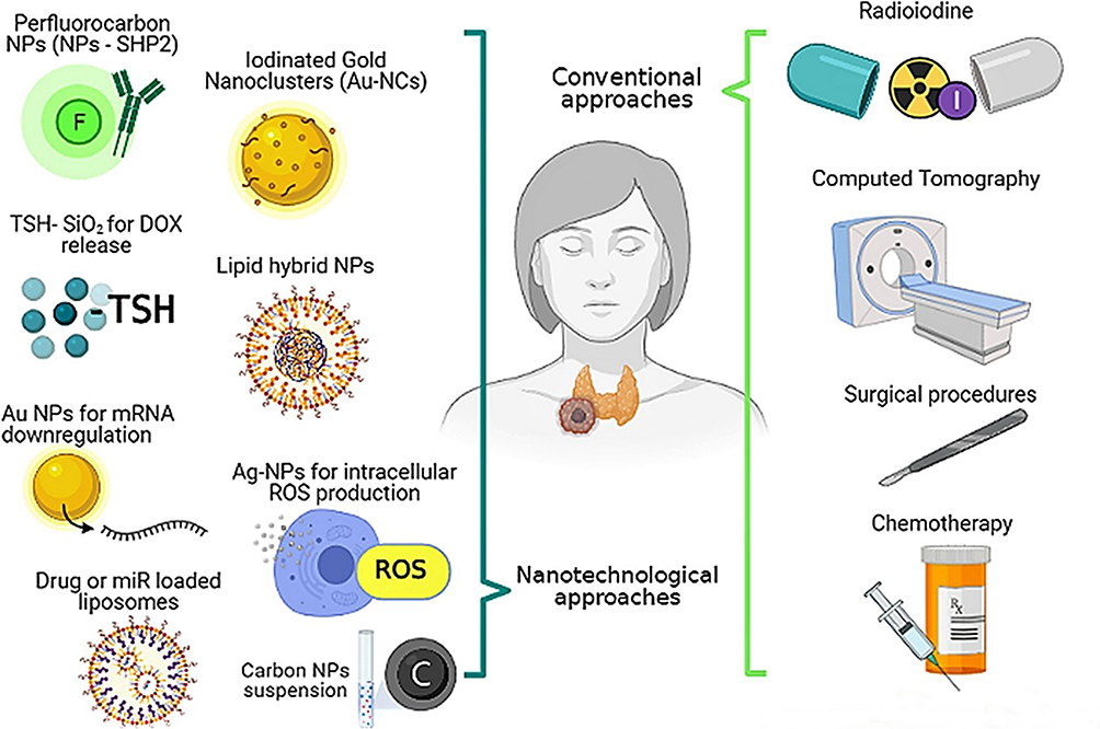 |
Figure 1 Schematic representation of the advantages of metallic nanomaterials in thyroid theranostics. Adapted with permission from Reference.13 |
In recent years, the concept of theranostics, which integrates therapeutic and diagnostic capabilities into a single platform, has gained prominence, especially in oncology and endocrinology.14,15 Metallic nanoparticles (MNPs), including gold (AuNPs), silver (AgNPs), and iron-oxide nanoparticles (IONPs), offer distinct advantages over conventional nanomedicine platforms due to their unique optical, magnetic, and plasmonic properties that enable multimodal imaging, photothermal ablation, and magnetic hyperthermia in a single construct16 (Figure 2). MNPs offer superior stability as compared to polymeric or lipid-based nanocarriers, tunable surface chemistry, and enhanced signal contrast across MRI, CT, facilitating early lesion detection and image-guided therapy.17–19 These properties facilitate enhanced imaging contrast across modalities such as ultrasound, magnetic resonance imaging (MRI), and computed tomography (CT), alongside enabling therapeutic interventions including photothermal therapy (PTT), magnetic hyperthermia, and controlled drug release.20,21 Gold nanoparticles (AuNPs) exhibit localized surface plasmon resonance (LSPR), making them highly effective contrast agents for imaging and efficient mediators of photothermal ablation, which can selectively destroy diseased thyroid tissue.22 Superparamagnetic iron-oxide nanoparticles (IONPs), predominantly Fe3O4-based, offer T2-weighted MRI contrast through superparamagnetism and enable localized magnetic hyperthermia (MH) upon exposure to alternating magnetic fields, inducing precise tumor ablation via heat generation while minimizing systemic impact.23 Their inherent magnetic properties also support targeted drug delivery and catalysis in tumor microenvironments. Composite architectures that fuse gold with iron-oxide (core–shell Fe3O4@Au or Janus designs) synergize imaging and therapy: the iron-oxide core enhances MRI and MH, while the gold shell adds CT contrast and NIR-mediated photothermal therapy (PTT), allowing dual-mode thermal treatment through both magnetic and optical triggers. In vivo studies demonstrate that Fe3O4/Au nanohybrids swiftly elevate tumor temperatures (~20 °C rise within 2 minutes) under combined alternating magnetic field and NIR stimulation, confirming enhanced heat dissipation and superior tumor ablation compared to either modality alone.24 Hybrid nanoparticles represent a flexible theranostic platform, supporting multimodal imaging (MRI/CT/optical), targeted hyperthermia, and controlled drug release, making them highly promising candidates for enhanced, image-guided thyroid cancer therapy.24–26
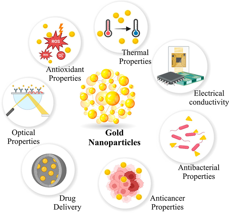 |
Figure 2 Key applications of gold nanoparticles across biomedical domains. Their unique physicochemical traits, strong localized surface plasmon resonance (LSPR), high biocompatibility, low toxicity, and ease of surface functionalization enable their use in imaging, biosensing, and cancer therapy, including as effective radiosensitizers. The figure is made by the authors themselves. |
Given these multifaceted capabilities, metallic nanoparticles offer a versatile platform that addresses many of the current challenges in thyroid disease management.27 This review systematically synthesizes literature published from 2020 onward, encompassing the diagnostic and therapeutic potentials of metallic nanoparticles across the spectrum of thyroid disorders, including benign, functional, and malignant pathologies. In the following sections, we will examine the suitability of specific nanoparticle types for different thyroid diseases, evaluating their physicochemical properties, targeting strategies, efficacy, and translational prospects.
Nanoparticle-Based Theranostics: Targeted Strategies for Diverse Thyroid Diseases
This section provides a comprehensive classification of metallic nanoparticles based on their diagnostic and therapeutic roles in different thyroid diseases, highlighting recent advances and disease-specific applications.
Benign Nodules and Goiter
Benign thyroid nodules and goiter represent the most prevalent thyroid abnormalities, affecting up to 50% of the adult population in iodine-deficient regions.28 It typically arises from dysregulated follicular cell proliferation, often driven by iodine deficiency, growth factor imbalances, or chronic inflammation, resulting in localized hyperplasia or hypertrophy. Current imaging modalities, such as conventional ultrasound, lack sufficient specificity to reliably differentiate benign from malignant lesions or assess treatment efficacy.29,30 Gold nanoparticles (AuNPs), owing to their tunable surface plasmon resonance, have demonstrated significant potential in enhancing ultrasound contrast for thyroid nodule characterization.31 Functionalization of AuNPs with thyroid-specific ligands, such as thyroglobulin antibodies, enables targeted accumulation within nodular tissues, thus improving image resolution and diagnostic accuracy. For instance, recent studies report that AuNP-enhanced ultrasound achieved a sensitivity increase of 20% compared to standard methods in ex vivo thyroid specimens.32
Magnetic nanoparticles, particularly iron-oxide nanoparticles (IONPs), offer an alternative modality by facilitating localized hyperthermia ablation of nodular tissues. When subjected to an alternating magnetic field, IONPs generate heat sufficient to induce apoptosis in hyperplastic cells via enhanced reactive-oxygen species (ROS) generation and lysosomal membrane permeabilization without damaging surrounding normal tissue. Preclinical rodent models have validated the efficacy of magnetic hyperthermia in reducing nodule volume by up to 60% within two weeks post-treatment.33 Hybrid nanoparticles combining gold and iron oxide cores merge diagnostic imaging and therapeutic functionalities into a single platform. Such composites enable simultaneous ultrasound and magnetic resonance imaging (MRI) with concurrent photothermal and magnetic hyperthermia ablation.16 A recent open-access study demonstrated that hybrid NPs significantly reduced nodular volume while enabling real-time imaging-guided therapy. These advancements represent a paradigm shift in minimally invasive management of benign thyroid disease, emphasizing the integration of diagnosis and therapy. Translational considerations, including heat generated by IONPs or hybrid systems, must be carefully calibrated to reliably induce apoptosis (rather than necrosis) in thyroid nodular tissue while preserving adjacent thyroid parenchyma and critical structures like the laryngeal nerve and trachea. The fate of nanoparticles (biodegradation, clearance, immune responses, and potential off-target accumulation in organs) must be addressed; for example, recent work showed alterations in hepatic enzyme function after MRI-contrast IONPs in animal models.34 Theranostic potential in benign conditions, the subsequent section explores metallic nanoparticle applications in hyperthyroidism, including Graves’ disease and toxic nodules.24,35
Hyperthyroidism (Graves’ Disease, Toxic Nodules)
Hyperthyroidism, such as Graves’ disease and toxic nodules, pathogenesis involves autoimmune stimulation of the thyroid-stimulating hormone receptor (TSHR), leading to excessive thyroid hormone synthesis and secretion (Figure 3).36–38 Graves’ disease is characterized by autoantibodies that mimic TSH, inducing diffuse glandular hyperactivity, whereas toxic nodules exhibit constitutive activation of intracellular signaling pathways independent of TSH regulation.38 Graves’ disease, an autoimmune hyperthyroidism, and toxic nodular goiter are principal etiologies requiring targeted intervention. Traditional treatment modalities such as antithyroid drugs, radioiodine therapy, and surgery have limitations, including adverse effects, recurrence, and hypothyroidism induction.39,40
 |
Figure 3 Schematic representation of the immunopathogenesis of Graves’ disease in the context of theranostic targeting. Adapted from the reference41 under the terms and conditions of a Creative Commons Attribution 4.0 International License (http://creativecommons.org/licenses/by/4.0/). |
Graves’ disease is an autoimmune hyperthyroid disorder characterized by autoreactive T and B lymphocyte infiltration and the production of TSH receptor–stimulating antibodies (TSHR-Ab), which mimic TSH and drive excessive thyroid hormone synthesis. Antigen-presenting cells (APCs) initiate T cell activation via MHC–antigen presentation and CD40–CD40L interactions, further propagating inflammation through cytokines such as IFN-γ, TNF-α, and interleukins. These immune responses lead to diffuse glandular hyperactivity. This pathogenic cascade presents multiple potential targets for metallic nanoparticle–based theranostics. Gold nanoparticles (AuNPs), conjugated with TSHR antibodies, enable targeted photothermal ablation of hyperfunctioning thyroid tissue, while iron oxide nanoparticles (IONPs) facilitate magnetic hyperthermia under an external alternating field. Combinatorial nanoparticle strategies may enhance selectivity and therapeutic efficacy in hyperthyroid disorders such as Graves’ disease and toxic nodular goiter.
Gold nanoparticles have been investigated extensively for their targeted photothermal therapeutic (PTT) applications in hyperthyroid tissue ablation. The LSPR properties of AuNPs facilitate efficient conversion of near-infrared (NIR) light into localized heat, enabling selective destruction of hyperfunctioning thyroid cells. Surface conjugation with thyroid-stimulating hormone receptor (TSHR) antibodies enhances nanoparticle specificity, minimizing collateral damage. In a recent preclinical trial, AuNP-mediated PTT achieved a 75% reduction in thyroid hormone levels in hyperthyroid rat models within 72 hours, demonstrating rapid therapeutic efficacy. Heat raises intracellular temperature to 43–47°C, initiating HSP (heat shock protein) suppression, membrane disruption, mitochondrial dysfunction, and ultimate apoptotic cell death.13,42,43 Iron oxide nanoparticles have been similarly employed in magnetic ablation strategies. Under an external alternating magnetic field, IONPs accumulate in hyperactive thyroid nodules and generate cytotoxic hyperthermia, offering a non-invasive alternative to surgery.44,45 Emerging studies also investigate combinatorial approaches where AuNPs and IONPs are co-administered to harness dual photothermal and magnetic hyperthermia effects, potentially overcoming limitations of monotherapy. These preclinical findings underscore the translational potential of metallic nanoparticle theranostics in hyperthyroid disorders.46,47
Hypothyroidism (Hashimoto’s Thyroiditis)
Hypothyroidism often stems from autoimmune destruction, as observed in Hashimoto’s thyroiditis, where lymphocytic infiltration leads to gradual follicular cell apoptosis and reduced hormone production. Chronic inflammation and fibrosis contribute to progressive glandular failure.30,48 Lifelong oral levothyroxine supplementation remains the standard of care, yet challenges persist, including variable absorption, compliance issues, and fluctuating serum hormone levels. Nanoparticle-based drug delivery systems offer a promising avenue for controlled, sustained release formulations to overcome these hurdles.49 Polymeric nanoparticles encapsulating levothyroxine have demonstrated improved bioavailability and prolonged therapeutic effects in animal models. For example, chitosan-coated AuNPs loaded with levothyroxine exhibited sustained drug release over 72 hours with enhanced intestinal uptake in rats, suggesting improved oral delivery efficiency. Further, iron/gold hybrid nanoparticles functionalized with inflammation-targeting ligands have been investigated for non-invasive imaging of residual thyroid tissue and monitoring of autoimmune inflammation using combined MRI and photothermal imaging modalities.50
Beyond endocrine regulation, chronic or inadequately managed hypothyroidism exerts significant systemic effects, particularly on skeletal health, as depicted in Figure 4. In adults, diminished thyroid hormone activity disrupts normal bone remodeling processes, while in pediatric populations, it can impede ossification and impair skeletal development.51,52 Recent advances suggest that metallic nanoparticles hold promise not only in enhancing hormone replacement strategies but also in targeting bone-specific molecular pathways, including thyroid hormone receptors (TRs) and the RANK/RANKL/OPG axis, for both diagnostic and therapeutic applications. Moreover, stimuli-responsive nanocarriers capable of releasing hormones in response to physiological cues local pH variations or inflammatory markers, are being actively investigated as part of a personalized approach to hypothyroidism management. Although these technologies remain in early developmental stages, they underscore the expanding potential of metallic nanoparticles in addressing a broader spectrum of thyroid-related dysfunctions beyond malignancies.13,53
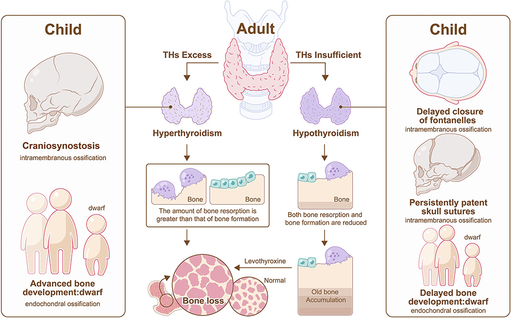 |
Figure 4 Skeletal effects of thyroid dysfunctions. In adults, hyperthyroidism induces bone loss via increased resorption, while hypothyroidism impairs remodeling; LT4 therapy may further contribute to loss. In children, hypothyroidism delays ossification, whereas hyperthyroidism causes craniosynostosis and stunted growth. These alterations involve targets such as thyroid hormone receptors (TRs), RANK/RANKL/OPG pathway, MMPs, and bone turnover markers, highlighting potential sites for metallic nanoparticle–based theranostic intervention in thyroid-related bone disorders. Adapted from the reference52 under the terms and conditions of a Creative Commons Attribution 4.0 International License (http://creativecommons.org/licenses/by/4.0/). Copyright© 2022 Zhu, Pang, Xu, Chen, Zhang, Wu and Gao. |
Thyroid Cancer
Thyroid cancer is the most prevalent malignant neoplasm of the cervical region and endocrine system, characterized by a discernible upward trend in incidence over recent years.54,55 Anaplastic thyroid cancer, characterized by high malignancy and aggressiveness, remains an unmet clinical need with no effective treatments available. Thyroid malignancies, including papillary, follicular, and anaplastic carcinomas, originate from genetic mutations affecting oncogenes (BRAF, RAS) and tumor suppressors (p53), driving uncontrolled cellular proliferation, invasion, and resistance to apoptosis.56,57 Tumor microenvironment factors such as hypoxia and immune evasion further complicate disease progression. Thyroid cancer, particularly papillary and anaplastic variants, demands precise diagnostic and therapeutic strategies due to variable prognosis and treatment resistance.58
Circulating biomarkers have emerged as powerful tools for the non-invasive detection and monitoring of thyroid cancer (TC), facilitated by advancements in liquid biopsy and nanotechnology.59 Among various nanomaterials, gold nanoparticles (AuNPs) are the most widely utilized due to their versatile signal enhancement capabilities across fluorescence, colorimetry, photoacoustics, surface-enhanced Raman scattering (SERS), electrochemical methods, dynamic light scattering (DLS), and localized surface plasmon resonance (LSPR).60 For thyroid-stimulating hormone (TSH) detection, anti-TSH antibody-conjugated horseradish peroxidase (HRP) immobilized on platinum nanoparticles demonstrated chemiluminescent detection with a threefold faster response and 100-fold greater sensitivity than commercial kits. Similarly, dendrimer-enhanced gold electrodes have enabled ultrasensitive TSH detection down to 0.026 mIU/L, outperforming the standard third-generation immunoassays (~0.02 mIU/L sensitivity), although further sensitivity is not routinely required in clinical settings. For thyroglobulin (Tg), a biomarker critical for post-treatment surveillance, a novel fluoroimmunodiagnostic nanoplatform using tannylated ferritin nanocages achieved a detection limit of 4.3 pg/mL, surpassing conventional assays such as Elecsys Tg II. Likewise, for medullary thyroid cancer, calcitonin detection has been enhanced using FRET-based AuNP thin films and graphene oxide-modified electrodes, achieving limits of detection as low as 0.7 pg/mL, markedly lower than most commercial platforms. Detection of genetic mutations, particularly BRAF V600E and KRAS exon 2, is also facilitated by nanotechnology.61 Circulating tumor DNA (ctDNA) and tumor cells can be isolated using magnetic nanoparticles (Fe3O4-based ferrofluids), with digital PCR achieving mutation detection limits as low as 0.0005%. Metallic nanoparticles, especially gold-based, have been extensively investigated for their theranostic capabilities in thyroid oncology. Surface-enhanced Raman spectroscopy (SERS) utilizing AuNPs enables ultra-sensitive detection of thyroid cancer biomarkers in serum and tissue samples, facilitating early diagnosis. Furthermore, AuNPs provide enhanced contrast in CT and MRI scans, improving tumor delineation. Therapeutically, AuNP-mediated photothermal therapy (PTT) achieves selective tumor ablation with minimal invasiveness. Radiosensitization effects of gold nanoparticles further potentiate radiotherapy outcomes by increasing DNA damage in cancer cells.62–65 In vivo imaging and theranostics for thyroid cancer (TC) integrate conventional modalities with nanoparticle-enhanced techniques to improve detection depth, specificity, and treatment efficacy. Standard imaging tools ultrasound, CT, MRI, and radionuclide scans using ^131/123I with SPECT-CT, remain foundational for staging and monitoring residual disease, while FDG-PET’s utility is less definitive.66
For medullary thyroid carcinoma (MTC), PET tracers like [^18F]-FDOPA and [^68Ga]-DOTATOC are preferred due to their superior sensitivity in neuroendocrine tissues.67 However, superficial thyroid tumors located just beneath the skin can benefit from imaging strategies optimized for limited penetration depths: NIR-based photothermal imaging and ultrasound both have tissue penetration limits (~2–3 cm for NIR, 3–6 cm for unfocused ultrasound at 2.5 MHz). These modalities are enhanced by gold nanoparticle (AuNP) contrast agents, enabling dual-mode CT/NIR fluorescence imaging.68 Mouse studies using BSA-coated, iodinated AuNP nanoclusters have detected thyroid tumors as small as ~2 mm2. Similarly, ^131I-labeled mesoporous silica nanoparticles (MSNs) targeting VEGF delivered clear SPECT signals following intratumoral injection, though systemic delivery faced rapid phagocytic clearance via the mononuclear phagocyte system. Low-intensity focused ultrasound (LIFUS) offers combined imaging and therapy for superficial tumors; when paired with phase-changeable, antibody-decorated PLGA nanoparticles (eg, SHP2-targeted) and contrast agents, LIFUS can both visualize and induce tumor-selective damage without harming adjacent tissue.69 Theranostic copper sulfide nanoparticles, PEG-coated,^64Cu-labeled CuS NPs, have been successfully used for PET-guided photothermal therapy (PTT) in anaplastic thyroid cancer (ATC) mouse models. Despite ~6% tumor accumulation post-injection, combined radiotherapy and NIR irradiation achieved tumor shrinkage of up to 83%, with no systemic toxicity. Similarly, polymeric NIR-emitting nanoparticles co-loaded with BRAF-silencing siRNA have been shown to both image BRAF-mutant xenografts and suppress tumor growth and metastasis in orthotopic mouse models. These “theranostic” platforms allow the spatial integration of diagnostics (imaging) and therapeutics (photothermal, gene silencing), although translation into clinical practice remains contingent upon the discovery of highly specific thyroid cancer markers and simplification of complex NP formulations. When available, patient-specific protein–corona profiling may help identify such markers and further personalize nanoparticle-assisted diagnosis.61 Magnetic nanoparticles, predominantly iron oxide, complement these approaches through MRI contrast enhancement and magnetic hyperthermia treatments (Figure 5). In papillary thyroid carcinoma models, IONP-induced hyperthermia significantly decreased tumor volume and inhibited metastasis. Composite nanoparticles integrating gold and iron-oxide cores have demonstrated synergistic effects, enabling multimodal imaging and combination therapy in both papillary and anaplastic thyroid cancer models. These platforms underscore a shift toward personalized, minimally invasive oncology management, although challenges in nanoparticle biodistribution, toxicity, and regulatory approval persist.70,71
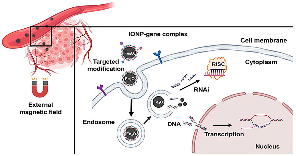 |
Figure 5 Schematic illustration of iron-oxide nanoparticles (IONPs) utilized for targeted gene delivery and tumor localization. This theranostic approach highlights IONP-mediated MRI contrast enhancement and magnetic hyperthermia, which have shown efficacy in reducing tumor volume and metastasis in papillary thyroid carcinoma models. Composite nanoparticles combining gold and iron oxide cores enable synergistic effects, supporting multimodal imaging and combination therapy in both papillary and anaplastic thyroid cancers. This strategy exemplifies a personalized and minimally invasive oncology paradigm, despite existing challenges in nanoparticle biodistribution, toxicity, and clinical translation. Adapted from the reference72 under the terms and conditions of a Creative Commons Attribution 4.0 International License (http://creativecommons.org/licenses/by/4.0/). |
Theranostic copper sulfide nanoparticles (CuS NPs), specifically PEG-coated and ^64Cu-labeled, have emerged as a promising dual-functional platform for the diagnosis and treatment of anaplastic thyroid cancer (ATC). These nanoparticles serve as both positron emission tomography (PET) imaging agents and photothermal therapy (PTT) mediators. Their synthesis involves incorporating ^64Cu directly into the CuS matrix during preparation, eliminating the need for external chelators and enhancing their stability and imaging efficacy. In preclinical studies, these CuS NPs demonstrated significant tumor accumulation and retention, facilitating effective PET imaging. Upon near-infrared (NIR) laser irradiation, they induced localized hyperthermia, leading to substantial tumor cell apoptosis and necrosis. Combined treatment with radiotherapy and NIR-induced PTT resulted in up to 83% tumor shrinkage without evident systemic toxicity. These findings underscore the potential of PEG-[^64Cu]CuS NPs as a versatile theranostic tool, offering precise imaging capabilities alongside targeted therapeutic effects, enhancing the clinical management of ATC.73
Comparative Analysis of Nanoparticle Platforms Across Thyroid Disorders
To comprehensively evaluate the translational potential of metallic nanoparticles in thyroid diseases, this section presents a side-by-side comparison of nanoparticle types, functionalization strategies, and disease-specific outcomes. Using data derived from recent preclinical and clinical research, diagnostic and therapeutic capabilities, and advancement toward clinical application are discussed in Table 1.
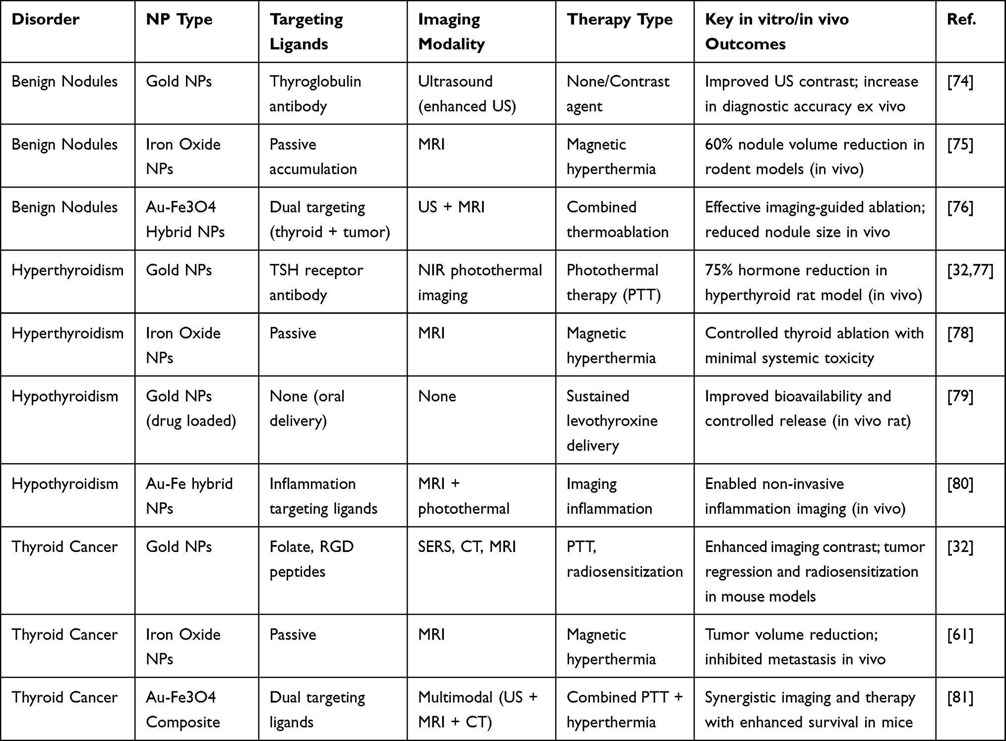 |
Table 1 Overview of Metallic Nanoparticle Platforms Used Across Different Thyroid Disorders, Detailing Nanoparticle Type, Targeting Strategy, Imaging and Therapeutic Functionalities, and Outcomes Observed in in vitro and in vivo Models |
Metallic nanoparticles vary in function across thyroid disorders, showing distinct imaging and therapeutic roles. To assess their clinical relevance, Table 2 outlines the current translational status, including preclinical outcomes and early-stage human trials.
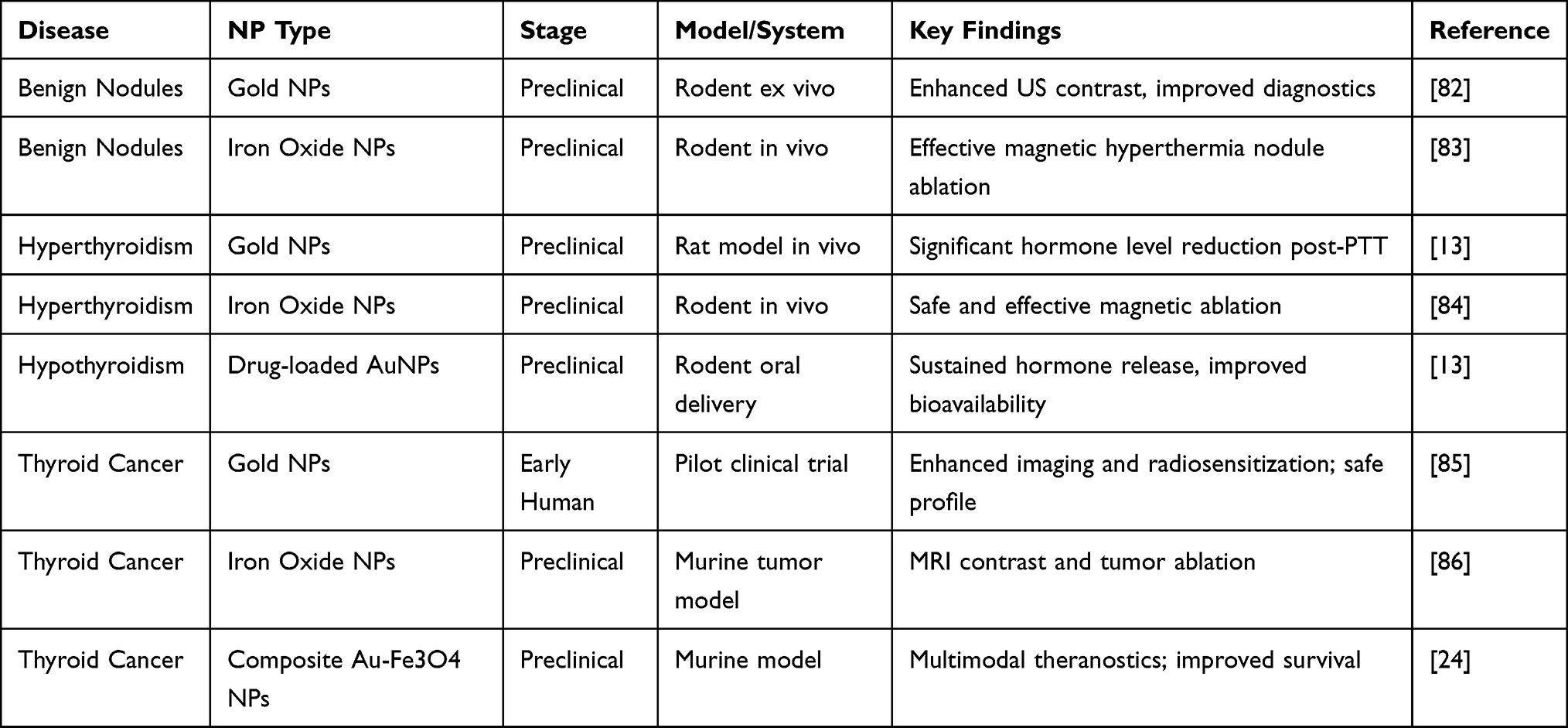 |
Table 2 Overview of Clinical Progress of Metallic Nanoparticles in Thyroid Disease Applications |
Recent advances in metallic nanoparticles highlight that AuNPs offer tunable size, strong plasmonic properties, and facile surface modification, enabling high-contrast imaging and photothermal therapy. IONPs, by contrast, provide intrinsic superparamagnetism for MRI, magnetic hyperthermia, and targeted drug delivery. A comparative summary of their physicochemical characteristics and clinical challenges is presented in Table 3.
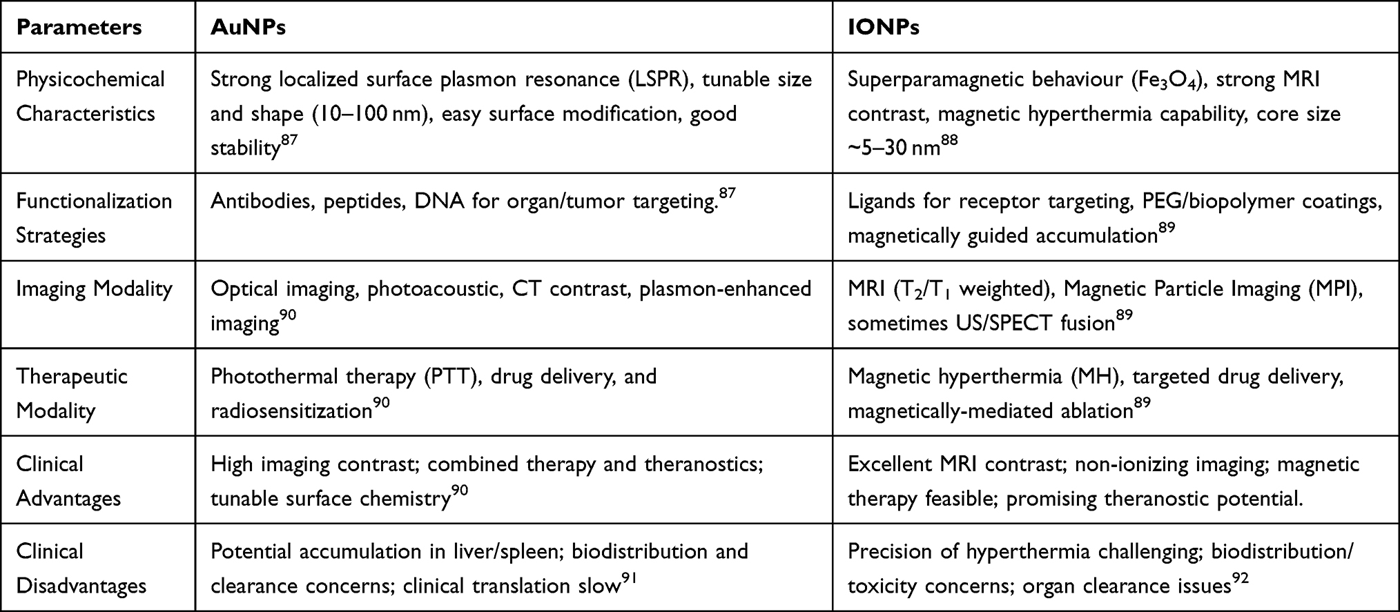 |
Table 3 Comparative Overview of AuNPs and IONPs Nanoparticles, Highlighting Their Physicochemical Properties |
Clinical research on metallic nanoparticles for thyroid diseases is still in its early stages, with most studies limited to Phase I trials focused primarily on thyroid cancer. Some of these emerging efforts, including those investigating carbon-based nanoparticle systems, are summarized in Table 4.
 |
Table 4 Ongoing and Completed Clinical Trials Involving Metallic Nanoparticles in Thyroid Cancer |
Despite promising preclinical data on metallic nanoparticles in thyroid oncology, clinical translation remains limited. Current trials, as outlined in Table 3, are restricted to Phase I studies employing carbon-based nanoparticles for lymphatic mapping and surgical guidance in papillary thyroid carcinoma. While these platforms offer biocompatibility and procedural utility, they lack the multifunctional theranostic capabilities of metallic systems such as gold and iron oxide nanoparticles. The absence of metallic nanoparticle-based trials reflects unresolved challenges related to toxicity, biodistribution, and regulatory compliance. Systematic data on biodistribution and endocrine safety in benign thyroid disorders remain limited. Regulatory bodies, including the U.S. Food and Drug Administration (FDA) and the European Medicines Agency (EMA), continue to evaluate metallic nanoplatforms under combination-product frameworks, highlighting the absence of standardized testing protocols for nanoparticle clearance, immunogenicity, and thyroid-specific long-term safety.93,94 Advancing these platforms into clinical settings requires rigorous standardization and integration into disease-specific therapeutic frameworks, particularly for aggressive thyroid malignancies.
Theranostic Potential and Translational Barriers
Nanoparticle-Driven Improvements
The integration of metallic nanoparticles (NPs) into thyroid disease management offers several compelling advantages.13,95 Primarily, these nanoplatforms enable multimodal diagnosis and therapy within a single system, allowing for simultaneous imaging, targeted drug delivery, and therapeutic intervention (Figure 6). For example, gold and iron oxide nanoparticles have been effectively utilized for combined photoacoustic imaging and photothermal therapy, which facilitates precise localization and ablation of pathological tissue. Such multifunctionality not only improves diagnostic accuracy but also enhances therapeutic efficacy.96 Further, the enhanced targeting capabilities of metallic NPs significantly reduce systemic side effects commonly associated with conventional therapies. By functionalizing nanoparticles with disease-specific ligands, accumulation at the pathological site is maximized, sparing healthy thyroid and surrounding tissues.97 This specificity is crucial across the disease spectrum from benign nodules and hyperthyroidism to malignant thyroid cancers, resulting in improved patient outcomes. Moreover, metallic nanoparticles facilitate real-time monitoring of treatment response through imaging modalities such as MRI and CT, providing clinicians with timely feedback to adjust therapeutic strategies dynamically.98,99 This capability supports personalized medicine approaches and can shorten treatment cycles by promptly identifying ineffective interventions.
 |
Figure 6 Schematic representation of the multifunctional advantages offered by gold nanoparticle (GNP) nanohybrids in theranostics. These platforms enable simultaneous imaging, targeted drug delivery, and therapeutic intervention within a single nanostructure. Specifically, in thyroid disease management, GNP-based hybrids have been effectively employed for photoacoustic imaging and photothermal therapy, enabling precise tumor localization and ablation. Functionalization with disease-specific ligands enhances targeted accumulation and reduces off-target toxicity. Adapted from the reference100 under the terms and conditions of a Creative Commons Attribution 4.0 International License (http://creativecommons.org/licenses/by/4.0/). |
Translational Barriers
While metallic nanoparticles (MNPs) hold considerable promise for theranostic applications in thyroid disorders, several unresolved challenges continue to hinder their effective clinical translation. One of the primary limitations lies in the heterogeneity of nanoparticle biodistribution across different thyroid pathologies. Inflammatory conditions such as Hashimoto’s thyroiditis exhibit altered immune microenvironments and vascular permeability compared to focal nodules or neoplastic lesions.101,102 These microenvironmental variations significantly impact nanoparticle localization, internalization, and subsequent therapeutic efficacy. Consequently, a one-size-fits-all approach to nanoparticle design proves inadequate.103–105 Instead, precision-engineered nanostructures that can dynamically respond to pathological cues such as pH shifts, enzymatic activity, or immune markers are increasingly necessary to ensure disease-specific targeting and functionality. Furthermore, the physicochemical properties of MNPs, including particle size, surface charge, and ligand functionalization, directly influence their systemic behavior and tissue-specific accumulation.106,107 For example, particles under 20 nm often exhibit enhanced renal clearance but may also penetrate non-target tissues more readily, increasing the risk of off-target effects.108 In contrast, larger particles accumulate preferentially in reticuloendothelial organs such as the liver and spleen, raising concerns about chronic toxicity and long-term retention.109 Meta-analyses reported metallic nanoparticle cores persist in spleen and liver for months to years, with measurable deposits leading to oxidative stress, mitochondrial dysfunction, and low-grade inflammation in animal models. Investigations also reveal MNP exposure modulates immune signaling, with upregulation of IL-6, TNF-α and complement activation products even at sub-therapeutic doses, suggesting that immune responses as translational barrier.110
Oxidative stress induced by metal-based nanoparticles, particularly through reactive oxygen species (ROS) generation, has been implicated in mitochondrial dysfunction, DNA damage, and pro-inflammatory responses. Addressing these safety concerns necessitates comprehensive in vivo toxicokinetic profiling that includes dose-dependent clearance studies, biodegradability assessments, and immunocompatibility evaluations.111–113 Recent advances in nanomedicine emphasize the functionalization of metallic nanoparticles with polyethylene glycol (PEG), often via monophosphonic or multiphosphonic acid linkers, to create a stealth-like surface that resists protein corona formation and prolongs systemic circulation.114 Surface modifications like anti-PEG antibodies may accelerate clearance of PEGylated nanoparticles and evoke hypersensitivity responses, revealing immune clearance and cost of modification as a translational challenge.115 Multivalent phosphonic-PEG coatings have demonstrated superior stability and robustness in physiological serum, maintaining dispersity over months, compared to monovalent analogs that tend to degrade more rapidly. In thyroid theranostics, such stealth coatings reduce recognition and clearance by the mononuclear phagocyte system, enabling higher tumor-to-normal organ accumulation.116 Moreover, PEGylated nanoscale metal-organic frameworks (NMOFs) labeled with 131I have shown significantly prolonged retention in thyroid tumors and enhanced therapeutic outcomes in murine models, underscoring their potential in differentiated thyroid carcinoma treatment.117 Beyond regulatory constraints, the clinical deployment of metallic nanoparticle formulations is further impeded by translational limitations related to manufacturing scalability, process reproducibility, and economic sustainability. Reproducing lab-scale formulations under Good Manufacturing Practice (GMP) conditions requires stringent control over critical quality attributes, including hydrodynamic size, polydispersity index, zeta potential, and surface functionalization density, all of which significantly influence in vivo performance. Batch-to-batch variability, particularly in multi-component platforms integrating targeting ligands or theranostic payloads, remains a formidable challenge. Emerging manufacturing technologies, such as microfluidic-based nanoprecipitation and scalable flash nanoprecipitation systems, have demonstrated promise in enhancing reproducibility while preserving nanoparticle functionality. However, these approaches demand substantial capital investment, rigorous GMP validation, and high-purity input materials, all of which contribute to elevated production costs. Until robust, cost-efficient production platforms are established and validated at clinical scale, the widespread adoption of nanoparticle-based therapies in endocrine oncology and benign thyroid disease will likely remain constrained.118–120
Emerging Trends and Strategic Directions
Beyond the promising stimuli-responsive functionalities, the clinical translation of metallic nanoparticles in thyroid theranostics critically depends on their ability to maintain physicochemical integrity during large-scale production and in vivo circulation. A growing body of research has focused on lipid–polymer hybrid nanocarriers incorporating redox-responsive disulfide or diselenide linkers, which remain stable under extracellular oxidative conditions but undergo cleavage within the reductive intracellular environment, enabling site-specific release of therapeutic payloads. These smart platforms facilitate precise drug delivery to thyroid tissues characterized by dysregulated redox homeostasis, a feature often observed in aggressive or inflammatory thyroid microenvironments.121,122
Thermosensitive iron oxide-based nanoparticles, often coated with polydopamine or PEGylated polymer matrices, have demonstrated dual-functional capabilities, combining magnetic resonance imaging (MRI) contrast enhancement with localized photothermal therapy (PTT). This multifunctionality not only improves the precision of tumor localization but also enhances therapeutic selectivity through heat-induced cytotoxicity in hypervascular thyroid lesions. However, translating such nanoplatforms into clinical practice necessitates rigorous optimization of key performance parameters, including lower critical solution temperature (LCST), linker density, and thermal or magnetic activation thresholds, which must be reproducibly maintained across Good Manufacturing Practice (GMP)-compliant batches. Moreover, comprehensive preclinical validation encompassing dual-trigger activation studies, biodistribution mapping, renal and hepatic clearance kinetics, and immunological profiling is essential to meet regulatory requirements and ensure clinical safety. Only through such multidimensional design and validation strategies can nanoparticle-based therapies achieve personalized, high-fidelity intervention in the complex landscape of thyroid disease.120
Interdisciplinary collaboration between endocrinologists, radiologists, and nanomedicine researchers has emerged as a key enabler of clinical translation. Recent studies in thyroid imaging show that AI-augmented ultrasound and multimodal radiomics platforms developed by teams of radiologists and computational scientists achieved high sensitivity and specificity in nodule classification of a deep-learning model combining B-mode ultrasound and strain elastography validation for TI-RADS4 nodules.123 Advanced deep learning architectures, such as convolutional neural networks (CNNs), have been trained to perform automated nodule detection, segmentation, and classification tasks with sensitivity and specificity comparable to expert radiologists.124,125 For instance, studies applying AI to thyroid ultrasound have demonstrated enhanced sensitivity in identifying malignant nodules, reducing false-negative rates while maintaining specificity at a clinically acceptable level.126 When combined with nanoparticle-derived imaging agents that amplify contrast or provide functional cues (pH-sensitive or enzyme-responsive probes), AI algorithms can exploit high-dimensional radiomic features as texture, heterogeneity, and contrast kinetics to discriminate lesion subtypes and predict molecular phenotypes, even those not visible to the naked eye.127 The integration of artificial intelligence (AI) into nanoparticle-enhanced imaging modalities such as ultrasound and magnetic resonance imaging (MRI) represents a paradigm shift in diagnostic precision. State-of-the-art convolutional neural networks (CNNs) have been trained on high-resolution image datasets to autonomously detect and classify thyroid nodules with sensitivity and specificity approaching expert radiologists.128–130 For instance, contrast-enhanced ultrasound combined with AI-powered segmentation algorithms has significantly improved discrimination between benign and malignant lesions by analyzing perfusion dynamics at a microvascular level. When coupled with nanoparticle contrast agents such as gold-based agents in ultrasound or superparamagnetic iron oxide nanoparticles (SPIONs) in MRI, the resulting platforms generate rich, multifunctional microscale signatures.131 AI algorithms exploit radiomic features like signal intensity variation, temporal contrast kinetics, and textural heterogeneity to infer underlying histopathological properties capabilities often beyond human visual assessment.132–134
Methodology
A systematic literature search was conducted using PubMed, Google Scholar, and Web of Science for articles published between 2015 and 2025. Keywords: combined thyroid disorders (thyroid cancer, hyperthyroidism) with metallic nanoparticles and theranostics. Peer-reviewed original research and reviews were included. ClinicalTrials.gov was searched for ongoing trials. Duplicate and non-peer-reviewed studies were excluded. Data were extracted to provide a comprehensive overview of recent advances, challenges, and prospects in metallic nanoparticle theranostics for thyroid diseases.
Conclusion
This review underscores the significant theranostic potential of metallic nanoparticles across the diverse spectrum of thyroid diseases, ranging from benign nodules and hyperthyroidism to hypothyroidism and thyroid malignancies. The multifunctional capabilities of these nanoplatforms, including enhanced imaging, targeted drug delivery, and localized therapy, offer promising avenues to overcome the limitations of conventional diagnostic and therapeutic approaches. Despite significant preclinical successes and emerging early-phase clinical trials, the translation of metallic nanoparticles into routine clinical practice remains at an incipient stage. Challenges related to biodistribution variability, toxicity concerns, and regulatory complexities necessitate robust, multidisciplinary research efforts to optimize nanoparticle design and ensure patient safety. Given the heterogeneity of thyroid disorders and the distinct requirements for managing benign versus malignant conditions, broad-based nanotheranostic research is urgently needed to develop tailored solutions that address disease-specific pathophysiology. This pursuit demands collaboration across disciplines, integrating expertise in nanotechnology, endocrinology, radiology, and clinical oncology. In conclusion, advancing metallic nanoparticle theranostics for thyroid diseases holds the potential to revolutionize patient care, providing more precise, effective, and personalized interventions. Realizing this promise will require concerted efforts to navigate translational barriers and foster innovative strategies that bridge the gap between laboratory innovation and clinical application.
Funding
This study was supported by the Medical Foundation of Jieping Wu (No. 320.6750.2024-11-10).
Disclosure
The Authors declare that they have no competing financial or non-financial interests or any other interests that might be perceived to influence the results and/or discussion reported in this paper.
References
1. Al-Suhaimi EA, Khan FA. Thyroid Glands: Physiology and Structure, in Emerging Concepts in Endocrine Structure and Functions. Springer; 2022:133–18.
2. Sai Kumar BAA. Hormonal regulation of metabolism, water, and minerals. In: Textbook of Veterinary Physiology. Springer; 2023:391–415.
3. Mariotti S, Beck-Peccoz P. Physiology of the Hypothalamic-Pituitary-Thyroid Axis. 2021. Endotext [Internet].
4. Nilsson M, Fagman H. Development of the thyroid gland. Development. 2017;144(12):2123–2140. doi:10.1242/dev.145615
5. Shah N, Ursani TJ, Shah NA, Raza HM. 3. Prevalence and etiology of thyroid disease: a review. Pure Appl Biol. 2021;10(3):691–702.
6. Dahiya V, Vasudeva N, Sharma S, Kumar A. Role of dietary supplements in thyroid diseases. Endoc Metabol Immun Disord Drug Target. 2022;22(10):985–996.
7. Dayal D, Gupta BM, Gupta A. Thyroid disorders in children and adolescents: systematic mapping of global research over the past three decades. Thyroid Res Pract. 2021;18(1):23–30. doi:10.4103/trp.trp_5_21
8. Jiang T, Bin L, Liu H, et al. Global, regional, and national burden of thyroid cancer in women of child-bearing age, 1990 to 2021 and predictions to 2035: an analysis of the global burden of disease study 2021. Front Endocrinol. 2025;16:1555841. doi:10.3389/fendo.2025.1555841
9. Miranda-Filho A, Lortet-Tieulent J, Bray F, et al. Thyroid cancer incidence trends by histology in 25 countries: a population-based study. Lancet Diab Endocrinol. 2021;9(4):225–234. doi:10.1016/S2213-8587(21)00027-9
10. Vargas-Uricoechea H. Molecular mechanisms in autoimmune thyroid disease. Cells. 2023;12(6):918. doi:10.3390/cells12060918
11. Lin Y, Lai S, Wang P, et al. Performance of current ultrasound-based malignancy risk stratification systems for thyroid nodules in patients with follicular neoplasms. Eur Radiol. 2022;32(6):3617–3630. doi:10.1007/s00330-021-08450-3
12. Hamidi S, Hofmann M-C, Iyer PC, et al. New treatments for advanced differentiated thyroid cancers and potential mechanisms of drug resistance. Front Endocrinol. 2023;14:1176731. doi:10.3389/fendo.2023.1176731
13. Sahare P, Ruiz-Manriquez LM, Anguiano B, et al. Recent advances in nanomedicine for the diagnosis and therapy of thyroid disorders. 3 Biotech. 2025;15(3):67. doi:10.1007/s13205-025-04234-4
14. Baldwin CK, Natter MB, Patel KN, et al. Minimally invasive techniques for the management of thyroid nodules. Endocrinol Metabol Clin. 2022;51(2):323–349. doi:10.1016/j.ecl.2022.01.001
15. Chakrabarty N, Mahajan A, Basu S, et al. Comprehensive review of the imaging recommendations for diagnosis, staging, and management of thyroid carcinoma. J Clin Med. 2024;13(10):2904. doi:10.3390/jcm13102904
16. Singh P, Pandit S, Balusamy SR, et al. Advanced nanomaterials for cancer therapy: gold, silver, and iron oxide nanoparticles in oncological applications. Adv Healthc Mat. 2025;14(4):2403059. doi:10.1002/adhm.202403059
17. Kiani MN, Khaliq H, Abubakar M, et al. Advancing the potential of nanoparticles for cancer detection and precision therapeutics. Med Oncol. 2025;42(7):239. doi:10.1007/s12032-025-02782-6
18. Andrade RG, Veloso SR, Castanheira EM, Rodrigues LR. Magnetic lipid-based nanoparticles: recent advances in therapeutic applications. In: Magnetic Polymer Composites and Their Emerging Applications. 2024;314–334.
19. Wang Y, Xu Y, Song J, et al. Tumor cell-targeting and tumor microenvironment–responsive nanoplatforms for the multimodal imaging-guided photodynamic/photothermal/chemodynamic treatment of cervical cancer. Int J Nanomed. 2024;Volume 19:5837–5858. doi:10.2147/IJN.S466042
20. Agnihotri TG, Gomte SS, Jain A. Emerging theranostics to combat cancer: a perspective on metal-based nanomaterials. Drug Dev Indust Pharm. 2022;48(11):585–601. doi:10.1080/03639045.2022.2153862
21. Karthikeyan L, Sobhana S, Yasothamani V, et al. Multifunctional theranostic nanomedicines for cancer treatment: recent progress and challenges. Biomed Engineer Adv. 2023;5:100082. doi:10.1016/j.bea.2023.100082
22. Mehta K, Rajput S, Sharma S. Low-Dimensional Nanomaterials as an Emerging Platform for Cancer Diagnosis and Therapy. In: Nanoparticles in Cancer Therapy. CRC Press;2024:76–97
23. Gade R, Dwarampudi LP, Yamuna K, Maraba N, Fufa G. Advanced Nanomaterials in Imaging and Diagnostics. In Exploring Nanomaterial Synthesis, Characterization, and Applications. IGI Global; 2025:79–100.
24. Tarkistani MAM, Komalla V, Kayser V. Recent advances in the use of iron–gold hybrid nanoparticles for biomedical applications. Nanomaterials. 2021;11(5):1227. doi:10.3390/nano11051227
25. Żuk M, Gawęda W, Majkowska-Pilip A, et al. Hybrid radiobioconjugated superparamagnetic iron oxide-based nanoparticles for multimodal cancer therapy. Pharmaceutics. 2021;13(11):1843. doi:10.3390/pharmaceutics13111843
26. Khani T, Alamzadeh Z, Sarikhani A, et al. Fe3O4@ Au core–shell hybrid nanocomposite for MRI-guided magnetic targeted photo-chemotherapy. Lasers Med Sci. 2022;37(5):2387–2395. doi:10.1007/s10103-021-03486-9
27. Zhang Y, Tang N, Zhou H, et al. Surface engineered multifunctional nano-systems for localised drug delivery against thyroid cancer: a review of current practices. Biomed Pharmacother. 2024;176:116840. doi:10.1016/j.biopha.2024.116840
28. Karunarathna I, Rodrigo PN, Ranasinghe S, et al. Thyroid nodules: comprehensive evaluation, diagnosis, and management.
29. Basolo F, Proietti A, Ugolini C. Thyroid cysts. In: Endocrine Pathology. Springer; 2022:779–780.
30. Sakr M. Benign Thyroid Disease, in Head and Neck and Endocrine Surgery: From Clinical Presentation to Treatment Success. Springer; 2024:279–339.
31. Li J, Dou J, Li H, et al. Contrast enhancement ultrasound improves diagnostic accuracy for thyroid nodules: a prospective multicenter study. J Endocr Soc. 2024;8(1):bvad145. doi:10.1210/jendso/bvad145
32. Li L, Wang Z, Guo H, et al. Nanomaterials: a promising multimodal theranostics platform for thyroid cancer. J Mat Chem B. 2023;11(32):7544–7566. doi:10.1039/D3TB01175E
33. Ghazi R, Ibrahim TK, Nasir JA, et al. Iron oxide based magnetic nanoparticles for hyperthermia, MRI and drug delivery applications: a review. RSC Adv. 2025;15(15):11587–11616. doi:10.1039/D5RA00728C
34. Mirzajani F, Rostamzadeh A, Tahmasian Z, et al. Effects of MRI magnetic iron oxide nanoparticles on the structural and enzymatic properties of liver-related enzymes. Micro Nano Syst Lett. 2024;12(1):13. doi:10.1186/s40486-024-00200-6
35. Fazal S, Paul-Prasanth B, Nair SV, et al. Theranostic iron oxide/gold ion nanoprobes for MR imaging and noninvasive RF hyperthermia. ACS Appl Mat Interfaces. 2017;9(34):28260–28272. doi:10.1021/acsami.7b08939
36. Bryliński Ł, Kostelecka K, Woliński F, et al. Effects of trace elements on endocrine function and pathogenesis of thyroid diseases—a literature review. Nutrients. 2025;17(3):398. doi:10.3390/nu17030398
37. Pinto CM, Romero JLG, Carrasco MG. Graves’ Disease, in Autoimmune Disease Diagnosis: Systemic and Organ-Specific Diseases. Springer; 2025:355–360.
38. Deshmukh D, Anjum N, Sahu B, Deshmukh N. The thyrotropin receptor and the regulation of thyrocyte activity and proliferation. J Pharmacol Genet Mol Biol. 2025;65–79.
39. Lanzolla G, Marinò M, Menconi F. Graves disease: latest understanding of pathogenesis and treatment options. Nat Rev Endocrinol. 2024;20(11):647–660. doi:10.1038/s41574-024-01016-5
40. Ciaccio M, Agnello L, Bivona G, Ciaccio AM, et al. Endocrine System. In: Clinical and Laboratory Medicine Textbook. Springer; 2024: 317–382.
41. Gallo D, Piantanida E, Bombelli R, et al. Natural killer cells in graves’ disease: increased frequency but impaired degranulation ability compared to healthy controls. Int J Mol Sci. 2025;26(3):977. doi:10.3390/ijms26030977
42. Gomes SM, Gaspar MM, Coelho JM, et al. Targeting superficial cancers with gold nanoparticles: a review of current research. Therapeut Deliv. 2024;15(10):781–799. doi:10.1080/20415990.2024.2395249
43. Hu Y, Zhou W, Xu S, et al. Thermal ablation for the treatment of malignant thyroid nodules: present and future. Int J Hyperther. 2024;41(1):2379983. doi:10.1080/02656736.2024.2379983
44. Venturini J, Chakraborty A, Baysal MA, Tsimberidou AM. Developments in nanotechnology approaches for the treatment of solid tumors. Exp Hematol Oncol. 2025;14(1):1–47.
45. Salehirozveh M, Dehghani P, Mijakovic I. Synthesis, functionalization, and biomedical applications of Iron Oxide Nanoparticles (IONPs). J Funct Biomat. 2024;15(11):340. doi:10.3390/jfb15110340
46. Sabale V, Dubey S, Sabale P. Theranostic nanoagents: future of personalized nanomedicine. In: Photophysics and Nanophysics in Therapeutics. Elsevier; 2022:349–378.
47. Shukla RP. Targeting of Tumor Microenvironment Through Nano-Biomaterial-Based Chemotherapy. Biomaterial-Inspired Nanomedicines for Targeted Therapies; 2024:147–182.
48. Wrońska K, Hałasa M, Szczuko M. The role of the immune system in the course of Hashimoto’s thyroiditis: the current state of knowledge. Int J Mol Sci. 2024;25(13):6883. doi:10.3390/ijms25136883
49. Tywanek E, Michalak A, Świrska J, et al. Autoimmunity, new potential biomarkers and the thyroid gland—the perspective of Hashimoto’s thyroiditis and its treatment. Int J Mol Sci. 2024;25(9):4703. doi:10.3390/ijms25094703
50. Liu H, Li W, Zhang W, et al. Levothyroxine: conventional and novel drug delivery formulations. Endocr Rev. 2023;44(3):393–416. doi:10.1210/endrev/bnac030
51. Gani A, Zakaria I. Bone maturnity delay in congenital hypothyroid. Budapest Int Res Exact Sci. 2021;3(1):13–21.
52. Zhu S, Pang Y, Xu J, et al. Endocrine regulation on bone by thyroid. Front Endocrinol. 2022;13:873820. doi:10.3389/fendo.2022.873820
53. Pradhan M, Swain S, Rautray TR, Kwon TY. Polymers for Smart Drug Delivery, In: Smart Micro-and Nanomaterials for Drug Delivery. CRC Press;2024:47–73.
54. Rocha J, Rocha L, Maniçoba NB. Thyroid cancer. In: Medical Oncology Compendium. Elsevier; 2025:535–549.
55. Forma A, Kłodnicka K, Pająk W, et al. Thyroid cancer: epidemiology, classification, risk factors, diagnostic and prognostic markers, and current treatment strategies. Int J Mol Sci. 2025;26(11):5173. doi:10.3390/ijms26115173
56. Zou Z, Zhong L. Anaplastic thyroid cancer: genetic roles, targeted therapy, and immunotherapy. Genes Dis. 2024;12:101403.
57. Fagin JA, Krishnamoorthy GP, Landa I. Pathogenesis of cancers derived from thyroid follicular cells. Nat Rev Canc. 2023;23(9):631–650.
58. Papachristos AJ, Serrao-Brown H, Gill AJ, et al. Medullary thyroid cancer: molecular drivers and immune cellular milieu of the tumour microenvironment—implications for systemic treatment. Cancers. 2024;16(13):2296. doi:10.3390/cancers16132296
59. Sergeeva OV, Luo L, Guiseppi-Elie A. Cancer theragnostics: closing the loop for advanced personalized cancer treatment through the platform integration of therapeutics and diagnostics. Front Bioengineer Biotechnol. 2025;12:1499474. doi:10.3389/fbioe.2024.1499474
60. Siddiqua A, Fatima N, Naz R, et al. Biomedical application of gold nanoparticles in different cancers. Front Chem Sci. 2023;4(2):36–47. doi:10.52700/fcs.v4i2.74
61. Fröhlich E, Wahl R. Nanoparticles: promising auxiliary agents for diagnosis and therapy of thyroid cancers. Cancers. 2021;13:4063. doi:10.3390/cancers13164063
62. Goulis T. The role of nanoparticles in radiation therapy of Glioblastoma Multiforme (GBM). 2021.
63. Chuang Y-C, Lee H-L, Chiou J-F, et al. Recent advances in gold nanomaterials for photothermal therapy. J Nanotheranostics. 2022;3(2):117–131. doi:10.3390/jnt3020008
64. Omidian H, Chowdhury SD. Advances in photothermal and photodynamic nanotheranostics for precision cancer treatment. J Nanotheranostics. 2024;5(4):228–252. doi:10.3390/jnt5040014
65. da Silva RLC, de Oliveira Gonçalves K, Courrol LC, et al. Study of the interactions of gold nanoparticles functionalized with aminolevulinic acid in membrane models. Colloid Surf B. 2021;205:111849. doi:10.1016/j.colsurfb.2021.111849
66. Hu Z, Yang B, Li T, et al. Thyroid cancer detection by ultrasound molecular imaging with SHP2‐targeted perfluorocarbon nanoparticles. Contrast Media Mol Imag. 2018;2018(1):8710862. doi:10.1155/2018/8710862
67. Giovanella L, Treglia G, Iakovou I, et al. EANM practice guideline for PET/CT imaging in medullary thyroid carcinoma. Eur J Nuclear Med Mol Imag. 2020;47:61–77. doi:10.1007/s00259-019-04458-6
68. Chen X, Zhu H, Huang X, et al. Novel iodinated gold nanoclusters for precise diagnosis of thyroid cancer. Nanoscale. 2017;9(6):2219–2231. doi:10.1039/C6NR07656D
69. Zhang R, Zhang Y, Tan J, et al. Antitumor effect of 131 I-Labeled Anti-VEGFR2 targeted mesoporous silica nanoparticles in anaplastic thyroid cancer. Nanoscal Res Lett. 2019;14:1–11. doi:10.1186/s11671-019-2924-z
70. García-Soriano D, Milán-Rois P, Lafuente-Gómez N, et al. Multicore iron oxide nanoparticles for magnetic hyperthermia and combination therapy against cancer cells. J Colloid Interface Sci. 2024;670:73–85. doi:10.1016/j.jcis.2024.05.046
71. Christou E, Pearson JR, Beltrán AM, et al. Iron–gold nanoflowers: a promising tool for multimodal imaging and hyperthermia therapy. Pharmaceutics. 2022;14(3):636. doi:10.3390/pharmaceutics14030636
72. Zhang J, Zhang T, Gao J. Biocompatible iron oxide nanoparticles for targeted cancer gene therapy: a review. Nanomaterials. 2022;12(19):3323. doi:10.3390/nano12193323
73. Zhou M, Chen Y, Adachi M, et al. Single agent nanoparticle for radiotherapy and radio-photothermal therapy in anaplastic thyroid cancer. Biomaterials. 2015;57:41–49. doi:10.1016/j.biomaterials.2015.04.013
74. Sayed-Pathan NI, Jadon RS, Gajbhiye KR, Gajbhiye V. Tailored gold nanoparticles for improved control over drug release. In: Stimuli-Responsive Nanocarriers. Elsevier; 2022:283–318.
75. Nowak-Jary J, Machnicka B. Comprehensive analysis of the potential toxicity of magnetic iron oxide nanoparticles for medical applications: cellular mechanisms and systemic effects. Int J Mol Sci. 2024;25(22):12013. doi:10.3390/ijms252212013
76. De La Encarnación C, de Aberasturi DJ, Liz-Marzán LM. Multifunctional plasmonic-magnetic nanoparticles for bioimaging and hyperthermia. Adv Drug Deliv Rev. 2022;189:114484. doi:10.1016/j.addr.2022.114484
77. Lee SS, Oudjedi F, Kirk AG, et al. Photothermal therapy of papillary thyroid cancer tumor xenografts with targeted thyroid stimulating hormone receptor antibody functionalized multiwalled carbon nanotubes. Cancer Nanotechnol. 2023;14(1):31. doi:10.1186/s12645-023-00184-9
78. Sun N, Wang T, Zhang S. Radionuclide-labelled nanoparticles for cancer combination therapy: a review. J Nanobiotechnol. 2024;22(1):728. doi:10.1186/s12951-024-03020-3
79. Alhawari HH, Abuhamdan RM, Alrashdan M, et al. Development and in vivo evaluation of sustained release microparticles loaded with levothyroxine for hypothyroidism treatment. J Pharmaceut Sci. 2024;113(6):1566–1571. doi:10.1016/j.xphs.2024.01.004
80. Ailuno G, Iacobazzi RM, Lopalco A, et al. The pharmaceutical technology approach on imaging innovations from Italian research. Pharmaceutics. 2021;13(8):1214. doi:10.3390/pharmaceutics13081214
81. Lankoff AM, Czerwińska M, Kruszewski M. Advances in nanotheranostic systems for concurrent cancer imaging and therapy: an overview of the last 5 years. Molecules. 2024;29(24):5985. doi:10.3390/molecules29245985
82. Gui Y, Cheng K, Wang R, et al. Photoacoustic detection of follicular thyroid carcinoma using targeted Nano-Au-Tripods. Chin J Chem Engineer. 2022;44:1–7. doi:10.1016/j.cjche.2021.06.013
83. Yan X, Li S, Yan H, et al. IONPs-based medical imaging in cancer care: moving beyond traditional diagnosis and therapeutic assessment. Int J Nanomed. 2023;18:1741–1763. doi:10.2147/IJN.S399047
84. Kraus S, Rabinovitz R, Sigalov E, et al. Self-regulating novel iron oxide nanoparticle-based magnetic hyperthermia in swine: biocompatibility, biodistribution, and safety assessments. Arch Toxicol. 2022;96(9):2447–2464. doi:10.1007/s00204-022-03314-1
85. Yao L, Bojic D, Liu M. Applications and safety of gold nanoparticles as therapeutic devices in clinical trials. J Pharmaceut Analys. 2023;13(9):960–967. doi:10.1016/j.jpha.2023.06.001
86. Ng TS, Gunda V, Li R, et al. Detecting immune response to therapies targeting PDL1 and BRAF by using ferumoxytol MRI and macrin in anaplastic thyroid cancer. Radiology. 2021;298(1):123–132. doi:10.1148/radiol.2020201791
87. Zhang P, Hou H, Xu S, Wen Y, Zhang Y, Xing F. Localized surface plasmon resonance sensing based on monometallic gold nanoparticles: from material preparation to detection of bioanalytes. Anal Meth. 2025;17(5):892–915.
88. Fahim YA, Hasani IW, Ragab WM. Promising biomedical applications using superparamagnetic nanoparticles. Eur J Med Res. 2025;30(1):441. doi:10.1186/s40001-025-02696-z
89. Patrick PS, Stuckey DJ, Zhu H, et al. Improved tumour delivery of iron oxide nanoparticles for magnetic hyperthermia therapy of melanoma via ultrasound guidance and 111 In SPECT quantification. Nanoscale. 2024;16(42):19715–19729. doi:10.1039/D4NR00240G
90. Kesharwani P, Ma R, Sang L, et al. Gold nanoparticles and gold nanorods in the landscape of cancer therapy. Mol Cancer. 2023;22(1):98. doi:10.1186/s12943-023-01798-8
91. Haute DV, Berlin JM. Challenges in realizing selectivity for nanoparticle biodistribution and clearance: lessons from gold nanoparticles. Therapeut Deliver. 2017;8(9):763–774. doi:10.4155/tde-2017-0057
92. Poonia N, Kumar V, Subudhi RN, et al. Iron oxide nanoparticles: a versatile nanoplatform for the treatment and diagnosis of ovarian cancer. Therapeut Deliver. 2025;16(4):379–392. doi:10.1080/20415990.2024.2442301
93. Amutha C, Gopan A, Pushbalatatha I, Ragavi M, Reneese JA. Nanotechnology and governance: regulatory framework for responsible innovation. In: Nanotechnology in Societal Development. Singapore:Springer; 2024:481–503.
94. Zayerzadeh E, Koohi MK. A review on the importance of standardization in nanotoxicology for promoting safe and sustainable nanotechnology: benefits, challenges, and solutions. Nanomed Res J. 2024;9(4):339–347.
95. Zhao X, Chen W, Wu J, et al. Application of biomimetic cell membrane-coated nanocarriers in cardiovascular diseases. Int J Nanomed. 2025;20:8249–8289. doi:10.2147/IJN.S531558
96. Xie D, Sun L, Wu M, et al. From detection to elimination: iron-based nanomaterials driving tumor imaging and advanced therapies. Front Oncol. 2025;15:1536779. doi:10.3389/fonc.2025.1536779
97. Sanchez-Cano C, Alvarez-Puebla RA, Abendroth JM, et al. X-ray-based techniques to study the nano–bio interface. ACS Nano. 2021;15(3):3754–3807. doi:10.1021/acsnano.0c09563
98. Truong TT, Mondal S, Doan VHM, et al. Precision-engineered metal and metal-oxide nanoparticles for biomedical imaging and healthcare applications. Advanc Colloid Interface Sc. 2024;332:103263. doi:10.1016/j.cis.2024.103263
99. Butt A, Bach H. Advancements in nanotechnology for diagnostics: a literature review, part II: advanced techniques in nuclear and optical imaging. Nanomedicine. 2025;20(2):183–206. doi:10.1080/17435889.2024.2439778
100. Ali AA, Abuwatfa WH, Al-Sayah MH, et al. Gold-nanoparticle hybrid nanostructures for multimodal cancer therapy. Nanomaterials. 2022;12(20):3706. doi:10.3390/nano12203706
101. Yan Y, Cai H, Yang M. The application of nanotechnology for the diagnosis and treatment of endocrine disorders: a review of current trends, toxicology and future perspective. Int J Nanomed. 2024;Volume 19:9921–9942. doi:10.2147/IJN.S477835
102. Bolcaen J, Kleynhans J, Nair S, et al. A perspective on the radiopharmaceutical requirements for imaging and therapy of glioblastoma. Theranostics. 2021;11(16):7911. doi:10.7150/thno.56639
103. Hamdy NM, Basalious EB, El-Sisi MG, et al. Advancements in current one-size-fits-all therapies compared to future treatment innovations for better improved chemotherapeutic outcomes: a step-toward personalized medicine. Curr Med Res Opin. 2024;40(11):1943–1961. doi:10.1080/03007995.2024.2416985
104. Hristova-Panusheva K, Xenodochidis C, Georgieva M, et al. Nanoparticle-mediated drug delivery systems for precision targeting in oncology. Pharmaceuticals. 2024;17(6):677. doi:10.3390/ph17060677
105. Singh D. Tailored therapies: exploring macromolecule based delivery science for personalized medicine. J Macromol Sci. 2025;64(8):948–967. doi:10.1080/00222348.2024.2372946
106. Khalid M, BS AK, NS D. Mechanism of interaction between nanoparticles and the body: molecular, cellular, and tissular levels-a review. Trend Pharmaceuti Sci. 2025;11(1).
107. Tripathi D, Pandey P, Sharma S, Rai AK, Bh MP. Advances in Nanomaterials for Precision Drug Delivery: Insights Into Pharmacokinetics and Toxicity. Vol. 15. BioImpacts: BI; 2024:30573.
108. Havelikar U, Ghorpade KB, Kumar A, et al. Comprehensive insights into mechanism of nanotoxicity, assessment methods and regulatory challenges of nanomedicines. Discover Nano. 2024;19(1):165. doi:10.1186/s11671-024-04118-1
109. Oh C, Jung HN, Park J, et al. Nanomedicine and Spleen-Targeting Strategies for Precision Immunomodulation: Advances, Challenges, and Future Perspectives. ACS Nano. 2025;19(26):23491–23516.
110. Shahalaei M, Azad AK, Sulaiman WMAW, et al. A review of metallic nanoparticles: present issues and prospects focused on the preparation methods, characterization techniques, and their theranostic applications. Front Chem. 2024;12:1398979. doi:10.3389/fchem.2024.1398979
111. Summer M, Ashraf R, Ali S, et al. Inflammatory response of nanoparticles: mechanisms, consequences, and strategies for mitigation. Chemosphere. 2024;363:142826. doi:10.1016/j.chemosphere.2024.142826
112. Wang X, Xiong X. Mitochondrial Reactive Oxygen Species (mROS) generation and cancer: emerging nanoparticle therapeutic approaches. Int J Nanomed. 2025;20:6085–6119. doi:10.2147/IJN.S510972
113. Rana S. Mechanistic paradigms of immunotoxicity, triggered by nanoparticles–a review. Toxicol Mechan Meth. 2025;35(3):262–278. doi:10.1080/15376516.2024.2431687
114. Rampado R, Crotti S, Caliceti P, et al. Recent advances in understanding the protein Corona of nanoparticles and in the formulation of “stealthy” nanomaterials. Front Bioeng Biotechnol. 2020;8:166. doi:10.3389/fbioe.2020.00166
115. Venturini J, Chakraborty A, Baysal MA, Tsimberidou AM. Developments in nanotechnology approaches for the treatment of solid tumors. Exp Hematol Oncol. 2025;14(1):76.
116. Yang Q, Parker CL, McCallen JD, et al. Addressing challenges of heterogeneous tumor treatment through bispecific protein-mediated pretargeted drug delivery. J Control Rel. 2015;220:715–726. doi:10.1016/j.jconrel.2015.09.040
117. Hertig JB, Shah VP, Flühmann B, et al. Tackling the challenges of nanomedicines: are we ready? Am J Health-Syst Pharm. 2021;78(12):1047–1056. doi:10.1093/ajhp/zxab048
118. Catalano E. Biophysical interaction, nanotoxicology evaluation, and biocompatibility and biosafety of metal nanoparticles. 2021. arXiv preprint arXiv:2108.05964.
119. Peña Q, Wang A, Zaremba O, et al. Metallodrugs in cancer nanomedicine. Chem Soc Rev. 2022;51(7):2544–2582. doi:10.1039/d1cs00468a
120. Adhana S, Chaudhary J, Mavi AK. Monoclonal Antibodies for Targeted Drug Delivery. In: Nanoparticles in Cancer Theranostics. CRC Press; 2024:270–293.
121. Acharya S, Lad N, Navale A, et al. PEGylated Nanocarrier as a Promising Tool for Site-Specific Delivery of Therapeutics. In: PEGylated Nanocarriers in Medicine and Pharmacy. Springer; 2025:195–238.
122. Almurisi SH, Rao PSN, Madheswaran T. Stimuli-Responsive PEGylated Nanocarriers. In: PEGylated Nanocarriers in Medicine and Pharmacy. Springer; 2025:423–453.
123. Zhan J, Zhang J, Zhu S, et al. Diagnostic performance of ultrasound characteristics-based artificial intelligence models for thyroid nodules: a systematic review and meta-analysis. Front Oncol. 2025;15:1614603. doi:10.3389/fonc.2025.1614603
124. Gao C, Wu L, Wu W, et al. Deep learning in pulmonary nodule detection and segmentation: a systematic review. Eur Radiol. 2025;35(1):255–266. doi:10.1007/s00330-024-10907-0
125. Aishwarya KV, Asuntha A. A survey on comparative study of lung nodules applying machine learning and deep learning techniques. Multimedia Tools Appl. 2025;84(5):2127–2181. doi:10.1007/s11042-024-20009-0
126. Tang X, Zhou H, Liu Y, et al. Diagnostic performance of the ultrasound-based artificial intelligence diagnostic system in predicting cervical lymph node metastasis in patients with thyroid cancer: a systematic review and meta-analysis. Sci Prog. 2025;108(2):00368504251346906. doi:10.1177/00368504251346906
127. Cao C-L, Li Q-L, Tong J, et al. Artificial intelligence in thyroid ultrasound. Front Oncol. 2023;13:1060702. doi:10.3389/fonc.2023.1060702
128. David E, Grazhdani H, Tattaresu G, et al. Thyroid nodule characterization: overview and state of the art of diagnosis with recent developments, from imaging to molecular diagnosis and artificial intelligence. Biomedicines. 2024;12(8):1676. doi:10.3390/biomedicines12081676
129. Yousefi M, Maleki SF, Jafarizadeh A, et al. Advancements in radiomics and artificial intelligence for thyroid cancer diagnosis. arXiv preprint arXiv:2404.07239. 2024.
130. Boruah K, Dutta L, Pathak MK. Modern thyroid cancer diagnosis: a review of ai-powered algorithms for detection and classification. Int J Next-Gen Computing. 2024;15(3):1.
131. Kumar PPP, Mahajan R. Gold polymer nanomaterials: a promising approach for enhanced biomolecular imaging. Nanotheranostics. 2024;8(1):64. doi:10.7150/ntno.89087
132. Yang L, Wang X, Zhang S, et al. Research progress on artificial intelligence technology-assisted diagnosis of thyroid diseases. Front Oncol. 2025;15:1536039. doi:10.3389/fonc.2025.1536039
133. Jain P, Mohanty SK, Saxena S. AI in radiomics and radiogenomics for neuro-oncology: achievements and challenges. Radiomics Radiogenom Neuro-Oncol. 2025;2025:301–324.
134. Bourdillon AT. Radiomics & Pathognomics. Artificial Intelligence in Otolaryngology. Otolaryngolog Clin North Am. 2024;57(5):719. doi:10.1016/j.otc.2024.05.003
 © 2026 The Author(s). This work is published and licensed by Dove Medical Press Limited. The
full terms of this license are available at https://www.dovepress.com/terms
and incorporate the Creative Commons Attribution
- Non Commercial (unported, 4.0) License.
By accessing the work you hereby accept the Terms. Non-commercial uses of the work are permitted
without any further permission from Dove Medical Press Limited, provided the work is properly
attributed. For permission for commercial use of this work, please see paragraphs 4.2 and 5 of our Terms.
© 2026 The Author(s). This work is published and licensed by Dove Medical Press Limited. The
full terms of this license are available at https://www.dovepress.com/terms
and incorporate the Creative Commons Attribution
- Non Commercial (unported, 4.0) License.
By accessing the work you hereby accept the Terms. Non-commercial uses of the work are permitted
without any further permission from Dove Medical Press Limited, provided the work is properly
attributed. For permission for commercial use of this work, please see paragraphs 4.2 and 5 of our Terms.