Back to Journals » Therapeutics and Clinical Risk Management » Volume 16
The Technique of Intradiscal Injection: A Narrative Review
Authors Migliore A ![]() , Sorbino A, Bacciu S, Bellelli A
, Sorbino A, Bacciu S, Bellelli A ![]() , Frediani B, Tormenta S, Pirri C
, Frediani B, Tormenta S, Pirri C ![]() , Foti C
, Foti C ![]()
Received 26 February 2020
Accepted for publication 3 August 2020
Published 9 October 2020 Volume 2020:16 Pages 953—968
DOI https://doi.org/10.2147/TCRM.S251495
Checked for plagiarism Yes
Review by Single anonymous peer review
Peer reviewer comments 2
Editor who approved publication: Professor Garry Walsh
Alberto Migliore,1 Andrea Sorbino,2 Serenella Bacciu,2 Alberto Bellelli,3 Bruno Frediani,4 Sandro Tormenta,3 Carmelo Pirri,2 Calogero Foti2
1Rheumatology, S. Pietro Fatebenefratelli Hospital, Rome, Italy; 2Physical and Rehabilitation Medicine, University of Rome “Tor Vergata”, Rome, Italy; 3Radiology, Fatebenefratelli S. Pietro Hospital, Rome, Italy; 4Research Centre of Systemic Autoinflammatory Diseases Behcet Disease Clinic and Rheumatology-Ophthalmology Collaborative Uveitis Centre Department of Medical Sciences Surgery and Neurosciences, University of Siena, Siena, Italy
Correspondence: Andrea Sorbino Email [email protected]
Background: Low back pain (LBP) is one of the most common spine diseases and represents the most frequent cause of absence from work in developed countries. Approximately 40% of chronic LBP is related to discogenic origin. The goal of the study is producing a review of literature to describe analytically the techniques of intradiscal injections.
Methods: PubMed database was searched for clinical studies with the different key terms: “intradiscal”, “injection”, “steroid” “procedures”, “techniques”, “CT”, “MRI”, “fluoroscopy”, “fluoroscopic”, “guidance”, “ozone”, “ultrasound”, “images”. Only studies written in English, French, or Italian in which the intradiscal injection represents the main procedure for the low back discopathy treatment on humans were considered. We excluded the articles that do not mention this procedure; those which indicated that the intradiscal injection had happened accidentally during other treatments; those reporting the patient’s pain was determined by other causes than the discopathy (facet joint syndrome, tumor, spondylodiscitis).
Results: Thirty-one articles dated from 1969 to 2018 met the criteria. The examined population was 6843 subjects, 52.3% male and 47.7% female, with a mean age of 45.9± 10.1 years. The techniques are highly variable in terms of procedure: different operators, needle guidance, injection sites, drugs, tilt angle of the needle).
Conclusion: The efficacy and the safety of the intradiscal procedures are not easily comparable due to different types of studies and their limited number. Further studies are needed to standardize the intradiscal injection technique/procedure to improve safety, repeatability and effectiveness, and last but not least to reduce peri- and postoperative care and health-care costs.
Keywords: discopathy, guidance, injection, intradiscal injections, low back pain, safety
Background
Low Back Pain (LBP) is one of the most common spine diseases and represents the most frequent reason of absence from work in developed countries. Around 80% of adults suffer from LBP during their lifetime, and 55% suffer from back pain associated with radicular syndrome.1 Chronic LBP is often responsible for a low quality of life due to pain, for disability and loss of work productivity and, in addition, for high health-care costs for society.2–4 Regarding its etiology, in literature it has been reported that approximately 40% of chronic LBP has a discogenic origin.5,6 Currently in the advanced phases of discopathy and in high symptomatic subjects, the elective treatment still remains spinal surgery. In the other less complicated cases, the therapeutical steps could vary from a simple pharmacological therapy to a physical therapy, as the low back traction, over to spinal injection (epidural, periradicular, intradiscal and intra-articular procedures). Moreover, intervertebral disk decompression techniques are minimally invasive outpatient procedures for the treatment of disk herniation. Under imaging guidance and via a percutaneous approach, a needle is inserted in the nucleus pulposus of the herniated disk. A variety of decompressive device of thermic, chemical or mechanical nature are introduced inside the nucleus pulpous with minimal disruption of the surrounding tissues, assuring its partial removal and a significant decrease of intradiscal pressure. Thermal decompression is achieved using laser fiber, plasma, energy electrode, and radiofrequency coil/electrode. Chemical decompression is achieved by alcohol gel or ozone intradiscal injection, which causes dehydration and breakdown of the nucleus pulposus. Lately, there has been a trend for biomaterial implantation (hydrogel, platelet-rich plasma and stem cell therapy) aiming for intervertebral disk regeneration. Symptomatic intervertebral disk herniation (refractory to 4–6 weeks of a conservative therapy course), occupying less than one third of the spinal canal, as confirmed by MRI (magnetic resonance imaging), is an indication for percutaneous decompressive disk therapies. The mean success rate for all techniques is approximately 85%. The mean complication rate (infections like spondylodiscitis, allergic reaction, hemorrhage, neurologic injury) is <0.5%.7 The goal of the study is producing a review of literature to describe analytically the actual techniques of intradiscal injections, the type of intervention performed, the used imaging guidance, the inoculated drug, the approach to the intervertebral disc, the patient’s position, the specialty of the operator performing the procedure, the type of anesthesia and the use of antibiotic prophylaxis.
Methods
Search Strategy
The PubMed database was searched for clinical studies with the following key terms: “intradiscal”, “injection”, “steroid” “procedures”, “techniques”, “CT” (computerized tomography), “MRI”, “fluoroscopy”, “fluoroscopic”, “guidance”, “ozone”, “ultrasound”, “images”. We made our research throw the combination of this terms, inserted between the Boolean operators “AND”/“OR”. We limited the research to studies on humans and types of articles were: case reports, clinical trials, controlled clinical trials, reviews, comparative studies, multicenter studies, and randomized controlled trials. The search was expanded through the bibliography within recruited texts (Figure 1).
Inclusion and Exclusion Criteria
For our review, we only considered studies written in English, French, or Italian in which the intradiscal injection represents the main procedure for the low back discopathy treatment, both isolated and in combination. We excluded the articles that did not mention that procedure or those which indicated that the intradiscal injection had happened accidentally during other treatments (ie during transforaminal injection). We excluded the articles where the patient’s pain was due to other causes than the discopathy (facet joint syndrome, tumor, spondylodiscitis), and also those articles that described the treatment performed on an animal species. We checked the bibliography to make sure that the articles were compatible with our research.
Results
Initially using the term “intradiscal injection” as a search key on PubMed, we found 385 articles; the results were reduced when we added other search keys or other selection criteria, as we showed in the description of our strategy of research. Depending on the abstracts or full texts we excluded the studies that did not satisfy the inclusion criteria. Moreover, in this review we included other articles shown as bibliography in previous research. The final result consisted of 31 articles4,8-26,35–44 dated from 1969 to 2018, and the examined population was 6843 subjects (Table 1). We did not consider the number of patients treated in the Giurazza et al study38,40 because being a review, it considered not only intradiscal, but also paravertebral injections. We also decided to cite other authors that described the varied and numerous procedures that are available to the image-guided interventions who may provide these therapies for the spine.27–31
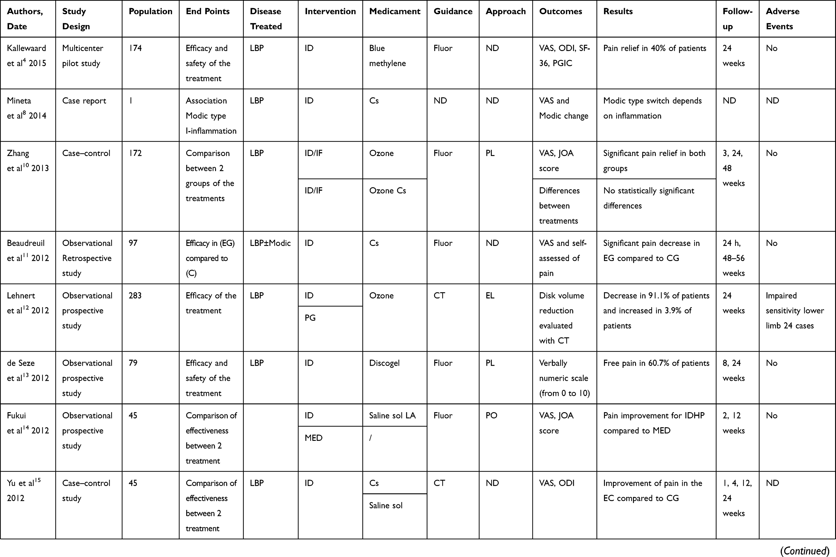 | 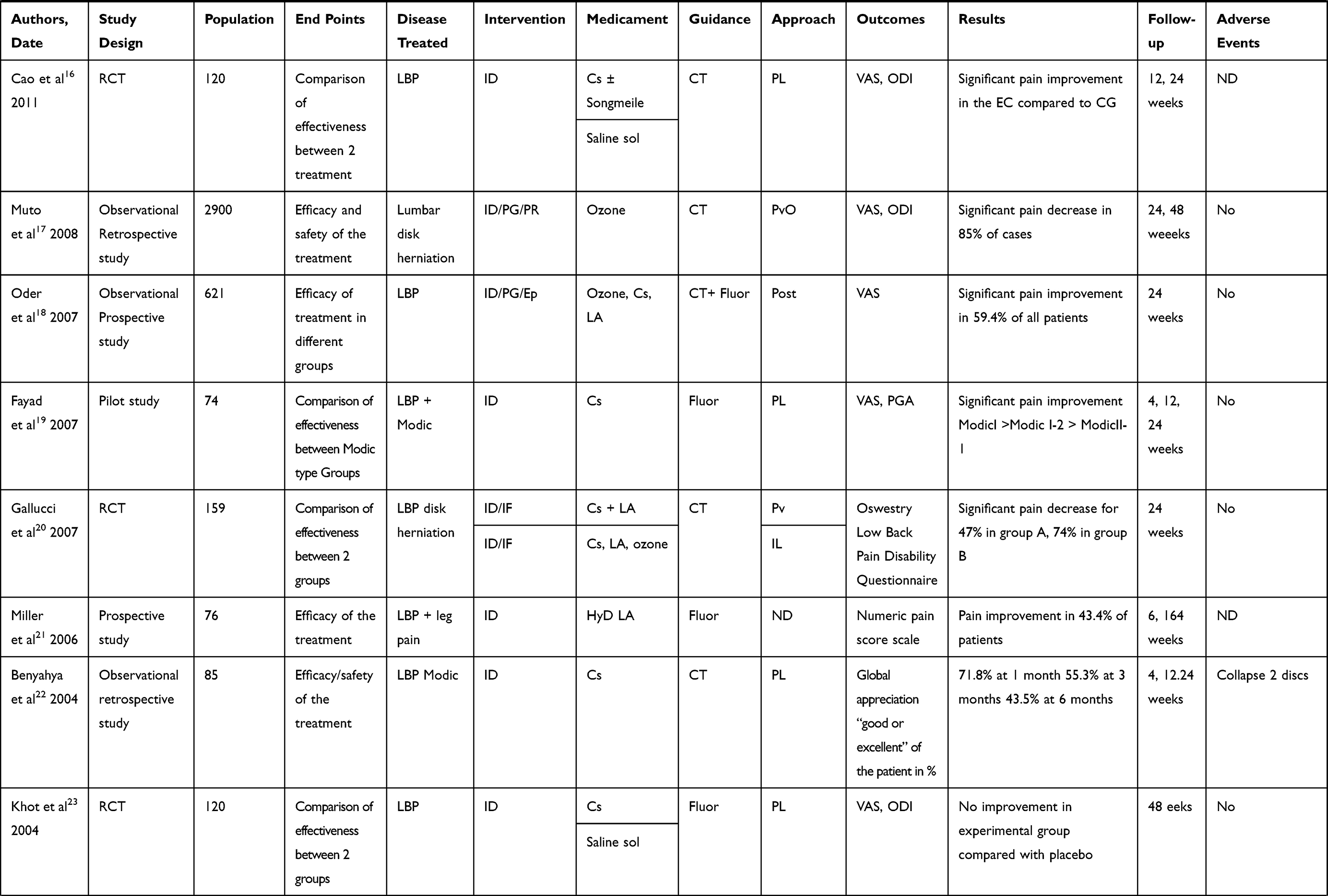 | 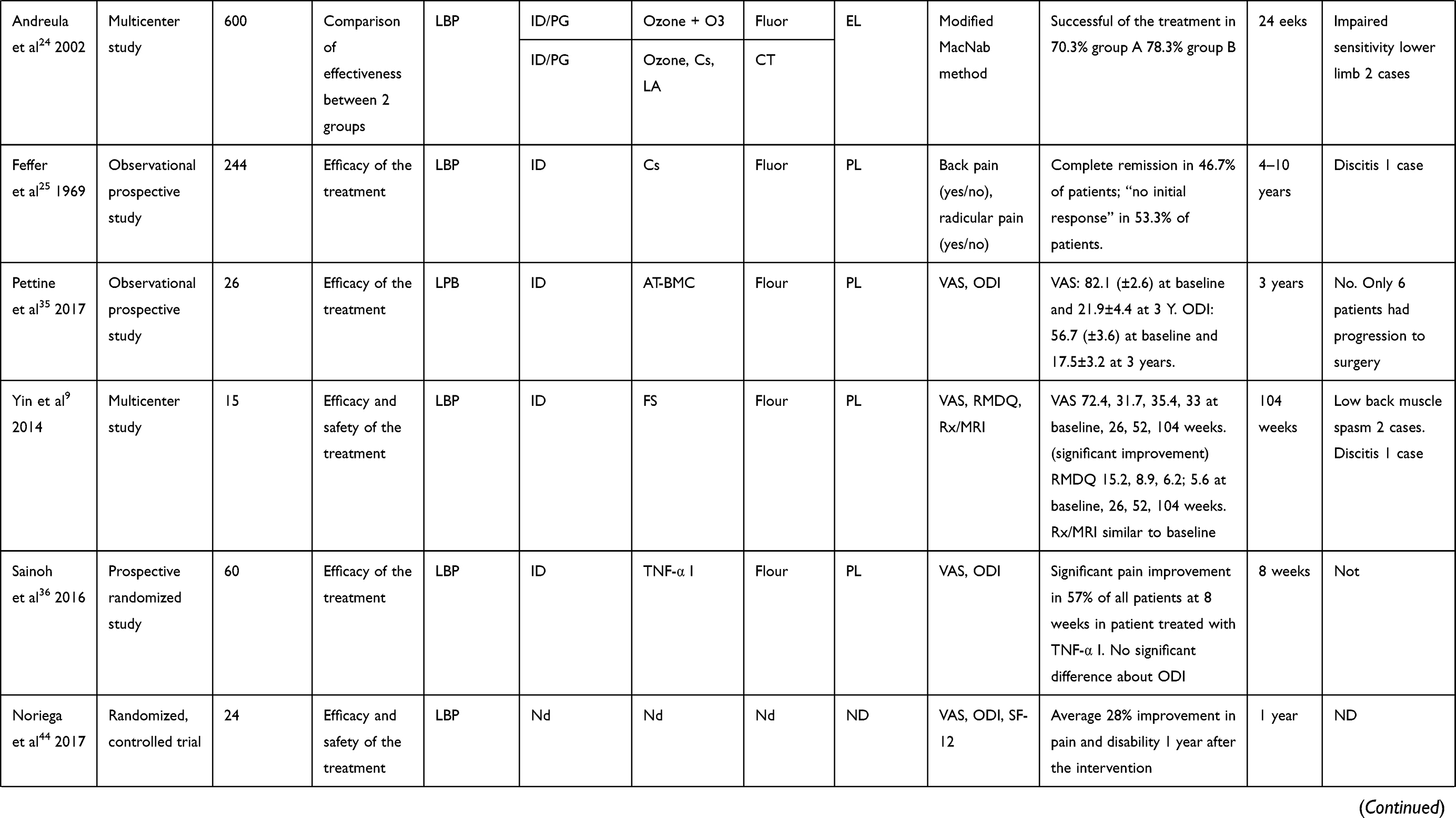 | 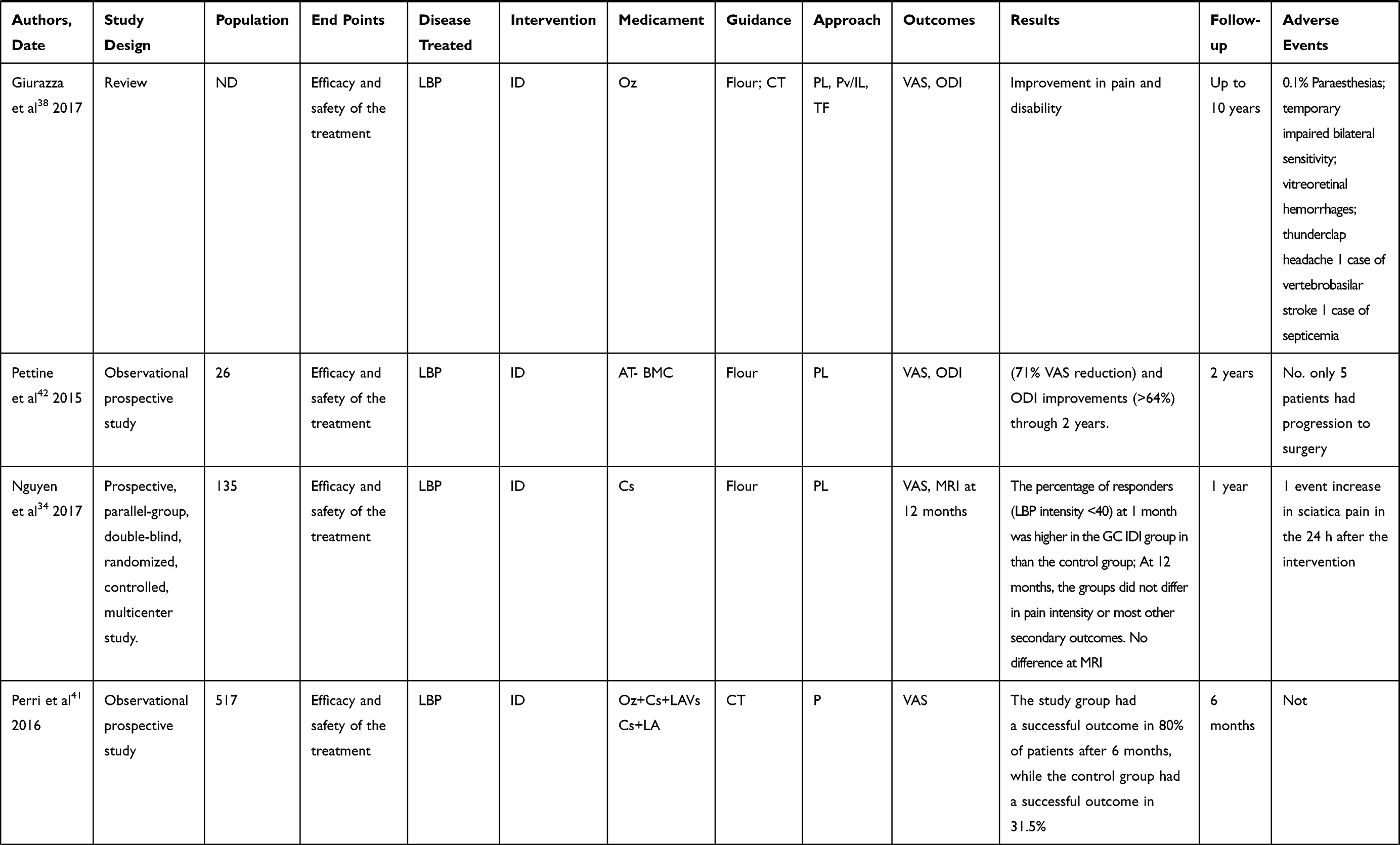 |  |  |
Table 1 Clinical Characteristics of Trials Employment of Intradiscal Injection |
Characteristics of Included Articles (Table 1)
The review includes three observational retrospective studies,12,18,23 12 observational prospective studies,13–15,19,22,26,36-38,41,43,44 two multicenter pilot studies,4,10 two case–control studies,11,16 six randomized controlled trials,4,8,17,21,24,35,42 one multicenter study25 two pilot studies,20,43 one case report,9 one single arm phase I clinical trial44 and one review.40
Population
Our population is composed of 6843 subjects, 52.3% male and 47.7% female, with a mean age of 45.9±10.1 years.
End Points
The aim of the study was to review literature for scientific evidence of intradiscal injections, to describe analytically the actual techniques, the type of intervention performed, the used imaging guidance, the inoculated drug, the approach to the intervertebral disc, the patient’s position, the operator who performed the treatment, the type of anesthesia used, antibiotic prophylaxis, if used.
Treated Disease
In all selected articles,4,9-26,35–38,39–44 the patients suffered from lumbar discopathy.
Type of Procedure
Different types of treatments are reported in the studies: intradiscal injection,4,8-26,35–44 epidural19 intraforaminal11,21 and facet joint injection, selective nerve block (SNRB),8 intradiscal high pressure injection (IDHP),15 microendoscopic discectomy (MED).16
Intradiscal Injection
The technique of intradiscal injection is reported in 31 articles. In 24 studies,4,9,10,12,14,16,17,20,22-24,26,35–44 it is the only treatment while, in the remaining seven studies11,13,15,18,19,21,25 it is compared to, or in association with, other minimally invasive procedures. (Table 2). In 19 articles the procedure was realized under fluoroscopy,4,10-12,14,15,20,22,24,26,35–39,41,42,44 in seven articles under CT guide,13,16–18,21,23,43 and in three studies both by fluoroscopy and CT guided, in comparison25 or in association;19,40 in two articles was not specified9,44 (Figure 2).
 |
Table 2 Characteristics of Intradiscal Injection Techniques |
The patients of the de Seze et al trial,13 Levi et al46 and Giurazza et al38 works were subjected to neurosedation, in the trials by Khot et al23 and Oder et al18 they were subjected to conscious sedation. In the studies by Fayad et al19 and Andreula et al24 and another five works34,37–39 the patients were neither sedated nor subjected to local anesthesia. In eleven trials,10,11,13,19,21,36,37,41–44 all patients were subjected to local anesthesia. The antibiotic prophylaxis was used in eight trials.4,10,19,21,41–44 The interventions were performed by highly experienced operators in the Lehnert’s trial12 two clinicians (two authors) in the Cao et al’s study;16 Fayad et al,19 Benyahya et al,22 Noriega et al,44 Giurazza et al,38 and Nguyen et al34 report the experienced radiologists, Gallucci et al20 and Perri et al40 neuroradiologists, Tuakli-Wosornu et al39 and Levi40,46 physiatrist and Khot et al23 two senior authors.
In 17 articles the required patients position was described according to the procedure: in 11 studies a prone position was used,15,18,19,21,36,38,40–44 Nguyen et al34 and Sainoh et al36 propose a lateral decubitus and Zhang et al10 advised use of a pillow under the waist to get the widest intervertebral spaces.
For the procedure spinal needles of 18- (n=4), 20- (n=2), 21- (n=1), 22- (n=17) and 23- (n=1) gauge were used, with variable length to 7 from 17.8 cm. For example the Muto et al’s study17 mentioned a 22-gauge spinal needle with paravertebral oblique access, Lehnert et al’s study12 mentioned an extraspinal lateral approach with a 22-gauge 17.8-cm spinal needle and Gallucci et al20 a paravertebral/interlaminar approach with a 9- or 15-cm 22-gauge spinal needle Five articles13,14,21,22,25 have specified that the side of the injection was chosen on the basis of the main location of symptoms.
The percutaneous approach is always posterior for the lumbar access: in 15.10,11,14,17,20,23,24,26,35,36,38,40–44 out of 31 articles it was specified as a posterolateral access, in other studies it was extralaminar12 or paravertebral access,20 posterior-oblique14 paravertebral-oblique,17 or anterolateral access. In the Gallucci et al’s study20 the intradiscal and intraforaminal injections were administered with a paravertebral approach in 92.4% of the patients and an interlaminar approach in 7.6% of the patients. The needle was advanced through the intraforaminal space, with an angle usually between 45° and 60°. In seven articles the point of access is not described.4,9,12,16,22,39,44 Oder et al’s study18 specified that the percutaneous approach was about 45° along the lateral margin of the inferior articular process of the vertebra and through the neuroforamen for preserving the nerve root. Muto et al17 used a needle inclination in a craniocaudal direction in the case of a lower herniation.
The site of injection is the center of the disc in 22 studies,7,10,11,13,16-22,35–44 the central third of the disc in the Yin et al’s study9 and in the mid portion of the herniated disc in the Fukui et al’s study.14 The position of the needle was confirmed by fluoroscopy using anteroposterior and lateral views in 20 studies;4,8,10–12,14,15,20,22,24,25,35–39,41,42,44 in Yin et al’s trial9 the procedures were performed with real time multiplanar fluoroscopy, with CT scan in seven other articles,13,16–18,21,23,43 in three articles with both fluoroscopy and CT,19,25,40 in two articles the position of the needle is not described.9,40
Some authors recommend the time remaining in supine position after injection: respectively 10 minutes;10 one hour,14 one, half an hour,9 two hours,4,21,25,39 three hours,13,16 four hours,41 six hours;12 12 h,18 12/24 h.11,37
Medicaments Injected
Several drugs have been injected, and were used individually or in association with each other: an oxygen-ozone mixture (O2O3) in eight studies,10,12,17,18,20,24,38,40 a saline solution in four studies;14–16,23 in 13 articles the steroids have been administered (methylprednisolone, acetate of prednisolone, hydrocortisone, betamethasone),8,10,11,15,16,18–20,22–25,34 and in six trials the local anesthetic (bupivacaine, lidocaine) were injected.4,14,20,21,24 For the remaining studies hypertonic dextrose,21 fibrin sealant,9 blue methylene,4,37 discogel,13 autologous bone marrow concentrate,35,42 allogenic mesenchymal stem cell and hyaluronic acid,41 tumour necrosis factor α1 inhibitor36 and songmeile16 (a kind of synthetic liquid of polypeptidic biological factors extracted from Chinese herbal medical ingredient) were used.
Outcomes Measures
Pain was the most frequently tested variable. It was expressed as percentage of patients with pain relief or as mean improvement on a continuous scale. The outcome measures shown in the studies were: VAS (visual analog score), NRS (numeric rating scale), McGill Pain Questionnaire. Outcome assessment of patient satisfaction are reported by “modified MacNab scale” or using Odom criteria (“Excellent”, “Good”, “Satisfactory” and “Poor”). Back-specific disability is expressed on a back-specific index, such as the Roland Disability Questionnaire or the Oswestry Disability Index and JOA score (widely used in Japan to evaluate disabilities associated with low-back pain and includes the following items: subjective symptoms; clinical signs; restriction of activities of daily living. JOA score ranges from 29 as the most positive score to minus six for the worst a global measure of improvement). Quality of life is measured by the SF-12, SF-36, and EuroQol.
Patients Global Impression of Change (PGIC) measured by a seven-point Likert scale. The evaluations of general health status or well being, disability for work, and patient satisfaction have all so been reported.
The disc volume was evaluated by MRI and CT images.
Clinical and/or radiologic short term follow-up were mainly performed at four or six weeks; the long-term follow-up were performed from 12, 24, 48, weeks up to 4–10 years.
Efficacy
The efficacy of the treatment is the target in 30 articles,4,9–25,34–44 The results are reported as clearly satisfactory in 27 out of the 30 articles,4,9–20,22,24,34–44 In the Muto et al’s study, for example, the results on 2900 patients, treated for LBP with intradiscal injection of O2–O3, were evaluated with the modified MacNab classification, the VAS and the Oswestry Disability Index at six and 12 months. Success rates were 75–80% for soft disc herniation, 70% for multiple-disc herniations and 55% for failed back surgery syndrome. None of the patients suffered early or late neurological or infectious complications.17 Benyahya et al22 made a retrospective study of medical records of 85 patients (55 women, mean age 49±9 years) to assess the effectiveness of intradiscal injection of acetate of prednisolone for the treatment of LBP. They used the global appreciation of the patient (excellent, good, mild, none, worse) concerning the result of the intradiscal injection, at one, three and six months. For effectiveness of intradiscal injection, the results showed that 71.8% of the patients considered the result good or excellent at one month, 55.3% at three months and 43.5% at six months.
Adverse Events
Six trials have reported the side effects,9,12,22,24,26,34 overall 32 cases for 6843 patients (0.47% of patients): three cases of discitis, two after injection of corticosteroid,25 one after injection of fibrin sealant;9 26 patients present impairment of sensitivity in the lower limb ipsilateral to the treatment with injection oxygen-ozone;12,24 two discs showed a collapse after injection of corticosteroid,22 1 case of increase in sciatica pain in the 24 hafter the intervention.34
Two trials were performed under CT guidance;12,22 two studies were performed by fluoroscopy,9,25 and only one case by both fluoroscopy and CT guide.24 Adverse events occurred in about 0.7% of the patients with CT guided injection and in 0.2% of the patients with fluoroscopic guided injection. In Yin et al’s trials,9 the patients have even been subjected to antibiotic prophylaxis, in others articles this was not described.
Yin et al, Lehnert et al, Benyahya et al, Andreula et al and Feffer et al9,12,22,24,25 report a posterolateral/extraspinal-lateral approach. Giurazza et al report that
The overall procedural complications rate is estimated around 0.1%. Have been reported in the literature: paresthesia on the anterolateral portion of the left leg and foot, suggesting nerve injury; few temporary episodes of impaired bilateral sensitivity; vitreoretinal hemorrhages; thunderclap headache related to pneumoencephalus as a consequence of inadvertent intrathecal puncture; and 1 case of vertebrobasilar stroke.38
Discussion
For the low back pain management, patients with a small or contained herniated disc with no response to medical treatments, can be candidates for one of the minimally invasive percutaneous techniques. Generally, the minimally invasive techniques offer good results with patient compliance and low cost, showing a very low side effects percentage.20 Only 0.47% of patients have manifested adverse events after intradiscal injection. The procedure is carried out on an outpatient basis by highly experienced operators such as radiologists,19,22 neuroradiologists,20 physiatrists46,39 and orthopedics.23 The procedure is of interest for many medical areas, for this reason standardizing this method allows it to be extended to various practitioners.
For preoperative management there is no consensus regarding sedation, local anesthesia, or antibiotic prophylaxis. Only seven authors mention antibiotic use,4,9,20,46–41 only two articles describe conscious sedation18,23 and three describe a deep sedation.13,38,46 Some unreviewed medical articles28,29 do not recommend local or general anesthesia because they could mask the nerve root puncture symptoms; the needle passes very close to the nerve root and may often touch it, causing a strong electric shock sensation which is quite harmless; if the patient is conscious they will feel the pain. About 0.19% of the patients subjected to antibiotic prophylaxis have had adverse events; while without antibiotics about 0.09% of the population have had side effects; current data do not allow a statistical analysis; for this reason prospective clinical trials are needed. Some authors advise setting up an aseptic room for anesthesiology care, ensuring peripheral access to the patient.29
A concordance has emerged about the patient position, the injection site and the needle type. The most included articles14,17,20,26,28–30,35,37-44 report a prone position as the best to increase the intervertebral space, also using a support under the abdomen to reduce lumbar lordosis. The lateral decubitus was reported in two works34,36 and in an unreviewed journal on chemiodiscolysis with ozone.28 According to five of the articles,12,13,20,21,24 de Santis et al29 recommend an access side at the same side as the symptoms.
The chosen injection site is the center of the disc, and the injection point was checked by fluoroscopic projections,4,9–11,13,14,19,21,23,25,34–39,41,43 CT scans;12,15–18,20,23,24,38,40 we highlight the need for trials to evaluate the more effective, safe, and less expensive methods, especially if using a toxic or very expensive drugs. For the safety, the data do not clarify which is the least injurious method, even though we have recorded a greater percentage of adverse events with the CT guided injection (Table 1). Clinical trials with same medication comparing the fluoroscopy and CT guided injections are needed.
During the procedure, the needle can be readily shifted a few millimeters to pass through without damaging the nerve. The approach and the needle inclination are essential criteria for a successful and safe procedure. In some articles it appears that the lumbar approach has a lateral inclination of 45° to 60° with respect to the axial line18,20 and that for the lower discs an additional cranial-caudal inclination is needed.20 We did not find accurate descriptions on the needle insertion procedure because the needle course was always evaluated radiographically and the access site was chosen accordingly. The authors recommend and/or use a fluoroscopy performed with the C-arm, that allows identification the trajectory of optimal access for needle placement into each disc.10,29,30 An image-guided procedure handbook30 describes a window of anatomical access to the intradiscal injection delineated by the superior articular process medially, the superior endplate below, and the traversing nerve root laterally and above. Staying close to the superior articular process could keep the needle as far as possible from the traversing nerve root.
The postintervention management was different between treatments, the authors have advised several rest times depending on the procedure (Table 2).
The efficacy and the safety of the intradiscal procedures are not easily comparable because the techniques are highly variable in terms of procedure (different operators, needle guidance, injection sites, drugs, tilt angle of the needle) (Table 2).
Conclusions
The efficacy and the safety of the intradiscal procedures are not easily comparable because of differences in the design of studies and their limited number.
The intradiscal injection is a technique widely used in the LBP management of patients with no response to rehabilitative and medical treatments. Differences of agreement between researchers are present on the technical aspects of the procedure in terms of imaging guidance, of injected substances, and efficacy of evaluation tools.
Further studies are needed in order to standardize the intradiscal injection technique/procedure as well as to improve efficacy, safety, repeatability, and to assess cost-effectiveness.
 |
Figure 1 Flow diagram illustrating published literature on intradiscal injection. |
 |
Figure 2 Different type of image guidance for intradiscal injection. |
Abbreviations
ADC, apparent diffusion coefficient; AL-MSC, allogenic mesenchymal stem cells; AT-BMC, autologous bone marrow concentrate; AT-MSC, adipose tissue mesenchymal stem cells; BM, blue methilene; C, control group; Cs, corticosteroid; CT, computerized tomography; EG, experimental group; EL, extraspinal lateral; Ep, epidural; EQ-5D, EuroQol; Fluor, fluoscopy; AL, anterolateral; FRI, Functional Rating Index; FS, fibrin sealant; HA, hyaluronic acid; HyD, hypertonic dextrose; ID, intradiscal injection; LA, local anesthetic; LBP, low back pain; MGPQ, McGill Pain Questionnaire; MIDPD, measurement of the intervertebral disc posterior dimension; MRI, magnetic resonance imaging; MSC, mesecnchymal stem cells; NASS, the modified North American Spine Society; NRS, Numeric Rating Scale; ODI, Oswestry Disability Index; P, periganglionic injection; PL, posterolateral; PO, posterior-oblique; Post, posterior; PR, periradicular injection; PRP, platelet-rich plasma; Pv/IL, paravertebral/interlaminar; PvO, paravertebral-Oblique; RCT, randomized controlled trial; RMDQ, Roland–Morris Disability Questionnaire; SANE, modified single assessment numeric evaluation; SF-36, short form-36; SNRB, selective nerve block; TF, transforaminal; TNF-α I, tumor nerosis factor α inhibitor; Treatm, treatment; VAS, visual analog scale.
Data Sharing Statement
All data analyzed during this study are included in this article.
Author Contributions
All authors made substantial contributions to conception and design, acquisition of data, or analysis and interpretation of data; took part in drafting the article or revising it critically for important intellectual content; gave final approval of the version to be published; and agree to be accountable for all aspects of the work.
Funding
This work is not supported by funds.
Disclosure
The authors declare that they have no competing interests in this work.
References
1. Guarnieri G, Vassallo P, Pezzullo MG, et al. A comparison of minimally invasive techniques in percutaneous treatment of lumbar herniated discs. Neuroradiol J. 2009;22(1):108–121. doi:10.1177/197140090902200116
2. Becker A, Held H, Redaelli M, et al. Implementation of a guideline for lowback pain management in primary care: a cost effectiveness analysis. Spine. 2012;37:701–710. doi:10.1097/BRS.0b013e31822b01bd
3. Leadley RM, Armstrong N, Lee YC, Allen A, Kleijnen J. Chronic diseases in the European Union: the prevalence and health cost implications of chronic pain. J Pain Palliat Care Pharmacother. 2012;26:310–325. doi:10.3109/15360288.2012.736933
4. Kallewaard JW, Geurts JW, Kessels A, Willems P, van Santbrink H, van Kleef M. Efficacy, safety, and predictors of intradiscal methylene blue injection for discogenic low back pain: results of a multicenter prospective clinical series. Pain Pract. 2015. doi:10.1111/papr.12283
5. Kuslich SD, Ulstrom CL, Michael CJ. The tissue origin of low back pain and sciatica: a report of pain response to tissue stimulation during operations on the lumbar spine using local anesthesia. Orthop Clin North Am. 1991;22:181–187.
6. Schwarzer AC, Aprill CN, Derby R, Fortin J, Kine G, Bogduk N. The relative contributions of the disc and zygapophyseal joint in chronic lowback pain. Spine. 1994;19:801–806. doi:10.1097/00007632-199404000-00013
7. Santiago FR, Kelekis A, Alvarez LG, Filippiadis DK. Interventional procedures of the spine. Semin Musculoskelet Radiol. 2014;18(3):309–317. doi:10.1055/s-0034-1375572
8. Mineta K, Higashino K, Sakai T, Fukui Y, Sairyo K. Recurrence of type I modic inflammatory changes in the lumbar spine: effectiveness of intradiscal therapy. Skeletal Radiol. 2014;43(11):1645–1649. doi:10.1007/s00256-014-1947-x
9. Yin W, Pauza K, Olan WJ, Doerzbacher JF, Thorne KJ. Intradiscal injection of fibrin sealant for the treatment of symptomatic lumbar internal disc disruption: results of a prospective multicenter pilot study with 24-month follow-up. Pain Med. 2014;15:16–31. Wiley Periodicals, Inc.
10. Zhang Y, Ma Y, Jiang J, Ding T, Wang J. Treatment of the lumbar disc herniation with intradiscal and intraforaminal injection of oxygen-ozone. J Back Musculoskelet Rehabil. 2013;26(3):317–322. doi:10.3233/BMR-130386
11. Beaudreuil J, Dieude P, Poiraudeau S, Revel M. Disabling chronic low back pain with modic type 1 MRI signal: acutereduction in pain with intradiscal corticotherapy. Ann Phys Rehabil Med. 2012;55:139–147. doi:10.1016/j.rehab.2012.01.004
12. Lehnert T, Naguib NN, Wutzler S, et al. Analysis of disk volume before and after CT-guided intradiscal and periganglionic ozone-oxygen injection for the treatment of lumbar disk herniation. J Vasc Interv Radiol. 2012;23(11):1430–1436. doi:10.1016/j.jvir.2012.07.029
13. De Seze M, Saliba L, Mazaux J-M. Percutaneous treatment of sciatica caused by a herniated disc: an exploratory study on the use of gaseous discography and discogel1 in 79 patients. Ann Phys Rehabil Med. 2013;56:143–154. doi:10.1016/j.rehab.2013.01.006
14. Fukui S
15. Yu Y, Liu W, Song D, Guo Q, Jia L. Diagnosis of discogenic low back pain in patients with probable symptoms but negative discography. Arch Orthop Trauma Surg. 2012;132(5):627–632. doi:10.1007/s00402-011-1448-5
16. Cao P, Jiang L, Zhuang C, et al. Intradiscal injection therapy for degenerative chronic discogenic low back pain with endplate modic changes. Spine J. 2011;11(2):100–106. doi:10.1016/j.spinee.2010.07.001
17. Muto M, Ambrosanio G, Guarnieri G, et al. Low back pain and sciatica: treatment with intradiscal-intraforaminal O(2)-O(3) injection. Our experience. Radiol Med. 2008;113(5):695–706. doi:10.1007/s11547-008-0302-5
18. Oder B, Loewe M, Reisegger M, Lang W, Ilias W, Thurnher SA. CT-guided/steroid therapy for the treatment of degenerative spinal disease–effect of age, gender, disc pathology and multi-segmental changes. Neuroradiology. 2008;50(9):777–785. doi:10.1007/s00234-008-0398-2
19. Fayad F, Lefevre-Colau MM, Rannou F, et al. Relation of inflammatory modic changes to intradiscal steroid injection outcome in chronic low back pain. Eur Spine J. 2007;16(7):925–931. doi:10.1007/s00586-006-0301-y
20. Gallucci M, Limbucci N, Zugaro L, et al. Sciatica: treatment with intradiscal and intraforaminal injections of steroid and oxygen-ozone versus steroid only. Radiology. 2007;242(3):907–913. doi:10.1148/radiol.2423051934
21. Miller Matthew R
22. Benyahya R, Lefevre-Colau MM, Fayad F, et al. Intradiscal injection of acetate of prednisolone in severe low back pain: complications and patients assessment of effectiveness. Ann Readapt Med Phys. 2004;47:621–626. doi:10.1016/S0168-6054(04)00193-X
23. Khot A, Bowditch M, Powell J, Sharp D. The use of intradiscal steroid therapy for lumbar spinal discogenic pain: a randomized controlled trial. Spine. 2004;29:833–836. doi:10.1097/00007632-200404150-00002
24. Andreula CF, Simonetti L, De Santis F, et al. Minimally invasive oxygenozone therapy for lumbar disk herniation. AJNR Am J Neuroradiol. 2003;24:996–1000.
25. Feffer HL. Therapeutic intradiscal hydrocortisone. A long-term study. Clin Orthop Relat Res. 1969;67:100–104. doi:10.1097/00003086-196911000-00015
26. Andreula C, Muto M, Leonardi M. Interventional spinal procedures. Eur J Radiol. 2004;50:112–119. doi:10.1016/j.ejrad.2003.10.013
27. Muto M, Andreula C, Leonardi M. Treatment of herniated lumbar disc by intradiscal and intraforaminal oxygen-ozone (O2-O3) injection. J Neuroradiol. 2004;31(3):183–189. doi:10.1016/S0150-9861(04)96989-1
28. Leonardi M. La puntura discale sotto fluoroscopia. Riv Ital Ossigeno Ozonoterapia. 2002;1:73–78.
29. De Santis F, Leonardi M, Simonetti L, Dall’Olio M, Princiotta C, Menetti F. Ossigeno-Ozonoterapia: la tecnica intradiscale. Int J Ozone Ther. 2009;8:138–146.
30. Mathis. Image-Guided spine Interventions. New York: Springer; 2004.
31. Migliore T, Laganà P, Granata B, et al. Safety of intra-articular hip injection of hyaluronic acid products by ultrasound guidance: an open study from ANTIAGE register. Eur Rev Med Pharmacol Sci. 2013;17(13):1752–1759.
32. Migliore A, Bizzi E, Massafra U, et al. A new technical contribution for ultrasound-guided injections of sacro-iliac joints. Eur Rev Med Pharmacol Sci. 2010;14(5):465–469.
33. Jee H, Lee JH, Park K
34. Nguyen C, Boutron I, Baron G, et al. Intradiscal glucocorticoid injection for patients with chronic low back pain associated with active dicopathy. Ann Intern Med. 2017. doi:10.7326/M16.1700
35. Pettine A, Suzuki RK, Sand TT, Murphy MB. Autologous bone marrow concentrate intradiscal injection for the treatment of degenerative disc disease with three-year follow up. Int Orthop. 2017;41:2097. doi:10.1007/s00264-017-3560-9
36. Sainoh T, Orita S, Miyagi M, et al. Single intradiscal administration of the tumor necrosis factor-alpha inhibitor, etanercept, for patients with discogenic low back pain. Pain Med. 2016;17:40–45. doi:10.1111/pme.12892
37. Zhang XJ, Hao J, Hu ZM, Yang HT. Clinical evaluation and magnetic resonance imaging assessment of intradiscal methylene blue injection for the treatment of discogenic low back pain. Pain Physician. 2016;19:E1189–E1195.
38. Giurazza F, Guarnieri G, Murphy KJ, Muto M. Intradiscal O2O3: rationale, injection technique, short- and long-term outcomes for the treatment of low back pain due to disc herniation. Can Assoc Radiol J. 2017;68:171–177. doi:10.1016/j.carj.2016.12.007
39. Tuakli-Wosornu YA, Terry A, Boachie-Adjei K, et al. Lumbar intradiskal Platelet-Rich Plasma (PRP) injections: a prospective, double-blind, Randomized Controlled Study. Pmrjournal. 2016;8:1–10.
40. Perri M, Marsecano C, Varrassi M, et al. Indications and efficacy of O2–O3 intradiscal versus steroid intraforaminal injection in different types of disco vertebral pathologies: a prospective randomized double‑blind trial with 517 patients. Radiol Med. 2016;121:463–471. doi:10.1007/s11547-015-0598-x
41. Kumar H, Ha DH, Lee EJ, et al. Safety and tolerability of intradiscal implantation of combined autologous adipose-derived mesenchymal stem cells and hyaluronic acid in patients with chronic discogenic low back pain: 1-year follow-up of a phase I study. Stem Cell Res Ther. 2017;8:262. doi:10.1186/s13287-017-0710-3
42. Pettine K, Suzuki R, Sand T, Murphy M. Treatment of discogenic back pain with autologous bone marrow concentrate injection with minimum two year follow-up. Int Orthop. 2015. doi:10.1007/s00264-015-2886-4
43. Centeno C, Markle J, Dodson E, et al. Treatment of lumbar degenerative disc disease‑associated radicular pain with culture‑expanded autologous mesenchymal stem cells: a pilot study on safety and efficacy. J Transl Med. 2017;15:197. doi:10.1186/s12967-017-1300-y
44. Noriega DC, Ardura F, Hernández-Ramajo R, et al. Intervertebral disc repair by allogeneic mesenchymal bone marrow cells: a randomized controlled trial. Transplantation. 2017;101(8):1945–1951. doi:10.1097/TP.0000000000001484
45. Hartung W, Ross CJ, Straub R, et al. Ultrasound-guided sacroiliac joint injection in patients with established sacroiliitis: precise IA injection verified by MRI scanning does not predict clinical outcome. Rheumatology (Oxford). 2010;49(8):1479–1482. doi:10.1093/rheumatology/kep424
46. Levi D, Horn S, Tyszko S, Levin J, Hecht-Leavitt C, Walko E. Intradiscal platelet-rich plasma injection for chronic discogenic low back pain: preliminary results from a prospective trial. Pain Med. 2016;17:1010–1022. doi:10.1093/pm/pnv053
 © 2020 The Author(s). This work is published and licensed by Dove Medical Press Limited. The
full terms of this license are available at https://www.dovepress.com/terms
and incorporate the Creative Commons Attribution
- Non Commercial (unported, 3.0) License.
By accessing the work you hereby accept the Terms. Non-commercial uses of the work are permitted
without any further permission from Dove Medical Press Limited, provided the work is properly
attributed. For permission for commercial use of this work, please see paragraphs 4.2 and 5 of our Terms.
© 2020 The Author(s). This work is published and licensed by Dove Medical Press Limited. The
full terms of this license are available at https://www.dovepress.com/terms
and incorporate the Creative Commons Attribution
- Non Commercial (unported, 3.0) License.
By accessing the work you hereby accept the Terms. Non-commercial uses of the work are permitted
without any further permission from Dove Medical Press Limited, provided the work is properly
attributed. For permission for commercial use of this work, please see paragraphs 4.2 and 5 of our Terms.