
Back to Journals » International Journal of Women's Health » Volume 17
The Relationship Between Serum Uric Acid and Gynecologic Cancer Risk: A Mendelian Randomization Study

Received 23 December 2024
Accepted for publication 25 April 2025
Published 5 May 2025 Volume 2025:17 Pages 1237—1245
DOI https://doi.org/10.2147/IJWH.S493564
Checked for plagiarism Yes
Review by Single anonymous peer review
Peer reviewer comments 2
Editor who approved publication: Dr Matteo Frigerio
Lei Cai, Chenmin Yang
Department of Obstetrics and Gynecology, Ruijin Hospital, School of Medicine, Shanghai Jiao Tong University, Shanghai, 200023, People’s Republic of China
Correspondence: Chenmin Yang, Department of Obstetrics and Gynecology, Ruijin Hospital, School of Medicine, Shanghai Jiao Tong University, No. 197 Second Ruijin Road, Huangpu District, Shanghai, 200023, People’s Republic of China, Tel +86-13917317396, Email [email protected]
Aim: High serum uric acid (UA) levels have been linked to cancer development through chronic inflammation and oxidative damage. Traditional epidemiological studies have shown inconsistent results regarding the relationship between uric acid and gynecological cancers. This study uses Mendelian randomization (MR) to explore the potential association between serum UA levels and various gynecological cancers.
Methods: In this two-sample MR study, summary statistical data of the genome-wide association studies (GWASs) on serum UA levels were extracted from the UK Biobank (UKB), and those on gynecological cancers were obtained from the FinnGen consortium, the Epidemiology of Endometrial Cancer Consortium (E2C2), and the Ovarian Cancer Association Consortium (OCAC). Inverse variance weighted (IVW), weighted median, MR-Egger, weighted mode, MR-Pleiotropy RESidual Sum and Outlier (MR-PRESSO), and MR-Radial methods were utilized to investigate the bidirectional causal associations of serum UA levels with gynecological cancers. The evaluation indexes were odds ratios (ORs) and confidence intervals (CIs). Tests for horizontal pleiotropism and heterogeneity of instrumental variables (IVs) were performed, respectively using MR-Egger test and Cochran’s Q statistics. In addition, leave-one-out and MR scatter plots were employed for sensitivity analyses.
Results: IVW estimates suggested that serum UA levels elevated 1 unit had a potential causal association with higher odds of both cervical cancer (CC) (OR=1.147, 95% CI: 1.020– 1.290) and invasive mucinous ovarian cancer (IMOC) (OR=1.199, 95% CI: 1.033– 1.393). Also, endometrial carcinoma (EC) had a potential causal association with it (OR=1.012, 95% CI: 1.000– 1.024). Additionally, sensitivity analyses showed the potential causal associations between UA and CC/IMOC were relatively robust.
Conclusion: An elevated serum UA level had potential associations with CC and IMOC, whereas patients with EC should pay attention to it in clinical practice, which may reduce the potential risk of gynecological cancers. However, further evidence is needed to clarify the true relationships between UA and gynecological cancers.
Keywords: serum UA level, gynecological cancers, Mendelian randomization study, causal association
Graphical Abstract:

Introduction
Gynecological cancers, including ovarian cancer (OC), cervical cancer (CC), and endometrial cancer (EC), pose a significant health challenge on a global scale.1 According to the estimates of GLOBOCAN 20202 on cancer incidence and mortality, out of the roughly 19.3 million new cancer cases and nearly 10 million cancer-related deaths worldwide in 2020, these three types of cancer make up approximately 7% of both new cases and deaths.2 Ovarian cancer is one of the most common cancers affecting women globally and has the highest mortality rate among all reproductive system cancers.3 Gynecological cancers pose a serious risk to women’s health. More research is focused on finding specific biomarkers and developing effective treatment methods for early detection and monitoring of the disease. The goal is to improve patient survival rates by better understanding of the causes and early warning signs of these cancers.
In addition to genetic susceptibility, metabolism-related factors are modifiable and influence cancer risk.4 Uric acid (UA) is the end product of purine metabolism in human, and both endogenous and exogenous purines are degraded to UA by xanthine oxidase. Serum UA has been considered as a substitute marker for metabolic disorders.5 Existing evidence suggests that high serum UA levels may contribute to the development of cancer through inducing chronic inflammation and increasing the production of reactive oxygen species (ROS).6 A previous cohort study found that high serum UA levels are positively associated with the risk of gynecological cancer but negatively associated with that of breast cancer (BC).7 A case-control study showed that hyperuricemia is an important factor in differentiating endometrial carcinoma (EC) from endometrial hyperplasia.8 It was suggested that uric acid levels may play a role in the diagnosis, severity assessment, and prognosis of various types of tumor, including gynecological cancers.9 At present, because of varieties in study design, data handling, and technical methods, conclusions on association between UA and cancer, based on observational studies, are still inconsistent. In addition, traditional epidemiological studies are susceptible to confounding factors and causal inversion, thus the true association of serum UA levels with the risk of gynecological cancer is unclear.
Mendelian randomization (MR), which exploits the single nucleotide polymorphisms (SNPs) as unconfounded instrumental variants (IVs), has been widely applied to investigate causal relationships of exposures with diseases.10 Due to Mendel’s law of separation and independent classification, it allows for a more comprehensive examination of cause and effect relationships compared to traditional observational studies, while also reducing the impact of bias caused by external factors.11,12 Meanwhile, MR is less susceptible to bias caused by reversed causation because the genetic code is not influenced by environmental factors or preclinical diseases. So far, the MR method has been utilized to explore the causal association between UA and the risk of BC, prostate cancer, and other cancers, and also found that UA has a significant positive correlation with the overall risk of cancer.13–15 Nevertheless, no study has discussed causal association of serum UA level with gynecological cancer on the basis of MR.
Herein, this two‐sample MR study aims to investigate causal associations of serum UA levels with different gynecological cancers: OC, EC, corpus uteri cancer (CUca), and CC. OC included endometrioid carcinomas of ovary (ENOC), epithelial ovarian cancer (EOC), invasive mucinous ovarian cancer (IMOC), clear cell ovarian cancer (CCOC), low grade serous ovarian cancer (LGSOC), and high grade serous ovarian cancer (HGSOC). We analyzed the possible reversed causal associations in order to provide some evidence-based foundation for the prevention and treatment of gynecological cancers and risk stratification management.
Methods
Data Sources
In this two-sample MR analysis, summarized data of UA and gynecological cancers were extracted from the genome-wide association studies (GWASs). Table 1 shows the data source of study variables. In brief, serum UA level data (437,354 samples) were obtained from the UK Biobank (UKB), with the unit of measurement μmol/L. More details are shown elsewhere: https://biobank.ndph.ox.ac.uk/showcase/field.cgi?id=30880. The study involved participants from diverse origins. EC data were collected by an expanded GWAS meta-analysis of 13 studies of endometrial cancer, the Epidemiology of Endometrial Cancer Consortium (E2C2)16 of 45 separate studies, and the UKB, and contained 12,906 cases and 108,979 controls. CC, CUca, and ENOC data were mainly accessed from the FinnGen consortium (https://www.finngen.fi/en/access_results). In addition, EOC data were extracted from the Ovarian Cancer Association Consortium (OCAC), and EOC with five major histotypes: HGSOC, LGSOC, MOC, ENOC, and CCOC.17
 |
Table 1 Data Source of Study Exposure and Outcomes |
Data from the involved GWASs are de-identified and fully open access. Informed consent from all the participants has been obtained. Since these GWASs have obtained ethical approval by the respective institutions, ethical approval has been waived by Ruijin Hospital, School of Medicine, Shanghai Jiao Tong University.
Single Nucleotide Polymorphisms Selection
Potential IVs were SNPs significantly associated with serum UA levels, with the selected threshold of P<5.0×10⁻⁸, and those with linkage disequilibrium (LD) need to be removed (LD threshold was r2 =0.001 and clumping distance of 10,000 kb). Also, SNPs being palindromic with intermediate allele frequencies should be deleted according to the MR principle that to ensure a same allele corresponds the effects between SNPs and the exposure, and that on the outcome. In addition, MR-Egger regression test, as one of the MR methods, was utilized to monitor potential horizontal pleiotropy effect, namely the confounding effect resulting from other diseases, which may violate the second assumption in MR analysis, and significant intercept item of MR-Egger represents the existence of pleiotropy (P<0.05).
The Assumptions of MR Analysis
MR study must conform to three assumptions to minimize the impact of bias on the results. The primary approach utilized was the IVW, selected for its ability to offer more cautious and dependable estimates in specific circumstances when compared to alternative methods.18 The other methods were used for validation purposes to confirm the reliability of the results. Firstly, IVs should be independent of confounders associated with exposures and outcomes. Secondly, IVs must be significantly linked to exposure, and the association strength was estimated via the following formulas: r2 = 2 × EAF × (1-EAF) × b × b / SE2; F = ((N - K - 1) / K) × (r2 / (1 - r2)), in which EAF represents effect allele frequency, b is the regression coefficient for serum UA levels and IVs, SE means standard error, K is the number of IVs, and N is sample size. When F<10, a weak association of IVs with exposure is recognized. Last, IVs only affect outcomes through exposures; this means no horizontal pleiotropy effect of IVs on outcome.
Statistical Analysis
Inverse variance weighted (IVW), weighted-median, MR-Egger,19 weighted mode,20 MR-Pleiotropy RESidual Sum and Outlier (MR-PRESSO), and MR-Radial21 methods were utilized to calculate causal effect values between serum UA levels and gynecological cancers. Specifically, IVW is the primary method to obtain unbiased estimates when horizontal pleiotropy was absent. Weighted-median can provide a relatively robust and consistent estimate of the causal effect, even if approximately 50% of genetic variants were invalid instruments. MR-Egger regression estimates generally exhibited low precision and might be influenced by outlying genetic variants. Weighted mode, which is also named the mode-based estimate (MBE), requires that the most common causal effect estimate is a consistent estimate of the true causal effect, even if the majority of instruments are invalid. In addition, MR-PRESSO analyses and MR-Radial detect and attempt to reduce horizontal pleiotropy by removing significant outliers. The effect size was expressed by odds ratios (ORs) with 95% confidence intervals (CIs). P<0.05 represents the statistical significance of evidence for potential causal effect. Test for heterogeneity used Cochran's Q test, IVs with P<0.05 were recognized heterogeneous.22 Furthermore, we conducted a “leave-one-out” analysis to evaluate the reliability of the findings. This involved systematically excluding one SNP at a time to determine if any individual SNP significantly influenced the conclusions regarding causal relationships.23 The statistical analyses were performed using R version 4.2.0 (Institute for Statistics and Mathematics, Vienna, Austria) with R package “TwoSampleMR”.
Results
Instrumental Variables Selection
Figure 1 is the flowchart of the study process. Table 2 shows the selection of IVs and tests on horizontal pleiotropy, heterogeneity, and strength. We initially identified 6933 SNPs just associated with gynecological cancers. After omitting LD and palindromic SNPs, there were respectively 301 SNPs for CC, 289 SNPs for EC, 288 SNPs for CUca, 278 SNPs for EOC, 286 SNPs for IMOC, 284 SNPs for CCOC, 280 SNPs for LGSOC, 286 SNPs for HGSOC, and 293 SNPs for ENOC as IVs. MR-Egger regression and MR-PRESSO outlier test, designed to assess potential bias, showed there was no horizontal pleiotropy or heterogeneity (all P>0.05). These findings strengthened confidence that the observed exposure-outcome association reflected a true causal effect.
 |
Table 2 IV Selection and Test of Horizontal Pleiotropy and Heterogeneity |
 |
Figure 1 Flowchart of the research process. |
Bidirectional Causal Association Between Serum UA Level and Gynecological Cancers
To examine potential reverse causal effects, we further explored bidirectional causal associations of serum UA levels with different gynecological cancers. Figure 2 shows the IVW results of associations between serum UA levels with gynecological cancers. The odds of CC and IMOC respectively increased 1.147 (95% CI: 1.020–1.290) and 1.199 (95% CI: 1.033–1.393) along with serum UA elevated 1 μmol/L. More details on results of other MR methods are shown in Table S1 which confirms same results. Similarly, more details of the reversed causal association between UA and gynecological cancers are shown elsewhere (Table S2). It was a critical step to ensure the validity of causal inferences. Besides, as it is shown in Figure 3, we only observed that EC had a potential causal association with serum UA level (OR=1.012, 95% CI: 1.000–1.024).
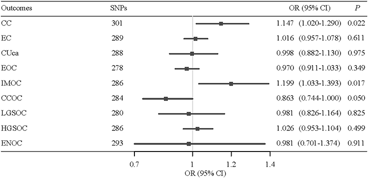 |
Figure 2 Potential causal associations of serum UA levels with different gynecological cancers. The P value of association between UA and CCOC is 0.050392. |
 |
Figure 3 Reversed potential causal association of serum UA levels with different gynecological cancers. |
Sensitivity analyses were performed on the potential causal associations of serum UA levels with CC and IMOC through the leave-one-out method (Figure S1). This method further supported that none of the identified causal associations was driven by any single IV. Figure S2 shows the MR scatter plots of the causal associations between serum UA levels and CC and IMOC respectively. According to these test results, no significant outliers have been found, indicating serum UA levels had a relatively robust potential causal association with CC and IMOC, which were stable results confirmed by different MR methods.
In summary, an increase of 1 μmol/L in serum UA levels was found to be potentially linked to higher odds of developing cervical cancer and invasive mucinous ovarian cancer. There was a potential causal association between UA levels and endometrial carcinoma.
Discussion
In the current study, we conducted a two-sample MR analysis to investigate the potential bidirectional causal associations of serum UA levels with gynecological cancers. The results showed that serum UA levels elevated for 1 μmol/L had a potential causal association with increased odds of both CC and IMOC. In addition, patients with EC seemed to have a higher level of serum UA.
In fact associations between serum UA levels and gynecological cancers are still unclear, and few studies have focused on associations of serum UA levels with CC or IMOC so far. A previous AMORIS study showed that a higher serum UC level was positively linked to the risk of gynecological cancer, but negatively associated with BC.7 In the present research, we observed potential causal associations between elevated serum UA levels and CC and IMOC based on MR methods, which aligns with Yiu and co-workers’ findings. Although underlying mechanisms that UA had potential causal associations with gynecological cancers are unclear, possible mechanisms that serum UA is involved in development of cancers have been reported. UA is a major product of purine metabolism catalyzed by xanthine oxidoreductase. Therefore, due to influencing the oxidation-reduction process in female reproductive disorders, UA may be involved in the pathogenesis of cancer by influencing oxidative stress.24,25 Besides, high levels of UA affect mitochondrial function, increase superoxide production, and then lead to lipid metabolism disorders, promoting the production of pro-inflammatory factors, which may further induce the development of cancers.26,27 There is a literature gap regarding the potential mechanism that UA had a causal association with CC. Basu et al28 found that plasma-reduced UA (PRUA) levels in women with cervical intraepithelial neoplasia (CIN) were significantly lower compared with controls, and the PRUA level was inversely linked to the histopathological graded severity of CIN. CIN is a precancerous condition of CC. A possible speculation was that during the progression of CIN to CC, along with the progression of inflammation and oxidative stress, the regulatory mechanism of the body’s clearance of peroxide or serum UA may be disturbed, thus further resulting in increased UA levels. The molecular structure of uric acid in its reduced state (PRUA) revealed its role as an antioxidant and increased alongside serum UA. As the end metabolic product of purine metabolism in vivo,29 UA has recently been found to have powerful antioxidant effects and can remove oxygen free radicals, singlet oxygen, and peroxynitrite. UA is also found in the follicular fluid of the female ovary.30 Hyperuricemia could promote the progress of hyperandrogenemia, insulin resistance, abnormal lipid metabolism, and complications in PCOS.31 Also, in patients with endometriosis (EM), interleukin (IL)-1β is produced when UA forms inflammatory bodies, which can induce the aggregation of inflammatory cells and activate the Th2-type immune response to produce local inflammation by promoting the activation of normal T cell expression and secretion factor mRNA expression.32–34 A MR analysis showed there was an association of genetic liability to EM with IMOC.35 In our study, women with EC had higher levels of serum UA, indicating that UA may be involved in the association between EM/EC and IMOC. However, the potential causal association between EC and serum UA level was not robust, and further studies are needed to clarify this relationship.
As mentioned before, MR is a relatively superior study design to observational researches on investigating the causal effect of potential risk factors on diseases of interest. Through exploring the bidirectional causal associations of serum UA levels with different gynecological cancers, this MR study may provide some reference for facilitating the recommendation of public health policies and clinical interventions, which could effectively reduce the incidence and social burden of gynecological cancers. We performed sensitivity analyses via leave-one-out and MR scatter plots to further explore the robustness of the potential causal associations of serum UA levels with CC and IMOC. Increased production of serum UA may follow consumption of diets high in purines, acute alcohol consumption, chronic fructose consumption, and severe exercise.29 Other chronic diseases, such as metabolic syndrome (MetS) is also associated with the aforementioned dietary factors that cause increased production of serum UA.36 Thus, in addition to timely screening for gynecological tumors, following a healthy dietary pattern, participating in appropriate physical activity, maintaining normal body mass index levels, and management of chronic diseases are also significant for maintaining normal serum UA levels and reducing the subsequent risk of gynecological cancers in clinical practice.
This study with two samples of MR suggested potential causal associations of serum UA levels with gynecological tumors, which to some extent overcame interference of confounding factors and reversed causal inference. GWAS data were obtained from large public databases of representative populations in Europe, and the selected IVs of study exposure were highly explanatory and representative. However, there are still some limitations of interpretation of the study results. Potential associations of serum UA levels with gynecological cancers were based on the European population, and whether these findings can be generalizable to other populations requires further evidence. The lack of individual data prevented the assessment of potential non-linear associations between UA and gynecological cancers because we used aggregated data from GWASs. In addition, data on lifestyle factors such as alcohol consumption, smoking status, and diet, which potentially impact UA levels,37 are lacking in existing databases. These variables should be accounted for in future research to ensure accurate analyses. Therefore, true relationships between UA and gynecological cancers need to be further validated.
This study suggests that targeting UA could be a promising strategy, but more research is needed to confirm its effectiveness before it can be used in clinical settings. UA modulation may be a valuable addition to current prevention strategies. It is important to focus on lowering UA in high-risk women and conducting mechanistic studies to understand its role before moving on to pilot trials. Collaboration between epidemiologists, oncologists, and pharmacologists will be essential for further advancement in this area.
Conclusion
Serum UA had a potential causal association with gynecological cancer risk, especially CC and IMOC, whereas EC was linked to a higher serum UA level. However, these potential causal relationships and underlying mechanisms still need further exploration. Limitations include lifestyle factors, population stratification, and histotype aggregation. Addressing these in the results and discussion would strengthen causal inference.
Data Sharing Statement
https://biobank.ndph.ox.ac.uk/showcase/field.cgi?id=30880. https://www.finngen.fi/en/access_results.
Ethics Approval and Informed Consent
This is an observational study and was approved by Ruijin Hospital ethics committee and adhered to the International Ethical Guidelines for Biomedical Research. Consents to participate were exempted.
Consent for Publication
The manuscript is original, unpublished, and not under consideration elsewhere. We confirm that the details of any images and tables can be published, and that the persons providing consent have been shown the article contents to be published.
Author Contributions
All authors made a significant contribution to the work reported, whether that is in the conception, study design, execution, acquisition of data, analysis and interpretation, or in all these areas; took part in drafting, revising or critically reviewing the article; gave final approval of the version to be published; have agreed on the journal to which the article has been submitted; and agree to be accountable for all aspects of the work.
Funding
This work was not supported by any grant.
Disclosure
The authors declare no competing interests in this work.
References
1. Siegel RL, Giaquinto AN, Jemal A. Cancer statistics, 2024. CA Cancer J Clin. 2024;74(1):12–49. doi:10.3322/caac.21820
2. Sung H, Ferlay J, Siegel RL, et al. Global cancer statistics 2020: GLOBOCAN estimates of incidence and mortality worldwide for 36 cancers in 185 countries. CA Cancer J Clin. 2021;71(3):209–249. doi:10.3322/caac.21660
3. Stewart C, Ralyea C, Lockwood S. Ovarian cancer: an integrated review. Semin Oncol Nurs. 2019;35(2):151–156. doi:10.1016/j.soncn.2019.02.001
4. Gyamfi J, Kim J, Choi J. Cancer as a metabolic disorder. Int J mol Sci. 2022;23(3):1155. doi:10.3390/ijms23031155
5. Kanbay M, Jensen T, Solak Y, et al. Uric acid in metabolic syndrome: from an innocent bystander to a central player. Eur J Intern Med. 2016;29:3–8. doi:10.1016/j.ejim.2015.11.026
6. Mi S, Gong L, Sui Z. Friend or Foe? An unrecognized role of uric acid in cancer development and the potential anticancer effects of uric acid-lowering drugs. J Cancer. 2020;11(17):5236–5244. doi:10.7150/jca.46200
7. Yiu A, Van Hemelrijck M, Garmo H, et al. Circulating uric acid levels and subsequent development of cancer in 493,281 individuals: findings from the AMORIS Study. Oncotarget. 2017;8(26):42332–42342. doi:10.18632/oncotarget.16198
8. Zhang H, Kong W, Han C, Liu T, Li J, Song D. Correlation of metabolic factors with endometrial atypical hyperplasia and endometrial cancer: development and assessment of a new predictive nomogram. Cancer Manag Res. 2021;13:7937–7949. doi:10.2147/CMAR.S335924
9. Dhankhar R, Dahiya K, Sharma TK, Ghalaut VS, Atri R, Kaushal V. Diagnostic significance of adenosine deaminase, uric acid and C-reactive protein levels in patients of head and neck carcinoma. Clin Lab. 2011;57(9–10):795–798.
10. Davey Smith G, Hemani G. Mendelian randomization: genetic anchors for causal inference in epidemiological studies. Hum Mol Genet. 2014;23(R1):R89–98. doi:10.1093/hmg/ddu328
11. Richmond RC, Davey Smith G. Mendelian randomization: concepts and scope. Cold Spring Harb Perspect Med. 2022;12(1):a040501. doi:10.1101/cshperspect.a040501
12. Davies NM, Holmes MV, Davey Smith G. Reading Mendelian randomisation studies: a guide, glossary, and checklist for clinicians. BMJ. 2018;362:k601. doi:10.1136/bmj.k601
13. Jiang M, Ren L, Chen S, Li G. Serum uric acid levels and risk of eight site-specific cancers: a Mendelian randomization study. Front Genet. 2021;12:608311. doi:10.3389/fgene.2021.608311
14. Kobylecki CJ, Afzal S, Nordestgaard BG. Plasma urate, cancer incidence, and all-cause mortality: a Mendelian randomization study. Clin Chem. 2017;63(6):1151–1160. doi:10.1373/clinchem.2016.268185
15. Li X, Meng X, Timofeeva M, et al. Serum uric acid levels and multiple health outcomes: umbrella review of evidence from observational studies, randomised controlled trials, and Mendelian randomisation studies. BMJ. 2017;357:j2376. doi:10.1136/bmj.j2376
16. Kamat MA, Blackshaw JA, Young R, et al. PhenoScanner V2: an expanded tool for searching human genotype-phenotype associations. Bioinformatics. 2019;35(22):4851–4853. doi:10.1093/bioinformatics/btz469
17. Phelan CM, Kuchenbaecker KB, Tyrer JP, et al. Identification of 12 new susceptibility loci for different histotypes of epithelial ovarian cancer. Nat Genet. 2017;49(5):680–691. doi:10.1038/ng.3826
18. Hemani G, Bowden J, Davey Smith G. Evaluating the potential role of pleiotropy in Mendelian randomization studies. Hum Mol Genet. 2018;27(R2):R195–R208. doi:10.1093/hmg/ddy163
19. Burgess S, Thompson SG. Interpreting findings from Mendelian randomization using the MR-Egger method. Eur J Epidemiol. 2017;32(5):377–389. doi:10.1007/s10654-017-0255-x
20. Hartwig FP, Davey Smith G, Bowden J. Robust inference in summary data Mendelian randomization via the zero modal pleiotropy assumption. Int J Epidemiol. 2017;46(6):1985–1998. doi:10.1093/ije/dyx102
21. Srolovitz DJ, Safran SA, Tenne R. Elastic equilibrium of curved thin films. Phys Rev E Stat Phys Plasmas Fluids Relat Interdiscip Topics. 1994;49(6):5260–5270. doi:10.1103/physreve.49.5260
22. Bowden J, Del Greco MF, Minelli C, et al. Improving the accuracy of two-sample summary-data Mendelian randomization: moving beyond the NOME assumption. Int J Epidemiol. 2019;48(3):728–742. doi:10.1093/ije/dyy258
23. Burgess S, Bowden J, Fall T, Ingelsson E, Thompson SG. Sensitivity analyses for robust causal inference from Mendelian randomization analyses with multiple genetic variants. Epidemiology. 2017;28(1):30–42. doi:10.1097/EDE.0000000000000559
24. Bannigida DM, Nayak BS, Vijayaraghavan R. Insulin resistance and oxidative marker in women with PCOS. Arch Physiol Biochem. 2020;126(2):183–186. doi:10.1080/13813455.2018.1499120
25. Aouache R, Biquard L, Vaiman D, Miralles F. Oxidative stress in preeclampsia and placental diseases. Int J mol Sci. 2018;19(5):1496. doi:10.3390/ijms19051496
26. Lanaspa MA, Sanchez-Lozada LG, Choi YJ, et al. Uric acid induces hepatic steatosis by generation of mitochondrial oxidative stress: potential role in fructose-dependent and -independent fatty liver. J Biol Chem. 2012;287(48):40732–40744. doi:10.1074/jbc.M112.399899
27. Ghaemi-Oskouie F, Shi Y. The role of uric acid as an endogenous danger signal in immunity and inflammation. Curr Rheumatol Rep. 2011;13(2):160–166. doi:10.1007/s11926-011-0162-1
28. Basu J, Mikhail MS, Ahn CW, et al. Plasma uric acid levels in women with cervical intraepithelial neoplasia. Nutr Cancer. 2005;51(1):25–31. doi:10.1207/s15327914nc5101_4
29. Kang DH, Ha SK. Uric acid puzzle: dual role as anti-oxidantand pro-oxidant. Electrolyte Blood Press. 2014;12(1):1–6. doi:10.5049/EBP.2014.12.1.1
30. Hu J, Xu W, Yang H, Mu L. Uric acid participating in female reproductive disorders: a review. Reprod Biol Endocrinol. 2021;19(1):65. doi:10.1186/s12958-021-00748-7
31. Liu YN, Luo H, Che X, Peng H, Li M, Liu KX. Uric acid metabolism in polycystic ovary syndrome. Clin Chim Acta. 2021;517:74–80. doi:10.1016/j.cca.2021.02.009
32. Lambert S, Santulli P, Chouzenoux S, et al. Endometriosis: increasing concentrations of serum interleukin-1beta and interleukin-1sRII is associated with the deep form of this pathology. J Gynecol Obstet Biol Reprod. 2014;43(9):735–743. Estonian. doi:10.1016/j.jgyn.2014.06.014
33. Urata Y, Osuga Y, Izumi G, et al. Interleukin-1beta stimulates the secretion of thymic stromal lymphopoietin (TSLP) from endometrioma stromal cells: possible involvement of TSLP in endometriosis. Hum Reprod. 2012;27(10):3028–3035. doi:10.1093/humrep/des291
34. Lebovic DI, Chao VA, Martini JF, Taylor RN. IL-1beta induction of RANTES (regulated upon activation, normal T cell expressed and secreted) chemokine gene expression in endometriotic stromal cells depends on a nuclear factor-kappaB site in the proximal promoter. J Clin Endocrinol Metab. 2001;86(10):4759–4764. doi:10.1210/jcem.86.10.7890
35. Yarmolinsky J, Relton CL, Lophatananon A, et al. Appraising the role of previously reported risk factors in epithelial ovarian cancer risk: a Mendelian randomization analysis. PLoS Med. 2019;16(8):e1002893. doi:10.1371/journal.pmed.1002893
36. Johnson RJ, Nakagawa T, Sanchez-Lozada LG, et al. Sugar, uric acid, and the etiology of diabetes and obesity. Diabetes. 2013;62(10):3307–3315. doi:10.2337/db12-1814
37. Zhang WZ, Peng Q, Cai XS, et al. A study on the correlation between hyperuricemia and lifestyle and dietary habits. Medicine. 2025;104(5):e41399. doi:10.1097/MD.0000000000041399
 © 2025 The Author(s). This work is published and licensed by Dove Medical Press Limited. The
full terms of this license are available at https://www.dovepress.com/terms
and incorporate the Creative Commons Attribution
- Non Commercial (unported, 4.0) License.
By accessing the work you hereby accept the Terms. Non-commercial uses of the work are permitted
without any further permission from Dove Medical Press Limited, provided the work is properly
attributed. For permission for commercial use of this work, please see paragraphs 4.2 and 5 of our Terms.
© 2025 The Author(s). This work is published and licensed by Dove Medical Press Limited. The
full terms of this license are available at https://www.dovepress.com/terms
and incorporate the Creative Commons Attribution
- Non Commercial (unported, 4.0) License.
By accessing the work you hereby accept the Terms. Non-commercial uses of the work are permitted
without any further permission from Dove Medical Press Limited, provided the work is properly
attributed. For permission for commercial use of this work, please see paragraphs 4.2 and 5 of our Terms.