
Back to Journals » International Journal of Nanomedicine » Volume 21
The Emerging Role of Nanocarrier-Based Delivery Systems for cGAS-STING Activation in Cancer Immunotherapy
Authors Zhu G, Li D, Zhang H, Wang J, Li X  , Zeng L, Zhang L , Yu L
Received 22 January 2026
Accepted for publication 22 May 2026
Published 3 June 2026 Volume 2026:21 598118
DOI https://doi.org/10.2147/IJN.S598118
Checked for plagiarism Yes
Review by Single anonymous peer review
Peer reviewer comments 7
Editor who approved publication: Dr Sachin Mali
Guanxiong Zhu,1,* Da Li,1,* Hongru Zhang,1 Jingyuan Wang,1 Xinyi Li,1 Liting Zeng,1 Lingmin Zhang,1,2 Lina Yu1
1Department of Preventive Dentistry, Affiliated Stomatology Hospital of Guangzhou Medical University, Guangdong Engineering Research Center of Oral Restoration and Reconstruction, Guangzhou Key Laboratory of Basic and Applied Research of Oral Regenerative Medicine, Guangzhou, 510182, People’s Republic of China; 2Guangzhou Municipal and Guangdong Provincial Key Laboratory of Molecular Target & Clinical Pharmacology, the State & NMPA Key Laboratory of Respiratory Disease, School of Pharmaceutical Sciences & the Fifth Affiliated Hospital, Guangzhou Medical University, Guangzhou, 511436, People’s Republic of China
*These authors contributed equally to this work
Correspondence: Lina Yu; Lingmin Zhang, Email [email protected]; [email protected]
Abstract: Activation of the cGAS-STING pathway is a potential strategy to potentiate anti-tumor immunity. However, clinical translation of STING agonists such as cGAMP is hampered by poor targeting, enzymatic degradation, and systemic off-target toxicity. Nanocarrier-based drug delivery systems (nano-DDS) overcome these limitations by improving stability, bioavailability, and tumor-specific delivery. This review summarizes recent advances in lipid-based, polymeric, metallic, mesoporous silica, and exosomal for STING agonist delivery. These nanoplatforms enable precise drug delivery and controlled release, thereby significantly augmenting anti-tumor immune responses. We further discuss combination strategies of STING nanoagonists with radiotherapy, chemotherapy, phototherapy, and immune checkpoint inhibitors. Finally, we outline current challenges and future directions, including protein corona effects, scalable manufacturing, and personalized therapy.
Keywords: nanocarrier-based delivery systems, cGAS-STING, immune activation, cancer immunotherapy
Introduction
Malignant tumors are major diseases that seriously threaten human health. The 5-year survival rate of many solid tumors has remained stagnant for a long time, posing a severe challenge to clinical practice. Particularly challenging is that many tumors exhibit typical immunosuppressive phenotypic characteristics - a severe lack of cytotoxic T lymphocytes in the tumor microenvironment, significantly impaired antigen-presenting function, and a rich variety of immunosuppressive cell populations.1 The unique immunosuppressive characteristics of this microenvironment directly leads to the unsatisfactory objective response rate of immune checkpoint inhibitors represented by PD-1/PD-L1 inhibitors in some patients, seriously restricting the clinical therapeutic effect.2,3
At present, the treatment strategies for immunosuppressive tumors mainly face the following bottlenecks: The mechanism of action of existing immunotherapeutic strategies relies on pre-existing tumor-specific T cells.4 However, in the immune desert phenotype, due to the defect of antigen presentation mechanism, abnormal secretion of chemokines, and the formation of immunosuppressive networks, naive T cells fail to become activated and are unable to infiltrate the tumor site.5 Although existing treatment methods such as radiotherapy and chemotherapy can control tumor progression to a certain extent, it is difficult to fundamentally reverse the immunosuppressive microenvironment.6 Therefore, although immunotherapy, which recognizes and eliminates cancer cells through the immune system, can significantly reduce the side effects of traditional therapies, its clinical efficacy remains constrained by the deficiencies of existing targets. Identifying new immunotherapy targets is crucial for breaking through the current bottlenecks in cancer treatment.7
The cyclic GMP-AMP synthase-stimulator of interferon genes (cGAS-STING) pathway is a natural immune signaling pathway. Its activation leads to the secretion of type I interferon (IFN) to promote dendritic cell (DC) maturation, subsequently increasing immune cell infiltration into the tumor microenvironment (TME), thereby initiating an anti-tumor immune response in vivo.8 The cGAS-STING pathway, by specifically targeting tumor cells and initiating anti-tumor immune responses in vivo, has become a new strategy to enhance anti-tumor immunity. However, the STING activator cGAMP is a dinucleotide prone to enzymatic degradation in the blood; it typically does not target tumors and is easily degraded in vivo, limiting its clinical application.9,10 Therefore, researchers employ carriers or chemical modifications to protect STING activators, thereby conferring higher enzymatic stability.
Nano-drug delivery systems are drug delivery platforms based on nanoparticle carriers, usually made from lipids, polymers, inorganic materials, or biological materials, with sizes generally between 1–200 nm.11 Their characteristic is to achieve precise delivery and controlled release by encapsulating, loading, or binding drugs onto nanoparticles and endowing them with targeting and functionality through surface modification, thereby achieving efficient treatment.11 Nano-DDS can selectively accumulate in tumor tissue through the enhanced permeability and retention (EPR) effect in cancer therapy and utilize their stimulus responsiveness to release drugs in specific microenvironments, reducing side effects on normal cells.12 Therefore, nanoparticles offer significant advantages for delivering activators of the cGAS-STING pathway, improving therapeutic efficacy and reducing side effects. Through targeted design of nanoparticles, cGAS-STING activators can be precisely delivered to tumor tissue or immune cells, reducing the impact on healthy tissues and thus lowering systemic toxicity. This article systematically reviews the cutting-edge progress of cGAS-STING combined with nanomaterials in the diagnosis and treatment of oral cancer, analyzes its mechanism of action and clinical challenges, and prospects future directions for interdisciplinary collaborative innovation to promote the development of precision medicine for oral cancer.
cGAS-STING Pathway Structure and Activation Mechanism
The cGAS-STING pathway is a key signaling pathway in the innate immune system that recognizes cytoplasmic DNA and initiates immune responses. This pathway consists of two core proteins: cyclic GMP-AMP synthase (cGAS) and stimulator of interferon genes (STING).13 cGAS, as a DNA sensor, has a structure that contains an N-terminal disordered region, a DNA-binding domain, and a C-terminal catalytic domain, enabling non-specific binding to double-stranded DNA (dsDNA) in the cytoplasm.14 When dsDNA released from tumors, viruses, bacteria, or damaged mitochondria binds to cGAS, it induces cGAS dimerization or oligomerization, exposing the catalytic active site, which then uses ATP and GTP to synthesize the second messenger 2′,3′-cGAMP.15 STING, as an endoplasmic reticulum transmembrane protein, consists of an N-terminal transmembrane region, a central linker region, and a C-terminal ligand-binding domain, usually existing as a dimer.16 When cGAMP binds to STING, it causes a conformational change in STING, transporting it from the endoplasmic reticulum to the Golgi apparatus and recruiting TANK-binding kinase 1 (TBK1) to form the STING-TBK1 signaling complex.17
The formation of the STING-TBK1 complex is a key step in initiating downstream signal transduction. After TBK1 activates through autophosphorylation, it further phosphorylates the C-terminal domain of STING, providing a binding site for the recruitment of interferon regulatory factor 3 (IRF3).18 Phosphorylated IRF3 forms dimers and translocates to the nucleus, initiating the transcription of type I interferons (IFN-α/β) and interferon-stimulated genes (ISGs).19 Simultaneously, STING can also activate the NF-κB signaling pathway through tumor necrosis factor receptor-associated factor 6 (TRAF6), promoting the production of pro-inflammatory cytokines.20 These immune molecules together constitute an anti-viral and anti-tumor immune defense network, which can not only directly inhibit pathogen replication but also activate immune cells such as NK cells and DCs, enhancing the body’s immune surveillance function.21,22 Figure 1 depicts the molecular mechanisms of the cGAS-STING Pathway.
 |
Figure 1 Schematic diagram of the molecular mechanism of the cGAS-STING signaling pathway in innate immunity for detecting cytoplasmic DNA. |
The activity of the cGAS-STING pathway is finely regulated at multiple levels to avoid tissue damage caused by excessive immune responses. At the molecular level, the cytoplasmic nuclease Trex1 can degrade free DNA, preventing sustained cGAS activation.23 E3 ubiquitin ligase RNF5-mediated ubiquitination and degradation of STING can terminate signal transduction; autophagy-related proteins such as ULK1 can also negatively regulate STING activity.24,25 Pathologically, functional defects in this pathway may lead to viral infection or tumor immune escape.26,27
Key Role of cGAS-STING Signal Transduction in Tumors
The cGAS-STING pathway is a key cytoplasmic DNA sensing mechanism that connects innate immunity with anti-tumor immune responses. Under pathological conditions such as genomic instability, mitochondrial damage, viral/bacterial infection, or cellular stress, accumulated dsDNA in the cytoplasm can be recognized by cGAS.28 As detailed in Section 2, activation of the cGAS-STING pathway triggers type I interferon production and pro-inflammatory cytokines, which play key roles in anti-tumor immunity. IFN-β plays a key role in activating tumor-specific T cells, thereby linking innate immune sensing with adaptive anti-tumor immunity. Due to its ability to recognize abnormal cytoplasmic DNA and initiate robust immune activation, cGAS-STING has become a key target for enhancing cancer immunotherapy.8 Simultaneously, this pathway helps activate dendritic cells, T cells, and natural killer cells, enhancing anti-tumor immune responses.29 The pathway can also promote apoptosis and senescence of cancer cells, enhancing the protective effects of cytotoxic T cells and natural killer cells.14 Activation of the cGAS-STING pathway can improve the efficacy of immune checkpoint inhibitors and other immunotherapies. Mechanistically, cGAS-STING signaling can promote antigen presentation, enhance T cell infiltration, and reduce immunosuppressive cell populations, thereby overcoming key resistance mechanisms in immunotherapy. Preclinical and clinical evidence indicate that combining cGAS-STING agonists with ICIs can significantly improve treatment efficacy while reducing the risk of primary and acquired resistance, providing new strategies to expand the clinical benefits of cancer immunotherapy.30
First, the cGAS-STING pathway prevents cancer cell proliferation through the DNA damage response (DDR) and senescence-associated secretory phenotype (SASP); second, STING induces autophagy to clear DNA from the cytoplasm, preventing excessive immune activation; furthermore, the cGAS-STING pathway also promotes cancer cell apoptosis by upregulating pro-apoptotic proteins and downregulating anti-apoptotic proteins.31–34 Simultaneously, the cGAS-STING pathway enhances T cell anti-tumor responses by activating DCs and natural NK cells, promoting the generation of tumor-specific immune responses.35 These mechanisms work together, making the cGAS-STING pathway play a key role in anti-tumor immunity, providing new strategies and targets for cancer immunotherapy. Activation of the cGAS-STING pathway can enhance tumor immune surveillance, promote anti-tumor T cell responses, and improve the immune microenvironment of tumors by inducing the production of immune mediators such as interferons.36 These mechanisms are crucial for the treatment of tumors, as the disease is often difficult to treat due to immune escape. However, although activation of the cGAS-STING pathway can promote immune responses, some tumor cells can evade the activity of this pathway through various mechanisms. For example, tumors may evade the effect of this pathway by deleting the STING gene, inhibiting cGAS activity, or upregulating immune escape factors, thereby avoiding immune surveillance and attack.37 To overcome this immune escape mechanism, researchers have proposed the possibility of combination immunotherapy. Using STING agonists to directly activate the cGAS-STING pathway can not only enhance immune responses in the tumor microenvironment but also be used in combination with immune checkpoint inhibitors to further improve the effectiveness of immunotherapy.38 Through this combination therapy, T cells can be activated and the immune response to tumor cells enhanced, thereby overcoming tumor immune escape and improving treatment efficacy.39 In summary, the cGAS-STING pathway plays a role in promoting immune clearance in the tumor immune response. The treatment strategy combining STING agonists with immune checkpoint inhibitors provides new therapeutic perspectives and potential breakthroughs for tumor immunotherapy.
STING Agonists and Nanomaterial-Based Delivery/Activation Strategies
As an important breakthrough in the field of tumor immunotherapy, STING agonists are currently mainly divided into two categories: cyclic dinucleotides (CDNs) and their derivatives, and non-nucleotide agonists.40 Among CDN derivatives, endogenous 2′,3′-cGAMP and its analogs ADU-S100 (MIW815) and MK-1454 have entered clinical research stages. These compounds exert anti-tumor effects by mimicking natural second messengers.41,42 Among them, ADU-S100 has shown encouraging therapeutic effects in clinical trials for solid tumors like melanoma. On the other hand, non-nucleotide agonists include synthetic small molecules and specific metal ions. This type of agonist has attracted much attention due to its unique chemical structure and mechanism of action43(Table 1).
 |
Table 1 STING Agonists in Clinical Trials and Their Combinations |
The clinical application of STING agonists faces a series of key challenges. Natural cyclic dinucleotide agonists like cGAMP exhibit significant pharmacokinetic defects; their phosphodiester backbone is readily degraded by the extracellular enzyme ENPP1, while their strong polarity limits cell membrane permeability, significantly limiting bioavailability.55 Furthermore, lack of tumor-specific targeting during systemic administration may cause systemic immune overactivation, leading to severe dose-limiting toxicity.56 These fundamental problems prompt researchers to seek breakthroughs from both molecular design and delivery technology dimensions, providing clear research directions for subsequent nanocarrier development and structural optimization. Current research hotspots focus on the optimization of agonist delivery systems, including the development of nanocarrier technology and targeted delivery strategies. These innovative methods significantly improve drug accumulation in tumors and therapeutic effects. In terms of clinical translation, STING agonists not only show therapeutic potential as single agents, but combination regimens with immune checkpoint inhibitors show even more synergistic effects.57,58 New-generation STING agonists have achieved significant breakthroughs in stability and selectivity through structural optimization, providing new intervention means for tumor immunotherapy.58 These advances make STING agonists one of the most potential research directions in the field of tumor immunotherapy.
CDNs and Their Derivatives
CDNs are natural immune signaling molecules formed by the cyclization of two nucleotides via phosphodiester bonds. 2′,3′-cGAMP is the natural STING ligand synthesized by mammalian cells after cGAS recognizes cytoplasmic DNA. 2′,3′-cGAMP activates the STING pathway via the canonical mechanism described in Section 2, leading to type I interferon expression and anti-tumor immunity. Besides mammals, bacteria can also secrete natural CDNs, including c-di-GMP (cyclic diguanylate monophosphate), c-di-AMP (cyclic diadenylate monophosphate), and cAMP-GMP (cyclic adenosine monophosphate–guanosine monophosphate), which can activate the host’s STING pathway.59 Recent research discovered a novel 2′,3′-cGAMP STING agonist compound-8d, which exhibits higher binding affinity to human STING compared to mammalian CDN-2′3′-cGAMP and strongly triggers the expression of type I IFN and pro-inflammatory cytokines.60 Besides natural CDNs, there are also a series of synthetic CDN derivatives. The first CDN derivative to enter clinical trials, ADU-S100, activates STING in the tumor microenvironment, inducing a strong anti-tumor immune response, including CD8+ T cell activation and tumor regression, demonstrating superior STING activation ability compared to natural CDNs. When combined with immune checkpoint inhibitors, it shows enhanced anti-tumor effects.49 As the first intravenous STING agonist—MK-1454, when injected into immunocompetent mice with syngeneic tumors, it showed robust tumor cytokine upregulation and effective anti-tumor activity. Furthermore, combination therapy with the anti-mouse PD-1 antibody mDX400 in animal models further enhanced tumor shrinkage in models intrinsically resistant to monotherapy.41 Recent research identified a CDN derivative specifically designed for human STING——IMSA101, that avoids responses in mouse models, which is significant for human clinical application61 Meanwhile, IMSA101 can continuously induce IFN-β for over 72 hours after subcutaneous injection. This long-lasting interferon induction helps maintain a sustained immune response, enhancing the anti-tumor effect.45 Although CDNs and their derivatives show the aforementioned significant potential in anti-tumor immunotherapy, their clinical application still faces multiple challenges. Pharmacokinetic defects are a major limitation; natural CDNs are easily degraded by extracellular nucleases, with a plasma half-life of less than 5 minutes. Even thiophosphorylated derivatives struggle to break the stability bottleneck of a few hours.42,62 Delivery obstacles are equally prominent; these large negatively charged molecules have difficulty penetrating cell membranes, tumor accumulation rates are generally low upon systemic administration, and intratumoral injection cannot cover metastatic lesions.9
Non-CDN Agonists
Current research on non-CDN classes is also very active. Compared to traditional CDN agonists, non-CDN agonists have advantages: (1) They have significantly improved physicochemical properties, enabling effective anti-tumor activity through oral administration, and greatly enhanced chemical stability, allowing long-term storage at room temperature, completely overcoming the limitations of CDNs requiring low-temperature storage and injection administration.63 (2) Through ingenious molecular design, they can specifically recognize different human STING haplotypes and stabilize the STING dimerization intermediate state, extending signal activation time to 72 hours (CDNs typically <12 hours), while avoiding false positive interference in preclinical models like mice.61,64 (3) Small molecule agonists significantly activate the STING signaling pathway by directly binding and stabilizing the STING protein, and show good anti-tumor effects and tolerance in mouse tumor models.65 They can also be rationally designed to construct bifunctional molecules that simultaneously activate pathways and degrade negative regulators.66 These advantages make non-nucleotide agonists a key driving force for the development of STING-targeted therapy towards precision and convenience.
As the first non-nucleotide STING agonist——DMXAA, can induce the production of tumor necrosis factor, serotonin, and nitric oxide, leading to tumor hemorrhagic necrosis.47 The oral non-CDN agonist MSA-2 can bind to STING in a non-covalent dimer form, has nanomolar affinity, and increases cellular uptake and retention in the acidic tumor microenvironment, thereby enhancing anti-tumor efficacy, overcoming the limitations of traditional CDN agonists.48 SR-717 is a non-nucleotide small molecule STING agonist targeting the cGAS-STING pathway in THP1 cells. It can directly activate the STING pathway by mimicking the “closed” conformation of the natural STING ligand cGAMP, thereby promoting the activation of CD8+ T cells, natural killer cells, and dendritic cells, and promoting antigen cross-priming, thus enhancing anti-tumor immune responses. It can also induce the expression of clinically relevant targets like PD-L1 through a STING-dependent mechanism.43,56 In recent research, VB-85247 is a novel non-CDN agonist for treatment via intravesical administration to provide sustained exposure time to bladder cancer cells while avoiding potential problems associated with intratumoral injection of STING agonists. The effective dose was well tolerated and induced an immune response with immune memory, preventing rechallenge without further treatment.50
Besides the aforementioned non-nucleotide agonists, metal ions have also been proven to be a class of potential STING agonists, particularly Mn2⁺, which were found to significantly enhance the activation of the STING pathway, promote the production of type I interferon (IFN-I), thereby promoting the maturation of DCs and the infiltration of antigen-specific T cells, thus enhancing anti-tumor immune responses.51 Besides Mn2⁺, Zn2⁺ are also common metal ion agonists. Zn2⁺ enhances the activity of the cGAS enzyme directly, by promoting phase separation of cGAS from DNA, helping it faster convert intracellular DNA danger signals into the immune signaling molecule cGAMP, thereby activating innate immune signaling.52 Besides the aforementioned metal ions that directly activate the STING pathway, there are also metal ions that indirectly activate it. For example, Cu2⁺ overload causes mitochondrial damage in astrocytes, leading to mitochondrial DNA leakage into the cytoplasm and activation of the cGAS-STING pathway, causing an immune response.53 Recent research shows that anions can also act as STING agonists to activate the cGAS-STING pathway. For instance, MoO42− can effectively activate the cGAS enzyme, enhancing its ability to detect abnormal DNA, thereby improving cGAMP production efficiency. Meanwhile, the slow release and intracellular accumulation characteristics of MoO42− enable it to continuously activate the cGAS-STING pathway, promote type I interferon secretion, and enhance anti-tumor immune responses.54 MoO42− to further amplify the activation of the STING pathway, forming a mechanism that significantly inhibits tumor growth and metastasis.67
Although non-CDN agonists like MSA-2 exhibit superior immune activation capabilities in tumors, the challenge of balancing efficacy and toxicity remains the primary limitation. Systemic administration may lead to excessive immune activation, resulting in systemic inflammation and cytokine release syndrome (CRS), with grade 3–4 fever and hepatotoxicity events observed clinically.68,69 Simultaneously, some non-CDN agonists may unexpectedly activate other immune pathways, such as TLR4, which could lead to undesired immune responses, causing overstimulation of the immune system, subsequently leading to decreased immune cell function, ie, immune exhaustion, and also promoting tumor metastasis.66 The issue of insufficient tumor targeting is equally prominent and requires further modification and optimization.
Nanomaterials for the Delivery of Agonists
Nanomaterials, due to their unique properties such as high affinity, controlled release, targeting, and extended drug half-life, serve as ideal carriers or direct agonists for STING agonists. In recent years, nanotechnology has made significant progress. These technologies enable nanomaterials, after modification and optimization, to greatly improve drug solubility in water, extend half-life, and promote maximum therapeutic effects while minimizing adverse reactions.70–72 Common nanomaterials include liposomes, polymeric nanoparticles, metal nanoparticles, and carbon-based nanomaterials. They can protect STING agonists from degradation and achieve precise delivery to tumors or infection sites, while maintaining long-term immune activation effects through sustained-release mechanisms (Table 2). Furthermore, the multifunctionality of nanomaterials allows them to be combined with other therapies to achieve synergistic treatment, significantly enhancing anti-tumor effects73(Table 3).
 |
Table 2 Nanomaterials Used for Delivering STING Agonists |
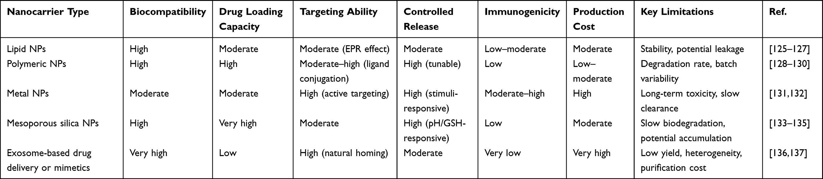 |
Table 3 Comparison of Different Nanocarriers for STING Agonist Delivery |
Lipid Nanoparticles
Lipid nanoparticles (LNPs) are nanoscale delivery systems composed of phospholipids, cholesterol, helper lipids, and polyethylene glycol (PEG)-modified lipids.138 Their unique lipid bilayer structure can efficiently encapsulate various drug molecules, including small molecule drugs, nucleic acids, and proteins, while protecting the drugs from enzymatic degradation or clearance by the immune system.139 LNPs have good biocompatibility and degradability. They can achieve controlled drug release by adjusting lipid composition and achieve specific delivery through surface modification with targeting ligands.127,138 These properties make LNPs ideal carriers in the field of drug delivery, widely used in vaccine development, cancer treatment, and gene therapy. For example, efficiently formulating cGAS agonists with lipid nanoparticles enables the development of therapeutic agents for cancer immunotherapy, allowing for the efficient delivery of cGAS agonists to tissues, cells, and the cytoplasm.80 Furthermore, by encapsulating the negatively charged STING agonist cGAMP in positively charged cationic liposomes, the cellular binding, endosomal uptake, and cytoplasmic delivery of cGAMP can be enhanced. Meanwhile, cationic liposomes have the ability to promote positive anti-tumor immune effects and can also be designed to selectively target the overexpression of mannose receptors in cancer and immune cells, including cationic particle charge and embedded muramyl dipeptide (MDP).76
In response to the demand for tumor immunotherapy, researchers have further developed intelligent lipid nanoparticles based on manganese ions and their nanocomplexes. These nanoparticles can specifically release active components in response to the tumor microenvironment, thereby achieving precise activation of the cGAS-STING pathway and immune enhancement. Jiawei Sun et al created a novel manganese lipid nanoparticle (LNM). LNM can activate the cGAS-STING signaling pathway without the need for combination with chemotherapeutic drugs or other immune activators, inducing the production of pro-inflammatory cytokines, and inhibiting tumor development.78 (As depicted in Figure 2.78) Besides encapsulating Mn2⁺ alone, lipid nanoparticles can also encapsulate FePt alloy and MnO nanocrystals to prepare ALFM. ALFM responds to the weakly acidic tumor microenvironment and degrades, releasing FePt and Mn2⁺ ions. The released Mn2⁺ ions not only increase cellular ROS levels through Fenton-like reactions but also activate the cGAS-STING signaling pathway, stimulating anti-tumor immune responses.74 These studies illustrate the evolutionary design of lipid nanoparticles—progressing from passive drug loading to active activation and environmental responsiveness—thereby significantly enhancing treatment specificity and efficacy.
 |
Figure 2 The schematic illustration of Lipid nanoparticles (A) Preparation process of LNM nanoparticles. (B) Transmission electron microscopy (TEM) image of LNM nanoparticles. (C) Mn2⁺ content in MC38 cells at different time periods. (D) Expression of P-STING protein under the action of different concentrations of LNM. (E) Expression of P-IRF3 protein under the action of different concentrations of LNM. (F) Tumor volume in mice. (G) Tumor weight in mice. (H) Statistical analysis of CD8 T cell positive rate in mouse splenocytes. (I) Statistical analysis of CD4 T cell positive rate in mouse splenocytes. (J) Expression of IFN-γ in mouse serum. (K) Expression of IL-6 in mouse serum.(ns, no significance;*p < 0.05, **p < 0.01,***p < 0.001, ****p < 0.0001).78 |
The core mechanism of LNPs as nanoscale delivery systems lies in their ability to enrich at tumor or inflammation sites through the EPR effect and enter cells through endocytosis.140,141 In the acidic environment of tumors, the lipid bilayer of LNPs undergoes structural changes, releasing drug molecules into the cytoplasm, thus achieving efficient delivery.142 Furthermore, liposomes can be functionalized with folate (FA) to enhance the accumulation and penetration of cGAS agonists in tumors, promote therapeutic effects, modulate the tumor microenvironment, and activate the cGAS-STING innate immune signaling pathway to improve immune function.143
To further enhance the targeting and responsiveness, researchers have also developed lipid nanoparticles based on cell membrane mimetic technology, enabling precise responses to tumor lesions and specific therapeutic signals. Wei et al developed a biomimetic nano-drug delivery system called PB, which utilizes platelet membrane (PM) fused liposomes to load cGAS agonists. After intravenous administration, PB selectively targets cancer cells. Irradiation can further promote the accelerated release of cGAS agonists, activate the cGAS-STING pathway in cancer cells, and ultimately enhance radiotherapy outcomes and anti-tumor immune responses.77 Zheng et al designed novel nanovesicles manufactured through hybridization of tumor cell membrane and phospholipids for the delivery of doxorubicin and reversine.124 These studies collectively represent the evolution of lipid nanoparticle technology towards biomimetic intelligent delivery. By mimicking the characteristics of natural cells, they significantly enhance their active targeting ability and response performance to specific physical signals or biological environments.
Although LNPs show great potential in drug delivery and immunotherapy, they still have some limitations. First, the stability and drug loading efficiency of LNPs need further optimization to avoid drug leakage or degradation during delivery.144 Second, LNPs may cause immunogenic reactions or toxicity, especially with long-term use or high doses, which may limit their clinical application.145 Furthermore, the targeting of LNPs still needs improvement. Although surface modification can improve specificity, precise delivery remains challenging in the complex human environment.146 Finally, the cost and technical difficulties of large-scale production are also important factors restricting their commercialization. Despite these shortcomings, nano-DDS loading cGAS agonists with LNPs still have broad prospects in the future. Through advances in materials science and nanotechnology, smarter and more stable LNP carriers can be developed to achieve precise release and efficient delivery of drugs.82
Polymeric Nanoparticles
Polymeric nanoparticles (PNPs) also have significant potential in delivering cGAS-STING activators in tumor immunotherapy. Polymeric nanoparticles are nanoscale delivery systems formed from natural or synthetic polymers through methods such as self-assembly, emulsion polymerization, or nanoprecipitation, with particle sizes typically between 10–1000 nm.147,148 The core structure of PNPs consists of polymer chains. Common polymer materials include polylactic acid (PLA), poly actic-co-glycolic acid (PLGA), polycaprolactone (PCL), polyethyleneimine (PEI), and polyethylene glycol (PEG).149
The core mechanism of PNPs as nanoscale delivery systems lies in their ability to effectively deliver drugs to specific targets, improve drug stability, and control drug release.150 Their design can overcome biological barriers by adjusting physicochemical properties, such as rapid clearance, complex hemodynamics, suboptimal biodistribution, etc.130 Additionally, the sustained-release properties of PNPs can avoid side effects caused by drug burst release while maintaining long-term drug concentration and improving therapeutic efficacy.151 Mohamed et al used a polymer platform to deliver cGAMP into tumors and activate the STING pathway in the TME, exerting its immunotherapeutic function. As shown in Figure 3 It demonstrates the activation of the cGAS-STING pathway in PNPs and its inhibition of tumors.96
 |
Figure 3 (A) Schematic diagram of polymer nanoparticle synthesis. (B) TEM images of polymeric nanoparticles at pH 7.0 and pH 5.0. (C) pH-responsive cGAMP release kinetics of polymeric nanoparticles. (D) Nanoparticles promoted the ability of cGAMP to induce murine IFN‐β (mIFN‐β) in DC2.4 cells. (E) Nanoparticles outperformed controls to induce mIFN‐β) in DC2.4 cells. (F) The dose‐dependent cGAMP‐selective IFN response.(G) The superior INF induction ability of nanoparticles than controls. (H) Tumor volume in mice. (I and J) Representative flow cytometry plots (I) and quantification (J). (K) Tumor weight in mice.(ns, no significance;*p < 0.05, **p < 0.01,***p < 0.001, ****p < 0.0001).96 |
Metal NPs
Metal nanoparticles (MNPs) are nanoscale particles composed of metal elements such as gold, silver, iron, and platinum, with particle sizes typically between 1–100 nm. They possess unique physicochemical properties, such as surface plasmon resonance effects and superparamagnetism.152,153 These properties give MNPs excellent performance in optics, magnetism, and catalysis, while also providing a foundation for their applications in biomedicine. The structures of MNPs are diverse, including spherical, rod-shaped, cubic, and core-shell structures. Their surfaces can be chemically modified to introduce functional molecules, thus achieving multifunctionality.154 Furthermore, MNPs have high surface area and reactivity, enabling efficient loading of drug molecules or catalyzing chemical reactions, while also having good biocompatibility and degradability, making them ideal choices for nanoscale delivery systems.155 Among many metal nanoparticles, manganese, as a STING agonist, activates the innate immune system in vivo through the cGAS-STING signaling pathway, therefore it is most commonly used in the construction of metal nanoparticles. Zhu et al developed a manganese nanoplatform that integrates a micro-precipitated manganese ion core and a phospholipid bilayer shell. Then, co-loading of glucose oxidase and paclitaxel forms manganese-based nanoparticles. This nanoplatform showed good synergistic effects in the 4T1 tumor-bearing mouse model and significantly reduced the dosage of chemotherapeutic drugs.112 Meng et al developed a novel delivery system based on a manganese-phenolic coordination network for loading the hydrophilic STING agonist ADU-S100. It is prepared by coordination-driven self-assembly of tannic acid and manganese ions, possessing unique pH/glutathione (GSH)-responsive degradation characteristics. It not only overcomes the stability challenges of STING agonists but also ensures efficient intracellular delivery. The core advantage of this system lies in its dual mechanism of action: the released ADU-S100 directly activates the STING pathway, while the synchronously released Mn2⁺ ions act as STING signal sensitizers, synergistically amplifying the immune activation effect.97
Mn can also be used to construct nanoparticles with other metals.156 Li et al constructed MnO2-modified zeolitic imidazolate framework-8 nanoparticles. Due to the high expression of glutathione and low pH in tumors, ZIF-8@MnO2 is decomposed to release Mn2⁺ and Zn2⁺ ions, inducing oxidative stress and cytoplasmic leakage of fragmented mitochondrial double-stranded DNA, further increasing the sensitivity of cGAS to dsDNA as an immunostimulatory signal.113 There are also CM NPs constructed with Mn and Ca, designed by Luo et al inspired by the biological functions of Ca2⁺ and Mn2⁺. They constructed a dual-iron overload nanoplatform (B16F10@CaCO3--CU@MnO2, called CM NPs) coated with B16F10 cell membrane. B16F10 cancer cell membrane-coated nanoparticles have active targeting ability, promoting efficient cellular uptake and tumor enrichment. pH-responsive released Ca2⁺ and Cu2⁺ induce intracellular calcium overload, triggering immunogenic cell death (ICD) and increased ROS, while Mn2⁺ remodels the TME by alleviating hypoxia and further amplifies ROS generation. Additionally, Mn2⁺ promotes immune cell proliferation and maturation, thereby activating systemic anti-tumor immunity. This dual-iron overload strategy exhibits excellent therapeutic effects. When combined with immune checkpoint inhibitors, it can markedly improve immune responses, providing a highly potential combination strategy for cancer treatment.104
Besides Mn2⁺ directly activating the cGAS-STING signaling pathway as a STING agonist, some metal ions can also damage mitochondrial DNA (mtDNA), leading to cytoplasmic DNA leakage, thereby activating the cGAS-STING pathway. Yu et al designed a mitochondria-targeting ruthenium complex, whose molecular structure contains a β-carboline alkaloid-derived main ligand and a triphenylphosphine-modified alkyl chain auxiliary ligand. This special structure allows Ru1 to self-assemble into stable nanoparticles through multivalent intermolecular interactions. Notably, the high charge density of the triphenylphosphine group gives it the ability to specifically target mitochondria. Under photoactivation conditions, Ru1 can induce mitochondrial DNA damage, leading to the release of cytoplasmic double-stranded DNA, thereby effectively activating the cGAS-STING pathway.101 Furthermore, Zn2⁺ can also promote phase separation of the cGAS protein and enhance the enzymatic catalytic activity of cGAS. They can also cause the accumulation of endogenous ROS within cells. Excessive intracellular ROS may also lead to mitochondrial damage and the release of mtDN, which is a dsDNA. Zhang et al, inspired by the STING activation potential of Zn2⁺ and the natural COX-2 inhibitory properties of CPC, developed a novel nanoreactor system by integrating cyclopentyl carbamate into zinc-based metal-organic frameworks. The Zn2⁺ released after nanoparticle decomposition has a dual function: generating ROS to induce tumor cell apoptosis and activating the STING pathway to promote dendritic cell maturation and anti-tumor immunity. CPC can effectively inhibit the COX-2/PGE2 pathway, reverse treatment-induced immunosuppression, and enhance Zn2⁺-mediated immune activation. The synergistic cytotoxic and immunomodulatory effects provide an innovative strategy for combined chemo-immunotherapy.110 (As depicted in Figure 4). Beyond serving as direct STING agonists for antitumor effects, manganese‑based nanoparticles can also enhance immunotherapy by modulating immune cell functions. A recent study repolarized neutrophils into an antitumor phenotype via MnO2 nanoparticle‑activated STING pathway, significantly potentiating Salmonella‑mediated tumor immunotherapy.157 This strategy offers a new perspective for manganese‑based nanomaterials in modulating the tumor immune microenvironment through STING pathway activation.
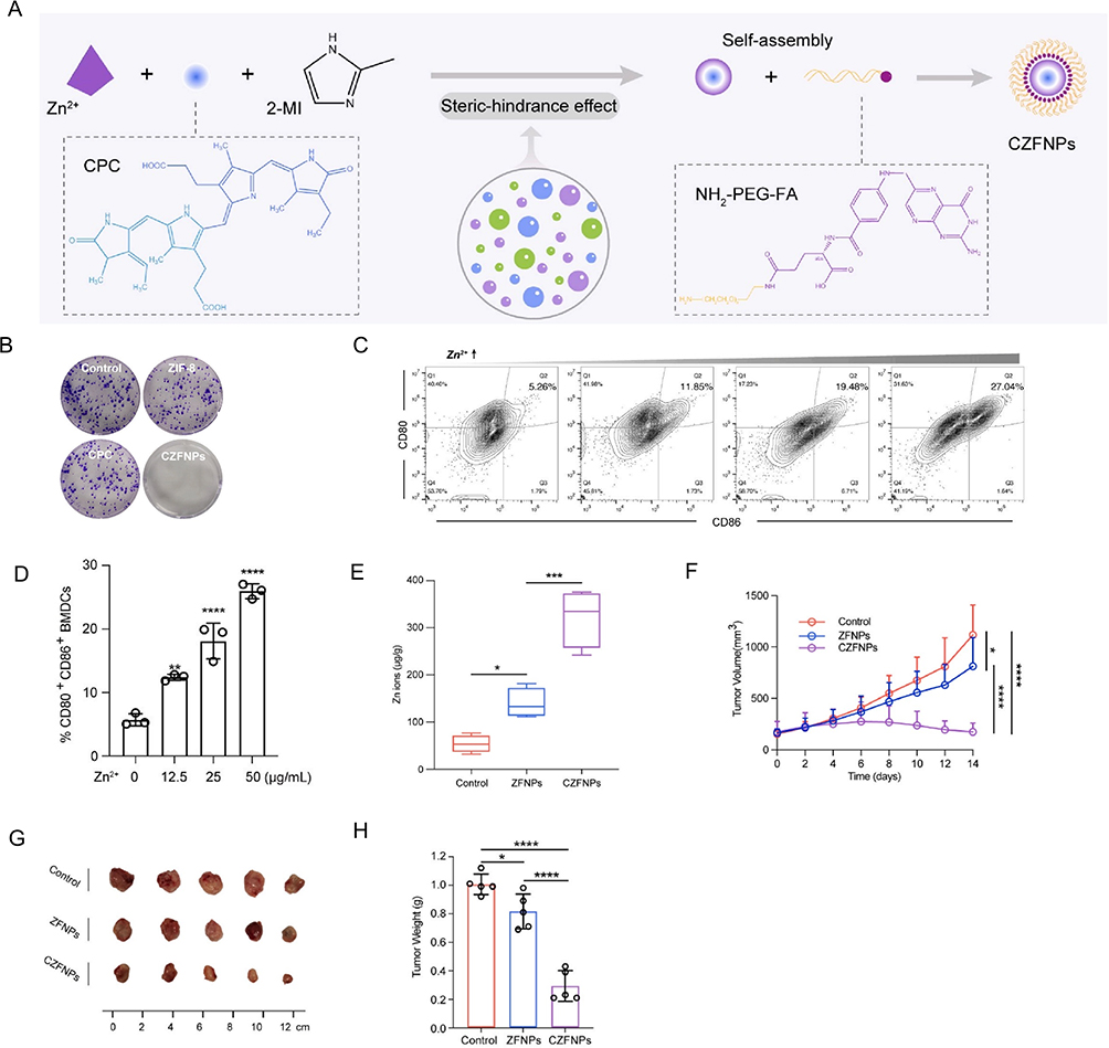 |
Figure 4 The schematic illustration of metal nanoparticles (A) Preparation process of metal nanoparticles. (B) Representative images of MDA-MB-231 cell clone formation under different treatments. (C and D) Representative images (C) and quantification (D) of DC maturation (CD80, CD86 of CD45 BMDCs) induced by supernatant from Zn2⁺-treated cancer cells. (E) Zn2⁺ concentration in mouse tumor tissue. (F) Tumor volume in mice. (G) Photographs of tumors excised from mice. (H) Tumor weight in mice.(*p < 0.05, **p < 0.01,***p < 0.001, ****p < 0.0001).110 |
MNPs as nano-DDS for cGAS agonists, although showing significant advantages in targeting, stimulus responsiveness, and multifunctionality, still have some limitations. First, the biocompatibility and long-term toxicity issues of MNPs have not been fully resolved. Some metal nanoparticles may be toxic to normal cells at high concentrations.158 Second, the metabolism and clearance mechanisms of MNPs in vivo are not yet clear, which may lead to long-term accumulation and potential side effects.159 Finally, the stability of MNPs may be affected by the complex in vivo environment; for example, protein adsorption may cause particle aggregation or functional loss.160 Despite these challenges, the potential of MNPs in cGAS agonist delivery remains enormous. Future research should focus on developing safer metal nanomaterials and improving their biocompatibility and stability through surface modification. Simultaneously, optimize the preparation process of MNPs to achieve low-cost, large-scale production, and In-depth study their metabolism and clearance mechanisms in vivo to ensure the safety of their clinical application.
Mesoporous Silica NPs
Mesoporous silica nanoparticles (MSNs) are inorganic nanomaterials with a regular mesoporous structure, primarily composed of SiO2. They possess highly ordered mesoporous channels, a large specific surface area, tunable pore size and pore volume, and excellent surface modification capability.134 The specific surface area of MSNs usually exceeds 1000 m2/g, enabling them to efficiently load various hydrophilic or hydrophobic drugs, including small molecule chemotherapeutic drugs, genetic drugs, proteins, and immunomodulators.161 Furthermore, their surfaces are rich in silanol groups, which can be modified with targeting ligands, polymers, or responsive groups to achieve targeted transport and controlled release, improving the therapeutic effect of drugs. Compared to organic nano-DDS, the inorganic nature of MSNs has higher stability. They can slowly degrade into non-toxic Si(OH)4 in physiological environments, metabolized and excreted by the body, exhibiting good biocompatibility and greatly reducing the potential toxicity issues that may arise from long-term accumulation.162
As part of nano-drug delivery systems, MSNs show great potential in cancer treatment and immunotherapy due to their exceptional drug loading capacity, targeted delivery, and intelligent responsive release mechanisms. Their nanoscale size and modifiable surface functionality enable them to specifically enter tumor cells or immune cells through receptor-mediated endocytosis, thereby improving drug delivery efficiency.163 Biodegradable PMMR@MH, after being internalized by tumor cells, decomposes in response to the acidic-reductive intracellular environment, rapidly releasing SR-717. Meanwhile, under glutathione (GSH)-rich conditions, the internal PMM slowly decomposes to perform sustained release of cisplatin and Mn2⁺. Under the sequential pre-activation by SR-717 and subsequently released Mn2⁺, cytosolic dsDNA fragments are easily recognized by cGAS in tumor cells and DCs, thus achieving cascade cGAS-STING activation to activate immune cells and amplify anti-tumor immune responses.116 (As depicted in Figure 5). Furthermore, MSNs often utilize their regular mesoporous structure, high specific surface area, and controllable size and morphology to guide the growth or coating of other materials, ultimately serving as templates for nanoparticle production, especially after removing the MSNs to form the target nanostructure. They are widely used in the preparation of nanomaterials with hollow structures or specific morphologies. For example, monodisperse H-MnO2 with uniform morphology was synthesized by in situ growth of MnO2 on solid silica nanoparticles, followed by removal of the silica core. Its interior shows abundant pores, remaining stable during surface modification and N-methyl-N-nitroaniline (NMA) loading processes.115
 |
Figure 5 The schematic illustration of Mesoporous silica nanoparticles (MSNs) (A) Schematic diagram of mesoporous silica nanoparticle preparation. (B) Representative TEM images of mesoporous silica nanoparticles incubated for 12 and 72 h under different conditions. (C) Flow cytometry of 4T1 cell apoptosis after different treatments. (D) Flow cytometry analysis of mature DC cells after incubation with 4T1 cells subjected to different treatments. (E) Whole-animal in vivo bioluminescence images of 4T1 luciferase-expressing mice after different treatments. (F) Mesoporous silica nanoparticles increased the expression of IFN-β, IL-6, IFN-γ, and TNF-α in vivo. (G) Tumor volume change curves in mice under different treatments. (**p < 0.01,***p < 0.001, ****p < 0.0001).116 |
MSNs still have some limitations in loading cGAS agonists. First, the biodegradation rate of MSNs is relatively slow, which may lead to prolonged retention in vivo, potentially causing chronic inflammatory reactions or long-term accumulation risks.135 Second, the strong negative charge on the MSN surface may affect interactions with cell membranes, reducing drug delivery efficiency. Therefore, further modification is usually needed to enhance targeting ability.164 Furthermore, the loading and release mechanisms of cGAS agonists in MSNs still need optimization. Although their mesoporous structure can efficiently load cGAMP, how to precisely control its release in the tumor microenvironment or within immune cells remains a challenge. For example, some designs may lead to premature drug leakage or insufficient release before lysosomal degradation, affecting immune activation.165 Simultaneously, the immunogenicity and in vivo metabolic pathways of MSNs need to further research to ensure their long-term safety and reduce potential side effects.135 Therefore, chemically modifying the surface of MSNs to improve their targeting and drug release controllability will be an important direction for future research.164 There is also a need to develop multifunctional MSN systems that can respond to multiple stimuli to achieve more precise drug release and therapeutic effects.166
Exosomes and Their Biomimetic Nanovesicles
Exosomes are composed of a lipid bilayer membrane enriched with specific membrane proteins. These proteins not only participate in the formation and secretion of exosomes but also mediate their interaction with target cells.167 The internal cavity of exosomes can carry various bioactive molecules, including proteins, lipids, RNA, and DNA, which play crucial roles in intercellular communication and signal transmission.168 Exosomes can protect their internal biomolecules from degradation and remain stable in the circulatory system. They possess excellent biocompatibility and low immunogenicity, allowing safe use in vivo, while also having natural targeting ability. They can recognize and bind to target cells through surface-expressed membrane proteins and receptors, thereby achieving precise delivery.136
Exosome-based drug delivery or mimetics can enter cells through endocytosis or membrane fusion, releasing their internal biomolecules, thus achieving efficient delivery.169,170 The natural targeting of exosomes enables them to recognize and bind to specific cell types, such as tumor cells or immune cells, enabling precise therapy.171 Study found that fusing M1 macrophage-derived extracellular vehicles with REV and liposomes loaded with SR780Fe (REV@SR780Fe@Lip) formed REV@SR780Fe@LEV hybrid nanovesicles. Further modification with RS17 peptide for tumor targeting combined photodynamic therapy, ferroptosis, and cGAS-STING pathway activation, thereby enhancing anti-tumor efficacy through synergistic effects. After laser irradiation, REV@SR780Fe@LEV-RS17 showed anti-tumor effects in the 4T1 breast cancer model, including inhibition of lung and liver metastasis and prevention of tumor recurrence.120 (As depicted in Figure 6).
 |
Figure 6 The schematic illustration of Exosome nanoparticles (A) Preparation of exosome nanoparticles. (B) TEM images of exosome nanoparticles at pH 7.4 and pH 5.5. (C) Expression of cGAS-STING pathway-related proteins in 4T1 cells after different treatments. (D–F) Release of CRT (D), HMGB1 (E), and ATP (F) from 4T1 cell supernatant after different treatments. (G) 4T1 Tumor growth curves of 4T1 tumor-bearing mice with various treatments. (H–J) Representative FCM (H) and quantification analysis (I and J) of CTLs in tumor. (ns, no significance;***p < 0.001, ****p < 0.0001).120 |
Due to their biocompatibility, exosomes are easily taken up by cancer cells and can better target siRNA-mediated oncogenic Kras. When exosomes are administered to tumor-bearing mice, the CD47 molecule on their surface reduces their clearance through phagocytosis in the circulation, thereby enhancing the anti-tumor response.121 Simultaneously, extracellular vehicles have been identified as natural mediators of signal transduction between cancer cells and tumor-resident APCs. Su et al designed a nanoplatform named exoSTING, an innovative engineered extracellular vesicle therapeutic candidate encapsulating a CDN STING agonist. Compared to free CDN, this EV-based delivery system demonstrated significantly enhanced therapeutic potential: efficacy increased by more than 100-fold in preclinical tumor models, while significantly improving tumor retention and reducing systemic cytokine release.122,172 The amount of naturally extracted exosomes is too low, and purification is time-consuming and costly. Therefore, researchers developed an exosome mimetic (EM) nanocarrier to address these shortcomings. Yawen Guo et al used exosome mimetic nanoparticles to load REV and DOX, named EM@REV@DOX. These drug-loaded nanoparticles can be easily produced in large quantities. EM@REV@DOX showed effective accumulation at the tumor site and was retained there due to EPR and homing targeting effects.123
Exosome nanoparticles although showing significant advantages in biocompatibility, natural targeting, and ability to cross biological barriers, have low yield, making large-scale preparation difficult. The isolation and purification processes are complex and costly, which limits the scalability of their clinical application.137 Second, the drug loading efficiency of exosomes is relatively low, especially for large molecules or hydrophobic drugs, requiring further optimization of encapsulation technology to improve drug loading capacity and stability.173 Furthermore, exosomes are highly heterogeneous; exosomes from different cell sources may differ in composition and function, which may lead to inconsistencies in delivery.174 Finally, the metabolism and clearance mechanisms of exosomes in vivo are not yet fully understood, which may affect their safety and effectiveness for long-term use.
The Emerging Role of cGAS-STING Activation by Nanoagonists in Cancer Immunotherapy
The development of OSCC typically progresses from normal epithelium to precancerous lesions eventually evolving into invasive cancer. During this process, genetic mutations may alter the immune microenvironment, inhibiting the function of tumor-infiltrating lymphocytes (TILs), preventing them from clearing precancerous and cancer cells.175 Specifically, mutations in classic head and neck squamous cell carcinoma driver genes, such as TP53, CDKN2A, and NOTCH1, are associated with the formation of an immunosuppressive tumor microenvironment.176–179 These mutations may induce the infiltration of regulatory T cells and myeloid-derived suppressor cells, as well as the phenotypic switch of tumor-associated macrophages from M1 to M2 type, thereby inhibiting the function of cytotoxic T lymphocytes.180 TP53 is the most frequently mutated gene in solid cancers and has a profound impact on multiple hallmarks of cancer. TP53 mutations are common in young adult OSCC, with 45% of tumors showing truncating mutations.181 Mutant p53 not only interferes with the infiltration of myeloid cells and T cells into the tumor microenvironment but also promotes an immunosuppressive state by interacting with TBK1 to drive tumor progression.182 For example, mutant p53R172H can construct a complex cytokine transcriptome network that inhibits the infiltration of cytotoxic CD8+ T cells while promoting the intratumoral recruitment of regulatory T cells and M2 macrophages.183 Furthermore, tumors carrying the p53R172H mutation are infiltrated by numerous CD8+ and CD4+ T cells that express programmed cell death protein 1. These cells showed some responsiveness to immune checkpoint inhibitors and stimulator of interferon genes 1 agonist therapy.184 These findings reveal the key role of p53 mutation in shaping the tumor immune microenvironment and provide new ideas for the development of related treatment strategies. In addition to genetic mutation‑driven immunosuppression, emerging biomarkers in OSCC can inform prognosis and predict immunotherapy response. A recent study identified cuproptosis‑related lncRNA signatures that correlate with patient prognosis and immunotherapy outcomes in OSCC.185 Given the mechanistic intersection between cuproptosis and the cGAS‑STING pathway, this finding suggests that STING nanoagonist design should consider multiple tumor metabolic and immune pathways to achieve more precise therapeutic effects.
The cGAS-STING pathway is often in a low-activity state in tumors.186 Studies have shown that oral cancer cells inhibit the activation of the cGAS-STING pathway through various mechanisms, including reducing cGAS expression, increasing STING protein degradation, or inhibiting downstream signal transduction.186–188 This dysregulation leads to reduced production of type I interferons in the tumor microenvironment, further weakening anti-tumor immune responses. For example, ARID 1A-regulated DNA repair pathways and interferon-STAT 1-induced TREX 1 expression reduce the level of DNA in tumor cells and limit the type I interferon produced by the cGAS-STING pathway. N-MYC reduces STING oligomerization, USP 35 deubiquitinates STING to limit STING signaling in cancer.189–191 Simultaneously, inhibiting IL-6 or its downstream JAK2/STAT3 signaling pathway can restore the responsiveness of DU145 cells to STING agonists.192 In mouse TRAMP-C2 prostate cancer cells, STING activity is crucial for tumor rejection and immune cell infiltration. Although endogenous STING agonists present in TRAMP-C2 cells contribute to tumor rejection, administration of cGAMP can further inhibit tumor growth.82 In summary, STING contributes to tumor rejection in tumor cells, but its function is often partially suppressed by the JAK2 and STAT3 pathways. Therefore, restoring the activity of the cGAS-STING pathway may be a key strategy to overcome the immunosuppressive microenvironment of oral cancer. Similarly, sustained activation of the cGAS-STING pathway in cancer leads to chronic inflammation, releasing pro-inflammatory factors such as IL-6 and TNF-α, creating an immunosuppressive microenvironment that weakens anti-tumor immune responses;82 furthermore, cytoplasmic DNA accumulation caused by chromosomal instability can chronically activate the cGAS-STING signaling pathway, which in this case promotes tumor growth and metastasis.193 Therefore, interventions targeting this pathway in cancer treatment require careful consideration of its dual pro-inflammatory and anti-tumor effects. Recent studies have highlighted that cancer cells exhibit an abnormal accumulation of dsDNA in the cytoplasm, resulting from genomic instability and mitochondrial dysfunction, which in turn activates the cGAS/STING signaling pathway to trigger anti-tumor immune responses. But to evade immune surveillance, cancer cells often inhibit cGAS/STING signaling through epigenetic silencing, gene mutation or deletion, interference by viral oncoproteins and oncogene proteins, and other methods. Furthermore, cancer cells evade immune attack through mechanisms such as loss of antigen presentation, induction of immune checkpoint molecules, recruitment of immunosuppressive cells during the immune editing process. Mitochondrial DNA leakage also prompts cancer cells to selectively inhibit STING expression to avoid STAT1-mediated cytotoxicity.7,27,194–196 These mechanisms collectively lead to the suppression of cGAS/STING signals, allowing cancer cells to evade immune surveillance, promote tumor progression, and cause resistance to immunotherapy. Understanding these mechanisms is crucial for developing effective immunotherapy strategies.
Combination Therapy Based on STING Nano-Agonists
Combination therapy based on STING nano-agonists is an important research direction in the field of cancer immunotherapy in recent years. Although monotherapy has achieved certain results in tumor treatment, its efficacy still has limitations and hard to meet clinical needs.8 In contrast, multimodal combination therapy shows significant advantages in cancer treatment. Nanotechnology has enhanced the stability, targeting, and controlled release capabilities of STING agonists, addressing issues such as rapid degradation, low delivery efficiency, and systemic toxicity associated with traditional agonists. STING nano-agonists can be combined with various treatment methods. They not only significantly improve treatment effects through synergistic effects but also reduce toxic side effects by lowering drug doses and effectively overcome tumor drug resistance issues.8,30 This article focuses on typical combination therapy strategies based on cGAS-STING pathway activation, including the combined application of STING nano-agonists with immunotherapy, chemotherapy, and radiotherapy. These synergistic treatment plans provide new ideas and potential solutions for cancer immunotherapy based on cGAS-STING pathway activation.Figure 7 illustrates various combined treatment options.
 |
Figure 7 Enhancing anti-tumor immune response: Mechanism diagram of the combined effect of STING agonists with various treatment regimens. |
STING Agonist Combined with Radiotherapy
Radiotherapy is a traditional cancer treatment method that uses high-energy radiation to destroy tumor cell DNA, inhibiting tumor growth by directly killing tumor cells or inducing their apoptosis.197,198 However, the effect of radiotherapy is not limited to local killing; it can also activate the immune system by releasing tumor antigens and damage-associated molecular patterns, triggering systemic anti-tumor immune responses, a phenomenon known as the “abscopal effect”199 Nevertheless, when used alone, radiotherapy’s ability to activate the immune system is limited and may be compromised by immunosuppression in the tumor microenvironment.197 Combining STING agonists with radiotherapy can significantly enhance this immune activation effect. Radiotherapy-induced DNA damage can lead to the accumulation of cyclic dinucleotides in the cytoplasm, thereby activating the STING pathway, further promoting the production of type I interferons and pro-inflammatory factors, enhancing antigen presentation and T cell infiltration.14 Furthermore, the addition of STING agonists can overcome the immunosuppressive state of the tumor microenvironment, enhance the abscopal effect of radiotherapy, and simultaneously produce synergistic anti-tumor effects on primary and metastatic lesions.200 This combination strategy not only improves the local therapeutic effect of radiotherapy but also inhibits distant metastases through systemic immune activation, providing a more comprehensive solution for cancer treatment.201 For example, the high electron density of functionalized hafnium oxide nanoparticles (NBTXR3) allows a high probability of interaction with incoming ionizing radiation and increases energy dose deposition within cancer cells. Due to this physical mode of action, treatment with RT-activated NBTXR3 compared to RT alone increases cancer cell death and better local tumor growth control.202 Simultaneously, Hf 4⁺ can successfully activate the cGAS stimulator of STING pathway, promote DC maturation and the production of IFN and other cytokines, thereby further strengthening the RT-induced immune response.90 Furthermore, different doses of RT can induce varying degrees of radioimmunotherapy. High-dose radiation (>10 Gy) may lead to immunosuppression, while medium dose (4–10 Gy), although able to alleviate immunosuppression caused by high-dose radiation, is usually insufficient to effectively kill tumor cells. Recently, Jian et al proposed a novel multifunctional nanoplatform RMLF, integrating a Ru (II) complex into folate-functionalized liposomes with BSA-MnO2 nanoparticles. Orthogonal experiment optimization enhanced radiosensitization by increasing accumulation in cancer cells, increasing ROS, and contributing to the dual enhancement of the cGAS-STING-dependent type I IFN signaling pathway, aiming to overcome the common DAMP deficiency in conventional RT at 4 Gy.143
New nanomaterials are developed for radiosensitization through three main methods. First, atoms with high atomic numbers tend to enhance the energy deposition of ionizing radiation, so these elements can be applied for radiosensitization. Second, modulating the tumor microenvironment is also a strategy to improve treatment efficiency. Finally, nanomedicines that normalize blood vessels can accelerate blood flow in the tumor area, thereby alleviating hypoxia and increasing the sensitivity of tumor cells to ionizing irradiation. Liu et al constructed a new nanozyme using MnCO and built it onto dendritic mesoporous silica nanoparticles, with TPP linked to the surface (MDP). MDP can not only stimulate POD-like activity but also CAT-like activity under tumor-mimetic conditions, meaning MDP produces toxic ·OH from H2O2 and induces mitochondrial death due to TPP surface modification. Meanwhile, MDP can convert H2O2 to O2, alleviating hypoxia to overcome radioresistance. Ru, as a high Z atom, enhanced the local energy of X-rays. More importantly, results showed that when MDP was applied for radiosensitization, cGAS-STING was upregulated.118 Similarly, Mengling Shen et al collected OMVs and performed biomineralization with MnO2 to obtain OM@MnO2-PEG nanoparticles, enhancing tumor radioimmunotherapy by increasing RT-induced ICD and cGAS-STING activation. In the acidic tumor microenvironment, OM@MnO2-PEG nanoparticles can react with H2O2, then producing a large amount of Mn2⁺, O2, and OM fragments. The alleviated tumor hypoxia improved tumor radiosensitivity, amplifying RT-induced ICD and cGAS-STING activation. Meanwhile, oxygen/OMs promote macrophage polarization to the anti-tumor M1 type, and OM fragments improve the efficiency of tumor antigen presentation by recognizing lipopolysaccharide (LPS) and toll-like receptors (TLR). Notably, the long-term high content of manganese ions in the tumor maintained the activated state of the cGAS-STING pathway. Figure 8 shows the therapeutic effect of the combined treatment of cGAS-STING and radiotherapy.77,90,107,118
 |
Figure 8 Potential of STING nanoparticles in combination therapy with radiotherapy. (A and B) TEM images of STING nanoparticles. (C) Image of cancer cell survival obtained by incubating with different concentrations of STING nanoparticles for 24 hours, then irradiating with different doses of X-rays. (D) Representative images of colony cluster staining after receiving various treatments. (E) Survival rate of cancer cells in different treatment groups. (F)Viability of cancer cells after treatment with different treatment groups. (G) Tumor weight in mice. (H) Image of tumors collected from mice after treatment with various drugs. (**p < 0.01,***p < 0.001, ****p < 0.0001).77,90,107,118 |
STING Agonist Combined with Chemotherapy
Chemotherapy is a traditional cancer treatment method that uses chemical drugs to kill or inhibit the growth of cancer cells. These drugs work by interfering with DNA replication, cell division, or other key metabolic processes of cancer cells. However, the primary limitation of chemotherapy is its lack of specificity. It not only eliminates tumor cells but also damages healthy tissues, leading to severe side effects such as immunosuppression, nausea, and hair loss. Moreover, tumor cells can develop resistance to chemotherapeutic agents, further compromising treatment efficacy.203
In recent years, combination chemotherapy strategies based on STING nano-agonists have offered new approaches to overcoming these limitations. STING agonists activate the cGAS-STING pathway to enhance anti-tumor immune responses. The application of nanotechnology further improves the stability and targeting of STING agonists, enabling their precise delivery to the tumor microenvironment.84 When combined with chemotherapy, chemotherapeutic drugs induce immunogenic cell death of tumor cells, releasing tumor antigens, while STING agonists activate antigen-presenting cells and T cells, enhancing systemic immune responses. This synergistic effect not only improves the therapeutic effect of chemotherapy but also reduces side effects by lowering the dose of chemotherapeutic drugs and overcomes tumor drug resistance.204,205 Lewicky et al developed a multi-target mannosylated cationic liposome immunomodulatory system containing low-dose chemotherapeutic cytarabine (Ara-C). They demonstrated that embedding non-cytotoxic doses of Ara-C in DS enhanced its ability to induce DNA double-strand breaks in human ovarian and colorectal cancer cell lines and various immune cells. They also proved that Ara-C/DS-induced DNA damage translates into cGAS-STING axis activation, highlighting the broad immunotherapeutic potential of Ara-C/DS through enhanced tumor-directed inflammatory responses.76
Another recent study on platinum drugs is that Xiang et al designed composite nanoparticles (NP-Pt-IDOi) from a ROS-sensitive amphiphilic polymer with a thiol-ketone bond in the backbone to encapsulate a Pt (IV) prodrug (Pt (IV)-C12) and an indoleamine-(2,3)-dioxygenase (IDO) inhibitor (IDOi, NLG919). Once NP-Pt-IDOi enters cancer cells, the polymeric nanoparticles dissociate due to intracellular ROS and release Pt (IV)-C12 and NLG919. Pt (IV)-C12 induces DNA damage and activates the cGAS-STING pathway, increasing the infiltration of CD8+ T cells in the tumor microenvironment. Furthermore, NLG919 inhibits tryptophan metabolism and enhances CD8+ T cell activity, ultimately activating anti-tumor immunity and enhancing the anti-tumor effect of platinum drugs.85
STING Agonist Combined with Phototherapy
Phototherapy is a non-invasive method that uses light energy for disease treatment, widely used in cancer treatment. Its principle is that photosensitizers absorb light of specific wavelengths, producing photodynamic effects (PDT) or photothermal effects (PTT), thereby inducing tumor cell death.206 The photodynamic effect directly kills tumor cells by generating ROS, while the photothermal effect destroys tumor tissue through local heating.207 The advantage of phototherapy lies in its precision and controllability, enabling selective action on the tumor site and reducing damage to normal tissues.208 The limitation of phototherapy is its limited immune activation ability and may be affected by immunosuppression in the tumor microenvironment.209 Combining STING agonists with phototherapy can significantly enhance the treatment effect. Phototherapy-induced tumor cell death releases tumor antigens and damage-associated molecular patterns (DAMPs), activating the immune system, while STING agonists, via STING pathway activation, further potentiate the immune effects of phototherapy.27,210
To synergistically utilize the photothermal effect and immune activation, researchers have designed a manganese-based composite nanomaterial. Through a single treatment, it can simultaneously achieve local thermal killing, alleviate tumor immune suppression, and systematically activate anti-tumor immunity. Zheng et al constructed Mn-rich photonic nanomedicine (MnPB-MnO2). By utilizing manganese-rich photonic nanomedicine with ideal biocompatibility, they effectively activated the cGAS-STING pathway with sufficient Mn2⁺, combined with abundant TAAs derived from damaged cancer cells due to the dual blow of hyperthermia and ROS, synergistically enhancing innate and adaptive anti-tumor immunity, achieving enhanced local photothermal/immunotherapy effects and triggering systemic immune therapy effects.108 Recently, the AMOPs (gold nanoparticles-Au NPs/manganese dioxide-MnO2 composite) developed by Xian An’s team revealed the synergistic anti-tumor mechanism of STING agonists and PTT. In the TME, AMOPs catalyze the decomposition of H2O2 into O2, alleviating hypoxia to enhance cytotoxic T lymphocyte (CTL) function. Meanwhile, the high concentration of GSH in tumor cells triggers the PTT effect of AMOPs, inducing ICD and promoting CTL infiltration. Furthermore, Mn2⁺ released from the decomposition of MnO2 can activate the STING pathway, further enhancing the immune response. This combined effect of STING agonist and PTT not only inhibits primary tumor growth but also establishes immune memory, preventing recurrence and metastasis.103 These studies demonstrate that integrating STING agonists into photothermal nanodevices can transform local physical ablation into a combined therapy that not only directly eliminates the lesion but also remodels the tumor immune microenvironment and stimulates a systemic immune response, thereby expanding the application prospects of phototherapy in immunotherapy.
Secondly, in terms of PDT, a new generation of nanomaterials with organelle-level precision has important prospects in maximizing the efficacy of targeted tumor therapy. Hao Tian ‘s team reported a novel amphiphilic phenolic polymer (PF) for mitochondria-targeted PDT, which can trigger excessive mtDNA damage through the synergistic effect of oxidative stress and furan-mediated DNA cross-linking. The phenolic units on PF can further self-assemble with Mn2⁺ through metal-phenolic coordination to form metal-phenolic nanomaterials (PFM). It emphasized that PFM promotes cGAS-STING-dependent immunity at the organelle level, thus having effective anti-tumor efficacy.97
Furthermore, the MOF-based nanoagonist (DZ@A7) researched and developed by Xun Guo et al achieves tumor-specific activation of the STING pathway through NIR light-triggered decomposition. Under NIR irradiation, DZ@A7 generates mitochondria-targeting ROS, enhancing the tumor-killing effect of PDT, while consuming GSH to amplify oxidative damage. Additionally, NIR-triggered STING activation synergizes with PDT, significantly enhancing anti-tumor immunity, providing a new strategy for the combination of STING agonists and PDT.105
Recently, photothermal ablation (PTA) has garnered widespread attention due to its ability to induce local thermal damage through light absorption by suitable agents. The Xia team reported an intelligent glutathione (GSH)-responsive photothermal nanosystem composed of Prussian blue (PB) nanoparticles supported by Mn (III) to generate a synchronous strategy to enhance the immune response treatment of incomplete photothermal ablation. This nanosystem ablates tumors through the photothermal effect of PB under 808 nm near-infrared laser irradiation. Then the exposed Mn (III) consumes GSH present in the tumor tissue. The interaction between Mn (III) and GSH leads to the release of Mn (II). Since the formation of cytoplasmic DNA was observed during photothermal ablation therapy, the synchronous release of Mn (II) is expected to further increase the sensitivity of cGAS to double-stranded DNA (dsDNA), which should promote the activation of the cGAS-STING pathway and remodel the tumor immune microenvironment to improve anti-tumor efficiency.102 Figure 9 shows the therapeutic effect of the combination of cGAS-STING and various phototherapies.102,108,109
 |
Figure 9 Potential of STING nanoparticles in combination therapy with phototherapy. (A) Schematic diagram of STING nanoparticle synthesis and its TEM image. (B) Flow cytometry analysis of apoptosis levels in cancer cells after different treatments. (C) Infrared thermal images of tumor models after different treatments. (D) Photographic images of mouse tumors. (E) Relative tumor growth curves of various treatment groups. (F) Temperature profiles of PTA tumor models.102,108,109 |
Combination Therapy of STING Agonists with Other Immunotherapies
Combination therapy based on STING nano-agonists with other immunotherapies is an important research direction in the field of cancer treatment in recent years. Other immunotherapies include immune checkpoint inhibitors (ICI), ICD inducers, and PD-L1 inhibitors. Immune checkpoint inhibitors restore the anti-tumor activity of T cells by blocking inhibitory signals between tumor cells and T cells, thereby enhancing the attack capability of the immune system.210 The efficacy of ICI depends on pre-existing T cell infiltration in the tumor microenvironment and has a limited effect on “cold tumors”211 ICD inducers activate dendritic cells and promote T cell-mediated immune responses by inducing tumor cells to release DAMPs and tumor antigens.212 PD-L1 inhibitors terminated the inhibition of T cells by blocking the binding of PD-L1 on the surface of tumor cells to PD-1 on T cells, enhancing their ability to kill tumors.213
STING nano-agonists significantly enhance the effects of these immunotherapies by activating the cGAS-STING pathway and inducing the production of type I interferons and pro-inflammatory factors. For example, ICI circumvent inhibitory checkpoint molecules on tumor cells, thereby enhancing T cell reactivity and preventing immune exhaustion, thus restoring immune surveillance. When STING agonists are combined with ICI, they can increase T cell infiltration in the tumor microenvironment, converting “cold tumors” into “hot tumors”, thereby improving the efficacy of ICI. To fully utilize the potential of ICI therapy, it is crucial to transform “cold” tumors into a more immunogenic and pro-inflammatory phenotype.36 This transformation can restore anti-tumor immunity, thereby improving the effectiveness of treatment. A particularly feasible method to achieve this goal is to enhance the activity of the cGAS-STING pathway. This pathway is a crucial component of the innate immune system, playing a vital role in anti-viral defense and anti-tumor responses. Recent studies have shown that the STING agonist SR-717 delivered based on an albumin nanocarrier (SH-NPs) can efficiently activate the STING pathway and reverse the immunosuppressive state of the TME. SH-NPs not only enhance T cell anti-tumor immunity but also significantly improve the efficacy of ICI, confirming the synergistic effect of STING agonists and ICI.214
Furthermore, as a special type of cell death, the immune response induced by ICD leads to dendritic cell maturation and activation of specific T cells, highlighting its potential as a target for anticancer therapy. The combination of STING agonists and ICD inducers can further enhance the release and presentation of tumor antigens, activating stronger anti-tumor immune responses.215 Recent studies have shown that nanoparticles (EM@REV@DOX) loaded with the STING agonist and doxorubicin can activate anti-tumor immunity through a dual mechanism. On the one hand, DOX induces ICD, releasing tumor antigens; on the other hand, REV activates the cGAS-STING pathway, promoting DC maturation and CTL infiltration. This synergistic effect of STING agonist and ICD significantly enhanced the immune response. Combined with PD-L1 blockade, it could further inhibit tumor growth.123 The research of Tianchuan Zhu et al found that the combination of the STING agonist cGAMP and PD-L1 inhibitor produced a synergistic anti-tumor effect. After co-delivery of cGAMP and anti-PD-L1 antibody (aPD-L1 NVs@cGAMP) via nanovesicles, STING pathway activation promoted IFN-β secretion and T cell infiltration, while PD-L1 blockade prevented T cell exhaustion. This combination strategy significantly enhanced the therapeutic effect of CAR-T cells on solid tumors by reshaping the tumor immune microenvironment.119 These combination strategies not only significantly improve treatment effects through synergy but also reduce side effects by lowering drug doses and overcoming tumor drug resistance, providing new ideas and potential solutions for cancer immunotherapy. Figure 10 shows the therapeutic potential of the combination of cGAS-STING and various immunotherapies.123,216
 |
Figure 10 Potential of STING nanoparticles in combination therapy with other immunotherapies. (A) SEM image of STING nanoparticles. (B and C) Release of HMGB1 and ATP from LLC cells after different treatments, indicating the effect of STING nanoparticles in achieving ICD. (D) Cytokine levels of TNF-α (i), and IFN-β (ii) in DCs after treatments with different formulations. (E) Representative corresponding quantification of CRT release in cells after different treatments. (F–K) Tumor pictures, average tumor growth curves, and survival curves of CT26 tumor-bearing mice after treatment with STING nanoparticles. (** p < 0.01,**** p < 0.0001).123,216 |
STING Agonists are Used in Combination with Various Treatment Methods
In some complex clinical scenarios, relying solely on one or two treatment strategies based on the cGAS-STING pathway may be difficult to fully address tumor heterogeneity and individualized patient needs. To this end, researchers have begun to explore integrating multiple treatment methods into combination platforms based on STING nano-agonists to achieve more comprehensive anti-tumor effects. In addition to the combinations discussed in Combination Therapy of STING Agonists with Other Immunotherapies, STING nano-agonists can be further integrated with multiple modalities simultaneously, such as chemotherapy, radiotherapy, and phototherapy.
Recently, it was found that using polyethylene glycol grafted poly copolymer (mPEG-PLGA, PP) as a biodegradable and biosafe micelle carrier, loading vadimezan (DMXAA), obtained PLGA-PEG/DMXAA (PPD) for multiple synergistic therapies of cancer and its metastasis/recurrence. After intravenous injection, PPD can effectively accumulate at the tumor site and trigger tumor-specific thrombosis. Furthermore, blood clots in the tumor can convert near-infrared light into heat and temporarily provide oxygen to tumor tissue to enhance PTT/RT. Besides PTT/RT, DMXAA-induced tumor vascular damage leads to starvation therapy for cancer.95 These multimodal combination strategies significantly improve treatment efficacy through synergistic effects while reducing drug doses and side effects, providing more flexible and efficient solutions for cancer treatment.
The strategy of combining STING nano-agonists with multiple treatment methods, although showing great potential in cancer treatment, still faces some challenges and limitations. First, the complexity of multifunctional combination nano-systems may hinder their large-scale production and clinical application. Complex preparation processes and high costs limit their feasibility in industrial production. Second, the synergistic mechanism of multiple treatment methods is not yet fully clear, making it difficult to accurately evaluate the specific contribution of each method in combination therapy, which brings difficulties to optimizing treatment plans. Furthermore, as the number of therapeutic drugs and combination methods increases, the risk of potential toxic side effects also rises, which may adversely affect patient tolerance. Therefore, when designing multi-mode treatment systems based on STING nano-agonists, it is necessary to comprehensively consider the drug dose, delivery efficiency, and biological safety, especially for complex systems involving multiple therapies.
Challenges and Prospects
Although nano-drug delivery systems have made significant progress, ensuring precise drug delivery to the tumor site without off-target effects remains a complex issue, such as the targeted delivery of STING agonists and maintaining their stability in the tumor microenvironment.9 The degradation of STING agonists in the blood and their limited penetration into tumor tissue can significantly reduce treatment efficacy. Furthermore, the immunosuppressive characteristics of the tumor microenvironment pose a major obstacle, as tumor cells often evade immune detection through mechanisms such as downregulating STING expression or upregulating immune checkpoint molecules. Overcoming these immune escape strategies is crucial for enhancing the therapeutic potential of STING agonists. Although nano-drug delivery systems improve the delivery and stability of STING agonists, potential toxicity and side effects remain concerns. The long-term impact of nanoparticles on normal tissues and the immune system requires in-depth study. The systemic release of cytokines caused by STING activation may lead to adverse reactions, such as cytokine release syndrome.217–219 The complexity of combining STING agonists with other therapies adds challenges to the treatment regimen. The synergistic effects of these combinations need careful balancing to avoid overactivation of the immune system or excessive toxicity. Optimizing dosing regimens and understanding the interactions between different treatment modes are crucial.220 Finally, the production of nano-drug delivery systems for STING agonists involves complex technologies and materials, which are costly and difficult to scale up. Developing cost-effective and scalable manufacturing processes is necessary to make these therapies accessible to a wider patient population.129
Several potential directions can address these challenges and enhance the efficacy of STING-based therapies. Future research should focus on developing next-generation nano-drug delivery systems with enhanced targeting capabilities and stimulus-responsive characteristics. For example, smart nanoparticles that release STING agonists only in the presence of specific tumor markers or microenvironment signals can improve precision and reduce off-target effects.221 Strategies to reprogram the tumor microenvironment and enhance immune cell infiltration are also crucial. Combining STING agonists with drugs that modulate the tumor microenvironment, such as inhibitors of immune checkpoint molecules or cytokines that promote dendritic cell maturation, can enhance the overall immune response. Additionally, personalized approaches based on the patient’s immune signature can optimize treatment outcomes. Exploring novel combinations of STING agonists with emerging therapies, such as oncolytic viruses or adoptive cell therapy, may open new avenues for cancer treatment. These combinations can leverage the advantages of each treatment modality, creating more robust and durable anti-tumor immune responses.222 Furthermore, combining STING agonists with precision medicine approaches, such as targeting specific genetic mutations, can further enhance treatment efficacy. Identifying biomarkers that predict response to STING agonists can enable more personalized and effective treatment strategies.223 Understanding the molecular and immunological characteristics of tumors that respond well to STING activation can guide patient selection and improve clinical outcomes. Efforts should also be made to streamline the clinical translation of STING therapies. This includes conducting large-scale clinical trials to determine safety and efficacy, as well as developing cost-effective production methods.42
Future research can explore the development of smart nanoparticles capable of releasing STING agonists based on specific signals in the TME. Such intelligent delivery systems can further improve treatment precision and reduce damage to normal tissues.224 Furthermore, besides using STING agonists, future research can explore how to reprogram the tumor microenvironment through gene editing or epigenetic regulation to shift it from an immunosuppressive state to an immune-activated state. This strategy can be used in combination with STING agonists to further potently boost antitumor immunity.225 Combining multimodal imaging techniques with STING agonist therapy can enable real-time monitoring of tumors and evaluation of treatment effects. This technology can help doctors adjust treatment plans based on dynamic changes in tumors, improving the individualization and precision of treatment. Future research can also explore combining STING agonists with tumor antigens to develop nanovaccines. Such vaccines can not only activate the STING pathway but also induce immune responses against specific tumor antigens, thereby enhancing anti-tumor effects.226 Finally, utilizing artificial intelligence and big data analytics can help better understand the differences in the efficacy of STING agonists among different patients and optimize treatment plans. By analyzing patient genomic, immune, and clinical data, AI can help predict treatment responses and develop personalized treatment strategies.227
Beyond the classical cGAS-STING signaling cascade, recent studies have revealed non-canonical functions of pathway components that warrant attention in future cancer immunotherapy.228 For instance, the second messenger cGAMP not only activates STING to induce type I interferon responses but also directly interacts with Rab18, promoting cell migration and tumor metastasis.229 This finding suggests that STING nanoagonist therapy may carry a potential risk of pro‑metastatic effects, or conversely, this mechanism could be harnessed to enhance immune cell chemotaxis toward tumor sites.84 Furthermore, cGAS and STING each have independent functions: cGAS directly regulates DNA damage repair and cellular senescence, whereas STING induces autophagy, reprograms metabolism, and modulates apoptosis.230–232 Activation or inhibition of these non‑canonical functions could produce dramatically different therapeutic outcomes depending on tumor type and microenvironment context. Therefore, future design of nanodelivery systems should consider selective modulation of these non‑canonical pathways to achieve more precise and safer cancer immunotherapy.
Conclusion
In summary, nanocarrier-based delivery systems for STING agonists show great promise in cancer immunotherapy. Lipid, polymeric, metallic, mesoporous silica, and exosomal nanoplatforms each have their own advantages and limitations, and should be selected based on therapeutic goals. Combining nanoagonists with radiotherapy, chemotherapy, phototherapy, and immune checkpoint inhibitors produces synergistic effects, improving efficacy and reducing toxicity. Although challenges such as protein corona formation, scalable manufacturing, and long-term safety remain, smart responsive nanomaterials, biomimetic designs, and personalized strategies position STING nanoagonists as key components of next-generation cancer immunotherapy.
Abbreviations
cGAS, Cyclic GMP-AMP synthase; STING, Stimulator of interferon genes; nano-DDSs, Nanocarrier-based drug delivery systems; cGAS-STING, cyclic GMP-AMP synthase-stimulator; IFN, type I interferon; DC, dendritic cell; TME, tumor microenvironment; NK, natural killer cells; CTL, cytotoxic T cells; EPR, enhanced permeability and retention; DsDNA, double-stranded DNA; TBK1, TANK-binding kinase 1; IRF3, interferon regulatory factor 3; ISGs, interferon-stimulated genes; CDNs, cyclic dinucleotides; 2′,3′-cGAMP, 2′,3′-cyclic guanosine monophosphate–adenosine monophosphate; c-di-GMP, cyclic diguanylate monophosphate; c-di-AMP, cyclic diadenylate monophosphate; cAMP-GMP, cyclic adenosine monophosphate–guanosine monophosphate; Mn2⁺, manganese ions; Zn2⁺, zinc ions; Cu2⁺, copper ion; MoO42−, molybdate ions; CRS, cytokine release syndrome; LNPs, Lipid nanoparticles; PEG, polyethylene glycol; MDP, muramyl dipeptide; PM, platelet membrane; DOX, doxorubicin; REV, reversine; PNPs, Polymeric nanoparticles; PLA, polylactic acid; PLGA, poly actic-co-glycolic acid; PCL, polycaprolactone; PEI, polyethyleneimine; MNPs, Metal nanoparticles; Gox, glucose oxidase; PTX, paclitaxel; ICD, immunogenic cell death; ROS, reactive oxygen species; TME, tumor microenvironment; mtDNA, mitochondrial DNA; CPC, cyclopentyl carbamate; Zn-MOFs, zinc-based metal-organic frameworks; MSNs, Mesoporous silica nanoparticles; EM, exosome mimetic; DDR, DNA damage response; SASP, senescence-associated secretory phenotype; TILs, tumor-infiltrating lymphocytes; PD-1, death protein 1; TLR, toll-like receptors; PDT, photodynamic effects; PTT, photothermal effects; DAMPs, damage-associated molecular patterns; CTL, cytotoxic T lymphocyte; PTA, photothermal ablation; ICI, immune checkpoint inhibitors.
Data Sharing Statement
No datasets were generated or analysed during the current study.
Disclosure
The authors declare no competing interests in this work.
References
1. watt RG, Daly B, Allison P, et al. Ending the neglect of global oral health: time for radical action. Lancet. 2019;394(10194):261–37. doi:10.1016/S0140-6736(19)31133-X
2. Hegde PS, Karanikas V, Evers S. The where, the when, and the how of immune monitoring for cancer immunotherapies in the era of checkpoint inhibition. Clin Cancer Res. 2016;22(8):1865–1874. doi:10.1158/1078-0432.CCR-15-1507
3. Kalbasi A, Ribas A. Tumour-Intrinsic Resistance to Immune Checkpoint Blockade. Nat Rev Immunol. 2020;20(1):25–39. doi:10.1038/s41577-019-0218-4
4. Galon J, Bruni D. Approaches to treat immune hot, altered and cold tumours with combination immunotherapies. Nat Rev Drug Discov. 2019;18(3):197–218. doi:10.1038/s41573-018-0007-y
5. Joyce JA, Fearon DT. T cell exclusion, immune privilege, and the tumor microenvironment. Science. 2015;348(6230):74–80. doi:10.1126/science.aaa6204
6. Weichselbaum RR, Liang H, Deng L, Fu Y-X. Radiotherapy and immunotherapy: a beneficial liaison? Nat Rev Clin Oncol. 2017;14(6):365–379. doi:10.1038/nrclinonc.2016.211
7. Sharma P, Hu-Lieskovan S, Wargo JA, Ribas A. Primary, adaptive, and acquired resistance to cancer immunotherapy. Cell. 2017;168(4):707–723. doi:10.1016/j.cell.2017.01.017
8. Wang Y, Luo J, Alu A, Han X, Wei Y, Wei X. cGAS-STING pathway in cancer biotherapy. Mol Cancer. 2020;19:136. doi:10.1186/s12943-020-01247-w
9. Shae D, Becker KW, Christov P, et al. Endosomolytic polymersomes increase the activity of cyclic dinucleotide STING agonists to enhance cancer immunotherapy. Nat Nanotechnol. 2019;14(3):269–278. doi:10.1038/s41565-018-0342-5
10. Kato K, Nishimasu H, Oikawa D, et al. Structural insights into cGAMP degradation by ecto-nucleotide pyrophosphatase phosphodiesterase 1. Nat Commun. 2018;9:4424. doi:10.1038/s41467-018-06922-7
11. Zhang Y, Wu Y, Du H, et al. Nano-drug delivery systems in oral cancer therapy: recent developments and prospective. Pharmaceutics. 2023;16(1):7. doi:10.3390/pharmaceutics16010007
12. Fan Q, He Z, Xiong J, Chao J. Smart drug delivery systems based on DNA nanotechnology. ChemPlusChem. 2022;87(3):e202100548. doi:10.1002/cplu.202100548
13. Pan J, Fei C-J, Hu Y, Wu X-Y, Nie L, Chen J. Current understanding of the cGAS-STING signaling pathway: structure, regulatory mechanisms, and related diseases. Zool Res. 2023;44(1):183–218. doi:10.24272/j.issn.2095-8137.2022.464
14. Zhang X, Bai X, Chen ZJ. Structures and mechanisms in the cGAS-STING innate immunity pathway. Immunity. 2020;53(1):43–53. doi:10.1016/j.immuni.2020.05.013
15. Shu C, Li X, Li P. The mechanism of double-stranded DNA sensing through the cGAS-STING pathway. Cytokine Growth Factor Rev. 2014;25(6):641–648. doi:10.1016/j.cytogfr.2014.06.006
16. Ergun SL, Fernandez D, Weiss TM, Li L. STING polymer structure reveals mechanisms for activation, hyperactivation, and inhibition. Cell. 2019;178(2):290–301.e10. doi:10.1016/j.cell.2019.05.036
17. Zhang C, Shang G, Gui X, Zhang X, Bai X, Chen ZJ. Structural basis of STING binding with and phosphorylation by TBK1. Nature. 2019;567(7748):394–398. doi:10.1038/s41586-019-1000-2
18. Zhao B, Du F, Xu P, et al. A conserved PLPLRT/SD Motif of STING mediates the recruitment and activation of TBK1. Nature. 2019;569(7758):718–722. doi:10.1038/s41586-019-1228-x
19. Yoneyama M, Suhara W, Fukuhara Y, Fukuda M, Nishida E, Fujita T. Direct triggering of the type I interferon system by virus infection: activation of a transcription factor complex containing IRF‐3 and CBP/P300. EMBO J. 1998;17(4):1087–1095. doi:10.1093/emboj/17.4.1087
20. Abe T, Barber GN. STING-dependent proinflammatory gene induction necessitates canonical NF-κB activation through TBK1. J Virol. 2014;88(10):5328–5341. doi:10.1128/JVI.00037-14
21. Vitiello GAF, Ferreira WAS, Cordeiro de Lima VC, Medina TDS. Antiviral responses in cancer: boosting antitumor immunity through activation of interferon pathway in the tumor microenvironment. Front Immunol. 2021;12:782852. doi:10.3389/fimmu.2021.782852
22. Rosato PC, Wijeyesinghe S, Stolley JM, et al. Virus-specific memory T cells populate tumors and can be repurposed for tumor immunotherapy. Nat Commun. 2019;10:567. doi:10.1038/s41467-019-08534-1
23. Mohr L, Toufektchan E, von Morgen P, Chu K, Kapoor A, Maciejowski J. ER-directed TREX1 limits cGAS activation at micronuclei. Molecular Cell. 2021;81(4):724–738.e9. doi:10.1016/j.molcel.2020.12.037
24. Yan J, Qiao G, Yin Y, et al. Black carp RNF5 inhibits STING/IFN signaling through promoting K48-linked ubiquitination and degradation of STING. Dev Comp Immunol. 2023;145:104712. doi:10.1016/j.dci.2023.104712
25. Konno H, Konno K, Barber GN. Cyclic dinucleotides trigger ULK1 (ATG1) phosphorylation of STING to prevent sustained innate immune signaling. Cell. 2013;155(3):688–698. doi:10.1016/j.cell.2013.09.049
26. Iampietro M, Dumont C, Mathieu C, et al. Activation of cGAS/STING pathway upon paramyxovirus infection. iScience. 2021;24(6):102519. doi:10.1016/j.isci.2021.102519
27. Kwon J, Bakhoum SF. the cytosolic dna-sensing cgas-sting pathway in cancer. Cancer Discov. 2020;10(1):26–39. doi:10.1158/2159-8290.CD-19-0761
28. Joshi B, Joshi JC, Mehta D. Regulation of cGAS activity and downstream signaling. Cells. 2022;11(18):2812. doi:10.3390/cells11182812
29. Ou L, Zhang A, Cheng Y, Chen Y. The cGAS-STING pathway: a promising immunotherapy target. Front Immunol. 2021;12:795048. doi:10.3389/fimmu.2021.795048
30. Li A, Yi M, Qin S, Song Y, Chu Q, Wu K. Activating cGAS-STING pathway for the optimal effect of cancer immunotherapy. J hematol oncol. 2019;12:35. doi:10.1186/s13045-019-0721-x
31. Loo TM, Miyata K, Tanaka Y, Takahashi A. Cellular senescence and senescence‐associated secretory phenotype via the cGAS‐STING signaling pathway in cancer. Cancer Sci. 2020;111(2):304–311. doi:10.1111/cas.14266
32. Li T, Chen ZJ. The cGAS–cGAMP–STING pathway connects DNA damage to inflammation, senescence, and cancer. J Exp Med. 2018;215(5):1287–1299. doi:10.1084/jem.20180139
33. Gui X, Yang H, Li T, et al. Autophagy induction via STING trafficking is a primordial function of the cGAS pathway. Nature. 2019;567(7747):262–266. doi:10.1038/s41586-019-1006-9
34. Gan Y, Li X, Han S, et al. The cGAS/STING pathway: a novel target for cancer therapy. Front Immunol. 2022;12:795401. doi:10.3389/fimmu.2021.795401
35. Du H, Xu T, Cui M. cGAS-STING signaling in cancer immunity and immunotherapy. Biomed Pharmacothe. 2021;133:110972. doi:10.1016/j.biopha.2020.110972
36. Huang M, Cha Z, Liu R, et al. Enhancing immunotherapy outcomes by targeted remodeling of the tumor microenvironment via combined cGAS-STING pathway strategies. Front Immunol. 2024;15:1399926. doi:10.3389/fimmu.2024.1399926
37. Wang Y, Li S, Hu M, et al. Universal STING mimic boosts antitumour immunity via preferential activation of tumour control signalling pathways. Nat Nanotechnol. 2024;19(6):856–866. doi:10.1038/s41565-024-01624-2
38. Hao M, Zhu L, Hou S, et al. Sensitizing tumors to immune checkpoint blockage via STING agonists delivered by tumor-penetrating neutrophil cytopharmaceuticals. ACS Nano. 2023;17(2):1663–1680. doi:10.1021/acsnano.2c11764
39. Zhu C, Li J, Yao M, Fang C. Potential for treatment benefit of STING agonists plus immune checkpoint inhibitors in oral squamous cell carcinoma. BMC Oral Health. 2021;21:506. doi:10.1186/s12903-021-01813-8
40. Vavřina Z, Perlíková P, Milisavljević N, et al. Synthesis, and biochemical and biological evaluation of novel 7-deazapurine cyclic dinucleotide analogues as STING receptor agonists. J Med Chem. 2022;65(20):14082–14103. doi:10.1021/acs.jmedchem.2c01305
41. Chang W, Altman MD, Lesburg CA, et al. Discovery of MK-1454: a potent cyclic dinucleotide stimulator of interferon genes agonist for the treatment of cancer. J Med Chem. 2022;65(7):5675–5689. doi:10.1021/acs.jmedchem.1c02197
42. Meric-Bernstam F, Sweis RF, Hodi FS, et al. Phase I dose-escalation trial of MIW815 (ADU-S100), an intratumoral STING agonist, in patients with advanced/metastatic solid tumors or lymphomas. Clin Cancer Res. 2022;28(4):677–688. doi:10.1158/1078-0432.CCR-21-1963
43. Chin EN, Yu C, Vartabedian VF, et al. Antitumor activity of a systemic STING-activating non-nucleotide cGAMP mimetic. Science. 2020;369(6506):993–999. doi:10.1126/science.abb4255
44. Foote JB, Kok M, Leatherman JM, et al. A STING agonist given with OX40 receptor and PD-L1 modulators primes immunity and reduces tumor growth in tolerized mice. Cancer Immunol Res. 2017;5(6):468–479. doi:10.1158/2326-6066.CIR-16-0284
45. Hamouda AEI, Schulze K, Ebensen T, Guzmán CA, Lirussi D. IFN-α/β signaling is required for CDG-mediated CTL generation and B lymphocyte activation. Pharmaceutics. 2022;14(12):2821. doi:10.3390/pharmaceutics14122821
46. Liang Y, Tang H, Guo J, et al. Targeting IFNα to tumor by anti-PD-L1 creates feedforward antitumor responses to overcome checkpoint blockade resistance. Nat Commun. 2018;9:4586. doi:10.1038/s41467-018-06890-y
47. Ching LM, Goldsmith D, Joseph WR, Körner H, Sedgwick JD, Baguley BC. Induction of intratumoral Tumor Necrosis Factor (TNF) synthesis and hemorrhagic necrosis by 5,6-Dimethylxanthenone-4-Acetic Acid (DMXAA) in TNF knockout mice. Cancer Res. 1999;59(14):3304–3307.
48. Liu J, Huang X, Ding J. Identification of MSA-2: an oral antitumor non-nucleotide STING agonist. Sig Transduct Target Ther. 2021;6(1):18. doi:10.1038/s41392-020-00459-2
49. Gogoi H, Mansouri S, Jin L. The age of cyclic dinucleotide vaccine adjuvants. Vaccines. 2020;8(3):453. doi:10.3390/vaccines8030453
50. Prabagar MG, McQueney M, Bommireddy V, et al. STING agonist VB-85247 induces durable antitumor immune responses by intravesical administration in a non–muscle-invasive bladder cancer. Cancer Res. 2025;85(7):1287–1296. doi:10.1158/0008-5472.CAN-24-1022
51. Yang L, Wang Y, Song Y, et al. Metal coordination nanotheranostics mediated by nucleoside metabolic inhibitors potentiate STING pathway activation for cancer metalloimmunotherapy. J Control Release. 2024;370:354–366. doi:10.1016/j.jconrel.2024.04.042
52. Du M, Chen ZJ. DNA-induced liquid phase condensation of cGAS activates innate immune signaling. Science. 2018;361(6403):704–709. doi:10.1126/science.aat1022
53. Shi W, Zhou Q, Lu L, et al. Copper induced cytosolic escape of mitochondrial DNA and activation of cGAS-STING-NLRP3 pathway-dependent pyroptosis in C8-D1A Cells. Ecotoxicol Environ Saf. 2024;285:117085. doi:10.1016/j.ecoenv.2024.117085
54. Hu X, Ma Z, Zhang B, et al. A single-atom Mn/MoO3- x nanoagonist for cascade cGAS/STING activation in tumor-specific catalytic metalloimmunotherapy. Small. 2025;21(10):2407214. doi:10.1002/smll.202407214
55. Su M, Zheng J, Gan L, Zhao Y, Fu Y, Chen Q. Second messenger 2′3′-Cyclic GMP–AMP (2′3′-cGAMP): synthesis, transmission, and degradation. Biochem Pharmacol. 2022;198:114934. doi:10.1016/j.bcp.2022.114934
56. Melero I, Castanon E, Alvarez M, Champiat S, Marabelle A. Intratumoural administration and tumour tissue targeting of cancer immunotherapies. Nat Rev Clin Oncol. 2021;18(9):558. doi:10.1038/s41571-021-00507-y
57. Nakamura T, Sato T, Endo R, et al. STING agonist loaded lipid nanoparticles overcome anti-pd-1 resistance in melanoma lung metastasis via NK cell activation. J Immunother Cancer. 2021;9(7):e002852. doi:10.1136/jitc-2021-002852
58. Wang B, Yu W, Jiang H, Meng X, Tang D, Liu D. Clinical applications of STING agonists in cancer immunotherapy: current progress and future prospects. Front Immunol. 2024;15.
59. Danilchanka O, Mekalanos JJ. Cyclic dinucleotides and the innate immune response. Cell. 2013;154(5):962–970. doi:10.1016/j.cell.2013.08.014
60. Xie X, Liu J, Wang X. Design, synthesis and biological evaluation of (2′,5′ and 3′5′-Linked) cGAMP analogs that activate Stimulator of Interferon Genes (STING). Molecules. 2020;25(22):5285. doi:10.3390/molecules25225285
61. Amouzegar A, Chelvanambi M, Filderman JN, Storkus WJ, Luke JJ. STING agonists as cancer therapeutics. Cancers. 2021;13(11):2695. doi:10.3390/cancers13112695
62. Carozza JA, Cordova AF, Brown JA, et al. ENPP1’s regulation of extracellular cGAMP is a ubiquitous mechanism of attenuating STING signaling. Proc Natl Acad Sci U S A. 2022;119(21):e2119189119. doi:10.1073/pnas.2119189119
63. Pan B-S, Perera SA, Piesvaux JA, et al. An orally available non-nucleotide STING agonist with antitumor activity. Science. 2020;369(6506):eaba6098. doi:10.1126/science.aba6098
64. Wu J, Zhao L, Hu H, Li W, Li Y. Agonists and inhibitors of the STING pathway: potential agents for immunotherapy. Med Res Rev. 2020;40(3):1117–1141. doi:10.1002/med.21649
65. Song Z, Wang X, Zhang Y, et al. Structure–activity relationship study of amidobenzimidazole analogues leading to potent and systemically administrable Stimulator of Interferon Gene (STING) agonists. J Med Chem. 2021;64(3):1649–1669. doi:10.1021/acs.jmedchem.0c01900
66. Cherney EC, Zhang L, Lo J, et al. Discovery of non-nucleotide small-molecule STING agonists via chemotype hybridization. J Med Chem. 2022;65(4):3518–3538. doi:10.1021/acs.jmedchem.1c01986
67. Lei H, Li Q, Li G, et al. Manganese molybdate nanodots with dual amplification of STING activation for “cycle” treatment of metalloimmunotherapy. Bioact Mater. 2023;31:53–62. doi:10.1016/j.bioactmat.2023.07.026
68. Frey N, Porter D. Cytokine release syndrome with chimeric antigen receptor T cell therapy. Biol Blood Marrow Transplant. 2019;25(4):e123–e127. doi:10.1016/j.bbmt.2018.12.756
69. Zhang S, Chen W, Zhou J, et al. The benefits and safety of monoclonal antibodies: implications for cancer immunotherapy. J Inflamm Res. 2025;18:4335–4357. doi:10.2147/JIR.S499403
70. Siddique S, Chow JCL. Application of nanomaterials in biomedical imaging and cancer therapy. Nanomaterials. 2020;10(9):1700. doi:10.3390/nano10091700
71. Chow JCL. Application of nanomaterials in biomedical imaging and cancer therapy II. Nanomaterials. 2024;14(20):1627. doi:10.3390/nano14201627
72. Zhou J, Ventura CJ, Fang RH, Zhang L. Nanodelivery of STING agonists against cancer and infectious diseases. Mol Aspects Med. 2022;83:101007. doi:10.1016/j.mam.2021.101007
73. Kemp JA, Shim MS, Heo CY, Kwon YJ. “Combo” nanomedicine: co-delivery of multi-modal therapeutics for efficient, targeted, and safe cancer therapy. Adv Drug Delivery Rev. 2016;98:3–18. doi:10.1016/j.addr.2015.10.019
74. Wang X, Yang Y, Wang P, et al. Oxygen Self-supplying nanoradiosensitizer activates cGAS-STING pathway to enhance radioimmunotherapy of triple negative breast cancer. J Control Release. 2025;385:113958. doi:10.1016/j.jconrel.2025.113958
75. Wu H, Du X, Xu J, et al. Multifunctional biomimetic nanoplatform based on photodynamic therapy and DNA repair intervention for the synergistic treatment of breast cancer. Acta Biomater. 2023;157:551–565. doi:10.1016/j.actbio.2022.12.010
76. Lewicky JD, Martel AL, Fraleigh NL, et al. Exploiting the DNA damaging activity of liposomal low dose cytarabine for cancer immunotherapy. Pharmaceutics. 2022;14(12):2710. doi:10.3390/pharmaceutics14122710
77. Ping W, Tang H, Dou H, Zhu D, Li X, Zhang N. Biomimetic liposome amplifying mitochondrial damage to potential cancer radio-immunotherapy. Colloids Surf B. 2024;242:114091. doi:10.1016/j.colsurfb.2024.114091
78. Sun J, Gong J, Gong L, et al. High manganese content of Lipid NanoMn (LNM) by microfluidic technology for enhancing anti-tumor immunity. Pharmaceutics. 2024;16(4):556. doi:10.3390/pharmaceutics16040556
79. Zhong H, Chen G, Li T, et al. Nanodrug augmenting antitumor immunity for enhanced TNBC therapy via pyroptosis and cGAS-STING activation. Nano Lett. 2023;23(11):5083–5091. doi:10.1021/acs.nanolett.3c01008
80. Zhou S, Su T, Cheng F, et al. Engineering cGAS-Agonistic oligonucleotides as therapeutics for cancer immunotherapy. Mol Ther Nucleic Acids. 2024;35(1):102126. doi:10.1016/j.omtn.2024.102126
81. Yang J, Yang K, Du S, et al. Bioorthogonal reaction‐mediated tumor‐selective delivery of CRISPR/Cas9 system for dual‐targeted cancer immunotherapy. Angew Chem Int Ed. 2023;62(37):e202306863. doi:10.1002/anie.202306863
82. Hou L, Tian C, Yan Y, Zhang L, Zhang H, Zhang Z. Manganese-Based nano-activator optimizes cancer immunotherapy via enhancing innate immunity. ACS Nano. 2020;14(4):3927–3940. doi:10.1021/acsnano.9b06111
83. Zheng C, Song Q, Zhao H, et al. A nanoplatform to boost multi-phases of cancer-immunity-cycle for enhancing immunotherapy. J Control Release. 2021;339:403–415. doi:10.1016/j.jconrel.2021.10.011
84. Wehbe M, Wang-Bishop L, Becker KW, et al. Nanoparticle delivery improves the pharmacokinetic properties of cyclic dinucleotide STING agonists to open a therapeutic window for intravenous administration. J Control Release. 2021;330:1118–1129. doi:10.1016/j.jconrel.2020.11.017
85. Xiang D, Han X, Li J, et al. Combination of IDO inhibitors and platinum(IV) prodrugs reverses low immune responses to enhance cancer chemotherapy and immunotherapy for osteosarcoma. Mater Today Bio. 2023;20:100675. doi:10.1016/j.mtbio.2023.100675
86. Li Q, Chen Q, Yang X, et al. Cocktail strategy based on a dual function nanoparticle and immune activator for effective tumor suppressive. J Nanobiotechnology. 2022;20:84. doi:10.1186/s12951-022-01241-y
87. Cao L, Tian H, Fang M, et al. Activating cGAS-STING pathway with ROS-responsive nanoparticles delivering a hybrid prodrug for enhanced chemo-immunotherapy. Biomaterials. 2022;290:121856. doi:10.1016/j.biomaterials.2022.121856
88. Cui M, Tang D, Wang B, Zhang H, Liang G, Xiao H. Bioorthogonal guided activation of cGAS‐STING by AIE photosensitizer nanoparticles for targeted tumor therapy and imaging. Adv Mater. 2023;35(52):2305668. doi:10.1002/adma.202305668
89. Li Z, Chu Z, Yang J, et al. Immunogenic cell death augmented by manganese zinc sulfide nanoparticles for metastatic melanoma immunotherapy. ACS Nano. 2022;16:15471–83.
90. Liu N, Zhu J, Zhu W, et al. X‐ray‐induced release of nitric oxide from hafnium‐based nanoradiosensitizers for enhanced radio‐immunotherapy. Adv Mater. 2023;35(29):2302220. doi:10.1002/adma.202302220
91. Podojil JR, Cogswell AC, Chiang M-Y, et al. Biodegradable nanoparticles induce cGAS/STING-dependent reprogramming of myeloid cells to promote tumor immunotherapy. Front Immunol. 2022;13:887649. doi:10.3389/fimmu.2022.887649
92. Li Y, Yi J, Ma R, et al. A polymeric nanoplatform enhances the cGAS-STING pathway in macrophages to potentiate phagocytosis for cancer immunotherapy. J Control Release. 2024;373:447–462. doi:10.1016/j.jconrel.2024.07.039
93. Li Q, Dong Z, Cao Z, et al. A general biomineralization strategy to synthesize autologous cancer vaccines with cGAS-STING activating capacity for postsurgical immunotherapy. ACS Nano. 2023;17(11):10496–10510. doi:10.1021/acsnano.3c01404
94. Tao H, Tan J, Zhang H, et al. cGAS‐STING pathway activation and systemic anti‐tumor immunity induction via photodynamic nanoparticles with potent toxic platinum dna intercalator against uveal melanoma. Adv Sci. 2023;10(33):2302895. doi:10.1002/advs.202302895
95. Gu J, Liu X, Ji Z, et al. Tumor vascular destruction and cGAS‐STING activation induced by single drug‐loaded nano‐micelles for multiple synergistic therapies of cancer. Small. 2023;19(46):2303517. doi:10.1002/smll.202303517
96. Su T, Cheng F, Qi J, et al. Responsive multivesicular polymeric nanovaccines that codeliver STING agonists and neoantigens for combination tumor immunotherapy. Adv Sci. 2022;9(23):2201895. doi:10.1002/advs.202201895
97. Tian H, Li W, Wang G, et al. Metal‐phenolic nanomaterial with organelle‐level precision primes antitumor immunity via mtDNA‐dependent cGAS‐STING activation. Angew Chem Int Ed. 2024;63(50):e202411498. doi:10.1002/anie.202411498
98. Meng Y, Huang J, Ding J, Zhou H, Li Y, Zhou W. Mn-phenolic networks as synergistic carrier for STING agonists in tumor immunotherapy. Mater Today Bio. 2024;26:101018. doi:10.1016/j.mtbio.2024.101018
99. Yang J, Guo W, Huang R, et al. Self-assembled albumin nanoparticles induce pyroptosis for photodynamic/photothermal/immuno synergistic therapies in triple-negative breast cancer. Front Immunol. 2023;14:1173487. doi:10.3389/fimmu.2023.1173487
100. Huang Z, Huang S, Song S, et al. Two-dimensional coordination risedronate-manganese nanobelts as adjuvant for cancer radiotherapy and immunotherapy. Nat Commun. 2024;15:8692. doi:10.1038/s41467-024-53084-w
101. Ling -Y-Y, Li Z-Y, Mu X, et al. Self-assembly of a ruthenium-based cGAS-STING photoactivator for carrier-free cancer immunotherapy. Eur J Med Chem. 2024;275:116638. doi:10.1016/j.ejmech.2024.116638
102. Xia J, Wang L, Shen T, et al. Integrated manganese (III)-doped nanosystem for optimizing photothermal ablation: amplifying hyperthermia-induced STING pathway and enhancing antitumor immunity. Acta Biomater. 2023;155:601–617. doi:10.1016/j.actbio.2022.11.019
103. An X, Chen Z, Luo Y, et al. Light‐activated in situ vaccine with enhanced cytotoxic T lymphocyte infiltration and function for potent cancer immunotherapy. Adv Sci. 2024;11(33):2403158. doi:10.1002/advs.202403158
104. Luo G, Li X, Lin J, et al. Multifunctional calcium–manganese nanomodulator provides antitumor treatment and improved immunotherapy via reprogramming of the tumor microenvironment. ACS Nano. 2023;17(16):15449. doi:10.1021/acsnano.3c01215
105. Guo X, Tu P, Wang X, et al. Decomposable nanoagonists enable NIR‐elicited cGAS‐STING activation for tandem‐amplified photodynamic‐metalloimmunotherapy. Adv Mater. 2024;36(21):2313029. doi:10.1002/adma.202313029
106. Hu H, Zheng S, He C, et al. Radiotherapy-sensitized cancer immunotherapy via cGAS-STING immune pathway by activatable nanocascade reaction. J Nanobiotechnol. 2024;22:234. doi:10.1186/s12951-024-02502-8
107. Yu B, Lu X, Feng X, et al. Gadolinium oxide nanoparticles reinforce the fractionated radiotherapy-induced immune response in tri-negative breast cancer via cGAS-STING pathway. Int J Nanomed. 2023;18:7713. doi:10.2147/IJN.S428044
108. Zheng Y, Chen J, Song X-R, et al. Manganese-enriched photonic/catalytic nanomedicine augments synergistic Anti-TNBC photothermal/nanocatalytic/immuno-therapy via activating cGAS-STING pathway. Biomaterials. 2023;293:121988. doi:10.1016/j.biomaterials.2022.121988
109. Xia Y, Shi B, Wang K, et al. A trinity STING-activating nanoparticle harnesses cancer cell STING machinery for enhanced immunotherapy. J Control Release. 2025;377:256–266. doi:10.1016/j.jconrel.2024.11.035
110. Zhang P, Zhong D, Yu Y, et al. Integration of STING activation and COX-2 inhibition via steric-hindrance effect tuned nanoreactors for cancer chemoimmunotherapy. Biomaterials. 2024;311:122695. doi:10.1016/j.biomaterials.2024.122695
111. Zhang X, Wei M, Zhang Z, et al. Risedronate-functionalized manganese-hydroxyapatite amorphous particles: a potent adjuvant for subunit vaccines and cancer immunotherapy. J Control Release. 2024;367:13–26. doi:10.1016/j.jconrel.2024.01.033
112. Zhu C, Ma Q, Gong L, et al. Manganese-based multifunctional nanoplatform for dual-modal imaging and synergistic therapy of breast cancer. Acta Biomater. 2022;141:429–439. doi:10.1016/j.actbio.2022.01.019
113. Sun L, Gao H, Wang H, et al. Nanoscale metal–organic frameworks‐mediated degradation of mutant P53 proteins and activation of cGAS‐STING pathway for enhanced cancer immunotherapy. Adv Sci. 2024;11(12):2307278. doi:10.1002/advs.202307278
114. Li J, Ma S, Lin Q, et al. Orchestrated copper-loaded nanoreactor for simultaneous induction of cuproptosis and immunotherapeutic intervention in colorectal cancer. Mater Today Bio. 2024;29:101326. doi:10.1016/j.mtbio.2024.101326
115. Bao Y, Ge Y, Wu M, Mao Z, Ye J, Tong W. Record‐high ultrasound‐sensitive NO nanogenerators for cascade tumor pyroptosis and immunotherapy. Adv Sci. 2023;10(26):2302278. doi:10.1002/advs.202302278
116. Li H, Zhang C, Chen Y, Xu Y, Yao W, Fan W. Biodegradable long-circulating nanoagonists optimize tumor-tropism chemo-metalloimmunotherapy for boosted antitumor immunity by cascade cGAS-STING pathway activation. ACS Nano. 2024;18(34):23711–23726. doi:10.1021/acsnano.4c08463
117. Wang L, Zhang T, Zheng Y, et al. Combination of irinotecan silicasome nanoparticles with radiation therapy sensitizes immunotherapy by modulating the activation of the cGAS/STING pathway for colorectal cancer. Mater Today Bio. 2023;23:100809. doi:10.1016/j.mtbio.2023.100809
118. Liu S, Jiang Y, Cheng X, et al. Mitochondria-targeting nanozyme for catalytical therapy and radiotherapy with activation of cGAS-STING. Colloids Surf B. 2024;244:114137. doi:10.1016/j.colsurfb.2024.114137
119. Zhu T, Xiao Y, Chen Z, et al. Inhalable nanovesicles loaded with a STING agonist enhance CAR-T Cell activity against solid tumors in the lung. Nat Commun. 2025;16:262. doi:10.1038/s41467-024-55751-4
120. Guo Y, Lv T, Li Z, et al. Acidity-activatable dynamic hybrid nanoplatforms derived from extracellular vesicles of M1 macrophages enhance cancer immunotherapy through synergistic triple immunotherapy. J Nanobiotechnology. 2024;22:430. doi:10.1186/s12951-024-02719-7
121. McAndrews KM, Che SP, LeBleu VS, Kalluri R. Effective delivery of STING agonist using exosomes suppresses tumor growth and enhances antitumor immunity. J Biol Chem. 2021;296:100523. doi:10.1016/j.jbc.2021.100523
122. Jang SC, Economides KD, Moniz RJ, et al. ExoSTING, an extracellular vesicle loaded with STING agonists, promotes tumor immune surveillance. Commun Biol. 2021;4:497. doi:10.1038/s42003-021-02004-5
123. Guo Y, Qian R, Li Z, et al. Tumor-derived nanovesicles enhance cancer synergistic chemo-immunotherapy by promoting cGAS/STING pathway activation and immunogenetic cell death. Life Sci. 2024;348:122687. doi:10.1016/j.lfs.2024.122687
124. Qian R, Guo Y, Wang R, et al. Cell membrane hybrid lipid nanovesicles enhance innate immunity for synergistic immunotherapy by promoting immunogenic cell death and cGAS activation. Biomater Res. 2024;28:0038. doi:10.34133/bmr.0038
125. Hao Y, Ji Z, Zhou H, et al. Lipid-based nanoparticles as drug delivery systems for cancer immunotherapy. MedComm. 2023;4(4):e339. doi:10.1002/mco2.339
126. Cheng Z, Fobian S-F, Gurrieri E, et al. Lipid-based nanosystems: the next generation of cancer immune therapy. J Hematol Oncol. 2024;17(1):53. doi:10.1186/s13045-024-01574-1
127. Tenchov R, Bird R, Curtze AE, Zhou Q. Lipid nanoparticles─from liposomes to mRNA vaccine delivery, a landscape of research diversity and advancement. ACS Nano. 2021;15(11):16982–17015. doi:10.1021/acsnano.1c04996
128. Wei Y, Weng X, Wang Y, Yang W. Stimuli-responsive polymersomes: reshaping the immunosuppressive tumor microenvironment. Biomacromolecules. 2024;25(8):4663–4676. doi:10.1021/acs.biomac.4c00706
129. Mitchell MJ, Billingsley MM, Haley RM, Wechsler ME, Peppas NA, Langer R. Engineering precision nanoparticles for drug delivery. Nat Rev Drug Discov. 2021;20(2):101–124. doi:10.1038/s41573-020-0090-8
130. Song C, Noh Y-W, Lim YT. Polymer nanoparticles for cross-presentation of exogenous antigens and enhanced cytotoxic t-lymphocyte immune response. Int J Nanomed. 2016;11:3753–3764. doi:10.2147/IJN.S110796
131. Wu Z, Jiang G, Jiang Z, et al. pH-triggered Mn-ZIF8 nanoreactor enhances tumor immunotherapy by amplifying the cGAS-STING pathway through a self-reinforcing gas-metal synergy strategy. J Nanobiotechnology. 2025;24(1):105. doi:10.1186/s12951-025-03950-6
132. Wang L, Lin D, Li M, et al. Bioactive metallic nanoparticles for synergistic cancer immunotherapy. Acta Pharm Sin B. 2025;15(4):1869–1911. doi:10.1016/j.apsb.2025.02.022
133. Gu B, Zhao Q, Ao Y. Advances in immunomodulatory mesoporous silica nanoparticles for inflammatory and cancer therapies. Biomolecules. 2024;14(9):1057. doi:10.3390/biom14091057
134. Zhou Y, Quan G, Wu Q, et al. Mesoporous silica nanoparticles for drug and gene delivery. Acta Pharm Sin B. 2018;8(2):165–177. doi:10.1016/j.apsb.2018.01.007
135. Stephen S, Gorain B, Choudhury H, Chatterjee B. Exploring the role of mesoporous silica nanoparticle in the development of novel drug delivery systems. Drug Deliv Transl Res. 2022;12(1):105–123. doi:10.1007/s13346-021-00935-4
136. Tenchov R, Sasso JM, Wang X, Liaw W-S, Chen C-A, Zhou QA. Exosomes—nature’s lipid nanoparticles, a rising star in drug delivery and diagnostics. ACS Nano. 2022;16(11):17802. doi:10.1021/acsnano.2c08774
137. Kimiz-Gebologlu I, Oncel SS. Exosomes: large-scale production, isolation, drug loading efficiency, and biodistribution and uptake. J Control Release. 2022;347:533–543. doi:10.1016/j.jconrel.2022.05.027
138. Duan Y, Dhar A, Patel C, et al. A brief review on solid lipid nanoparticles: part and parcel of contemporary drug delivery systems. RSC Adv. 2020;10(45):26777–26791. doi:10.1039/d0ra03491f
139. Ezzati Nazhad Dolatabadi J, Valizadeh H, Hamishehkar H. Solid lipid nanoparticles as efficient drug and gene delivery systems: recent breakthroughs. Adv Pharm Bull. 2015;5(2):151–159. doi:10.15171/apb.2015.022
140. Dong X, Chu D, Wang Z. Leukocyte-mediated delivery of nanotherapeutics in inflammatory and tumor sites. Theranostics. 2017;7(3):751–763. doi:10.7150/thno.18069
141. Sayers EJ, Peel SE, Schantz A, et al. Endocytic profiling of cancer cell models reveals critical factors influencing LNP-Mediated mRNA delivery and protein expression. Mol Ther. 2019;27(11):1950–1962. doi:10.1016/j.ymthe.2019.07.018
142. Monteiro LOF, Malachias Â, Pound-Lana G, et al. Paclitaxel-loaded ph-sensitive liposome: new insights on structural and physicochemical characterization. Langmuir. 2018;34(20):5728–5737. doi:10.1021/acs.langmuir.8b00411
143. Peng J, Quan D-L, Yang G, et al. Multifunctional nanocomposites utilizing ruthenium (II) Complex/Manganese (IV) dioxide nanoparticle for synergistic reinforcing radioimmunotherapy. J Nanobiotechnol. 2024;22:735. doi:10.1186/s12951-024-03013-2
144. Li Y, Li X, Yi J, et al. Nanoparticle-mediated STING activation for cancer immunotherapy. Adv Healthcare Mater. 2023;12(19):2300260. doi:10.1002/adhm.202300260
145. Kedmi R, Ben-Arie N, Peer D. The systemic toxicity of positively charged lipid nanoparticles and the role of toll-like receptor 4 in immune activation. Biomaterials. 2010;31(26):6867–6875. doi:10.1016/j.biomaterials.2010.05.027
146. Chen Y, Li R, Duan Q, et al. A DNA‐modularized STING agonist with macrophage‐selectivity and programmability for enhanced anti‐tumor immunotherapy. Adv Sci. 2024;11(32):2400149. doi:10.1002/advs.202400149
147. Pulingam T, Foroozandeh P, Chuah J-A, Sudesh K. Exploring various techniques for the chemical and biological synthesis of polymeric nanoparticles. Nanomaterials. 2022;12(3):576. doi:10.3390/nano12030576
148. Zielińska A, Carreiró F, Oliveira AM, et al. Polymeric nanoparticles: production, characterization, toxicology and ecotoxicology. Molecules. 2020;25(16):3731. doi:10.3390/molecules25163731
149. Shifrina ZB, Matveeva VG, Bronstein LM. Role of polymer structures in catalysis by transition metal and metal oxide nanoparticle composites. Chem Rev. 2020;120(2):1350–1396. doi:10.1021/acs.chemrev.9b00137
150. Baby T, Liu Y, Yang G, Chen D, Zhao C-X. Microfluidic synthesis of curcumin loaded polymer nanoparticles with tunable drug loading and ph-triggered release. J Colloid Interface Sci. 2021;594:474–484. doi:10.1016/j.jcis.2021.03.035
151. Li Z, Ho W, Bai X, et al. Nanoparticle depots for controlled and sustained gene delivery. J Control Release. 2020;322:622–631. doi:10.1016/j.jconrel.2020.03.021
152. Dulińska-Litewka J, Łazarczyk A, Hałubiec P, Szafrański O, Karnas K, Karewicz A. Superparamagnetic iron oxide nanoparticles—current and prospective medical applications. Materials. 2019;12(4):617. doi:10.3390/ma12040617
153. Lee T-H, Hirst DJ, Kulkarni K, Del Borgo MP, Aguilar M-I. Exploring molecular-biomembrane interactions with surface plasmon resonance and dual polarization interferometry technology: expanding the spotlight onto biomembrane structure. Chem Rev. 2018;118(11):5392–5487. doi:10.1021/acs.chemrev.7b00729
154. Saeb MR, Rabiee N, Mozafari M, Mostafavi E. Metal-Organic Frameworks (MOFs)-based nanomaterials for drug delivery. Materials. 2021;14(13):3652. doi:10.3390/ma14133652
155. Zhou Z, Vázquez-González M, Willner I. Stimuli-responsive metal–organic framework nanoparticles for controlled drug delivery and medical applications. Chem Soc Rev. 2021;50(7):4541–4563. doi:10.1039/D0CS01030H
156. Wang Y, Xu Y, Song J, et al. Tumor cell-targeting and tumor microenvironment-responsive nanoplatforms for the multimodal imaging-guided photodynamic/photothermal/chemodynamic treatment of cervical cancer. Int J Nanomed. 2024;19:5837–5858. doi:10.2147/IJN.S466042
157. Wang X, Sun K, Dong J, et al. Carrier-free nanoparticles based on natural products trigger dual “synergy and attenuation” for enhanced phototherapy of liver cancer. Mater Today Bio. 2025;35:102278. doi:10.1016/j.mtbio.2025.102278
158. Wang Y-L, Lee Y-H, Chou C-L, Chang Y-S, Liu W-C, Chiu H-W. Oxidative stress and potential effects of metal nanoparticles: a review of biocompatibility and toxicity concerns. Environ Pollut. 2024;346:123617. doi:10.1016/j.envpol.2024.123617
159. Huang H, Lei P, Yu H, et al. Micro/nano plastics in the urinary system: pathways, mechanisms, and health risks. Environ Int. 2024;193:109109. doi:10.1016/j.envint.2024.109109
160. Yallapu MM, Chauhan N, Othman SF, et al. Implications of protein corona on physico-chemical and biological properties of magnetic nanoparticles. Biomaterials. 2015;46:1–12. doi:10.1016/j.biomaterials.2014.12.045
161. Tang F, Li L, Chen D. Mesoporous silica nanoparticles: synthesis, biocompatibility and drug delivery. Adv Mater. 2012;24(12):1504–1534. doi:10.1002/adma.201104763
162. Xu C, Lei C, Yu C. Mesoporous silica nanoparticles for protein protection and delivery. Front Chem. 2019;7:290. doi:10.3389/fchem.2019.00290
163. Vallet-Regí M, Colilla M, Izquierdo-Barba I, Manzano M. Mesoporous silica nanoparticles for drug delivery: current insights. Molecules. 2017;23(1):47. doi:10.3390/molecules23010047
164. Kovtareva S, Kusepova L, Tazhkenova G, Mashan T, Bazarbaeva K, Kopishev E. Surface modification of mesoporous silica nanoparticles for application in targeted delivery systems of antitumour drugs. Polymers. 2024;16(8):1105. doi:10.3390/polym16081105
165. Oh JY, Yang G, Choi E, Ryu J-H. Mesoporous silica nanoparticle-supported nanocarriers with enhanced drug loading, encapsulation stability, and targeting efficiency. Biomater Sci. 2022;10(6):1448–1455. doi:10.1039/D2BM00010E
166. Song Y, Li Y, Xu Q, Liu Z. Mesoporous silica nanoparticles for stimuli-responsive controlled drug delivery: advances, challenges, and outlook. Int J Nanomed. 2016;12:87–110. doi:10.2147/IJN.S117495
167. Théry C, Ostrowski M, Segura E. Membrane vesicles as conveyors of immune responses. Nat Rev Immunol. 2009;9(8):581–593. doi:10.1038/nri2567
168. Isaac R, Reis FCG, Ying W, Olefsky JM. Exosomes as mediators of intercellular crosstalk in metabolism. Cell Metab. 2021;33(9):1744–1762. doi:10.1016/j.cmet.2021.08.006
169. Sawada S, Sato YT, Kawasaki R, et al. Nanogel hybrid assembly for exosome intracellular delivery: effects on endocytosis and fusion by exosome surface polymer engineering. Biomater Sci. 2020;8(2):619–630. doi:10.1039/C9BM01232J
170. Liu A, Yang G, Liu Y, Liu T. Research progress in membrane fusion-based hybrid exosomes for drug delivery systems. Front Bioeng Biotechnol. 2022;10:939441. doi:10.3389/fbioe.2022.939441
171. Li T, Li X, Han G, et al. The therapeutic potential and clinical significance of exosomes as carriers of drug delivery system. Pharmaceutics. 2022;15(1):21. doi:10.3390/pharmaceutics15010021
172. Yang J, Zhu J, Lu S, Qin H, Zhou W. Transdermal psoriasis treatment inspired by tumor microenvironment-mediated immunomodulation and advanced by exosomal engineering. J Control Release. 2025;382:113664. doi:10.1016/j.jconrel.2025.113664
173. Lu M, Huang Y. Bioinspired exosome-like therapeutics and delivery nanoplatforms. Biomaterials. 2020;242:119925. doi:10.1016/j.biomaterials.2020.119925
174. Willms E, Johansson HJ, Mäger I, et al. Cells release subpopulations of exosomes with distinct molecular and biological properties. Sci Rep. 2016;6:22519. doi:10.1038/srep22519
175. Duan W, Yang L, Liu J, et al. A TGF‐β signaling‐related lncRNA signature for prediction of glioma prognosis, immune microenvironment, and immunotherapy response. CNS Neurosci Ther. 2023;30(4):e14489. doi:10.1111/cns.14489
176. Lindemann A, Takahashi H, Patel AA, Osman AA, Myers JN. Targeting the DNA damage response in OSCC with TP53 mutations. J Dent Res. 2018;97(6):635–644. doi:10.1177/0022034518759068
177. Chai AWY, Lim KP, Cheong SC. Translational genomics and recent advances in oral squamous cell carcinoma. Semi Cancer Biol. 2020;61:71–83. doi:10.1016/j.semcancer.2019.09.011
178. Zheng Y, Wang Z, Xiong X, et al. Membrane-tethered notch1 exhibits oncogenic property via activation of EGFR–PI3K–AKT pathway in oral squamous cell carcinoma. J Cell Physiol. 2019;234(5):5940–5952. doi:10.1002/jcp.27022
179. Pelaez-Prestel HF, Gonzalez-Martin F, Ras-Carmona A, et al. Oral squamous cell carcinomas drive monocytes into immunosuppressive CD25CD163CD206 Macrophages+++. Oral Oncol. 2024;159:107078. doi:10.1016/j.oraloncology.2024.107078
180. Rangel R, Pickering CR, Sikora AG, Spiotto MT. Genetic changes driving immunosuppressive microenvironments in oral premalignancy. Front Immunol. 2022;13:840923. doi:10.3389/fimmu.2022.840923
181. Braakhuis B, Rietbergen M, Buijze M, et al. TP53 mutation and human papilloma virus status of oral squamous cell carcinomas in young adult patients. Oral Dis. 2014;20(6):602–608. doi:10.1111/odi.12178
182. Shi D, Jiang P. A different facet of P53 function: regulation of immunity and inflammation during tumor development. Front Cell Develop Biol. 2021;9:762651. doi:10.3389/fcell.2021.762651
183. Shi Y, Ren X, Cao S, et al. TP53 gain-of-function mutation modulates the immunosuppressive microenvironment in Non-HPV-associated oral squamous cell carcinoma. J Immunother Cancer. 2023;11(8):e006666. doi:10.1136/jitc-2023-006666
184. Ghaffari A, Peterson N, Khalaj K, et al. STING agonist therapy in combination with PD-1 immune checkpoint blockade enhances response to carboplatin chemotherapy in high-grade serous ovarian cancer. Br J Cancer. 2018;119(4):440–449. doi:10.1038/s41416-018-0188-5
185. Gong H, Liu Z, Yuan C, et al. Identification of cuproptosis-related lncRNAs with the significance in prognosis and immunotherapy of oral squamous cell carcinoma. Comput Biol Med. 2024;171:108198. doi:10.1016/j.compbiomed.2024.108198
186. Sasaki N, Homme M, Kitajima S. Targeting the loss of cGAS/STING signaling in cancer. Cancer Sci. 2023;114(10):3806. doi:10.1111/cas.15913
187. Zhao C, Guo S, Ge S. Epigenetic regulation of cGAS and STING expression in cancer. Int Immunopharmacol. 2024;138:112556. doi:10.1016/j.intimp.2024.112556
188. Kumar V, Bauer C, Stewart JH. Cancer cell-specific cGAS/STING signaling pathway in the era of advancing cancer cell biology. Eur J Cell Biol. 2023;102(3):151338. doi:10.1016/j.ejcb.2023.151338
189. Bakr A, Corte GD, Veselinov O, et al. ARID1A regulates DNA repair through chromatin organization and its deficiency triggers DNA damage-mediated anti-tumor immune response. Nucleic Acids Res. 2024;52(10):5698–5719. doi:10.1093/nar/gkae233
190. Tani T, Mathsyaraja H, Campisi M, et al. TREX1 inactivation unleashes cancer cell STING-interferon signaling and promotes anti-tumor immunity. Cancer Discov. 2024;14(5):752–765. doi:10.1158/2159-8290.CD-23-0700
191. Miranda A, Pattnaik S, Hamilton PT, et al. N-MYC impairs innate immune signaling in high-grade serous ovarian carcinoma. Sci Adv. 2024;10(20):eadj5428. doi:10.1126/sciadv.adj5428
192. Suter MA, Tan NY, Thiam CH, et al. cGAS–STING cytosolic DNA sensing pathway is suppressed by JAK2-STAT3 in tumor cells. Sci Rep. 2021;11:7243. doi:10.1038/s41598-021-86644-x
193. Bakhoum SF, Ngo B, Laughney AM, et al. Chromosomal instability drives metastasis through a cytosolic DNA response. Nature. 2018;553(7689):467–472. doi:10.1038/nature25432
194. Hong C, Schubert M, Tijhuis AE, et al. cGAS-STING drives the IL-6-dependent survival of chromosomally instable cancers. Nature. 2022;607(7918):366–373. doi:10.1038/s41586-022-04847-2
195. Hegde PS, Chen DS. Top 10 challenges in cancer immunotherapy. Immunity. 2020;52(1):17–35. doi:10.1016/j.immuni.2019.12.011
196. Jin X, Gao J, Zheng R, et al. Antagonizing circRNA_002581–miR-122–CPEB1 axis alleviates NASH through restoring PTEN–AMPK–mTOR pathway regulated autophagy. Cell Death Dis. 2020;11(2):123. doi:10.1038/s41419-020-2293-7
197. Chen Q, Hu Q, Dukhovlinova E, et al. Photothermal therapy promotes tumor infiltration and antitumor activity of CAR T Cells. Adv Mater. 2019;31(23):e1900192. doi:10.1002/adma.201900192
198. Zhang Z, Liu X, Chen D, Yu J. Radiotherapy combined with immunotherapy: the dawn of cancer treatment. Signal Transduct Target Ther. 2022;7:258. doi:10.1038/s41392-022-01102-y
199. Li H, Chen X, Zeng W, et al. Radiation-enhanced expression of CCL22 in nasopharyngeal carcinoma is associated with CCR4+ CD8 T cell recruitment. Int J Radiat Oncol Biol Phys. 2020;108(1):126–139. doi:10.1016/j.ijrobp.2020.05.001
200. Deng L, Liang H, Xu M, et al. STING-dependent cytosolic DNA sensing promotes radiation-induced type i interferon-dependent antitumor immunity in immunogenic tumors. Immunity. 2014;41(5):843–852. doi:10.1016/j.immuni.2014.10.019
201. Liu Y, Crowe WN, Wang L, et al. An inhalable nanoparticulate STING agonist synergizes with radiotherapy to confer long-term control of lung metastases. Nat Commun. 2019;10:5108. doi:10.1038/s41467-019-13094-5
202. Bonvalot S, Rutkowski PL, Thariat J, et al. NBTXR3, a first-in-class radioenhancer hafnium oxide nanoparticle, plus radiotherapy versus radiotherapy alone in patients with locally advanced soft-tissue sarcoma (Act.In.Sarc): a multicentre, phase 2-3, randomised, controlled trial. Lancet Oncol. 2019;20(8):1148–1159. doi:10.1016/S1470-2045(19)30326-2
203. Ma Y, Yang L, Ma J, et al. Rutin attenuates doxorubicin-induced cardiotoxicity via regulating autophagy and apoptosis. Biochim Biophys Acta Mol Basis Dis. 2017;1863(8):1904–1911. doi:10.1016/j.bbadis.2016.12.021
204. Yang H, Lee WS, Kong SJ, et al. STING activation reprograms tumor vasculatures and synergizes with VEGFR2 blockade. J Clin Invest. 2019;129(10):4350–4364. doi:10.1172/JCI125413
205. Wang-Bishop L, Wehbe M, Shae D, et al. Potent STING Activation Stimulates Immunogenic Cell Death to Enhance Antitumor Immunity in Neuroblastoma. J Immunother Cancer. 2020;8(1):e000282. doi:10.1136/jitc-2019-000282
206. Abrahamse H, Hamblin MR. New Photosensitizers for Photodynamic Therapy. Biochem. J. 2016;473(4):347–364. doi:10.1042/BJ20150942
207. Lucky SS, Soo KC, Zhang Y. Nanoparticles in photodynamic therapy. Chem Rev. 2015;115(4):1990–2042. doi:10.1021/cr5004198
208. Li X, Lovell JF, Yoon J, Chen X. Clinical development and potential of photothermal and photodynamic therapies for cancer. Nat Rev Clin Oncol. 2020;17(11):657–674. doi:10.1038/s41571-020-0410-2
209. Hu Z, Ott PA, Wu CJ. Towards personalized, tumour-specific, therapeutic vaccines for cancer. Nat Rev Immunol. 2018;18(3):168–182. doi:10.1038/nri.2017.131
210. Sharma P, Allison JP. The future of immune checkpoint therapy. Science. 2015;348(6230):56–61. doi:10.1126/science.aaa8172
211. Chen DS, Mellman I. Elements of cancer immunity and the cancer-immune set point. Nature. 2017;541(7637):321–330. doi:10.1038/nature21349
212. Kroemer G, Galluzzi L, Kepp O, Zitvogel L. Immunogenic cell death in cancer therapy. Annu Rev Immunol. 2013;31(1):51–72. doi:10.1146/annurev-immunol-032712-100008
213. Pardoll DM. The blockade of immune checkpoints in cancer immunotherapy. Nat Rev Cancer. 2012;12(4):252–264. doi:10.1038/nrc3239
214. Miao Z, Song X, Xu A, et al. Targeted delivery of STING agonist via albumin nanoreactor boosts immunotherapeutic efficacy against aggressive cancers. Pharmaceutics. 2024;16(9):1216. doi:10.3390/pharmaceutics16091216
215. Galluzzi L, Buqué A, Kepp O, Zitvogel L, Kroemer G. Immunogenic cell death in cancer and infectious disease. Nat Rev Immunol. 2017;17(2):97–111. doi:10.1038/nri.2016.107
216. Li Q, Yan Y, Wang C, et al. Biomineralization-inspired synthesis of autologous cancer vaccines for personalized metallo-immunotherapy. iScience. 2024;27(7):110189. doi:10.1016/j.isci.2024.110189
217. Xia T, Konno H, Ahn J, Barber GN. Deregulation of STING signaling in colorectal carcinoma constrains DNA-damage responses and correlates with tumorigenesis. Cell Rep. 2016;14(2):282–297. doi:10.1016/j.celrep.2015.12.029
218. Nel A, Xia T, Mädler L, Li N. Toxic potential of materials at the nanolevel. Science. 2006;311(5761):622–627. doi:10.1126/science.1114397
219. Baird JR, Friedman D, Cottam B, et al. Radiation therapy combined with novel STING-targeting oligonucleotides results in regression of established tumors. Cancer Res. 2016;76(1):50–61. doi:10.1158/0008-5472.CAN-14-3619
220. Wang H, Hu S, Chen X, et al. cGAS is essential for the antitumor effect of immune checkpoint blockade. Proc Natl Acad Sci U S A. 2017;114(7):1637–1642. doi:10.1073/pnas.1621363114
221. Cheng N, Watkins-Schulz R, Junkins RD, et al. A nanoparticle-incorporated STING activator enhances antitumor immunity in PD-L1–insensitive models of triple-negative breast cancer. JCI Insight. 2018;3(22):e120638. doi:10.1172/jci.insight.120638
222. Demaria O, De Gassart A, Coso S, et al. STING activation of tumor endothelial cells initiates spontaneous and therapeutic antitumor immunity. Proc Natl Acad Sci U S A. 2015;112(50):15408–15413. doi:10.1073/pnas.1512832112
223. Sivick KE, Desbien AL, Glickman LH, et al. Magnitude of therapeutic STING activation determines CD8 T cell-mediated anti-tumor immunity+. Cell Rep. 2018;25(11):3074–3085.e5. doi:10.1016/j.celrep.2018.11.047
224. Li X, Pan Y, Chen C, et al. Hypoxia-responsive gene editing to reduce tumor thermal tolerance for mild-photothermal therapy. Angew Chem Int Ed. 2021;60(39):21200–21204. doi:10.1002/anie.202107036
225. Terry S, Faouzi Zaarour R, Hassan Venkatesh G, et al. Role of hypoxic stress in regulating tumor immunogenicity, resistance and plasticity. Int J Mol Sci. 2018;19(10):3044. doi:10.3390/ijms19103044
226. Hanson MC, Crespo MP, Abraham W, et al. Nanoparticulate STING agonists are potent lymph node–targeted vaccine adjuvants. J Clin Invest. 2015;125(6):2532–2546. doi:10.1172/JCI79915
227. Jiang P, Gu S, Pan D, et al. Signatures of T cell dysfunction and exclusion predict cancer immunotherapy response. Nat Med. 2018;24(10):1550–1558. doi:10.1038/s41591-018-0136-1
228. Huang Z, Zhu J, Zhou YL, Shi J. The cGAS-STING pathway: a dual regulator of immune response in cancer and therapeutic implications. J Transl Med. 2025;23(1):766. doi:10.1186/s12967-025-06843-2
229. Deng Y, Hahn Q, Yu L, et al. 2’3’-cGAMP interactome identifies 2’3’-cGAMP/Rab18/FosB signaling in cell migration control independent of innate immunity. Sci Adv. 2024;10(42):eado7024. doi:10.1126/sciadv.ado7024
230. Jiang H, Xue X, Panda S, et al. Chromatin-bound cGAS is an inhibitor of DNA repair and hence accelerates genome destabilization and cell death. EMBO J. 2019;38(21):e102718. doi:10.15252/embj.2019102718
231. Li X, Li X, Xie C, et al. cGAS guards against chromosome end-to-end fusions during mitosis and facilitates replicative senescence. Protein Cell. 2022;13(1):47–64. doi:10.1007/s13238-021-00879-y
232. Liu Y, Xu P. cGAS, an innate dsDNA sensor with multifaceted functions. Cell Insight. 2025;4(3):100249.
 © 2026 The Author(s). This work is published and licensed by Dove Medical Press Limited. The
full terms of this license are available at https://www.dovepress.com/terms
and incorporate the Creative Commons Attribution
- Non Commercial (unported, 4.0) License.
By accessing the work you hereby accept the Terms. Non-commercial uses of the work are permitted
without any further permission from Dove Medical Press Limited, provided the work is properly
attributed. For permission for commercial use of this work, please see paragraphs 4.2 and 5 of our Terms.
© 2026 The Author(s). This work is published and licensed by Dove Medical Press Limited. The
full terms of this license are available at https://www.dovepress.com/terms
and incorporate the Creative Commons Attribution
- Non Commercial (unported, 4.0) License.
By accessing the work you hereby accept the Terms. Non-commercial uses of the work are permitted
without any further permission from Dove Medical Press Limited, provided the work is properly
attributed. For permission for commercial use of this work, please see paragraphs 4.2 and 5 of our Terms.