
Back to Journals » Therapeutics and Clinical Risk Management » Volume 21
The Effect of Induced Hypothermia on Postoperative Outcomes Following Hyperthermic Intraperitoneal Chemotherapy: A Negative Finding
Authors Yang MC, Lin KL, Chung KC, Chou SE, Chien M, Hsu CY 
Received 16 July 2025
Accepted for publication 15 October 2025
Published 22 October 2025 Volume 2025:21 Pages 1485—1498
DOI https://doi.org/10.2147/TCRM.S551927
Checked for plagiarism Yes
Review by Single anonymous peer review
Peer reviewer comments 2
Editor who approved publication: Professor Garry Walsh
Ming-Chi Yang,1 Kai-Lieh Lin,1 Kuan-Chih Chung,1 Sheng-En Chou,2 Min Chien,3 Chih-Yi Hsu1
1Department of Anesthesiology, Kaohsiung Chang Gung Memorial Hospital, Kaohsiung, Taiwan; 2Department of Trauma Surgery, Kaohsiung Chang Gung Memorial Hospital, Kaohsiung, Taiwan; 3Administrative Department, Min-Jhong Hospital, Pingtung, Taiwan
Correspondence: Chih-Yi Hsu, Department of Anesthesiology, Kaohsiung Chang Gung Memorial Hospital, No. 123, Dapi Road, Niaosong District, Kaohsiung, 83301, Taiwan, Tel +886-87636672, Email [email protected]
Purpose: The optimal strategy for body temperature management during cytoreductive surgery (CRS) with hyperthermic intraperitoneal chemotherapy (HIPEC) remains controversial. This study aimed to assess whether intentionally cooling the core body temperature (CBT) to hypothermia (< 35°C) before the HIPEC phase improves postoperative outcomes.
Patients and Methods: In this retrospective cohort study, we analyzed 73 patients who underwent CRS plus HIPEC, grouped by CBT immediately before HIPEC: CBT ≥ 35°C (n=51) and CBT < 35°C (n=22). Primary outcomes including time to extubation and Clavien-Dindo classification. Secondary outcomes including length of stay (LOS), ICU stay, postoperative acute kidney injury (AKI) and reintubation event. Intraoperative parameters such as hemodynamic status, blood loss, transfusion requirements, and intravenous (IV) fluid amount were also compared.
Results: Compared to the normothermia group, patients in the hypothermia group had significantly longer time to extubation (median 11.5 vs 8.0 hours, p = 0.0314), greater blood loss (median 350 vs 150 mL, p = 0.0045), higher leukocyte-poor red blood cells transfusion units (p = 0.0016) and increased total IV fluid amount (p = 0.0049). Delayed extubation, defined as occurring more than 12 hours after surgery, appeared to be independently associated with hypothermia (odds ratio [OR] 6.31, 95% confidence interval [CI] 1.11– 35.70, P = 0.037) and total IV fluid administration (per 100 mL; OR 1.05, 95% CI 1.00– 1.10, P = 0.042) in multivariate analysis.
Conclusion: Actively inducing hypothermia before the HIPEC phase did not demonstrate improved postoperative outcomes and may be associated with delayed extubation, greater blood loss, higher transfusion requirements, and increased IV fluid administration.
Keywords: body temperature, cytoreduction surgical procedures, perioperative care, airway extubation
Introduction
Peritoneal surface malignancies (PSMs) include primary peritoneal tumors, such as peritoneal mesothelioma, primary peritoneal cancer, and peritoneal metastases.1 These metastases typically arise from gastrointestinal cancers, including malignancies of the colon, stomach, small intestine, and appendix. Additionally, they may originate from gynecological cancers or sarcomas, with rare occurrences at non-peritoneal sites.1–3 Peritoneal disease is characterized by rapid progression, poor prognosis, and significantly shortened survival, with systemic chemotherapy offering limited effectiveness in certain cases. As a result, locoregional therapies have emerged, combining aggressive surgical interventions such as cytoreductive surgery (CRS) with peritonectomy and intraperitoneal chemotherapy (IPC).4 When integrated with modern multidisciplinary strategies, these approaches have substantially improved outcomes for selected patients with peritoneal surface malignancies, even presenting the possibility of cure.5,6
The key prognostic determinants in successful management include the completeness of CRS and the burden of peritoneal disease, which can be quantified by the Peritoneal Cancer Index (PCI).7–9 Numerous studies have suggested that CRS, especially when combined with intraperitoneal chemotherapy such as hyperthermic intraperitoneal chemotherapy (HIPEC), can transform the treatment of peritoneal metastases, markedly enhance survival rates and even achieve curative results in some cases.5,6,9–11 As systemic chemotherapy is often associated with severe dose-limiting toxicity in many such patients, CRS with the addition of HIPEC has become a treatment standard for various subsets of peritoneal surface malignancies including primary peritoneal carcinomatosis, or peritoneal metastasis from gynecological or gastrointestinal cancer.1,12–14 It offers an opportunity for the eradication of macroscopic disease and treatment of microscopic disease, with a benefit of a decreased risk of systemic toxicity and prolongation of survival.15–17 The procedure typically involves multiple organ resections, peritonectomies, and the instillation of heated chemotherapy into the abdominal cavity. Numerous mechanisms by which HIPEC affects tumor cells and enhances cytotoxicity have been proposed, such as 1) hyperthermia-induced increased permeability of chemotherapeutic agents into tumor cells, 2) increased drug-induced DNA damage, 3) inhibition of the repair of drug-induced DNA damage, 4) and the expression of heat shock proteins by tumor cells, which ultimately potentiates the effect of Natural Killer cells (antitumor response).15,18–26 Thus, the goal of HIPEC is to target any residual microscopic cancer cells and to prevent recurrence.
However, temperature imbalance remains a major challenge for anesthesiologists during this type of surgery. During the HIPEC phase, heated chemotherapy agent instillation often causes body temperature elevation and may induce hyperdynamic circulation, increased oxygen demand, heart rate, end-tidal CO2 levels, and metabolic acidosis/increased lactate values.27–30 As shown in Figure 1, some studies demonstrated that elevated core body temperature (CBT) may cause complications including cardiac arrhythmias, intravascular depletion, cardiovascular collapse, acute lung injury, immunosuppression, poor neurologic outcomes, renal failure, coagulopathies, seizures, and an increased risk of severe 30-day postoperative complications.31–36
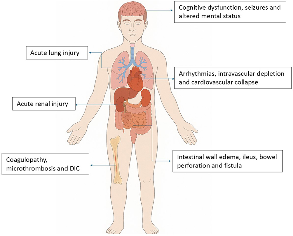 |
Figure 1 Multisystem complications associated with intraoperative hyperthermia. Hyperthermia during cytoreductive surgery with hyperthermic intraperitoneal chemotherapy (HIPEC) can lead to systemic complications, including cognitive dysfunction and seizures, acute lung and renal injury, cardiovascular collapse, intestinal wall edema and perforation, and coagulopathy with disseminated intravascular coagulation (DIC). |
Many experts have debated how to overcome such deals. Some centers intentionally cooled patients by using forced air blankets (Bair Hugger ®), cooled intravenous fluid, ice packs in the axillae of patients, or reducing ambient temperature settings to a lower degree before starting HIPEC to prevent the following hyperthermia.36–38 However, no consensus has been reached regarding whether cooling CBT to a lower degree before HIPEC can improve postoperative outcomes. Perioperative hypothermia may induce several catastrophic adverse events, including myocardial ischemia, coagulopathy, wound infection and poor healing. Prevention of hypothermia is also recommended by Guidelines for Perioperative Care in Cytoreductive Surgery with or without Hyperthermic Intraperitoneal Chemotherapy and Enhanced Recovery After Surgery (ERAS®) in gynecologic oncology.
Current evidences still have debates on whether to cool CBT before HIPEC phase. This study aimed to investigate whether active cooling CBT below 35°C before the HIPEC phase during CRS is associated with differences in intraoperative parameters, postoperative mortality and morbidity rates, and postoperative outcomes. We hypothesize that induced cooling before the HIPEC phase may be associated with better outcomes.
Materials and Methods
We retrospectively enrolled 90 patients who underwent CRS plus HIPEC at the Kaohsiung Chang Gung Memorial Hospital between 2018 and 2023. Eligible patients were adults aged > 18 years with an American Society of Anesthesiologists (ASA) score of ≤ 3. Eight patients were excluded due to incomplete data. An additional nine patients were excluded because HIPEC was not performed after CRS was decided by the surgeon due to unexpected disease progression. A flowchart of patient selection is shown in Figure 2. This study was approved by the Institutional Review Board of the Chang Gung Memorial Hospital, Kaohsiung, Taiwan (IRB No.202100987B0D001). The requirement for individual patient consent was waived because of the retrospective design, minimal risk to participants, and use of de-identified data. Patient confidentiality was strictly protected, and the study was conducted in accordance with the principles of the Declaration of Helsinki.
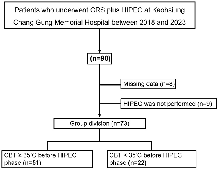 |
Figure 2 Patient enrollment flow chart. A total of 90 patients who underwent cytoreductive surgery (CRS) with planned hyperthermic intraperitoneal chemotherapy (HIPEC) were assessed for eligibility. Seventeen patients were excluded: 8 due to missing data and 9 because HIPEC was not performed. The remaining 73 patients were divided into two groups based on core body temperature (CBT) immediately prior to the HIPEC phase: the normothermia group (CBT ≥ 35°C, n = 51) and the hypothermia group (CBT < 35°C, n = 22). |
A core body temperature of 35°C is commonly considered the lower threshold of normal.39,40 Therefore, patients were divided into two groups based on their CBT immediately before the initiation of HIPEC: those with CBT ≥ 35°C and those with CBT < 35°C. A total of 51 patients were classified into the CBT ≥ 35°C group and 22 into the CBT < 35°C group.
The primary outcomes of this study were the time to extubation following surgery and the Clavien-Dindo classification. The secondary outcomes included length of stay (LOS), intensive care unit (ICU) stay time, incidence of postoperative acute kidney injury (AKI) according to the AKIN classification, reintubation events, and intraoperative parameters such as intraoperative intravenous fluid volume, intraoperative blood loss, leukocyte-poor red blood cells (LPR), fresh frozen plasma (FFP) transfusion unit, and albumin supplement amount.
The electronic medical records of the enrolled patients were reviewed to obtain clinical data. Preoperative characteristics included age, sex, body weight (BW), body mass index (BMI), history of hypertension (HTN), diabetes mellitus (DM), coronary artery disease (CAD), congestive heart failure (CHF), lung disease, cerebrovascular accident (CVA), chronic kidney disease (CKD), peripheral arterial occlusive disease (PAOD), ASA classification, prior chemotherapy or surgery, cancer type, creatinine (Cr), albumin, platelet count, international normalized ratio (INR), and activated partial thromboplastin time (APTT). Intraoperative variables included duration of surgery, peritoneal cancer index (PCI) score, completeness of cytoreduction (CC) score, intraoperative vital signs, central venous pressure (CVP), stroke volume (SV), systemic vascular resistance (SVR), stroke volume variation (SVV), intraoperative CBT, total volume of intravenous fluids administered, albumin supplementation amount, estimated blood loss, and blood product requirements. HIPEC-related data, such as perfusate temperature, duration of intraperitoneal chemotherapy, and the chemotherapeutic agents used, were also collected. Postoperative data, including LOS, ICU stay time, incidence of postoperative AKI according to the AKIN classification, time to extubation following surgery, reintubation event, and Clavien-Dindo classification, were also acquired. We have followed up on patient medical records to track delayed postoperative outcomes and complications until discharge or expiration.
Standard intraoperative monitoring, including heart rate, SpO2, and blood pressure, was recorded every 5 min. Esophageal temperature was measured using a temperature probe throughout the surgery and data were recorded every 15 min. We also monitored advanced hemodynamic data via the arterial line, central venous catheter, and the Flotrac system. The bispectral index (BIS) was used to monitor the depth of anesthesia. Arterial blood gas levels were periodically monitored throughout the procedure. Urine output was monitored hourly by using a Foley catheter. General anesthesia was administered to all patients using propofol, sevoflurane, and opioids in combination with muscle relaxants (cisatracurium). Sevoflurane was used to maintain anesthesia depth between BIS values of 40 and 60 throughout the surgery. Cisatracurium 2–4 mg was administered every 30 min to prevent undesired patient movement during the operation. Fluid therapy, including continuous infusion of crystalloids, was heated to 40°C before administration. Natural colloids (albumin) were infused as volume replacements. Intraoperative goal directed fluid therapy was guided by Flotrac system to keep SVV lower than 13. Blood products were transfused to maintain a hemoglobin concentration > 8 g/dL and heated to 37°C before transfusion.
Intraoperative body temperature was controlled using underbody mattresses, forced air underbody blankets (Bair Hugger ®), intravenous fluid, ice packs in the axillae of the patients, and ice pillows. Whether to actively cool patients to a low CBT level before HIPEC initiation was decided by the attending surgeons and anesthesiologists after discussion. The cooling protocol was initiated 1 h before HIPEC was initiated by targeting CBT 34–35°C, including 1) turning underbody mattresses to 4°C, 2) turning forced air blankets to 32°C, 3) cold intravenous lactate ringer (LR) administration at 4 °C via a 16-gauge peripheral IV line, and 4) using ice packs in the axillae and ice pillow. Ten minutes before HIPEC ended, targeted CBT returned to normothermia. The underbody mattresses and forced air blankets were reset to 36°C. Intravenous fluids were changed back to warm or ambient-temperature crystalloids depending on the patient’s CBT. Ice packs and pillows were removed. Otherwise, CBT was maintained by underbody mattresses, forced air blankets, and warm or ambient-temperature crystalloids throughout surgery with a targeting level of 35–38°C if attending surgeons and anesthesiologists decided not to actively cool patients to a low CBT level before HIPEC.
CRS was performed on the basis of the clinical judgement of the attending surgeon. Following CRS, intraoperative HIPEC was performed using the closed abdomen technique. Hyperthermic perfusion was performed using the Performer HT system (RanD S.r.l.; Medolla, Italy), a CE-certified device specifically designed for intraoperative chemotherapy delivery. The system features dual high-flow roller pumps (up to 2 L/min each), precise temperature control (28–46°C) via integrated plate warmers, and multiple safety components including pressure sensors, temperature probes, and automated alarms. The machine was controlled by attending surgeons, and all perfusion parameters were continuously monitored and recorded digitally throughout the procedure. Chemotherapeutics included cisplatin, taxotere, oxaliplatin, mitomycin C, and gemcitabine, which were heated to 42°C and instilled in the abdominal cavity.
After surgery, the patients were routinely sent to ICU for further monitoring and care. If vital signs were stable after the operation, extubation was performed in the operating room, depending on the discussion between surgeons and anesthesiologists. Neostigmine (50 mcg/kg plus atropine 1 mg) was used to reverse muscle relaxation. Otherwise, extubation would be performed in the ICU depending on the patient’s condition and ICU physician’s judgement. Delayed extubation was defined as postoperative mechanical ventilation lasting more than 12 hours in this research.
The Kolmogorov–Smirnov test was used to analyze the distribution of continuous variables. In the bivariate analysis, variables with normal distribution were analyzed using Student’s t-test, including age, BW, BMI, preoperative albumin level, preoperative Cr level, preoperative platelet count, operation time, intraoperative fluid administration (mL/kg/min), and postoperative Cr between the two groups of patients (CBT ≥ 35°C and < 35°C). Variables without a normal distribution were analyzed using the Mann–Whitney U-test. These included preoperative INR, PCI score, postoperative day 1 and day 2 Cr, intraoperative blood loss, intraoperative transfusion volumes of LPR and FFP, intraoperative albumin supplementation, postoperative time before extubation (h), postoperative ICU time, and length of hospital stay between the two groups of patients (CBT ≥ 35°C and < 35°C). Categorical variables such as sex, history of HTN, DM, CAD, CHF, CVA, asthma, chronic obstructive pulmonary disease (COPD), CKD, PAOD, ASA score, Clavien-Dindo complication grades, postoperative AKI, and reintubation events were compared using the chi-square or Fisher’s exact test. To evaluate the association between perioperative factors and delayed extubation (>12 hours), binary logistic regression analyses were performed. Odds ratios with 95% confidence intervals were calculated. Statistical significance was defined as a p-value < 0.05. All statistical analyses were performed using Statistical Package for the Social Sciences (SPSS) version 26 for Windows (SPSS Inc., Chicago, IL, USA).
Continuous variables are presented as mean ± standard deviation (SD) for normally distributed data, and as median with interquartile range (IQR) for non-normally distributed data. Categorical variables are expressed as numbers (percentages).
Results
Ninety patients scheduled to undergo CRS combined with HIPEC were enrolled. Eight patients were excluded because of incomplete data, and nine patients were excluded because HIPEC was not performed following CRS based on intraoperative findings of unexpected disease progression. Thus, 73 patients were included in the final analysis.
Among the 73 patients, 51 (69.8%) had a CBT ≥ 35°C before the initiation of HIPEC and 22 (30.1%) had a CBT < 35°C. The baseline characteristics of the patients are summarized in Table 1. The ASA of Anesthesiologists classification was significantly different between the two groups (p = 0.039). A higher proportion of ASA class II patients were observed in the CBT < 35°C group (72.7%), whereas ASA class III patients were more prevalent in the CBT ≥ 35°C group (56.9%). No significant differences were observed between the groups with respect to age (61.5 [52.0–65.8] vs 59.0 [50.0–66.0] years, p = 0.7406), sex distribution (31.8% vs 51.0% male, p = 0.210), body weight, BMI, preoperative creatinine level, platelet count, albumin level, and comorbidities, such as hypertension, diabetes mellitus, coronary artery disease, congestive heart failure, lung disease, cerebrovascular accident, chronic kidney disease, and peripheral arterial occlusive disease (all p > 0.05).
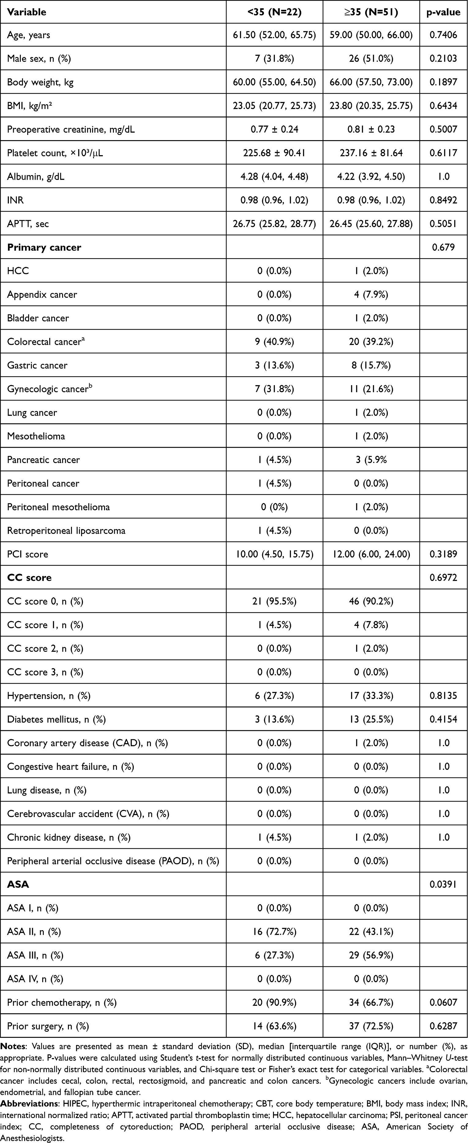 |
Table 1 Baseline Characteristics of Patients According to Pre-HIPEC Core Body Temperature (CBT) |
The intraoperative parameters are listed in Table 2. The CBT < 35°C group had a significantly greater total intraoperative fluid volume (7,600 [6,125–9,225] mL vs 5,600 [4,187.5–6,975] mL; p = 0.0049) and fluid administration rate normalized by body weight and operation time (0.17 [0.14–0.20] vs 0.11 [0.10–0.14] mL/kg/min; p < 0.0001) compared with the CBT ≥ 35°C group. Blood loss was significantly greater in the CBT < 35°C group (350 mL [200–600 mL]) than in the CBT ≥ 35°C group (150 mL [100–300 mL]; P = 0.0045). The number of LPR units transfused was also significantly higher in the CBT < 35°C group (1.00 [0.00–3.50] units) compared with the CBT ≥ 35°C group (0.00 [0.00–0.00] units; p = 0.0016). No significant differences were observed between the groups in operation time (p = 0.9521), HIPEC duration (p = 0.6807), FFP transfusion (p = 0.1542), albumin supplementation (p = 0.1492), or hemodynamic parameters including CVP, heart rate (HR), SV, SVR, and SVV before, during, and after HIPEC (all p > 0.05).
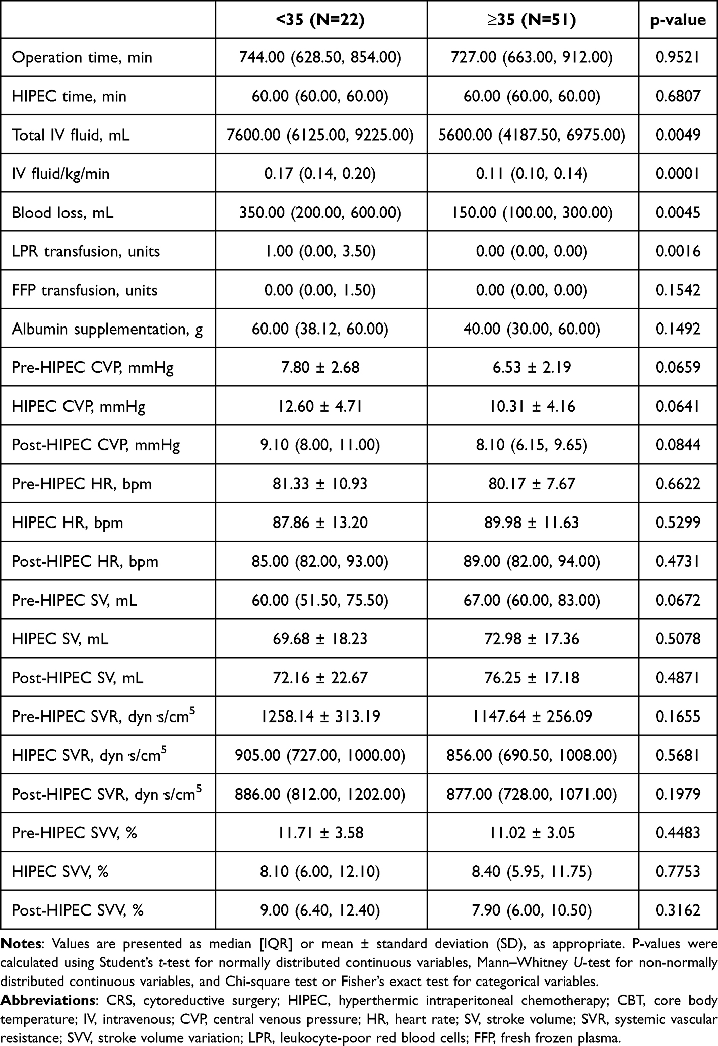 |
Table 2 Intraoperative Parameters During CRS and HIPEC According to Pre-HIPEC Core Body Temperature (CBT) |
The postoperative outcomes are summarized in Table 3. The CBT < 35°C group had a significantly longer time from the end of surgery to extubation than the CBT ≥ 35°C group (median [IQR]: 11.50 [7.50–14.75] min vs 8.00 [3.00–11.50] min; p = 0.0314, Mann–Whitney U-test). Postoperative creatinine was significantly lower in the CBT < 35°C group (0.67 ± 0.20 mg/dL) than in the CBT ≥ 35°C group (0.82 ± 0.24 mg/dL; p = 0.0113, Student’s t-test). Similarly, postoperative day (POD)1 creatinine was lower in the CBT < 35°C group (0.55 [0.48–0.78] mg/dL) compared to the CBT ≥ 35°C group (0.76 [0.54–0.99] mg/dL; p = 0.0446). No significant differences were observed between the two groups in terms of the POD2 creatinine level (p = 0.2111), length of hospital stay (p = 0.1038), ICU stay duration (p = 0.243), incidence of postoperative AKI (p = 1), reintubation events (p = 0.6629), or Clavien-Dindo complication grade (p = 0.157).
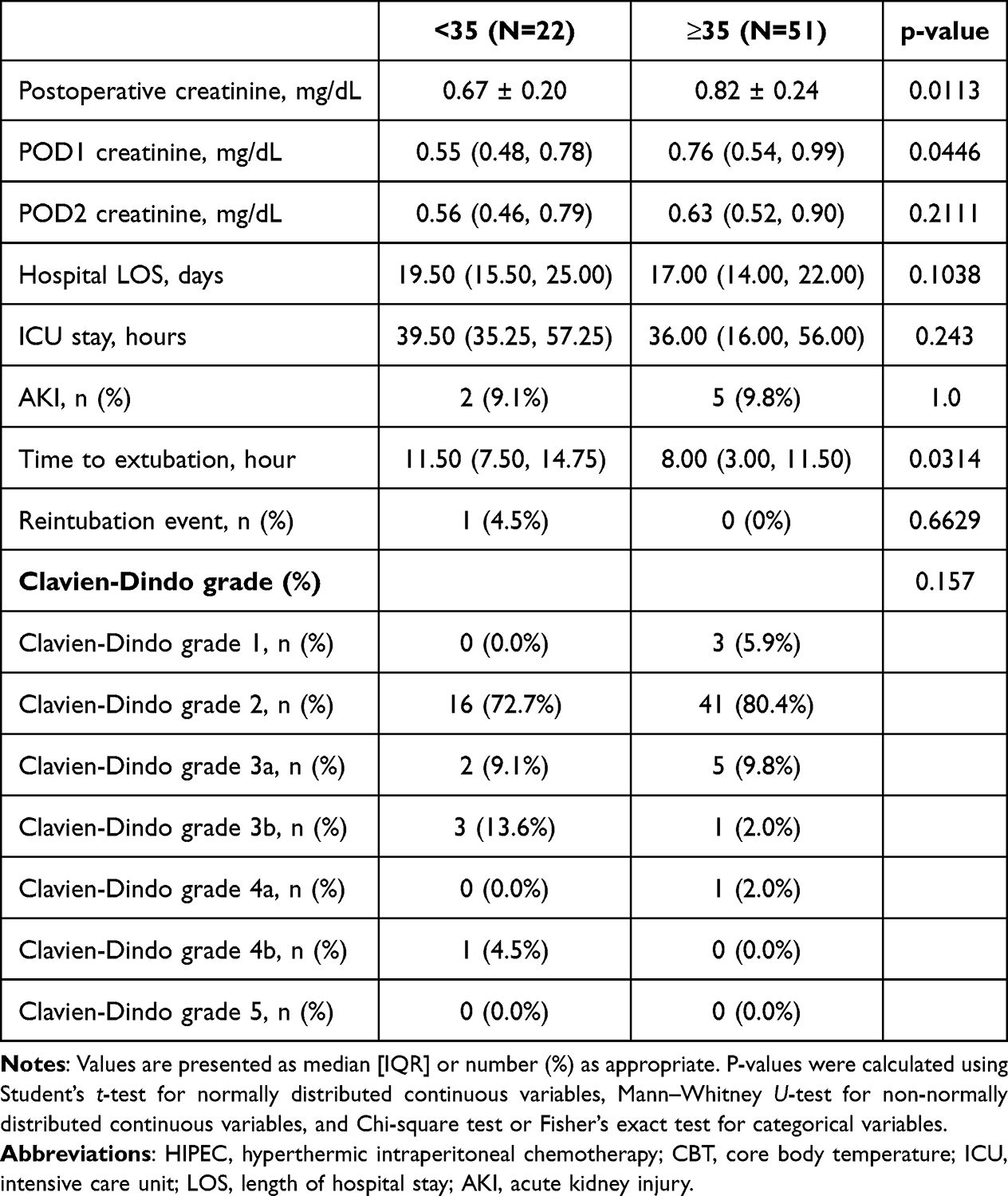 |
Table 3 Postoperative Outcomes According to Pre-HIPEC Core Body Temperature (CBT) |
Delayed extubation in this study was defined as extubation occurring more than 12 hours after the completion of surgery. In the univariate logistic regression analysis (Table 4), older age (odds ratio [OR] 1.08, 95% confidence interval [CI] 1.01–1.16, p = 0.021), pre-HIPEC CBT < 35 °C (OR 6.29, 95% CI 1.98–19.96, p = 0.002), higher total IV fluid administration (per 100 mL; OR 1.02, 95% CI 1.01–1.04, p = 0.010), and greater IV fluid per body weight per hour (OR 1.27, 95% CI 1.05–1.52, p = 0.013) were significantly associated with delayed extubation. In the multivariate model, after adjusting for potential confounders, pre-HIPEC CBT < 35 °C remained independently associated with delayed extubation (OR 6.31, 95% CI 1.11–35.70, p = 0.037), along with total IV fluid administration (per 100 mL; OR 1.05, 95% CI 1.00–1.10, p = 0.042).
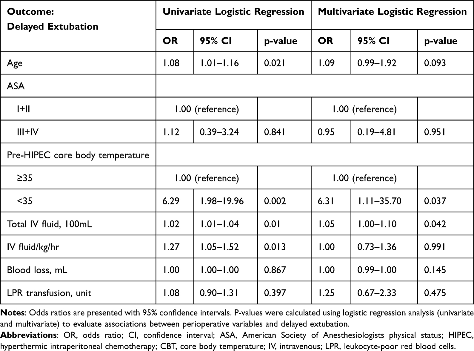 |
Table 4 Univariate and Multivariate Logistic Regression Analyses for Factors Associated with Delayed Extubation |
Discussion
The major finding of the present study was that patients who actively cooled to a lower CBT level before HIPEC had a significantly longer time to extubation after surgery (p = 0.0314). Logistic regression analysis further suggested that hypothermal group was associated with delayed extubation and appeared to be an independent predictor after adjusting for potential confounders.
No benefit was observed according to other postoperative outcomes, including postoperative AKI (p = 1), LOS (p = 0.1038), ICU stay time (p = 0.243), reintubation events (p = 0.6629), and Clavien-Dindo classification (p = 0.157).
In major abdominal surgery, delayed extubation or prolonged postoperative mechanical ventilation is strongly associated with adverse outcomes. Using the ACS-NSQIP database of 165,196 patients, Yang et al demonstrated that 5.8% of patients developed postoperative pulmonary complications (PPCs), including unplanned reintubation (2.8%) and prolonged ventilation (3.0%).41 These complications were closely linked to higher ASA class, longer operative times, and were strongly associated with increased rates of reoperation, sepsis, acute renal failure, prolonged ICU and hospital length of stay, and substantially higher mortality. Importantly, prolonged intubation predisposes patients to ventilator-associated pneumonia (VAP), with reported mortality rates up to 70%, while unplanned reintubation carries mortality rates of approximately 40%. Consistent findings have been reported in the field of liver transplantation. Xu et al showed that immediate postoperative extubation significantly reduced 30-day all-cause mortality, acute kidney injury, and moderate to severe pulmonary complications compared to ICU extubation, while also shortening both ICU and total hospital stays.42 Taken together, these data highlight that delayed extubation and prolonged intubation in major abdominal surgery are not merely markers of disease severity, but independent predictors of adverse outcomes. Promoting early extubation when clinically feasible is therefore an essential component of modern perioperative care and Enhanced Recovery After Surgery (ERAS) strategies.
Previous abdominal vascular surgery studies have consistently shown that outcomes deteriorate once postoperative ventilation extends beyond 12 hours. Both Zettervall et al and David et al reported increased complications and longer hospitalization in patients extubated at or after 12 hours, while Stone et al similarly observed worse trajectories beyond this cutoff.43–45 These findings support the use of 12 hours as a clinically meaningful and evidence-based definition of delayed extubation in our cohort.
Maintaining normothermia during surgery is widely accepted as standard practice to minimize perioperative complications. Perioperative hypothermia may induce several catastrophic adverse events including myocardial ischemia, coagulopathy, wound infection, and poor healing. However, the context of CRS combined with HIPEC is unique, because hyperthermia during the HIPEC phase may cause a substantial rise in body temperature. To date, no clear evidence has indicated whether active cooling before the HIPEC phase improves the postoperative outcomes. A study conducted by Balakrishnan and Survesan revealed that greater delta temperatures were statistically associated with longer ventilation and ICU stay.46 Prevention of hypothermia was also recommended by the Guidelines for Perioperative Care in Cytoreductive Surgery with or without Hyperthermic Intraperitoneal Chemotherapy and Enhanced Recovery After Surgery (ERAS®) in gynecologic oncology.47 Nevertheless, perioperative thermal management varies considerably across centers. Edward A. Levine et al shared their experience with 1,000 CRS plus HIPEC patients, they actively cooled their patients to a CBT of approximately 34°C to 35°C near the completion of CRS.31 Similar article written by Maria F. Ramirez and colleagues had their own cooling protocol before HIPEC started.36 A case report written by Namita Saraswat also cooled the patient before HIPEC initiation.38 Thus, the findings of our study may contribute additional insight for future practice.
Regarding intraoperative data, patients with CBT below 35°C before HIPEC had a significantly greater total blood loss (p = 0.0045). Several studies have demonstrated that hypothermia is related to excessive surgical blood loss.48–51 Increased surgical blood loss is caused by impaired platelet function and disruption of coagulation pathways. Hypothermia significantly impairs platelet function by inhibiting thromboxane A2 release, upregulating platelet surface protein GMP-140, and downregulating platelet glycoprotein Ib-IX complex.51 Prolonged prothrombin and partial thromboplastin times when assays are conducted at the patient’s actual core temperature rather than at 37°C.49 A study by Gupta et al showed that excessive blood loss was positively correlated with the need for postoperative ventilation and the length of ICU stay.52
The number of LPR transfusions (p = 0.0016) was significantly higher in the low-CBT group. This result might be related to the fact that greater total blood loss was observed in the low-CBT group. A study conducted by Schmied et al demonstrated that blood requirements increased in hypothermic patients undergoing total hip arthroplasty.51 Another study conducted by Zhuo Sun et al, involving 58,814 adults undergoing surgeries lasting more than 60 min, showed that hypothermia was associated with increased transfusion requirement.53 A review article by Rauch et al demonstrated that even mild hypothermia significantly increases blood loss and transfusion requirement.50
In the current study, the total amount of intraoperative intravenous fluid was significantly higher in the low-CBT group (p = 0.0049). The total amount of intraoperative intravenous fluid adjusted for body weight and operation time also showed a significant difference (p = 0.0001). This result may be related to the greater total blood loss observed in the low-CBT group. The additional cold fluid for decreasing CBT may also be related to this result. An Australian study conducted by Shamavonian et al showed that increasing intraoperative IV fluid administration was associated with an increase in patient morbidity in CRS plus HIPEC.54 Another study by Oliver S Eng al. demonstrated that intraoperative fluid administration was associated with a significant increase in perioperative morbidity in patients undergoing CRS plus HIPEC.55 In line with these findings, our multivariate logistic regression analysis further identified total intraoperative IV fluid volume as an independent risk factor for delayed extubation, reinforcing the evidence that fluid overload adversely affects postoperative outcomes in CRS plus HIPEC.
Postoperative and POD1 creatinine were significantly lower in low CBT group (p = 0.0113 and 0.0446 respectively). The median postoperative and POD1 creatinine level were all within normal range despite the statistical difference. There was also no significant difference between postoperative AKI of these two groups. We consider the statistical difference may lack clinical significance.
Overall, the findings of this study suggest that aggressive cooling before HIPEC may increase intraoperative bleeding and transfusion requirements, potentially leading to delayed post-operative extubation. Future studies are warranted to further evaluate the optimal thermal management strategies for CRS and HIPEC to improve patient outcomes.
Limitations
There are some limitations of our study. The first one is the relatively small sample size due to limited number of cases at our center. On the other hand, some physicians believed that active cooling would reduce the adverse effects of hyperthermia induced by HIPEC initially. However, as time went on, we found no significant benefit to postoperative outcomes. Consequently, we gradually abandoned the active cooling protocol. As a result, there are only 22 patients in the CBT < 35°C group. This retrospective study was conducted to confirm our hypothesis based on these observations. Second, the patients of the two groups are uneven. The decision to actively cool patients to a low CBT level before HIPEC was made by the attending surgeons and anesthesiologists after discussion. These personnel were not fixed and might have different opinion. Third, this is a retrospective study. Further prospective randomized trials are required to evaluate the optimal thermal management strategies during CRS plus HIPEC procedures. Nevertheless, the significant findings observed in this study remain robust. To the best of our knowledge, this is the first cohort observation study in Taiwan to investigate whether actively cooling CBT to a low level before HIPEC in patients received CRS plus HIPEC is beneficial to postoperative outcomes.
Conclusion
In this study, actively cooling CBT to < 35 °C before the HIPEC phase did not demonstrate improved postoperative outcomes. On the contrary, hypothermia appeared to be associated with delayed extubation, greater surgical blood loss, higher transfusion requirements, and increased intraoperative IV fluid administration. These findings should be interpreted with caution given the retrospective design. Further large-scale or prospective randomized studies are warranted to validate these observations and to establish evidence-based thermal management strategies for patients undergoing CRS with HIPEC.
Data Sharing Statement
The original contributions presented in the study are included in the article. Further inquiries can be directed to the corresponding author.
Ethics Statement
The study was approved by the Institutional Review Board of Chang Gung Memorial Hospital, Kaohsiung, Taiwan (IRB No.202100987B0D001). The requirement for individual patient consent was waived because of the retrospective design, minimal risk to participants, and use of de-identified data. Patient confidentiality was strictly protected, and the study was conducted in accordance with the principles of the Declaration of Helsinki.
Disclosure
The authors declare no conflicts of interest related to this study.
References
1. Kepenekian V, Bhatt A, Péron J, et al. Advances in the management of peritoneal malignancies. Nat Rev Clin Oncol. 2022;19(11):698–718. doi:10.1038/s41571-022-00675-5
2. Cortés-Guiral D, Hübner M, Alyami M, et al. Primary and metastatic peritoneal surface malignancies. Nat Rev Dis Primers. 2021;7(1):91. doi:10.1038/s41572-021-00326-6
3. Coccolini F, Gheza F, Lotti M, et al. Peritoneal carcinomatosis. World J Gastroenterol. 2013;19(41):6979–6994. doi:10.3748/wjg.v19.i41.6979
4. Bhatt A, Rousset P, Baratti D, et al. Patterns of peritoneal dissemination and response to systemic chemotherapy in common and rare peritoneal tumours treated by cytoreductive surgery: study protocol of a prospective, multicentre, observational study. BMJ Open. 2021;11(7):e046819. doi:10.1136/bmjopen-2020-046819
5. Sugarbaker PH. Peritonectomy procedures. Ann Surg. 1995;221(1):29–42. doi:10.1097/00000658-199501000-00004
6. Glehen O, Gilly FN, Boutitie F, et al. Toward curative treatment of peritoneal carcinomatosis from nonovarian origin by cytoreductive surgery combined with perioperative intraperitoneal chemotherapy: a multi‐institutional study of 1290 patients. Cancer. 2010;116(24):5608–5618. doi:10.1002/cncr.25356
7. Fallon EA, Awiwi MO, Bhutiani N, et al. Peritoneal cancer index correlates with radiographic assessment of colorectal carcinomatosis. Ann Surg Oncol. 2024;1–9.
8. Ji Z-H, Yu Y, Liu G, et al. Peritoneal cancer index (PCI) based patient selecting strategy for complete cytoreductive surgery plus hyperthermic intraperitoneal chemotherapy in gastric cancer with peritoneal metastasis: a single-center retrospective analysis of 125 patients. Eur J Surg Oncol. 2021;47(6):1411–1419. doi:10.1016/j.ejso.2020.11.139
9. Rawert FL, Luengas-Würzinger V, Claßen-Gräfin von Spee S, et al. The importance of the peritoneal cancer index (PCI) to predict surgical outcome after neoadjuvant chemotherapy in advanced ovarian cancer. Arch Gynecol Obstet. 2022;306(5):1665–1672. doi:10.1007/s00404-022-06527-y
10. Jayne D, Fook S, Loi C, Seow-Choen F. Peritoneal carcinomatosis from colorectal cancer. Br J Surg. 2002;89(12):1545–1550. doi:10.1046/j.1365-2168.2002.02274.x
11. Garach NR, Kusamura S, Guaglio M, Bartolini V, Deraco M, Baratti D. Comparative study of mucinous and non-mucinous appendiceal neoplasms with peritoneal dissemination treated by cyoreductive surgery and hyperthermic intraperitoneal chemotherapy (HIPEC). Eur J Surg Oncol. 2021;47(5):1132–1139. doi:10.1016/j.ejso.2020.08.017
12. Gronau F, Feldbruegge L, Oberwittler F, et al. HIPEC in peritoneal metastasis of gastric origin: a systematic review of regimens and techniques. J Clin Med. 2022;11(5):1456. doi:10.3390/jcm11051456
13. Klempner SJ, Ryan DP. HIPEC for colorectal peritoneal metastases. Lancet Oncol. 2021;22(2):162–164. doi:10.1016/S1470-2045(20)30693-8
14. Sugarbaker PH. A narrative review of what can HIPEC do. Eur J Surg Oncol. 2023;49(9):106976. doi:10.1016/j.ejso.2023.07.002
15. Lim P-Q, Han I-H, Seow K-M, Chen K-H. Hyperthermic intraperitoneal chemotherapy (HIPEC): an overview of the molecular and cellular mechanisms of actions and effects on epithelial ovarian cancers. Int J Mol Sci. 2022;23(17):10078. doi:10.3390/ijms231710078
16. Bhatt A, de Hingh I, Van Der Speeten K, et al. HIPEC methodology and regimens: the need for an expert consensus. Ann Surg Oncol. 2021;28(13):9098–9113. doi:10.1245/s10434-021-10193-w
17. Dellinger TH, Han ES. State of the science: the role of HIPEC in the treatment of ovarian cancer. Gynecol Oncol. 2021;160(2):364–368. doi:10.1016/j.ygyno.2020.12.029
18. Overgaard J. Effect of hyperthermia on malignant cellsin vivo: a review and a hypothesis. Cancer. 1977;39(6):2637–2646. doi:10.1002/1097-0142(197706)39:6<2637::AID-CNCR2820390650>3.0.CO;2-S
19. Warters RL, Henle KJ. DNA degradation in Chinese hamster ovary cells after exposure to hyperthermia. Cancer Res. 1982;42(11):4427–4432.
20. Chen T, Guo J, Han C, Yang M, Cao X. Heat shock protein 70, released from heat-stressed tumor cells, initiates antitumor immunity by inducing tumor cell chemokine production and activating dendritic cells via TLR4 pathway. J Immunol. 2009;182(3):1449–1459. doi:10.4049/jimmunol.182.3.1449
21. Skitzki JJ, Repasky EA, Evans SS. Hyperthermia as an immunotherapy strategy for cancer. Curr Opin Investig Drugs. 2009;10(6):550.
22. Hurwitz M, Stauffer P. Hyperthermia, radiation and chemotherapy: the role of heat in multidisciplinary cancer care. Semin Oncol. 2014;41(6):305–316. doi:10.1053/j.seminoncol.2014.09.014
23. Mantso T, Goussetis G, Franco R, et al. Effects of hyperthermia as a mitigation strategy in DNA damage-based cancer therapies. Semin Cancer Biol. 2016;40:71–80.
24. Galluzzi L, Buqué A, Kepp O, Zitvogel L, Kroemer G. Immunogenic cell death in cancer and infectious disease. Nat Rev Immunol. 2017;17(2):97–111. doi:10.1038/nri.2016.107
25. Grimmig T, Moll E-M, Kloos K, et al. Upregulated heat shock proteins after hyperthermic chemotherapy point to induced cell survival mechanisms in affected tumor cells from peritoneal carcinomatosis. Cancer Growth and Metastasis. 2017;10:1179064417730559. doi:10.1177/1179064417730559
26. Ni L-P, Sun H-T, Wang P, et al. Hyperthermia enhances the efficacy of chemotherapeutic drugs in heat-sensitive cells through interfering with DNA damage repair. Ann Transl Med. 2022;10(8):463. doi:10.21037/atm-22-955
27. Raspe C, Piso P, Wiesenack C, Bucher M. Anesthetic management in patients undergoing hyperthermic chemotherapy. Curr Opin Anaesthesiol. 2012;25(3):348–355. doi:10.1097/ACO.0b013e32835347b2
28. Raspé C, Flöther L, Schneider R, Bucher M, Piso P. Best practice for perioperative management of patients with cytoreductive surgery and HIPEC. Eur J Surg Oncol. 2017;43(6):1013–1027. doi:10.1016/j.ejso.2016.09.008
29. Owusu-Agyemang P, Arunkumar R, Green H, Hurst D, Landoski K, Hayes-Jordan A. Anesthetic management and renal function in pediatric patients undergoing cytoreductive surgery with continuous hyperthermic intraperitoneal chemotherapy (HIPEC) with cisplatin. Ann Surg Oncol. 2012;19(8):2652–2656. doi:10.1245/s10434-012-2319-1
30. Garg R. Cytoreductive surgery and hyperthermic intraperitoneal chemotherapy: fluid and temperature remain the culprit! Indian J Anaesth. 2018;62(3):162–165. doi:10.4103/ija.IJA_170_18
31. Putzu M, Casati A, Berti M, Pagliarini G, Fanelli G. Clinical complications, monitoring and management of perioperative mild hypothermia: anesthesiological features. Acta Bio Med Atenei Parmensis. 2007;78(3):163–169.
32. Cafiero T, Di Iorio C, Di Minno R, Sivolella G, Confuorto G. Non-invasive cardiac monitoring by aortic blood flow determination in patients undergoing hyperthermic intraperitoneal intraoperative chemotherapy. Minerva anestesiologica. 2006;72(4):207–215.
33. Schmidt C, Creutzenberg M, Piso P, Hobbhahn J, Bucher M. Peri-operative anaesthetic management of cytoreductive surgery with hyperthermic intraperitoneal chemotherapy. Anaesthesia. 2008;63(4):389–395. doi:10.1111/j.1365-2044.2007.05380.x
34. Goldenshluger M, Zippel D, Ben-Yaacov A, et al. Core body temperature but not intraabdominal pressure predicts postoperative complications following closed-system hyperthermic intraperitoneal chemotherapy (HIPEC) administration. Ann Surg Oncol. 2018;25(3):660–666. doi:10.1245/s10434-017-6279-3
35. Hendrix RJ, Kassira JP, Lambert LA. Elevated maximum core body temperature during hyperthermic intraperitoneal chemoperfusion (HIPEC) is associated with increased postoperative complications. Ann Surg Oncol. 2020;27(1):232–239. doi:10.1245/s10434-019-07495-5
36. Ramirez MF, Guerra-Londono JJ, Owusu-Agyemang P, Fournier K, Guerra-Londono CE. Temperature management during cytoreductive surgery with hyperthermic intraperitoneal chemotherapy. Front Oncol. 2023;12:1062158. doi:10.3389/fonc.2022.1062158
37. Levine EA, Stewart IVJH, Shen P, Russell GB, Loggie BL, Votanopoulos KI. Intraperitoneal chemotherapy for peritoneal surface malignancy: experience with 1,000 patients. J Am Coll Surg. 2014;218(4):573–585. doi:10.1016/j.jamcollsurg.2013.12.013
38. Saraswat N, Gupta A, Kaur M, Mahajan S. Anaesthesia concerns for peritonectomy with hyperthermic intraoperative peritoneal chemotherapy. Saudi J Anaesth. 2019;13(2):169–171. doi:10.4103/sja.SJA_12_17
39. Brown DJA, Brugger H, Boyd J, Paal P. Accidental hypothermia. N Engl J Med. 2012;367(20):1930–1938. doi:10.1056/NEJMra1114208
40. Duong H, Patel G. Hypothermia. StatPearls. StatPearls Publishing; 2024.
41. Yang CK, Teng A, Lee DY, Rose K. Pulmonary complications after major abdominal surgery: national surgical quality improvement program analysis. J Surg Res. 2015;198(2):441–449. doi:10.1016/j.jss.2015.03.028
42. Bartels K, Frendl G, Sprung J, et al. Postoperative pulmonary complications with adjuvant regional anesthesia versus general anesthesia alone: a sub-analysis of the perioperative research network study. BMC Anesthesiology. 2022;22(1):136. doi:10.1186/s12871-022-01679-5
43. Zettervall SL, Ultee KHJ, Soden PA, et al. Predictors of renal dysfunction after endovascular and open repair of abdominal aortic aneurysms. J Vasc Surg. 2017;65(4):1564–1571.e3. doi:10.1016/j.jvs.2016.06.113
44. David RA, de Guerre LEVM, Black JH, et al. Predictors of delayed extubation following open descending and thoracoabdominal aortic aneurysm repair. J Vasc Surg. 2017;65(2):325–334.
45. Stone WM, Hallett JW Jr, Pairolero PC, Cherry KJ Jr, Hallett JW. Respiratory failure and pneumonia after aortic surgery. J Cardiothorac Vasc Anesth. 1998;12(2):141–144.
46. Balakrishnan KP, Survesan S. Anaesthetic management and perioperative outcomes of cytoreductive surgery with hyperthermic intraperitoneal chemotherapy: a retrospective analysis. Indian J Anaesth. 2018;62(3):188–196. doi:10.4103/ija.IJA_39_18
47. Nelson G, Dowdy SC, Lasala J, et al. Enhanced recovery after surgery (ERAS®) in gynecologic oncology – practical considerations for program development. Gynecol Oncol. 2017;147(3):617–620. doi:10.1016/j.ygyno.2017.09.023
48. Rajagopalan S, Mascha E, Na J, Sessler D. The effects of mild perioperative hypothermia on blood loss and transfusion requirement. Anesthesiology. 2008;108(1):71–77. doi:10.1097/01.anes.0000296719.73450.52
49. Sessler DI. Complications and treatment of mild hypothermia. Anesthesiology. 2001;95(2):531–543. doi:10.1097/00000542-200108000-00040
50. Rauch S, Miller C, Bräuer A, Wallner B, Bock M, Paal P. Perioperative hypothermia—a narrative review. Int J Environ Res Public Health. 2021;18(16):8749. doi:10.3390/ijerph18168749
51. Schmied H, Reiter A, Kurz A, Sessler D, Kozek S. Mild hypothermia increases blood loss and transfusion requirements during total Hip arthroplasty. Lancet. 1996;347(8997):289–292. doi:10.1016/S0140-6736(96)90466-3
52. Gupta R, Gupta N, Sirohiya P, et al. Perioperative anaesthetic management in cytoreductive surgery (CRS) with hyperthermic intraperitoneal chemotherapy (HIPEC): a retrospective analysis in a single tertiary care cancer centre. Pleura Peritoneum. 2022;7(3):127–134. doi:10.1515/pp-2022-0001
53. Sun Z, Honar H, Sessler DI, et al. Intraoperative core temperature patterns, transfusion requirement, and hospital duration in patients warmed with forced air. Anesthesiology. 2015;122(2):276–285. doi:10.1097/ALN.0000000000000551
54. Shamavonian R, McLachlan R, Fisher OM, et al. The effect of intraoperative fluid administration on outcomes of patients undergoing cytoreductive surgery with hyperthermic intraperitoneal chemotherapy. J Gastrointest Oncol. 2019;10(2):235–244. doi:10.21037/jgo.2018.12.11
55. Eng OS, Dumitra S, O’Leary M, et al. Association of fluid administration with morbidity in cytoreductive surgery with hyperthermic intraperitoneal chemotherapy. JAMA Surgery. 2017;152(12):1156–1160. doi:10.1001/jamasurg.2017.2865
 © 2025 The Author(s). This work is published and licensed by Dove Medical Press Limited. The
full terms of this license are available at https://www.dovepress.com/terms
and incorporate the Creative Commons Attribution
- Non Commercial (unported, 4.0) License.
By accessing the work you hereby accept the Terms. Non-commercial uses of the work are permitted
without any further permission from Dove Medical Press Limited, provided the work is properly
attributed. For permission for commercial use of this work, please see paragraphs 4.2 and 5 of our Terms.
© 2025 The Author(s). This work is published and licensed by Dove Medical Press Limited. The
full terms of this license are available at https://www.dovepress.com/terms
and incorporate the Creative Commons Attribution
- Non Commercial (unported, 4.0) License.
By accessing the work you hereby accept the Terms. Non-commercial uses of the work are permitted
without any further permission from Dove Medical Press Limited, provided the work is properly
attributed. For permission for commercial use of this work, please see paragraphs 4.2 and 5 of our Terms.