Back to Journals » International Journal of Nanomedicine » Volume 21
Strategies, Challenges and Application Prospects for Exosome Engineering Modifications in Tumor Targeted Therapeutics
Authors Ge X, Shan S, Lu H, Wang W, Gao C, Fu S
Received 6 October 2025
Accepted for publication 8 January 2026
Published 30 January 2026 Volume 2026:21 572435
DOI https://doi.org/10.2147/IJN.S572435
Checked for plagiarism Yes
Review by Single anonymous peer review
Peer reviewer comments 3
Editor who approved publication: Professor Xing Zhang
Xiaoyan Ge,1,2,* Shuo Shan,1,* Haozhong Lu,2 Weihua Wang,1 Chao Gao,1 Shunli Fu1
1Tumor Immunology and Cytotherapy of Medical Research Center, Shandong Provincial Key Laboratory of Clinical Research for Pancreatic Diseases, The Affiliated Hospital of Qingdao University; Qingdao University, Qingdao, People’s Republic of China; 2Department of Pharmacy, Qingdao Mental Health Center, Qingdao, People’s Republic of China
*These authors contributed equally to this work
Correspondence: Shunli Fu, Tumor Immunology and Cytotherapy of Medical Research Center, Shandong Provincial Key Laboratory of Clinical Research for Pancreatic Diseases, The Affiliated Hospital of Qingdao University; Qingdao University, Qingdao, People’s Republic of China, Email [email protected] Chao Gao, Tumor Immunology and Cytotherapy of Medical Research Center, Shandong Provincial Key Laboratory of Clinical Research for Pancreatic Diseases, The Affiliated Hospital of Qingdao University; Qingdao University, Qingdao, People’s Republic of China, Email [email protected]
Abstract: Nanodrugs have significantly revolutionized tumor therapy. Nevertheless, conventional nanodrug delivery systems suffer from a critical limitation: only ~0.7% of administered nanoparticles effectively accumulate in solid tumors, severely restricting clinical therapeutic efficacy. In recent years, exosomes—natural extracellular vesicles—have emerged as highly promising candidates for tumor-targeted drug delivery. Endowed with inherent low immunogenicity, excellent biocompatibility, and intrinsic capacity to traverse biological barriers, exosomes offer distinct advantages over conventional nanocarriers. Their characteristic lipid bilayer membrane not only protects encapsulated cargo but also enables surface engineering for functional optimization. Through strategic engineering modifications, exosomes can be endowed with enhanced tumor-targeting specificity, tunable payload release profiles, and multimodal functionalities, thus enabling the development of “smart” therapeutic platforms. This review systematically outlines current methodologies for exosome isolation and characterization, with a particular focus on engineering strategies aimed at augmenting tumor targeting. We comprehensively analyze approaches based on physical manipulation, chemical conjugation, and biological engineering. Furthermore, we summarize recent advances in exosome-based targeted cancer therapies and discuss key challenges related to scalability, standardization, regulatory approval, and clinical translation. Finally, we highlight emerging opportunities and future perspectives for next-generation exosome-engineered therapeutic development, aiming to provide a robust technical and conceptual foundation for advancing tumor therapy.
Keywords: exosome, engineering modifications, tumor targeted therapeutics, nanotechnology
Introduction
Malignant tumors remain a major global health burden, with high incidence and mortality rates.1 Traditional treatments are often limited by tumor heterogeneity, immunosuppressive microenvironments, and biological barriers. As a result, only about 0.7% of intravenously administered drugs typically reach tumor sites, while off-target toxicity compromises therapeutic outcomes and patient quality of life.2 Tumor-targeted therapies, designed to maximize drug accumulation in tumors while sparing normal tissues, have thus become a key focus in oncology. Among these approaches, nanodrug delivery systems stand out for their ability to improve targeting efficiency.3 More than 90 nanocarrier systems have already reached clinical use, highlighting the rapid progress of the field.4
Although the nanocarrier systems, represented by liposomes and polymer nanoparticles, have been applied in clinical practice, they still exhibited limitations: Firstly, their material composition may trigger immune responses or cause unexpected toxicity; secondly, they are easily recognized and cleared by the immune system in the complex in vivo environment, resulting in a short circulation time; more importantly, they usually lack intrinsic biological targeting ability and mainly rely on passive targeting or simple ligands on the surface for active targeting, but this targeting is often not precise enough and difficult to overcome multiple biological barriers in the tumor microenvironment.5,6 In addition, the “cargo” encapsulation and release control of these nanocarrier systems still face challenges, such as premature leakage or insufficient release, which affects therapeutic efficacy and increases off-target toxicity.7 These deficiencies have prompted researchers to seek more biologically intelligent delivery platforms.
Exosomes—naturally secreted lipid bilayer vesicles (30–150 nm in diameter)—have emerged as particularly promising nanocarriers8 Derived from diverse sources (eg, animals, plants, and microorganisms),9 they exhibit superior biocompatibility compared to synthetic carriers such as liposomes or polymers. Their natural phospholipid membrane confers structural stability and reduces risks of systemic toxicity,10,11 while their small size facilitates blood–brain barrier penetration, offering unique advantages for brain tumor therapy.12 Exosomes also exhibit intrinsic targeting capacity and can be engineered for enhanced functionality. Beyond therapeutic delivery, their abundance in bodily fluids and stability make them attractive diagnostic biomarkers,13 enabling sensitive monitoring of disease progression and treatment response.14 A defining feature of exosomes is their biological plasticity: they can be readily modified with antibodies, peptides, or fluorescent probes to enable personalized delivery of bioactive agents.15 Furthermore, the specific protein profiles on the exosome membrane and target cell surface enable precise tissue or cell type homing through specific recognition.16,17 This facilitates the efficient delivery of their “cargo” into target cells, thereby regulating target cell functions and influencing physiological processes, such as immune responses and signal transduction. These properties provide a strong molecular basis for exosome-mediated cancer therapy.18–20
Animal-derived exosomes, particularly those from tumor cells, mesenchymal stem cells (MSCs), immune cells, endothelial progenitors, platelets, and fibroblasts, are most widely used in tumor-targeted applications. They are extractable from common bodily fluids including blood, urine, or milk, supporting large-scale preparation.21,22 Compared to animal-derived exosomes, plant-derived exosomes from sources such as grapes, ginger, and carrots exhibit superior immunotolerance and enhanced biocompatibility.23 For instance, ginseng-derived exosomes suppress tumor growth by modulating macrophage polarization through the toll-like receptor 4 (TLR4)-myeloid differentiation factor 88 (MyD88) pathway. Besides, ginger-derived exosome-like nanoparticles enhance anti-programmed cell death ligand 1 (PD-L1) therapy in melanoma by suppressing bacterial phospholipase C expression and increasing docosahexaenoic acid accumulation, underscoring the potential of plant-derived exosomes in reshaping tumor immunotherapy via host–microbiota interactions.24 Microbial exosomes, particularly those from gut microbiota, also play key roles in modulating intestinal immunity. They hold promise not only for intestinal disorders but also for systemic cancer therapy.25 For example, Kyong-Su Park et al combined E. coli-derived exosomes (SyBV) with tumor-derived exosomes for treatment. The results showed that SyBV activated dendritic cells (DCs) and exhibited reduced toxicity compared to native E. coli outer membrane vesicles (OMVs). This positions SyBV as a safe candidate for tumor immunotherapy that significantly enhances the efficacy of melanoma immunotherapy.26
Despite these advantages, exosomes face limitations in drug-loading capacity and targeting efficiency. To overcome the limitations of natural exosomes, engineering strategies (such as physical manipulation, chemical coupling, and genetic modification) provide effective solutions. Among them, exosome mimics and in vivo exosome biogenesis engineering are the two most promising directions at present.
Exosome mimics are artificial carriers prepared through artificial methods such as cell squeezing, liposome fusion, and membrane reorganization, which have the advantages of controllable yield, precise composition, drug loading increase and simple preparation, reducing the clinical transformation cost. Exosome mimics have to some extent solved the problem of large-scale production of natural exosomes, but the artificial carriers are easily recognized and cleared by immune cells in the body and lack the endogenous targeting property of natural exosomes.27,28
To further improve the precision and effectiveness of tumor treatment, in vivo exosome biogenesis engineering is a hot research direction. Exosome biogenesis engineering as an optimization strategy, its core principle is to regulate the exosome biogenesis process of tumor cells, stem cells, and other host cells in vivo through small molecule inhibitors, gene editing, and microenvironmental regulation (such as regulating the ESCRT pathway, HD-PTP, etc.)29 to achieve “on-demand generation of therapeutic exosomes”. The prominent advantages of this technology include: no need for an in vitro preparation and reinfusion process, significantly simplifying the treatment process. Endogenous exosomes retain the complete natural membrane structure and targeting proteins, and have better stability and targeting in the body. Using patient’s own cells as exosome parent cells can avoid the risk of immune rejection from heterologous carriers, and is particularly suitable for precise treatment of tumor heterogeneity. Currently, research in this field has achieved key breakthroughs, such as exosomes derived from malignant ascites of advanced gastric cancer patients containing abundant MET genes, which can promote enhanced tumor angiogenesis. By blocking the secretion of these exosomes, tumor growth can be slowed down.30 Exosome targeting may enhance the therapeutic effect of GC and provide inspiration for developing similar methods for other primary tumor types.
In summary, from the limitations of traditional drug delivery systems, such as poor targeting and strong toxicity, to the advantages of natural exosomes, such as superior biocompatibility and strong targeting, and to the technological iterations of exosome mimics (solving the problem of large-scale production) and in vitro/in vivo exosome biogenesis engineering (achieving in situ precise regulation), exosome-related delivery systems, with their core characteristics of precise targeting, low toxicity and high efficiency, and multi-functional synergy, gradually surpass traditional synthetic carriers and become the core carrier for tumor-specific treatment, promoting the advancement of tumor treatment towards precision and low toxicity. This review summarizes the current methods for exosome isolation and characterization, studies the engineering strategies for tumor-targeted exosomes, and summarizes the latest progress in exosome-based cancer treatment.
Preparation and Characterization of Exosomes
Isolation, purification, and characterization of exosomes are essential prerequisites for both basic research and clinical translation. Multiple methods have been developed based on different separation principles. Traditional approaches such as differential and density gradient centrifugation are widely used for preliminary separation, while more advanced techniques—including immunocapture and size-exclusion chromatography—enable higher purity and specificity. The choice of method depends on the research objective, sample type, and intended application. Characterization typically combines complementary techniques: transmission electron microscopy (TEM) is employed to analyze morphology, nanoparticle tracking analysis (NTA) to determine size distribution and concentration, and Western blotting to detect marker proteins like CD63 and CD81 (Figure 1). Optimizing and standardizing these processes is crucial to ensure reproducibility, support functional studies of exosomal cargo, and accelerate clinical application as diagnostic markers and therapeutic carriers. Moreover, the selection of source cells, preparation and isolation, surface modification, drug loading, characterization and quality control are the core steps in exosome engineering. The following systematically elaborates the technical key points, applicable scenarios and optimization strategies of each critical step, providing technical support for the application of exosomes in tumor treatment.
 |
Figure 1 Exosome isolation and characterization. (a) Isolation Methods. (b) Increase Production Methods. (c) Characterization Methods: i) Physical and chemical characterization; ii) Molecular component analysis; iii) Functional activity analysis. Created with Biorender. |
Selection of Source Cells
The type of the source cell directly determines the secretion quantity, biological compatibility, targeting potential and safety of exosomes, which is the fundamental prerequisite for exosome engineering. The selection should follow the three principles of “high secretion, low toxicity, and targeted adaptation”. Common source cells include:
Mesenchymal Stem Cells (MSCs)
The most widely used source cells in clinical research, which can be isolated from easily accessible tissues such as bone marrow, fat, and umbilical cord. They have strong proliferation ability and efficient exosome secretion efficiency. Their derived exosomes have excellent biological compatibility, low immunogenicity, and inherent tumor chemotaxis, and can migrate directionally through the tumor microenvironment (TME) based on inflammatory signals.31 However, it should be noted that MSCs from different donors may have batch differences, and the aging during in vitro expansion may affect the stability of exosome functions.32
Tumor Cells
Tumor cell-derived exosomes have a unique “homologous targeting property”, with tumor-specific antigens such as HER2 and CA125 retained on the surface, and can specifically bind to homologous tumor cells, significantly improving targeting accuracy.33 However, a core limitation is that they may carry oncogenic factors, such as metastasis-promoting miRNAs, which exert a facilitative effect on tumor growth.34
Immune Cells
Dendritic cells (DCs), macrophages, and natural killer (NK) cells are common choices. DC-derived exosomes carry MHC-peptide, CD80, and CD86, which can activate anti-tumor immune responses and are suitable for immunotherapy;35 M1-type macrophage-derived exosomes secrete inflammatory factors such as TNF-α and IL-6, which can reshape the immunosuppressive TME.36 However, immune cell culture is costly, and the secretion quantity of exosomes is limited, making it difficult to meet the requirements of large-scale production.
Plant/Microbial Cells
Exosomes derived from plants such as ginseng and ginger, and microorganisms such as Escherichia coli and Lactobacillus, have advantages such as low cost, easy large-scale extraction, and strong immune tolerance. For example, exosome-like nanoparticles derived from ginger can enhance the anti-PD-L1 treatment effect by regulating the intestinal microbiota, but their natural targeting property is weak and require subsequent surface modification to improve tumor targeting accuracy.37
Engineering Cell Lines
Through genetic modification to transform HEK293T and CHO cells, stable exosome-producing cell lines can be constructed. For example, by genetic modification, HEK-293T cells can be made to express the Lamp2b-iRGD - tyrosine fragment fusion membrane protein, allowing the exosomes secreted to carry the iRGD targeting peptide, and achieving precise targeting through binding to the αv integrin of target cells.38 Such cells can make up for the deficiencies of natural source cells, but the safety of genetic modification needs to be strictly evaluated.
Isolation and Purification of Exosomes
The isolation and purification of exosomes is the primary step in their applications. Although exosomes are abundant in bodily fluids, the high complexity of these fluids poses significant challenges for their separation.39 Currently, six primary methods for isolating and extracting exosomes exist: ultracentrifugation, density gradient centrifugation, filtration, differential centrifugation, immunomagnetic bead separation, and size exclusion chromatography.40 Here, each method possesses distinct advantages and limitations, often requiring a tradeoff between product purity and yield in practical applications (Table 1).
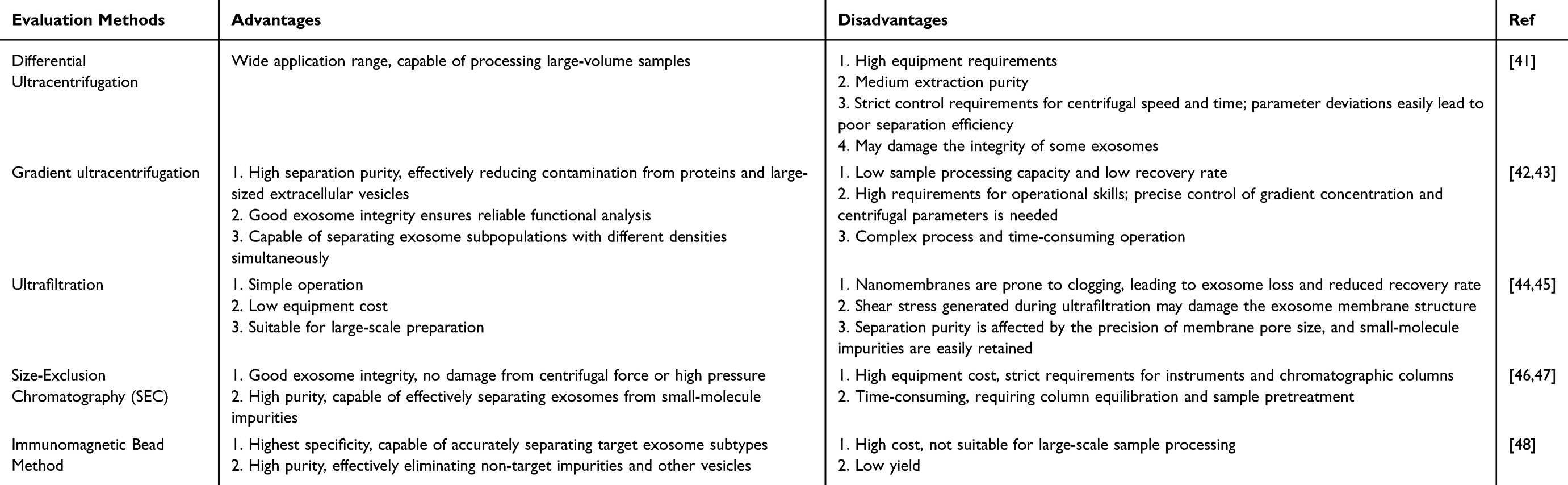 |
Table 1 Comparison of Common Methods for Isolating Exosomes |
Ultracentrifugation technology, based on high-speed centrifugation (100,000–150,000×g), enables the separation of exosomes across different size and density ranges.49 It currently serves as the gold standard for exosome isolation, with density gradient ultracentrifugation and differential speed ultracentrifugation being the two most common methods.50 Ultracentrifugation is applicable to cell culture media and various bodily fluids, enabling the acquisition of high-concentration exosomes. For instance, Yahang Liang et al successfully extracted exosome miR-106a-5p from highly metastatic colorectal cancer cells via ultracentrifugation and demonstrated its pivotal role in promoting liver metastasis.51 However, ultracentrifugation relies on expensive equipment, and high centrifugal forces may potentially damage exosome membranes.
Differential ultracentrifugation separates components based on sedimentation rates by progressively increasing centrifugal speeds: 500 × g to pellet contaminating cells, 2000 × g for cell debris, 10,000 × g for apoptotic bodies and macrovesicles, and finally 100,000 × g for exosomes.52,53 This approach is well-suited for large-volume samples and is cost-effective but requires careful control of speed and time to ensure proper separation, and it yields relatively low purity.
Density gradient centrifugation involves adding a concentrated sample to sucrose or iodixanol to form a density gradient.54 Through ultracentrifugation, exosomes (typically accumulating at densities of 1.13–1.19 g/mL) can be separated from other vesicles, reducing contamination from proteins and larger extracellular vesicles, thereby further purifying exosomes.55 However, this method has low throughput and is highly sensitive to centrifugation time.
Ultrafiltration uses pressure differences across a membrane to separate large particles like dead cells and proteins based on molecular weight cut-off, resulting in a concentrated sample that is further purified using membranes of different pore sizes for exosome isolation.56 This method is straightforward, economical, and applicable to large-scale preparation but may lead to losses due to membrane clogging, and shear stress may damage exosomes.
The immunomagnetic bead method relies on specific binding between exosome markers (eg, CD63, CD81) and antibodies, enabling the isolation of exosomes from specific sources with high purity.57,58 However, it is costly, and elution steps may damage exosomes.
Through gel filtration, SEC separates exosomes based on the differences in their particle sizes. Gels with pores of different sizes allow larger particles to elute first than smaller ones, separating components by elution time.59 Improved SEC protocols, such as the two-dimensional size-exclusion liquid chromatography (2D-SELC) platform with fully automated sampling and fraction collection, avoid human error.60 SEC maintains the natural state of exosomes with high purity and reproducibility, applicable to various samples, but equipment costs are high, and factors like pore size, packing, and flow rate affect separation.
Beyond these six approaches, alternative techniques such as polymer precipitation61 and microfluidics62 are being explored. Given the diversity of exosome sources and intended applications, no single method is universally applicable. Achieving uniform, homogeneous exosome populations will likely require hybrid strategies tailored to physicochemical properties and biological context.
Besides optimizing purification methods, strategies to boost exosome secretion are critical for large-scale applications. Modulating cell culture conditions can effectively promote exosome release; for example, subjecting cells to stress or hypoxia often induces more exosome secretion.63 Studies have confirmed that medium pH significantly affects exosome yield, with acidic conditions favoring production.64 In MSC culture systems, adding substances like fenoterol, norepinephrine or N-methyldopamine can increase exosome yield by up to threefold.64 Similarly, rapamycin and other small molecules (calcium phosphate particles and adiponectin) modulate signaling pathways such as mammalian target of rapamycin and autophagy to promote release.65–67 Advances in cell culture formats further support scalable production. Compared with conventional two-dimensional systems, three-dimensional (3D) cultures more closely mimic the in vivo microenvironment, promoting cell–cell and cell–matrix interactions. For example, umbilical cord-derived MSCs grown in microcarrier-based 3D systems secreted up to 20 times more exosomes than in 2D culture; combined with tangential flow filtration, yield increased up to 140-fold that of 2D culture, demonstrating strong potential for industrial-scale preparation.68 Additionally, exogenous physical stimulation is an effective means to promote exosome generation, including radiation and electrical stimulation. For instance, cells cultured with magnetic nanoparticles under magnetic force showed enhanced exosome generation by upregulating exosome transport and secretion pathways.69 Shanguo Zhang et al significantly increased MSC exosome secretion by applying electrical stimulation via a self-powered, portable, and efficient triboelectric nanogenerator.70
The isolation of exosomes requires multidimensional characterization to verify their physical properties, molecular composition, and functional activity, ensuring quality and application potential.
Method of Improving the Drug Delivery of Exosomes
The core bottlenecks of exosomes as drug delivery carriers include insufficient targeting accuracy, low drug loading efficiency, and short in vivo circulation half-life. These issues need to be optimized from multiple aspects such as carrier modification, drug loading optimization, and delivery assistance.
Enhancement of Targeting
By screening based on cell sources and surface engineering modification, the recognition and binding ability of exosomes to target tissues can be enhanced. Cells with natural homing properties naturally carry molecules related to the recognition of target tissues, which can significantly enhance targeting. For example, mesenchymal stem cells (MSCs) and tumor-derived cells can achieve targeted delivery without additional modification. In the experimental verification of the HT1080 cell nude mouse tumor transplantation model, exosomes derived from HeLa or HT1080 cell sources all demonstrated the ability to target and homing to the same tumor tissue.71 The membrane surface of mesenchymal stem cells is richly distributed with receptor molecules (CD29, CD90, CD105, etc.), covering various types of chemokine receptors and growth factor receptors. The signaling molecules released at the tumor site will precisely match the surface receptors of MSCs, thereby guiding MSCs to migrate towards the tumor microenvironment. Therefore, MSCs and their secreted exosomes have the potential to actively target tumor tissues and exert anti-tumor effects.72 To improve the targeting treatment effect of CD24-expressing tumors (such as ovarian cancer ID8 cells, triple-negative breast cancer 4T1 cells), Li et al transfected M0-type macrophages (Raw264.7) with Siglec-10 by lentivirus, induced polarization, and prepared targeted exosomes SM1Aexo by compression method. In vitro experiments confirmed that SM1Aexo had a significantly better CD24-blocking effect on ID8 cells and 4T1 cells than wild-type M1 exosomes (M1Aexo), and could increase the phagocytosis rate of bone marrow-derived macrophages (BMDMs) for EGFP-labeled tumor cells by 3.47 times. In vivo studies further demonstrated that when this targeted exosome was coupled with alginate oxide (OSA) to form a hydrogel delivery system, it could significantly inhibit ovarian cancer peritoneal metastasis and the growth of triple-negative breast cancer, providing a typical example for enhancing the efficiency of tumor delivery by targeted modification of exosomes.73
Optimization of Drug Loading Efficiency
Exogenous drugs can be introduced into exosomes through passive and active methods, such as co-incubation, ultrasound treatment, electroporation, and freeze-thaw cycles, and these have been well reviewed (Figure 2). 74 There are two types of passive loading. One is to incubate the drug with the source cells, and then the exosomes released by the source cells will contain the target drug (Figure 2ai). The other is to adopt a direct co-incubation strategy, where the exosomes and the drug are incubated in the same system, and the drug molecules can penetrate the membrane structure of the exosomes in a concentration-dependent manner,75 thereby completing the delivery into the interior of the vesicles (Figure 2aii). Previous studies have shown that co-incubating exosomes with drug-loaded liposomes can also result in higher drug loading and better targeting ability for hybrid exosomes.76 The electroporation technique can cause the exosome membrane to form micropore channels, creating conditions for the inward flow of drug molecules, and this method is currently regarded as the preferred strategy for loading nucleic acid substances (Figure 2bi). The ultrasonic mechanical shear force can cause the exosome membrane to deform,77 enabling effective loading of drugs (Figure 2bii). Detergents can remove cholesterol from the exosome membrane, reduce the membrane barrier, and facilitate the loading of macromolecular drugs, and are commonly used methods for loading hemolytic drugs (Figure 2biii). When loading exosomes with drugs using the compression method, lipid bilayer fragments can achieve self-assembly into spherical structures, and through treatment with filters of different pore diameters,78 it is conducive to preparing exosomes with uniform particle size (Figure 2biv). The freeze-thaw cycle is characterized by 3–5 cycles of room temperature and −80°C alternation,79 forming pores on the exosome membrane surface to help drugs enter the interior, but this method may damage the structure and properties of the exosomes (Figure 2bv).
 |
Figure 2 Various technologies for drug loading into Extracellular vesicles (EVs). (a) Passive Loading: i) Direct co-culture of drugs and cells: Achieved through varying drug concentration gradients, suitable for hydrophobic drugs and small-dose medications; ii) Incubation: Directly mix hydrophobic drugs and small molecule drugs with purified exosomes. (b) Active Loading: i) Electroporation: Membrane electroporation under electric current, which creates temporary pores, is suitable for the loading of hydrophilic drugs and is considered the preferred method for loading nucleic acid molecular. ii) Sonication: Capable of loading hydrophilic molecules, though some small molecules may reside on the exterior of EVs. iii) Detergent treatment: Detergents remove cholesterol from membranes, allowing for the loading of large molecules, typically used for hemolytic drugs. iv) Extrusion: Fusing drugs with the membranes of extruded and fragmented EVs. v) Freeze-thaw cycles: This method can load drugs into exosomes, but it may alter the properties of the EVs. Reprinted with permission from,74 Copyright 2024 MDPI. |
Extension of Intra-Body Circulation
Extending the in-body circulation time of exosomes is one of the key steps to enhance their drug delivery efficiency. It can effectively reduce the clearance by the reticuloendothelial system and provide sufficient time for exosomes to target and accumulate at the lesion site. In recent years, several studies have achieved significant extension of exosome in-body circulation through surface modification or functional fragment integration strategies. For instance, researchers constructed an “immune invisible” barrier by surface modification with biocompatible polymers. Using polyoxazoline (POx) as a substitute for traditional PEG, they modified extracellular vesicles (EVs) derived from mesenchymal stem cells (MSCs). This modification strategy successfully reduced the recognition and clearance by the immune system while retaining the inherent immune regulatory function of MSC-EVs, increasing the blood half-life of EVs after intravenous injection by 6 times.80 At the same time, the extension of circulation time significantly improved the targeting accumulation efficiency of EVs in tumor tissues, laying the foundation for the subsequent drug action. Integrating functional fragments to inhibit macrophage phagocytosis is beneficial for extending the retention time of the exosome system in the in-body environment. Exosomes are easily cleared by the liver and spleen mononuclear phagocyte system (MPS) in the body, with limited circulation time, which affects the targeted delivery efficiency. To solve this problem, Justin F. Creeden et al constructed CD9-engineered intelligent exosomes (ExoSmart), which were co-modified with CD47p110–130 functional fragments and RGD targeting peptides. CD47p110–130 can specifically bind to SIRPα on the surface of macrophages and inhibit phagocytosis by transmitting the “don’t eat me” signal, significantly reducing the clearance rate of exosomes in RES organs.81 The RGD peptide integrates with the αvβ3 integrin highly expressed in pancreatic ductal adenocarcinoma (PDAC) cells to achieve active targeting, converting the advantage of extended circulation into efficient enrichment at the lesion site. This study confirmed that through the anti-clearance modification mediated by the CD47-SIRPα axis, it is possible to maintain the natural carrier advantage of exosomes while optimizing their circulation stability, and when combined with targeted modification, it can significantly enhance the therapeutic effect of PDAC treatment, providing a reference for the improvement of the in-body circulation of the exosome drug delivery system.
Characterization of Exosomes
The reliable use of exosomes requires rigorous, multidimensional characterization to confirm their physical properties, molecular composition, and functional activity. Together, these analyses ensure sample quality, reproducibility, and application potential.
Physical Characterization
Imaging techniques provide direct insights into exosome morphology and size. TEM typically depicts the cup-shaped appearance of exosomes, while cryoelectron microscopy (cryo-EM) captures their native spherical form.82 Complementary techniques such as dynamic light scattering (DLS) and NTA assess size distribution and concentration, confirming the characteristic 30–150 nm range.83 Zeta potential analysis further evaluates surface charge, with the naturally negative membrane potential contributing to exosome stability during circulation.84
Molecular Composition Analysis
Detecting specific molecular markers is essential for confirming exosome origin and integrity. At the protein level, this includes transmembrane proteins such as CD63, CD82, and CD9, transport and fusion-related proteins such as annexins and heat shock proteins, and adhesion-related proteins such as integrins.85 Techniques including Western blotting, flow cytometry, and immunofluorescence allow both qualitative and quantitative assessment.86 At the nucleic acid level, exosomes carry diverse cargo including messenger RNA (mRNA) and other nucleic acid molecules. Reverse transcription-polymerase chain reaction, sequencing, and related methods identify and quantify these molecules, with expression profiles often reflecting the physiological state of source cells.87 Emerging omics approaches, particularly proteomics, are increasingly used to comprehensively map protein composition and abundance, supporting the establishment of quality control standards.
Functional Activity Analysis
Since therapeutic efficacy depends on biological function, functional assays are critical. These typically include evaluating cellular uptake efficiency, in vivo biodistribution, and therapeutic outcomes in relevant models. For example, the binding affinity between the modified exosomes and tumor cells was verified by using in vitro fluorescence microscopy and flow cytometry to confirm the targeting ability; in vivo, the enrichment effect in tumor tissues was verified through near-infrared fluorescence imaging and bioluminescence imaging. In vitro, the anti-tumor effect of the drug-loaded exosomes was evaluated by the CCK-8 cell viability assay; in vivo, the tumor suppression rate was verified through a tumor xenograft mo for immunotherapy-related exosomes, immune activation indicators such as DC maturation and CD8+T cell proliferation ability were detected by flow cytometry.88,89 Such studies verify whether exosomes not only retain structural integrity but also deliver their cargo effectively to target cells, thereby validating their use as drug carriers or diagnostic tools.
Comprehensive characterization of exosomes is an important part of exosome quality control. To ensure the therapeutic activity and homogeneity of exosomes, a systematic assessment of their purity, safety, and consistency is necessary. Among them, the purity of exosomes can be ensured by detecting the positive expression levels of typical exosome markers such as CD9 and CD63, and screening for the presence of plasma contaminants such as apolipoprotein A (ApoA), thereby eliminating the interference of impurities and ensuring the high purity of the samples. Through quality control, contaminants such as viruses and impurities in the culture medium can be avoided from contaminating exosomes, thus meeting the requirements of clinical applications.90 The batch stability of exosomes is easily affected by various factors, such as the type of source cells, the number of cell passages, and the method of drug loading.91 There may be differences between different batches. Therefore, the consistency check needs to be throughout the entire process of exosome quality control. By strictly standardizing the cultivation requirements of source cells, optimizing the steps of separation and purification, and other key operations, ensure that the exosomes from different batches maintain stable consistency in core indicators such as particle size, drug loading rate, and expression levels of characteristic markers, to ensure the reliability of therapeutic effects.
Engineering Methods to Enhance Exosome Tumor Targeting
Natural exosomes, as endogenous nanovesicles, although having low immunogenicity, good biocompatibility and the ability to cross biological barriers, have limited targeting ability, low drug loading efficiency, poor process controllability, lack of responsiveness to pathological microenvironments or the intelligence for collaborative diagnosis and treatment, their inherent limitations seriously limit the potential for clinical application. In addition, natural exosomes also exhibit significant heterogeneity and bottlenecks in large-scale production, making quality control and standardized application challenging. This highlights the necessity of engineering exosomes.
Engineered exosomes are nanovesicles modified to optimize the structure, composition, or function of natural exosomes. Through precise design, they can be endowed with improved targeting ability, greater drug-loading capacity, and enhanced bioactivity.92 Compared with their natural counterparts, engineered exosomes overcome key limitations such as low intrinsic targeting and limited loading efficiency, making them more suitable for therapeutic and diagnostic applications. Strategies for engineering exosomes generally fall into two categories: drug loading and surface modification. This section focuses on surface modifications, which are designed to enhance tumor-targeting efficiency. Approaches include chemical modification, physical modification, and genetic engineering of exosome-producing cells (Figure 3). These strategies enable the addition of targeting ligands, functional molecules, or responsive elements to exosome membranes, thereby improving their precision in recognizing and interacting with tumor cells.
 |
Figure 3 Engineering modifications for enhancing exosome tumor-targeted therapy. Created with Biorender. |
Chemical Modification
Chemical modification introduces targeting molecules onto the exosome surface through well-defined reactions, thereby enhancing their ability to selectively recognize tumor cells. A major advantage of this approach is the broad choice of targeting ligands—including antibodies, peptides, and aptamers—that can be tailored to different tumor types. For instance, human epidermal growth factor receptor2 (HER2) antibodies have been used to target HER2-overexpressing breast cancer,93 RGD peptides bind integrins commonly found on tumor cells,94 and the AP-9R aptamer specifically recognizes annexin A2 on lung cancer cells, supporting applications in diagnosis and therapy.95 Chemical modification methods are generally mild, controllable, and versatile, making them a cornerstone of exosome engineering. Two major strategies are used: covalent binding and non-covalent binding.
Covalent binding: Exosome membranes contain abundant reactive groups, such as thiols and amines, that allow covalent attachment of ligands via amidation reactions, thiol-maleimide reactions, and click chemistry. Amidation reactions link activated carboxyl groups on targeting ligands with amines on exosomal proteins, forming stable amide bonds. Jun Yeong Kim et al quantified the amount of amine groups in exosomal proteins using fluorescamine reagent, then activated the amines on exosomes at an amine/NHS molar ratio of 1:2 for 3 hours, facilitating easier binding to i-motif targeting heads.96 This method offers high modification efficiency and stable linkage between targeting molecules and exosomes, effectively improving exosome recognition and binding to tumor cells. Maleimide groups specifically react with thiols to form stable thioether bonds.97 This reaction is highly specific and selective, proceeding efficiently under mild physiological pH conditions. Chaoxiang Chen et al used TCEP (a mild reducing agent) to reduce disulfide bonds on milk-derived exosomes to thiols, then conjugated ligands to the exosome surface via thiol-maleimide reactions, demonstrating the feasibility of ligand modification without damaging vesicle structure.98 Common click chemistry reactions, including copper-catalyzed azide-alkyne cycloaddition and strain-promoted copper-free click reactions (eg, between azides and dibenzocyclooctyne (DBCO)), form stable triazole rings through azide-alkyne reactions.99 These reactions are efficient, highly specific, and occur under mild conditions, widely used in chemical biology. For example, Miao Deng et al labeled tumor cells and tumor-derived exosomes with azide groups while modifying nanoparticles with alkyne groups (DBCO); specific binding via bioorthogonal click reaction formed stable covalent bonds.100 This study tracked and downregulated exosomal macrophage migration inhibitory factor, inhibiting the role of tumor-derived exosomes in inducing pre-metastatic niche formation in the liver and promoting tumor metastasis, showing good therapeutic effects against pancreatic cancer. Dumei Ma et al successfully distinguished breast cancer patients from healthy individuals by detecting serum exosomes via click reaction.101
Non-covalent binding: Non-covalent approaches rely on electrostatic attraction and hydrophobic interactions. Positively charged ligands can associate with the negatively charged exosome membrane through electrostatic forces, while hydrophobic ligands insert into the lipid bilayer.102,103 1,2-Distearoyl-sn-glycero-3-phosphoethanolamine-Polyethylene Glycol (DSPE-PEG), a lipid-like material, integrates functional molecules into the exosome surface through hydrophobic interactions with membrane lipids and renders exosomes stealthy, reducing protein adsorption and enhancing tumor targeting.104 For instance, Rui Deng et al first reacted DSPE-PEG2000-maleimid (DSPE-PEG2000-Mal) with a thiol group of a hepatocyte-targeting cell-penetrating peptide (HTPP) to form DSPE-PEG2000-HTPP, then utilized the hydrophobicity of DSPE to fuse into the exosome lipid bilayer, ultimately modifying HTPP onto the exosome surface via a linker.105 This study showed that HTPP-modified exosomes significantly enhanced targeting to liver cancer. This method is relatively simple and has minimal impact on exosome bioactivity, but binding stability is poorer than covalent binding. Examples of exosome systems based on chemical modification for tumor therapy are summarized in Table 2.
 |
Table 2 Examples of Exosome Systems Based on Chemical Modification for Tumor Therapy |
Physical Modification
Physical modification employs external stimuli or mechanical processes to alter exosome membranes, enabling functional enhancement or hybridization with other nanostructures. Common approaches include freeze–thaw cycles, membrane extrusion, sonication, and magnetic guidance.111 The fusion with lipid nanoparticle (LNP) is a typical direction. Compared to LNPs, the endogenous lipid membrane structure of exosomes can significantly reduce immunogenicity and liver toxicity. The inherent tumor-targeting property of exosomes combined with surface engineering modifications (such as targeting peptide conjugation) can precisely target tumors, overcoming the problem of LNPs’ tendency to accumulate non-targetedly in the liver and spleen.112 At the delivery level, exosomes can simultaneously carry various bioactive molecules such as proteins, nucleic acids, and lipids, having better compatibility than the single loading characteristic of LNPs. However, there are also some issues with exosomes, such as the reliance on complex processes like ultracentrifugation for separation and purification, which are time-consuming and difficult to control in terms of purity, and the greater difficulty in large-scale production compared to LNPs.113 Exosomes are prone to aggregation during storage, while LNPs have an advantage in improving the stability of the formulation (such as PEG modification and optimization of lipid components).114 Based on this, the hybrid exosome - LNP can achieve complementary advantages through physical fusion (such as ultrasound-mediated membrane fusion or forced assembly by extrusion). By leveraging the structural stability and mature preparation process of LNPs, the problems of exosome storage and large-scale production can be improved. The low immunogenicity, targeting ability, and multi-load capacity carried by exosomes are conducive to the construction of an efficient delivery system with both biocompatibility and formulation stability. Zhuo et al’s cholesterol-enriched milk-derived exosomes (MEs) exemplify this complementary strategy. Modulating cholesterol content in the exosomal membrane enhances fusion with cancer cell membranes, preserving exosomes’ inherent low toxicity and precise targeting while enabling efficient cytosolic siRNA delivery.115 Xu et al incorporated macrophage-derived exosomes into ROS-responsive cationic liposomes to construct a biomimetic nanocomplex (E-cLip-DTX/si) for the co-delivery of DTX and Bcl-2 siRNA. In the mouse model of breast cancer, this nanosystem exhibited potent tumor growth inhibition and extended in vivo circulation time.116 Compared with chemical modification, these methods are simpler, avoid introducing reactive chemicals, and preserve most of the intrinsic properties of exosomes.
Freeze–Thaw Cycles
Repeated freezing and thawing destabilize lipid bilayers, promoting fusion between exosomes and other vesicles. Yifan Gao et al applied this method to fuse liposomes with γδ T cell–derived exosomes, achieving targeted photoimmunotherapy for melanoma while retaining the exosomes’ antiviral activity against Epstein–Barr virus–related tumors.117 Similarly, Lili Cheng et al reported a fusion efficiency of 97.4% when combining thermosensitive liposomes with genetically engineered exosomes through three freeze–thaw cycles.118
Membrane Extrusion
This widely used technique forces exosomes and other lipid vesicles (eg, liposomes) through polycarbonate membranes under mechanical pressure, inducing membrane remodeling and fusion.119 The resulting hybrid exosomes display uniform size and preserved biological function. Yang Yang et al demonstrated this by hybridizing Raw 264.7 cell–derived exosomes with liposomes, then loading thalidomide to regulate Treg cells for tumor immunotherapy.120 In another example, autologous breast cancer cell–derived exosomes were fused with liposomes via incubation–extrusion, yielding a hybrid system for combined chemo-, immuno-, and photothermal therapy.121 Beyond mammalian systems, Rufan Huang et al fused grapefruit-derived exosomes with engineered mesenchymal stem cell–derived exosomes, producing hybrid vesicles with strong inflammatory tissue tropism.122
Sonication
Ultrasound disrupts and reassembles membranes, enabling the fusion of exosomes with other vesicles. Yong-Yu Jhan et al sonicated liposome–exosome mixtures at different ratios, followed by extrusion, producing hybrid exosomes of ~100 nm. This method increased vesicle yield by 6- to 43-fold compared with conventional approaches.123
Magnetic Guidance
Incorporating magnetic nanoparticles into or onto exosomes enables external magnetic fields to direct their accumulation at tumor sites. Mengran Li et al coated exosome membranes onto magnetic mesoporous silica nanoparticles.124 These constructs not only evaded immune clearance and exhibited tumor tropism but also achieved rapid, magnetically controlled accumulation at tumor sites. Capture efficiency for homologous tumor cells reached 94.37% ± 1.3%.
Overall, physical modification offers a straightforward way to enhance exosome function while preserving their biochemical composition. However, to achieve optimal tumor targeting, these methods are often combined with chemical or genetic modifications. Representative examples of exosome systems based on physical modification for tumor therapy are summarized in Table 3.
 |
Table 3 Examples of Exosome Systems Based on Physical Modification for Tumor Therapy |
Genetic Modification
Genetic modification enhances tumor targeting by engineering the parent cells to express specific ligands or proteins on the exosome surface. These exosomes can then bind selectively to receptors on tumor cells or within the tumor microenvironment, improving delivery precision and therapeutic efficacy.
For example, Lingmin Zhang et al used CRISPR/Cas9 knock-in technology to fuse a tumor-associated macrophage (TAM)-targeting peptide (CRV) to the lysosome-associated membrane glycoprotein 2 gene.128 The resulting exosomes displayed high levels of CRV on their surface, effectively reprogramming M2-type TAMs into tumoricidal M1-type macrophages and suppressing metastasis. Similarly, Jung Hyun Park et al genetically modified adipose-derived stem cells with a pDisplay vector encoding the DE532 peptide, which binds gastric cancer MKN45 cells. The resulting DE532 exosomes, when loaded with alvespimycin hydrochloride (17-DMAG), achieved precise targeting and reduced drug-associated toxicity.129 In another study, Peishan Li et al transfected HEK293T cells with a constructed PD1 high-expression plasmid, selected stably transfected cell lines (PD1-HEK 293T cells) with puromycin, and obtained recombinant PD1 protein exosomes (PD1 Exo) after purification and isolation.130 PD1 Exo helps address key issues of immune exhaustion and escape in tumor immune checkpoint therapy.
The key advantage of genetic modification lies in the natural expression and assembly of targeting molecules during exosome biogenesis, ensuring high compatibility and preserving vesicle integrity. However, this strategy remains technically demanding, with challenges including high cost, complex procedures, and variable transfection efficiency.131 Representative exosome systems based on genetic modification for tumor therapy are summarized in Table 4.
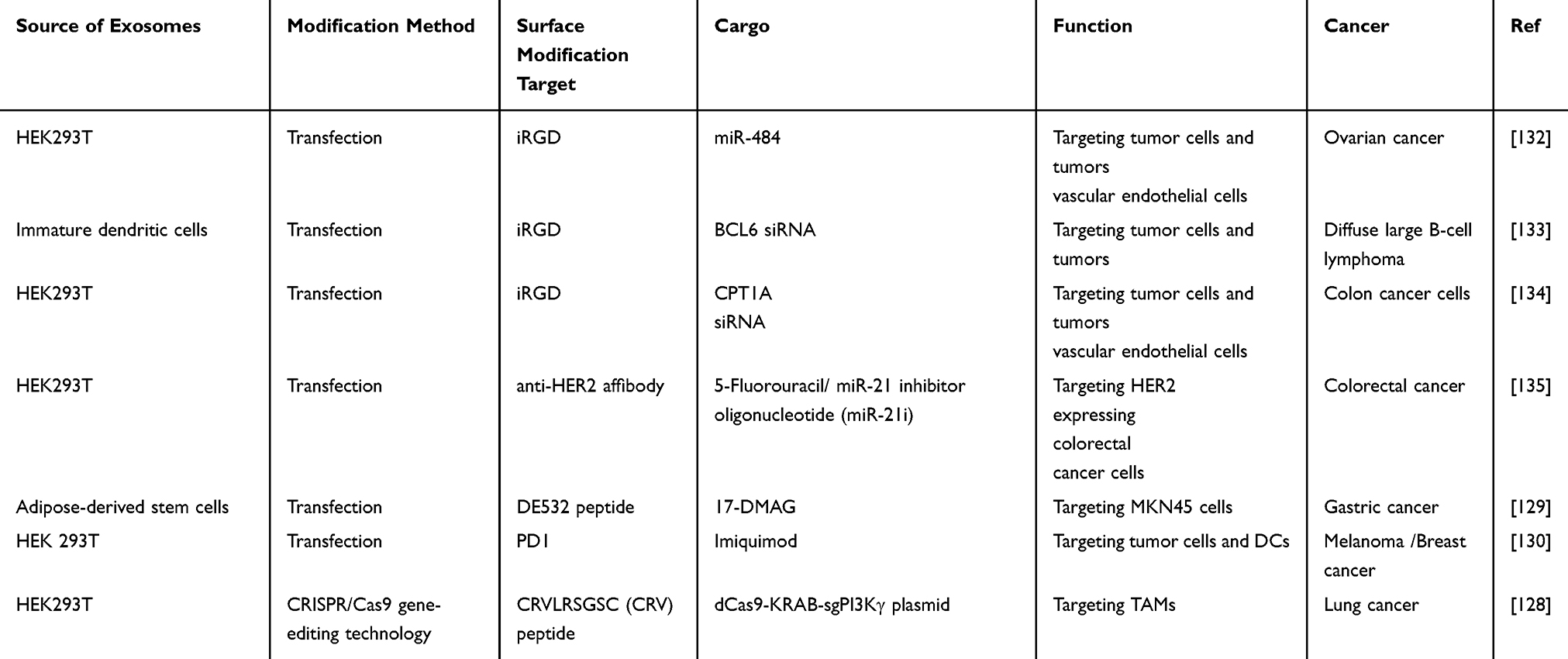 |
Table 4 Examples of Exosome Systems Based on Gene Modification for Tumor Therapy |
Applications of Exosomes in Tumor-Targeted Therapy
Exosome-based nanosystems, as natural vesicles, offer distinct advantages including excellent biocompatibility, minimal immunogenicity, and effective delivery of therapeutic cargo. These features make them highly attractive for tumor-targeted applications. By harnessing these properties, exosome platforms not only overcome limitations of conventional single-agent therapies but also create opportunities for synergistic effects in combination strategies. We have summarized and organized the advantages of the exosome delivery system compared to the traditional delivery system in different treatment methods in the form of a table (Table 5). Building on these strengths, exosome systems are increasingly being investigated across diverse therapeutic modalities. The following sections detail their specific roles in various treatment modalities from two dimensions: single therapy and combined immunotherapy (Figure 4).
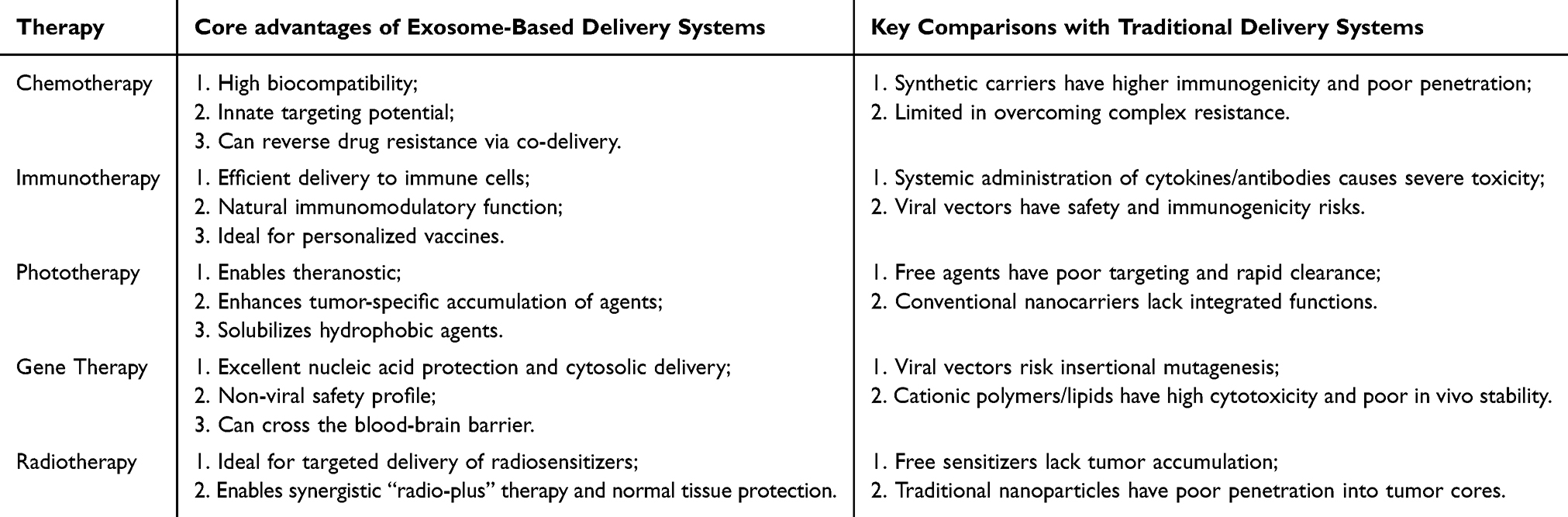 |
Table 5 The Advantages of Exosome-Based Delivery Systems in Different Tumor Treatment |
 |
Figure 4 Exosome-based nanosystem strategies for tumor therapy. Created with Biorender. |
Single Therapy Modalities
Chemotherapy
Exosomes can efficiently encapsulate chemotherapeutic drugs and deliver them to tumor tissues, thereby enhancing antitumor activity while minimizing systemic toxicity. This makes them highly suitable for chemotherapy across different cancer types. For instance, Tooba Soudi et al developed a co-delivery system using exosomes loaded with carvedilol and 5-fluorouracil, which synergistically improved treatment outcomes in breast cancer.136 Exosomes derived from healthy epithelial breast cells, loaded with lapatinib via electroporation, inhibited the proliferation and promoted apoptosis of SKBR3 breast cancer cells at low doses.137 This result confirms that exosomes as drug carriers can achieve low-dose breast cancer treatment, improve drug efficacy and chemotherapeutic index, and enhance cancer treatment effects.
Exosomes possess natural tumor tissue targeting tropism. Biomimetic artificial exosomes constructed based on this biological characteristic can enhance the accumulation of chemotherapeutic drugs in homotypic tumors. Artificial exosomes prepared by modifying phospholipid liposomes with cancer cell membrane proteins not only have high yield and are easy to prepare but also specifically deliver loaded doxorubicin and vorinostat, effectively inhibiting non-small cell lung cancer growth with no significant adverse reactions.138 Engineering modifications to add targeting molecules to the exosome surface can further improve the efficacy of tumor-targeted therapy. Pratiksha Tiwari et al modified exosomes with the laminin receptor-binding peptide YIGSR (YIGSR-Exo) and formed hybrid-Exo with lipid-polymer nanoparticles (LPNP) via freeze-thaw method, then loaded dasatinib (DST) to form DST-FuNP@YIGSR-Exo (Figure 5a). Confocal microscopy revealed that DST-FuNP@YIGSR-Exo selectively accumulated in cancer cells highly expressing laminin receptors via receptor-mediated endocytosis, ensuring effective drug delivery (Figure 5b–d).139
 |
Figure 5 (a) Schematic of Exosome fusion with LPNPs; (b) Confocal images of Nile red labeled localization after 2 and 6 hours of treatment in MDA-MB-231 cells; (c) Quantitative histogram data of Nile red labeled confocal images located in MDA-MB-231 cells after 2 and 6 hours of treatment; (d) Flow cytometry provided quantitative evaluation of cell uptake after 2 and 6 hours of incubation in MDA-MB-231 cells. Reprinted with permission from.139 Copyright 2025 John Wiley and Sons; (e) Schematic diagram of preparation of exosomes loaded with DOX and Boron clusters, and their application in treating DOX-Resistant breast cancer cells and promoting cell apoptosis. Reprinted with permission from.140 Copyright 2024 John Wiley and Sons. |
Notably, exosomes can cross the blood-brain barrier (BBB) through mechanisms such as receptor-mediated endocytosis and lipid raft-mediated micropinocytosis via specific proteins.141,142 Therefore, compared with traditional nanodelivery systems, exosomes have a natural advantage in drug delivery for brain tumors. Sana Shaikh et al demonstrated through in vivo computed tomography (CT) imaging experiments that loading temozolomide (TMZ) and gold nanoparticles (AuNPs) via M1 macrophage exosomes could deliver them to the brain, overcoming the BBB, and efficiently inhibit glioblastoma growth.143 Additionally, resistance induced by traditional chemotherapy often leads to tumor recurrence, a major cause of cancer death. Using exosome drug delivery systems can effectively overcome chemotherapy resistance. The mechanism of chemotherapy resistance is mainly related to the overexpression of P-glycoprotein (P-gp), which actively pumps drugs out of cells using ATP energy, leading to chemotherapy resistance.144 When exosomes are used as the delivery system, their endocytosis process also consumes ATP, which can competitively inhibit P-gp-mediated drug efflux, thereby alleviating resistance.145 Yi-Ru Bao et al formed a supramolecular complex of boron clusters and doxorubicin (DOX), increasing the drug loading capacity of DOX in exosomes, and utilized the ATP consumption effect of exosomes to alleviate DOX resistance in breast cancer cells (Figure 5e). 140
Immunotherapy
The core of immunotherapy is to utilize the human immune system to activate immune responses (especially CD4+/CD8+ T cells) to eliminate tumors.146 Some types of exosomes carry information that helps activate immune responses.147 Current exosome-based immunotherapies are mainly divided into cancer vaccines, immune checkpoint therapies, and adoptive cell therapies. Xinyi Zhang et al have previously reviewed this in detail.148 Tumor cell-derived exosome vaccines contain various tumor antigens and are promising vaccine development carriers. Yan Zou et al prepared a tumor nanovaccine platform (Exo-CpG) by loading CpG oligodeoxynucleotides (CpG) onto homologous exosomes derived from glioblastoma multiforme (GBM), which can effectively target lymph nodes, activate immune responses, and induce long-term immune memory, achieving a 100% survival rate in tumor-bearing mice within 5 months and effectively preventing metastasis.149 Similarly, Shanshan Li et al constructed a GBM tumor-derived exosome nanosystem loaded with CpG adjuvant (Exo-CpG) (Figure 6a). This system can effectively cross the BBB and accumulate in the brain (Figure 6b and c), activate the body’s innate immunity through facilitating the maturation of antigen-presenting DCs in lymph nodes and eliciting potent T-cell responses, achieving GBM proliferation inhibition and inducing durable protective immunity; when combined with anti-programmed cell death ligand-1 antibody (aPD-L1), it effectively inhibited the growth of GBM in immunocompetent mice with orthotopic primary GL261 models and immunosuppressive CT2A models, prolonging mouse survival (Figure 6d and e).150 Furthermore, engineering modifications of exosomes can enhance their immunogenicity. Lanxiang Huan et al genetically engineered allogeneic breast cancer-derived exosomes to overexpress α-lactalbumin (α-LA), enhancing exosome targeting and immunogenicity, and loaded them with immunogenic cell death (ICD) inducers human neutrophil elastase (ELANE) and Hiltonol (TLR3 agonist) to create HELA-Exos in situ vaccines. HELA-Exos target the tumor microenvironment, stimulate the maturation of cDC1s, improve CD8+T cell responses, and exhibit strong antitumor activity in mouse models and human breast cancer organoids.151
 |
Figure 6 (a) Schematic diagram of a multifunctional platform based on natural GBM-derived exosomes loaded with CpG for enhancing immune response in the treatment of primary GBM and its recurrences; (b) Fluorescence intensity of Exo CpG loaded with Cy5, as detected in the lower chamber of the in vitro BBB transwell model at different time intervals (n = 3, ****p < 0.0001); (c) Ex vivo fluorescence imaging; (d) Survival curves of the treated GL261-luc tumor-bearing mice (n = 3, *p <0.05, **p < 0.01); (e) Survival curves of the treated CT2A-luc tumor-bearing mice (n = 3, *p <0.1). Reprinted with permission from150. Copyright 2025 Elsevier. LN homing capacity of EmDEX@GA (f and g). (f) Representative in vivo fluorescence images of mice with intratumoral administration of distinct exosome groups; (g) Representative immunofluorescence images of inguinal TDLN of mice in EmDEX@GA group (blue: DAPI; yellow: CD3; green: CD11c; red: DiR-labeled exosomes; scale bar: 500 μm, 50 μm for (i, ii), 20 μm for (iii, iv)). Reprinted with permission from152. Copyright 2025 John Wiley and Sons. |
The expression of immunosuppressive molecules such as PD-1/PD-L1 and CTLA4 on tumor cells and myeloid-derived suppressor cells prevents T cells from recognizing and attacking tumors, leading to tumor development and metastasis.153 Immune checkpoint inhibitors have been widely used clinically to enhance antitumor immune responses. PD-1/PD-L1 immune checkpoint inhibitors are the most commonly used immunotherapy strategy and have achieved good therapeutic effects in tumors such as liver cancer, gastric cancer, non-small cell lung cancer, and breast cancer.154,155 Notably, besides tumor tissue, myeloid cells (eg, DCs and macrophages) in tumor-draining lymph nodes (TDLNs) also express PD-L1 during the initial phase of immune response activation, which directly limits T cell initiation and proliferation.156 Based on this, Yizhen Wang et al designed and developed engineered dendritic cell exosomes targeting TDLNs (EmDEX@GA). Mature DC-derived exosomes highly express CC chemokine receptor 7, which can bind to CC chemokine ligand 19 (CCL19) and CCL21, achieving LN homing. Moreover, they could make DC-derived exosomes overexpress PD-1 through genetic transfection for local PD-1/PD-L1 blockade and immune microenvironment remodeling in TDLNs. Finally, the stimulator of interferon genes (STING) agonist 2’,3’-cyclic GMP-AMP (cGAMP) was loaded into the exosomes to form EmDEX@GA. EmDEX@GA achieved TDLN targeting (Figure 6f and g), blocked DC-derived PD-L1, and enhanced antitumor immunity by activating the STING pathway. Compared with systemic immune checkpoint blockade, local immunotherapy using EmDEX@GA showed superior effects in inhibiting distant metastasis.152
Adoptive cell therapies, represented by CAR-T, have become new hopes for conquering tumors. CAR exosomes contain the same cytotoxic granules as their parent cells and show significant antitumor activity in vitro and in animal models.157 Exosomes derived from CAR-T cells carry CAR on their surface, highly express cytotoxic molecules, but do not express PD1; treatment with recombinant PD-L1 does not reduce their tumor therapeutic effect.158 Compared with CAR-T, exosomes deliver therapeutic agents to tumor cells more broadly and are less likely to cause adverse reactions.159
Phototherapy
Phototherapy mainly includes photothermal therapy (PTT) and photodynamic therapy (PDT), which use near-infrared light (NIR) to excite photosensitizers at the target site to generate heat and reactive oxygen species (ROS), respectively, for tumor treatment.160,161 Compared with other treatments, phototherapy is site-specific, as the light source only affects the irradiated tumor area, offering good safety. However, the penetration depth of visible light (<2 mm) and NIR light (~1 cm) into tumors is limited, so phototherapy is usually applied to superficial tumors (eg, skin cancer, breast cancer).162 Most clinical photosensitizers (eg, porphyrins, indocyanine green) are hydrophobic and prone to aggregation and precipitation in vivo, leading to reduced activity or delivery efficiency.163 Delivering photosensitizers via exosomes can improve the poor targeting, low biocompatibility, and insufficient tumor penetration issues associated with traditional photosensitizer delivery. Exosomes can load photosensitizers via hydrophobic interactions, forming stable exosome-photosensitizer complexes that avoid enzymatic degradation or rapid clearance, prolonging half-life. For example, Jing Liu et al loaded black phosphorus quantum dots (BPQD) into exosome membranes via electroporation. On one hand, this protected BPQD from degradation in vivo; on the other hand, the size of exosomes allowed better penetration into deep tumors, improving the efficiency of photothermal tumor therapy (Figure 7a). 164
 |
Figure 7 Exosome-Based phototherapy Systems: (a) Schematic Diagram of BE Synthesis via Packaging BPQD into EXO Carriers by Electroporation, and BE Enabling Effective Tumor Penetration and PTT During Treatment. Reprinted with permission from164. Copyright 2021 John Wiley and Sons; (b) Schematic Diagram of Cancer Cell-Derived Exosomes Co-Loaded with AIEgens and PPI for Tumor Glutamine Starvation Therapy and Enhanced Type I Photodynamic Therapy. Reprinted with permission from.165 Copyright 2022 Elsevier.; (c) Schematic of Stellate Plasmonic Exosomes Applied in Penetrative Targeting for Tumor NIR-II Thermo-Radiotherapy. Reprinted with permission from.166 Copyright 2020 American Chemical Society. |
Tumor cell-derived exosomes and engineered exosomes can enhance phototherapy efficacy by improving the tumor targeting and penetration of photosensitizers. Daoming Zhu et al designed a tumor-derived exosome system (CPT) co-loaded with aggregation-induced emission luminogens (AIEgens) and proton pump inhibitors (PPI). AIEgens can break through the limitations of the tumor hypoxic microenvironment and efficiently induce ROS generation; PPI targets the glutamine metabolic pathway, inhibiting this process to reduce glutathione (GSH) synthesis, thereby lifting GSH’s inhibition of ROS’s antitumor effects; exosomes, leveraging their natural tumor-targeting properties, precisely deliver the two therapeutic agents to the tumor region, achieving over 90% tumor inhibition rate through the dual therapy of photodynamics and glutamine starvation (Figure 7b). 165 Similarly, loading gold nanostars into CT-26 tumor cell exosomes achieved tumor-targeted aggregation for photothermal therapy and photoacoustic imaging (Figure 7c). 166
Besides using tumor cell-derived exosomes, exosomes from engineered cells are more suitable for genetic editing and engineering for phototherapy. Jianbing Du et al obtained exosomes with high surface expression of “don’t eat me” CD47 (ExosCD)47 by transfecting HEK293T cells. ExosCD47 loaded with ferroptosis inducer (Erastin, Er) and photosensitizer (Rose Bengal, RB), relying on CD47-mediated “don’t eat me” immune escape signals, effectively evaded recognition and clearance by the mononuclear phagocyte system (MPS), significantly increasing accumulation concentration in tumor tissue, building a stable “drug reservoir” for subsequent treatment.167 Jiwoong Cho et al used milk-derived exosomes to deliver the photosensitizer chlorin e6 (Ce6@mExo). After oral administration, it effectively crossed the intestinal epithelial barrier and BBB, delivering large amounts of photosensitizer to glioblastoma, enhancing photodynamic therapy for glioblastoma.168
Additionally, immune cell-derived exosomes show unique value in remodeling the tumor immune microenvironment and activating antitumor immune synergy. Tianyi Kang et al precisely incorporated optimized D-A coordinated Ir(III) photosensitizers into M1 phenotype macrophage-derived exosomes. Leveraging the targeting and immunomodulatory properties of exosomes, they not only efficiently delivered photosensitizers for photodynamic therapy but also drove the “reprogramming” of tumor-associated macrophages from an immunosuppressive state to an antitumor activated state, achieving deep synergy between photodynamics and immunotherapy, injecting new vitality into comprehensive tumor therapy.169
Gene Therapy
The core of gene therapy is to deliver exogenous genetic material into the patient’s target cells to correct or compensate for diseases caused by genetic defects and abnormalities, thereby achieving treatment or even cure.170 In the field of gene delivery, viral vectors are the preferred strategy due to their unique advantages, with nearly 70% of clinical research cases using viral vectors for gene delivery.171 Exosomes, with their high biocompatibility and low immunogenicity, are expected to become non-viral delivery systems for gene delivery. Aasa Shimizu et al focused on ovarian cancer (OC) and implemented a gene silencing strategy for the MET proto-oncogene and receptor tyrosine kinase (MET), designing and synthesizing c-Met siRNA and loading it into exosomes derived from omental fibroblasts of OC patients (constructed as Met-siExosomes) via electroporation. These engineered exosomes can leverage natural targeting to accurately deliver siRNA to intraperitoneal tumor sites; meanwhile, combined with the clinical reality of “routine omentectomy and easy collection of large numbers of fibroblasts” in OC diagnosis and treatment, it provides a new potential pathway for patient autologous exosomes to become efficient siRNA delivery vehicles for treating OC.172
The clustered regularly interspaced short palindromic repeats (CRISPR)/associated nuclease (Cas) system plays an important role in tumor treatment through gene editing.173 However, the selectivity and safety of the CRISPR/Cas9 system for in vivo applications remain challenging. Previous studies have shown that compared with epithelial cell-derived exosomes, cancer-derived exosomes, due to their cell tropism, can selectively aggregate in SKOV3 xenograft mouse ovarian cancer tumors. As carriers loaded with CRISPR/Cas9, they can inhibit PARP-1 expression to induce apoptosis in ovarian cancer cells and enhance their chemosensitivity to cisplatin, making them highly promising for future cancer therapy.174
Radiotherapy
The hypoxic microenvironment within tumor tissue is a key factor limiting the efficacy of radiotherapy (RT). Exosome systems designed based on synergistic mechanisms can effectively break through this limitation, significantly improving RT treatment efficiency. As shown in Figure 8a, Sun Xiurong et al constructed an exosome-based nanoplatform (MT) loaded with the bioreducing agent tirapazamine (TPZ) to enhance the efficacy of RT. This platform can reach the hypoxic regions inside tumors (Figure 8b) by virtue of the homotypic tumor-targeting property and excellent tumor tissue penetration ability of tumor cell-derived exosomes. After intravenous injection, this platform achieved selective enrichment at the tumor site, prompting specific activation of TPZ in the intratumoral hypoxic microenvironment to generate free radicals, thereby targeting and damaging tumor cells. Mechanistic studies showed that RT exacerbates intratumoral hypoxia, and the hypoxic environment can further promote TPZ activation to enhance bioreductive chemotherapy effects. Therefore, the efficacy of MT combined with 2 Gy low-dose RT was significantly better than that combined with 6 Gy high-dose RT (Figure 8c). 175 Additionally, exosomes can directly and efficiently deliver radiosensitizers to tumor tissue and reduce toxic side effects. For example, for manganese carbonyl (MnCO), a radiosensitizer-related substance, researchers constructed a 4T1 breast cancer cell-derived exosome nanoplatform loaded with MnCO (Figure 8d). When synergizing with 2 Gy low-dose RT, this platform achieved a 90% tumor growth inhibition rate, with efficacy superior to that combined with 6 Gy high-dose RT (Figure 8e). It also effectively addressed the issues of high toxicity and easy leakage of carbon monoxide (CO) generated by the reaction of MnCO with hydrogen peroxide, enabling the in situ controlled delivery of CO to the tumor site and thus avoiding systemic toxicity (Figure 8f). 176 To address the reduced efficacy of breast cancer RT due to high intracellular glutathione (GSH) and the severe side effects of high-dose RT, Gaili Chen designed a multifunctional nanozyme system CuPy-Au@EM using 4T1 tumor cell exosomes as carriers. The core component CuPy-Au nanozyme was prepared by in situ nucleation of copper (II) with polyvinyl alcohol (PVA) to form the basic structure, then depositing gold nanoparticles (AuNP) on its surface. This system relies on the targeting character of exosome membrane proteins to achieve precise enrichment at the tumor site. AuNP not only enhances the absorption efficiency of X-ray energy to induce cytotoxic ROS generation but also exerts glucose oxidase-like activity to increase the concentration of hydrogen peroxide (H2O2) in the tumor microenvironment. CuPy nanozyme accelerates the consumption of GSH in tumor cells and catalyzes the conversion of H2O2 to hydroxyl radicals (·OH). The above multiple effects synergistically enhance radiosensitivity. In vitro and in vivo research results showed that combining CuPy-Au@EM with 4 Gy medium-dose RT effectively inhibited tumor proliferation.177 The above research fully demonstrates the important value of exosomes in tumor RT by precisely delivering drugs, reducing toxicity, and synergistically enhancing therapeutic effects.
 |
Figure 8 (a) Schematic outline of utilizing bionic prodrugs to enhance low-dose radiotherapy; (b) Tumor weight in the indicated treatment groups as assessed over time (n=5, ***p < 0.001; Student’s t-test.); (c) Assessment of the colocalization between MT and intratumoral hypoxia (Green: PIMO, red: DiL labeled MT). Reprinted with permission from.175 Copyright 2021 Frontiers Media SA; (d) Schematic representation of Manganese Carbonyl delivery to the tumor microenvironment via tumor-derived exosomes, applied in cancer gas therapy and low-dose radiotherapy; (e) Tumor weight in the indicated treatment groups as assessed over time (n=5, ***p < 0.005; Student’s t-test.); (f) Fluorescence imaging for colocalization of tumor region to test CO production. (Blue: DAPI, green: FL-CO-1). Reprinted with permission from.176 Copyright 2021 Wiley. |
Combined Therapy Modalities
The tumor immune microenvironment, as a core regulatory factor in tumor development, treatment response, and prognosis, directly affects treatment outcomes due to its immunosuppressive state (eg, presence of many immunosuppressive cells like M2-type macrophages and regulatory T cells, and high concentrations of inhibitory cytokines like TGF-β and IL-10).178,179 Therefore, immunotherapy aimed at remodeling the immune microenvironment and activating antitumor immune responses has become a research hotspot and clinical breakthrough direction in current tumor therapy. In view of this, this section mainly introduces the application of targeted exosome preparations in three types of combined therapy strategies: chemoimmunotherapy, photoimmunotherapy, and gene immunotherapy, ultimately providing theoretical and practical references for the synergistic mechanisms and clinical translation of multimodal combined therapy (Table 6).
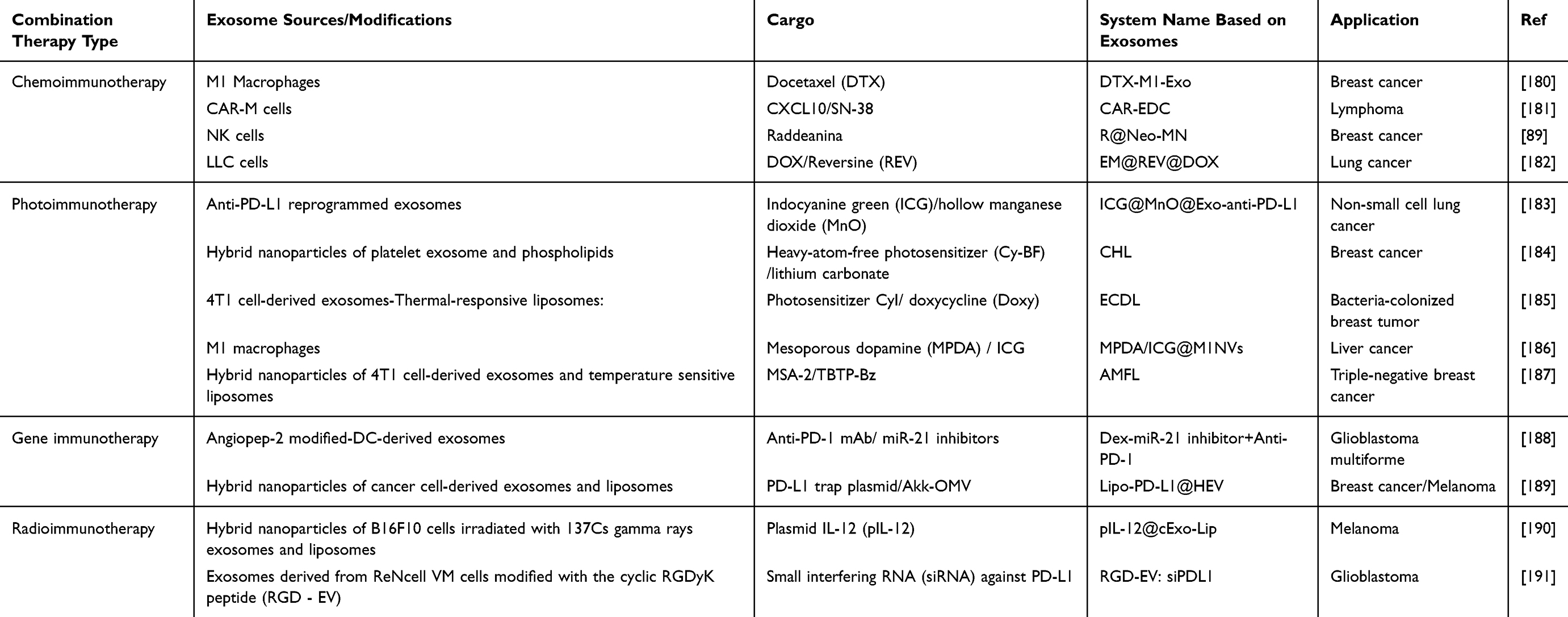 |
Table 6 Examples of Exosome-Mediated Targeted Combination Therapy for Tumors |
Chemoimmunotherapy
Chemoimmunotherapy, on one hand, directly kills tumor cells through chemotherapeutic drugs, and on the other hand, activates antitumor immunity, exerting both the “fast effect” of chemotherapy directly killing tumors and the “long effect” of immunotherapy activating long-term antitumor immunity, ultimately amplifying the therapeutic effect. It has significant advantages especially in overcoming drug resistance, inhibiting metastasis, and reducing recurrence.192,193 Qijun Lv et al first prepared exosomes overexpressing CD47 and mixed them with thermosensitive liposomes, then loaded DTX and GM-CSF (G/D-gETL NPs). Chemotherapy directly kills tumor cells, and the immune drug promotes the repolarization of M2-type tumor-associated macrophages to M1-type. Experimental results confirmed that G/D-gETL NPs can achieve effective aggregation at the tumor site, and when combined with hyperthermic intraperitoneal chemotherapy (HIPEC), they can further enhance the inhibitory effect on tumor growth (Figure 9a). 126
 |
Figure 9 Exosome-Based Chemoimmunotherapy Systems: (a) Schematic Diagram of CD47-Overexpressing Exosome-Thermosensitive Liposome Hybrid Nanoparticles (for GM-CSF/DTX Co-Delivery) in Metastatic Peritoneal Cancer Therapy. Reprinted with permission from.126 Copyright 2020 John Wiley and Sons.; (b) Schematic Diagram of Preparation of Hybrid Exosomes (Derived from M1 Macrophages and CD47-Overexpressing Tumor Cells) and Their Nanoplatform for Co-Delivery of SN38 and MnO2 to Improve the Immunosuppressive Tumor Microenvironment. Reprinted with permission from.194 Copyright 2023 John Wiley and Sons. |
Some chemotherapy drugs such as oxaliplatin (OXA), paclitaxel (PTX), and doxorubicin (DOX) can not only directly kill tumor cells but also induce immunogenic cell death (ICD) in tumor cells.195 This process releases a large number of tumor-associated antigens (TAAs) and damage-associated molecular patterns (DAMPs), presenting the “identity” of tumor cells to the immune system, promoting the maturation and antigen presentation of DCs, thereby activating adaptive immune responses. Therefore, combining such chemical drugs with immunotherapy will further enhance antitumor immunity and have a synergistic therapeutic effect on tumors. For example, chemotherapy drugs induce ICD, then combined application of PD-1 inhibitors alleviates immunosuppression, promoting T cells to kill tumor cells.196 Jie Wang et al focused on endoplasmic reticulum-targeted colorectal cancer chemoimmunotherapy.197 They first verified that celastrol can induce ICD by promoting endoplasmic reticulum stress and autophagy, then proposed a strategy using milk exosomes modified with KDEL peptide to co-deliver celastrol and PD-L1 small interfering RNA, which can effectively target the endoplasmic reticulum, downregulate PD-L1 expression, and ultimately enhance antitumor efficacy and immune response, providing an effective method for cancer chemoimmunotherapy. Lili Cheng et al designed multifunctional hybrid exosomes (SN/Mn@gHE) to activate the cGAS/STING pathway: fusing genetically engineered exosomes carrying tumor cell CD47 with M1 macrophage exosomes, then encapsulating SN38 and MnO2 (STING agonist). CD47 modification endows cGAS/STING with strong tumor targeting and long blood circulation capabilities. At the tumor site, it polarizes TAMs to the M1 phenotype, releases SN38 causing DNA damage and inducing ICD, Mn2⁺ stimulates cGAS/STING activation, promotes immune cell recruitment, and enhances cancer immunotherapy by improving the tumor immunosuppressive microenvironment, providing a new strategy for anticancer (Figure 9b). 194
Additionally, some chemotherapy drugs can reduce the number of immunosuppressive cells (eg, M2-type tumor-associated macrophages) in the tumor microenvironment and lower the levels of immunosuppressive factors (eg, IL-10, TGF-β), thereby improving the “immune desert” or “immune exclusion” state of tumor tissue. Studies have shown that low-dose cyclophosphamide treatment in mice with colon cancer liver metastasis can downregulate the expression levels of TGF-β and IL-10, while increasing the number of CD4⁺ and CD8⁺ T cells, effectively inhibiting the progression of liver metastasis lesions;198 in the mouse mammary tumor 4T1-Neu model, docetaxel can promote the activation of M1-type tumor-associated macrophages and enhance the immune response of cytotoxic T lymphocytes.199 These research findings provide an experimental basis for the development of chemoimmuno combination therapy strategies based on exosome nanoplatforms.
Photoimmunotherapy
Photoimmunotherapy leverages the direct killing effects of PTT and PDT, synergizing with ICD-induced immune activation to form a combined effect of “local ablation - systemic defense”. Nevertheless, the intensity of ICD induced by PDT is insufficient and susceptible to immunosuppressive factors in the tumor microenvironment. Therefore, combining it with immunotherapy can enhance immune stimulation effects, thereby effectively curbing tumor metastasis and recurrence.200,201 Lin Xun et al engineered microglia to overexpress LAG3 inhibitory antibodies on their surface and loaded them with AIE photothermal agents (EE@Fs-NPs). These anti-LAG3-carrying microglial exosomes promote the efficient penetration of EE@Fs-NPs across the BBB and target glioblastoma. In glioblastoma, anti-LAG3 can effectively prevent T cell exhaustion and inhibit heat shock protein expression to enhance the thermal sensitivity of tumor cells; meanwhile, the photothermal effect generated by the AIE photothermal agent significantly increases the infiltration of CD8⁺ T cells into the tumor.202 The synergistic effect of the two provides an effective platform for GMB treatment. Genetically engineered liver cancer cells can produce a special type of exosome (cypate@EXO-CXCL9) that both overexpresses CXCL9 and is loaded with cypate dye (Figure 10a). 203 After systemic administration, it achieves efficient aggregation in liver cancer tissue through homotypic targeting; upon NIR excitation, the photothermal effect generated by cypate, on one hand, disrupts the exosome structure, releasing CXCL9 to promote T cell migration to the tumor site, and on the other hand, induces immunogenic death of liver cancer cells, promoting the activation of recruited T cells. The synergistic effect of the two can effectively eliminate liver cancer tumors with no significant adverse reactions. Hybrid exosomes formed by membrane fusion of exosomes derived from human γδ-T cells (γδ-T exosomes) and liposomes loaded with chlorin e6 (Ce6) can specifically bind to melanoma via surface proteins such as CCR5 and PD-1 (Figure 10b). 117 γδ-T exosomes containing cytolytic molecules such as granzyme A, B, and perforin can specifically induce cancer cell apoptosis without damaging normal cells. Under light conditions, the ROS generated in melanoma cells works together with the above-mentioned cellular molecules, not only inducing apoptosis but also promoting immunogenic cell death. The damage-associated molecules released in this process can promote the maturation of dendritic cells, thereby stimulating antigen-specific CD4⁺ and CD8⁺ T cell responses, ultimately enhancing the body’s antitumor immune capacity.
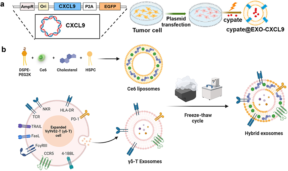 |
Figure 10 Exosome-Based Photoimmunotherapy Systems: (a) Schematic Diagram of Preparation of Cypate@EXO-CXCL9. Reprinted with permission from.203 Copyright 2025 American Chemical Society; (b) Schematic illustration of photosensitive γδ-T hybrid exosome preparation. Reprinted with permission from.117 Copyright 2025 American Chemical Society. |
Gene Immunotherapy
Exosomes can specifically recognize and be taken up by target cells (eg, tumor cells, immune cells) through naturally occurring targeting molecules on their surface (eg, integrins, membrane proteins) or artificially modified targeting ligands, achieving precise gene delivery. For example, Qinqin Cheng et al developed a genetically engineered multifunctional immunomodulatory exosome (GEMINI-Exos) that achieves precise immunotherapy for EGFR-positive triple-negative breast cancer (TNBC) through “targeting-immunomodulation” dual-function integration (Figure 11a). Using Expi293F cells as the source, through membrane protein gene fusion technology, the exosome surface was made to simultaneously express four functional molecules: αCD3 (targeting T cells), αEGFR (targeting tumor cells), PD-1 (blocking immunosuppression), and OX40L (enhancing T cell activation) (Figure 11b). GEMINI-Exos target cancer cells via “αEGFR+PD-1” and target and activate T cells via “αCD3+OX40L”, ultimately precisely guiding T cells to the vicinity of tumor cells and enhancing their killing ability, achieving a “targeting-activation-killing” therapeutic effect on TNBC (Figure 11c and d).204 Exosomes can be genetically engineered by modifying donor cells to secrete exosomes carrying specific immunomodulatory genes. Peishan Li et al genetically modified HEK 293T cells to stably express mouse PD1 and produced PD1 exosomes (PD1-Exo) in vitro to reverse the exhaustion of CD8⁺ T cells by blocking PDL1. Additionally, delivering the immune adjuvant imiquimod with PD1-Exo (PD1-Imi Exo) can activate DCs in the tumor microenvironment, and the combination therapy further enhanced the antitumor effect of CD8⁺ T lymphocytes.130
 |
Figure 11 Exosome-Based Geneimmunotherapy Systems: (a) Schematic of αCD3-αEGFR-PD-1- OX40L GEMINI-Exos for targeted cancer immunotherapy; (b) Immunoblot analysis showed that the fusion proteins were successfully expressed in GEMINI-Exos (1: Native Exos; 2: PD-1-OX40L-Exos; 3: αCD3-αEGFR-Exos; 4: ½αCD3-αEGFR-Exos+½PD-1-OX40L-Exos; 5: αCD3-αEGFR-PD-1-OX40L GEMINI-Exos); (c) Cell targeting effect of GEMINI Exos; (d) Time-dependent activation of human T cells by GEMINI-Exos. Data are shown as mean ± SD of triplicates. *p < 0.05, and ****p < 0.0001 (ordinary one-way ANOVA test). Reprinted with permission from.204 Copyright 2022 Elsevier. Abbreviation: ns, not significant. |
Exosomes can also directly load immune-related genes. For example, Tian-Yang Wang et al first chemically linked cyclic dinucleotides (CDN) with Pam3CSK4. Through the high lipophilicity of Pam3CSK4, tumor-derived exosomes can effectively load CDN, then load the Toll-like receptor 1/2 agonist (TLR1/2) to form EXO.TLR1,2-STING EXOTLR1,2-STING can effectively deliver therapeutic agents to antigen-presenting cells and tumor cells. Synergistic activation of the STING pathway by CDN and immune activation by TLR1/2 can effectively induce APC activation and kill tumor cells.205
Radioimmunotherapy
The immunosuppressive tumor microenvironment is prone to induces RT resistance. Radioimmunotherapy synergy can improve radiotherapy efficiency. Zhiran Chen et al delivered the STING agonist MSA-2 via exosomes (EXO) loaded on mesoporous microneedles: the microneedles enhanced tumor penetration, helping EXO to be continuously released and accumulate deep within the tumor. After internalization, EXO released MSA-2, which combined with ultra-high dose rate (FLASH) irradiation to promote activation of the STING pathway, increase type I interferon levels, promote DC maturation, and regulate the immunosuppressive microenvironment, showing antitumor effects in both primary and metastatic tumors, and is expected to optimize FLASH radiotherapy effects and alleviate radioresistance.206 Additionally, using exosomes themselves as carriers can also achieve synergy between radiotherapy and immunity. Fangming Zhang et al constructed an autologous cell-derived exosome engineered nanoagonist MnExo@cGAMP, loaded with Mn2⁺ (metal immunotherapeutic agent) and 2’,3’-cGAMP (STING agonist). Relying on autologous targeting, it enriched in tumor cells. After internalization, Mn2⁺ enhanced STING binding stimulus, making cGAS sensitive to radiotherapy-induced dsDNA, synergizing with 2’,3’-cGAMP to activate the cGAS-STING pathway, promote type I interferon overexpression, drive DC maturation, antigen presentation, and CD8⁺ T cell activation, overcome immunosuppression, and achieve efficient radioimmunotherapy for melanoma (Figure 12a). 207 Immune cell-derived exosomes have natural tissue targeting and immunomodulatory functions, giving them advantages for achieving radioimmuno combination. Addressing the three core challenges of solid tumor radiotherapy: tumor hypoxia (reducing radiation sensitivity), rapid DNA damage repair after radiotherapy (causing resistance), and radiotherapy-induced tumor immunosuppressive microenvironment, Xiaotu Ma et al designed engineered exosomes derived from M1 macrophages (M1Exos) to overcome treatment bottlenecks through multifunctional synergy. M1Exos were precisely designed: expressing catalase (CAT) inside the membrane to catalyze the generation of oxygen from high concentrations of H2O2 in the tumor microenvironment, directly alleviating solid tumor hypoxia; loaded with DNA damage repair inhibitors (DDRi, such as nedisertib) to block the DNA damage repair process in tumor cells after radiotherapy; expressing anti-PD-L1 nanobodies on the outer membrane, while themselves can repolarize M2-type tumor-infiltrating macrophages (M2-TAM) to the M1 phenotype. This design remodels the tumor immune microenvironment by alleviating hypoxia, inhibiting DNA repair, and relieving T cell immunosuppression, ultimately significantly improving the efficacy of radiotherapy (Figure 12b). 208
 |
Figure 12 Exosome-Based Radioimmunotherapy Systems: (a) Schematic Diagram of Engineered Homologous Cell-Derived Exosomes Loaded with Immune Agonists (Mn2⁺, 2’,3’-cGAMP) for Enhancing Immunotherapeutic Efficacy. Reprinted with permission from207. Copyright 2024 John Wiley and Sons.; (b) Schematic Diagram of Engineered M1Exos for Radiosensitization (i: O2 generation and increased DNA damage of radiotherapy; ii: Inhibition of DNA damage repair; iii: Macrophage polarity and remodeling of immunosuppressive microenvironment). Reprinted with permission from.208 Copyright 2024 John Wiley and Sons. |
Clinical Translation Progress of Exosomes
Exosomes have shown potential in various aspects of clinical tumor therapy, for example, as drug delivery carriers carrying chemotherapeutic drugs, gene therapy substances, and other therapeutic agents to target tumor cells. They can also activate antitumor immune responses through immune cell-derived exosomes, serve as tumor vaccines, or be used in combination with chemotherapy and phototherapy to exert synergistic therapeutic effects.
Besides the therapeutic field, exosomes, due to their stable presence in various body fluids such as blood, pleural effusion, and urine, and the biological molecules they carry (eg, nucleic acids, proteins) that can reflect the pathological state of the source cells, make them ideal biomarkers for non-invasive liquid biopsy. They are gradually becoming an important breakthrough in precise tumor diagnosis and treatment, early diagnosis, efficacy monitoring, and prognosis evaluation.209–211 Examples of clinical trials based on exosomes in tumor treatment and diagnosis are shown in Table 7 and Table 8.
 |
Table 7 Example of Clinical Trials Based on Exosomes in Tumor Treatment |
 |
Table 8 Example of Clinical Trials Based on Exosomes in Tumor Diagnosis |
The tumor-derived nucleic acid fragments carried by exosomes facilitate the detection of tumor-related gene mutations. Tumor-driven exosomal circRNAs have been confirmed to be widely involved in the pathological processes of various malignancies. For example, in liver cancer, gastric cancer, glioma, lung cancer, and other malignancies, exosomal circRNAs regulate target gene expression or participate in signaling pathways, serving as important regulatory factors in tumor progression.212 Multiple studies have confirmed the clinical value of exosomal markers in tumor diagnosis: for example, one study found that HDAC5 regulates the expression of radiotherapy-induced exosomal Maspin and miR-151a-3p through interaction with p53. These two exosomal molecules can serve as biomarkers for enhancing radiotherapy sensitivity in hepatocellular carcinoma, providing a theoretical basis for optimizing radiotherapy strategies for HCC. Another study showed that the expression quantity of plasma exosomal lnc-SNAPC5-3:4 is closely related to the efficacy of anlotinib treatment in patients with advanced non-small cell lung cancer and can serve as a potential biomarker for monitoring the efficacy of this treatment regimen.213 In the field of lung adenocarcinoma, Qu et al’s study of 52 lung adenocarcinoma patients further confirmed that exosomal DNA (exoDNA) in malignant pleural effusion can serve as a reliable DNA source for EGFR mutation detection, with a sensitivity of 100%, specificity of 96.55%, and a consistency of 98.08% (Kappa=0.961, P<0.001) with tumor tissue and cell block detection results, fully demonstrating the accuracy of exosomes in tumor molecular diagnosis.214 A newly identified mechanism reveals that tumor‑derived miR‑203a‑3p enhances ferroptosis by targeting ZEB1 and upregulating SLC11A2, offering a potential novel target for the screening and therapeutic intervention of pancreatic cancer cachexia.215 Additionally, proteins contained in exosomes are potential biomarkers for predicting clinical outcomes. Shayista Akbar et al found that exosomal immuno-oncology proteins/cytokines can serve as potential biomarkers for monitoring the response to immune checkpoint inhibitor therapy in non-small cell lung cancer patients, predicting clinical outcomes. Exosomal PD-L1 was present in 100% of sample patients, making it more reliable than tissue PD-L1, providing an important basis for optimizing patient stratification and efficacy monitoring for ICI therapy.216
In terms of clinical translation, exosome technology has made important progress. Early clinical trial results of mesenchymal stromal cell and DC exosomes showed that exosomes have good safety and also verified the feasibility of clinical-scale exosome production to a certain extent. This provides an important safety basis and practical support at the production level for the translation of exosomes from basic research to clinical applications.171 After 2016, two exosome detection products were successfully approved for marketing: ExoDx Prostate (IntelliScore) detects the expression levels of genes such as PCA3 and ERG in plasma exosomes for risk stratification and diagnosis of prostate cancer; ExoDx Lung (ALK) accurately screens for ALK fusion genes in non-small cell lung cancer based on exosomal RNA detection.217 The clinical application of these products not only verifies the clinical utility of exosomes as tumor diagnostic markers but also marks an important breakthrough in the translation of exosome technology from basic research to clinical application, indicating the broad prospects of exosomes in precise tumor diagnosis.
Clinical Translation Challenge and Prospective of Exosomes
As naturally derived nanovesicles, exosomes have become a research hotspot in the field of tumor diagnosis and treatment due to their good biocompatibility, low immunogenicity, ability to cross barriers, and targeting potential. This article systematically elaborates on the biological characteristics, extraction, isolation, and characterization methods of exosomes, focusing on their application modes in tumor therapy—from single targeted delivery therapies (eg, chemotherapeutic drugs, photosensitizers, gene editing) to combined applications with chemotherapy, phototherapy, and gene therapy—all demonstrating the unique advantages of exosomes as “natural nanocarriers” in improving drug delivery efficiency, reducing off-target toxicity, and enhancing tumor microenvironment regulation. Meanwhile, due to the tumor-specific biomarkers they carry (eg, nucleic acids, proteins), exosomes show great potential in early tumor diagnosis, efficacy monitoring, and prognosis evaluation, providing a new technical path for achieving “theranostics”.
Although exosomes have broad prospects in tumor diagnosis and treatment, moving from laboratory research to clinical application still faces many key bottlenecks, mainly reflected in the following aspects. In terms of large-scale preparation and standardization systems, the current main sources of exosomes are cell culture supernatants (eg, MSCs, tumor cells) or body fluids (blood, urine, ascites, etc.), which have limitations for large-scale production such as low yield, high cost, large batch differences, and lack of standardized isolation and purification methods. In terms of in vivo fate and biological safety, the in vivo behavior and potential risks of exosomes are not fully understood, which is particularly prominent in tumor-derived exosomes. For example, PD-L1 present on the surface of tumor-derived exosomes can bind to receptors on CD8⁺ T cells, leading to effector T cell exhaustion and weakening the body’s antitumor immune response;218 some tumor-derived exosomes can act as “information carriers”, transmitting biological signals from tumor cells to healthy cells or cells in an abnormal state, thereby increasing the possibility of tumor metastasis.219,220 Additionally, the role of tumor-derived exosomes in mediating tumor drug resistance cannot be ignored. For example, exosomes derived from cancer-associated fibroblasts (CAFs) can carry specific miRNAs and functional proteins, regulating drug resistance-related signaling pathways, ultimately leading to reduced drug sensitivity in tumor cells.221
Technological progress is dedicated to resolving the core contradiction from the laboratory to clinical application. The combination of cell expansion based on three-dimensional bioreactors with novel purification techniques such as tangential flow filtration (TFF) and size exclusion chromatography (SEC) has enabled stable mass production in line with pharmaceutical production quality management standards (GMP), significantly increasing output and reducing batch variations. At the same time, the integration of microfluidic chip technology offers the possibility of achieving integrated and automated production from cell culture, exosome separation to drug loading. Further advanced technologies such as nano-flow cytometry and single exosome proteomics analysis have laid a new foundation for quality control. The engineering modification strategy has evolved from simple drug loading to active design and precise programming of its biological functions. Hybrid exosome and engineered exosome strategies provide feasible paths for risk mitigation: hybrid exosomes reduce the cancer-promoting risk of single-source exosomes by integrating the advantages of exosomes from different sources (eg, low immunogenicity of stem cell exosomes and natural targeting of tumor cell exosomes); engineered exosomes achieve “de-risking” through genetic editing (eg, knocking out PD-L1 gene), surface modification (eg, conjugating PEG to evade phagocytosis), or loading functional molecules (eg, drug resistance reversal agents), laying the foundation for precise cancer therapy.
Looking ahead, the deep integration of emerging technologies such as artificial intelligence (AI), automation, and microfluidics will drive exosome therapy to shift from an experience-driven trial-and-error model to a data and model-driven rational design paradigm. In the basic research stage, AI can efficiently mine specific targets and diagnostic markers through deep learning analysis of multi-omics data, and intelligently design the optimal targeting ligands and drug delivery schemes; computational modeling can simulate drug loading, release kinetics, and membrane modification effects, providing precise simulations for experiments. In the production and quality control phases, AI-driven automated microfluidic platforms are expected to achieve intelligent optimization and real-time monitoring of the entire process from cultivation, separation to engineering modification, ensuring the standardization and consistency of the products. Ultimately, at the clinical translation level, by integrating real-time liquid biopsy and imaging data from patients, AI can construct dynamic models to guide individualized dosing strategies and even customize engineering plans, forming a “monitoring-analysis-intervention” precise treatment loop. This technological integration will not only systematically overcome the bottlenecks of exosomes in targeting, production, and personalized applications, but also may give rise to the next generation of “living drugs” that can sense and respond intelligently. Exosomes are expected to become a new generation of personalized medical models.
Conclusion
As a “natural nanodrug delivery platform”, exosomes demonstrate irreplaceable advantages in precise tumor treatment and early diagnosis. Their inherent low immunogenicity, excellent biocompatibility, and innate ability to cross biological barriers (such as the blood-brain barrier) provide fundamental solutions to overcome the shortcomings of traditional synthetic nanocarriers. In terms of treatment, exosomes not only can efficiently encapsulate and protect various therapeutic “goods” (including small molecule drugs, nucleic acids, and proteins), but their phospholipid bilayer membrane can also be functionally modified through surface engineering, enabling highly precise targeting of specific tumor cells or microenvironments, and designing responsive release mechanisms, significantly enhancing efficacy and reducing systemic toxicity.
In terms of diagnosis, exosomes serve as a stable “liquid biopsy” biomarker library. The membrane surface proteins on their membranes and the molecules (such as nucleic acids and proteins) contained within them faithfully reflect the physiological and pathological conditions of the source cells. By detecting tumor-specific exosomes and their molecular profiles, it is possible to achieve extremely early screening of cancer, molecular classification, dynamic monitoring of treatment responses and prognosis assessment, thus promoting the advancement of tumor diagnosis and treatment into a real-time and minimally invasive new stage.
Currently, with the increasing maturity of large-scale production and purification technologies (such as efficient separation based on microfluidic chips, high-yield stable cell lines modified by genetic engineering), as well as continuous breakthroughs in drug loading strategies (such as active drug loading, membrane fusion) and targeted function optimization (such as ligand engineering, biomimetic camouflage), the core bottlenecks faced by the clinical application of exosomes, including production standardization, drug loading efficiency, and targeted controllability, are being gradually overcome. Looking ahead, the exosome platform is expected to integrate diagnostic and therapeutic functions, evolving into a “diagnosis-treatment integration” tool. By using patient’s own cell sources or universal engineered exosomes, loading personalized therapeutic drugs and diagnostic markers, it will ultimately drive the tumor treatment model to evolve towards a truly efficient, low-toxicity, dynamic and precise new generation of personalized medicine.
Acknowledgments
Some figures were created by Biorender (www.biorender.com). ChatGPT assisted in the language editing of this paper, drawing on the latest Nature article, ‘Three ways ChatGPT helps me in my academic writing’222
Author Contributions
All authors made a significant contribution to the work reported, whether that is in the conception, study design, execution, acquisition of data, analysis and interpretation, or in all these areas; took part in drafting, revising or critically reviewing the article; gave final approval of the version to be published; have agreed on the journal to which the article has been submitted; and agree to be accountable for all aspects of the work.
Funding
This research was funded by Natural Science Foundation of Shandong Province (ZR2025QC1717) and Qingdao Natural Science Foundation (25-1-1-142-zyyd-jch).
Disclosure
The authors report no conflicts of interest in this work.
References
1. Siegel RL, Miller KD, Fuchs HE, Jemal A. Cancer statistics, 2022. CA Cancer J Clin. 2022;72(1):7–41. doi:10.3322/caac.21708
2. Wilhelm S, Tavares AJ, Dai Q, Ohta S, Chan WCW. Analysis of nanoparticle delivery to tumours. Nat Rev Mater. 2016;1(5):16014. doi:10.1038/natrevmats.2016.14
3. Huang X, He T, Liang X, et al. Advances and applications of nanoparticles in cancer therapy. MedComm – Oncol. 2024;3(1):e67. doi:10.1002/mog2.67
4. Jia Y, He Y, Jiang Y, et al. Approved nanomedicine against diseases. Pharmaceutics. 2023;15(3):774. doi:10.3390/pharmaceutics15030774
5. Rafiya K, Alam S, Nadaf A, Hasan N, Ahmad FJ. Cell membrane-engineered nanoparticles: a bionic platform for targeted cancer therapy. Biomat Adv. 2026;178:214471. doi:10.1016/j.bioadv.2025.214471
6. Chen L, Wang L, Zhu L, et al. Exosomes as drug carriers in anti-cancer therapy. Front Cell Dev Biol. 2022;10:728616. doi:10.3389/fcell.2022.728616
7. Jiang W, Cheng Y, Hou L, et al. A dual-prodrug nanogel combining Vorinostat and Pyropheophorbide a for a high efficient photochemotherapy. Int J Pharm. 2024;661:124422. doi:10.1016/j.ijpharm.2024.124422
8. Zhou X, Yan Y, Shen Y, Xu M, Xu W. Exosomes: emerging insights into the progression of pancreatic cancer. Int J Biol Sci. 2024;20(10):4098–4113. doi:10.7150/ijbs.97076
9. C-SL HSH. Exosome-integrated hydrogels for bone tissue engineering. Gels. 2024;10(12):762. doi:10.3390/gels10120762
10. Esteves M, Abreu R, Fernandes H, et al. MicroRNA-124-3p-enriched small extracellular vesicles as a therapeutic approach for Parkinson’s disease. Mol Ther. 2022;30(10):3176–3192. doi:10.1016/j.ymthe.2022.06.003
11. Panzarini E, Mariano S, Tacconi S, Carata E, Tata AM, Dini L. Novel therapeutic delivery of nanocurcumin in central nervous system related disorders. Nanomaterials. 2020;11(1):2. doi:10.3390/nano11010002
12. Basyoni AE, Atta A, Salem MM, Mohamed TM. Harnessing exosomes for targeted drug delivery systems to combat brain cancer. Cancer Cell Int. 2025;25(1):150. doi:10.1186/s12935-025-03731-z
13. Steiner L, Eldh M, Offens A, et al. Protein profile in urinary extracellular vesicles is a marker of malignancy and correlates with muscle invasiveness in urinary bladder cancer. Cancer Lett. 2025;609:217352. doi:10.1016/j.canlet.2024.217352
14. Yu D, Li Y, Wang M, et al. Exosomes as a new frontier of cancer liquid biopsy. Mol Cancer. 2022;21(1):56. doi:10.1186/s12943-022-01509-9
15. Liang Y, Duan L, Lu J, Xia J. Engineering exosomes for targeted drug delivery. Theranostics. 2021;11(7):3183–3195. doi:10.7150/thno.52570
16. Lindenbergh MFS, Stoorvogel W. Antigen presentation by extracellular vesicles from professional antigen-presenting cells. Annu Rev Immunol. 2018;36:435–459. doi:10.1146/annurev-immunol-041015-055700
17. Shao H, Im H, Castro CM, Breakefield X, Weissleder R, Lee H. New technologies for analysis of extracellular vesicles. Chem Rev. 2018;118(4):1917–1950. doi:10.1021/acs.chemrev.7b00534
18. Xu J, Cao W, Wang P, Liu H. Tumor-derived membrane vesicles: a promising tool for personalized immunotherapy. Pharmaceuticals. 2022;15(7):876. doi:10.3390/ph15070876
19. Yang Q, Li S, Ou H, et al. Exosome-based delivery strategies for tumor therapy: an update on modification, loading, and clinical application. J Nanobiotechnol. 2024;22(1):41. doi:10.1186/s12951-024-02298-7
20. Li L, Wang F, Zhu D, Hu S, Cheng K, Li Z. Engineering exosomes and exosome-like nanovesicles for improving tissue targeting and retention. Fundam Res. 2025;5(2):851–867. doi:10.1016/j.fmre.2024.03.025
21. Jin X, Zhang J, Zhang Y, et al. Different origin-derived exosomes and their clinical advantages in cancer therapy. Front Immunol. 2024;15:1401852. doi:10.3389/fimmu.2024.1401852
22. Wallen M, Aqil F, Spencer W, Gupta RC. Milk/colostrum exosomes: a nanoplatform advancing delivery of cancer therapeutics. Cancer Lett. 2023;561:216141. doi:10.1016/j.canlet.2023.216141
23. Liu Y, Ren C, Zhan R, et al. Exploring the potential of plant-derived exosome-like nanovesicle as functional food components for human health: a review. Foods. 2024;13(5):712. doi:10.3390/foods13050712
24. Ly NP, Han HS, Kim M, Park JH, Choi KY. Plant-derived nanovesicles: current understanding and applications for cancer therapy. Bioact Mater. 2023;22:365–383. doi:10.1016/j.bioactmat.2022.10.005
25. Zou Z, Li H, Xu G, Hu Y, Zhang W, Tian K. Current knowledge and future perspectives of exosomes as nanocarriers in diagnosis and treatment of diseases. Int J Nanomed. 2023;18:4751–4778. doi:10.2147/ijn.S417422
26. Park KS, Svennerholm K, Crescitelli R, Lässer C, Gribonika I, Lötvall J. Synthetic bacterial vesicles combined with tumour extracellular vesicles as cancer immunotherapy. J Extracell Vesicles. 2021;10(9):e12120. doi:10.1002/jev2.12120
27. Zhang Y, Bi J, Huang J, Du S, Li P. Exosome: A Review of Its Classification, Isolation Techniques, Storage, Diagnostic and Targeted Therapy Applications. Int J Nanomed. 2020;15:6917–6934. doi:10.2147/IJN.S264498
28. Xu X, Xu L, Wen C, Xia J, Zhang Y, Liang Y. Programming assembly of biomimetic exosomes: an emerging theranostic nanomedicine platform. Mat Today Bio. 2023;22:100760. doi:10.1016/j.mtbio.2023.100760
29. Han QF, Li WJ, Hu KS, et al. Exosome biogenesis: machinery, regulation, and therapeutic implications in cancer. Mol Cancer. 2022;21:207. doi:10.1186/s12943-022-01671-0
30. Hyung S, Ko J, Heo YJ, et al. Patient-derived exosomes facilitate therapeutic targeting of oncogenic MET in advanced gastric cancer. Sci Adv. 2023;9(47):eadk1098. doi:10.1126/sciadv.adk1098
31. Quan J, Liu Q, Li P, et al. Mesenchymal stem cell exosome therapy: current research status in the treatment of neurodegenerative diseases and the possibility of reversing normal brain aging. Stem Cell Res Ther. 2025;16(1):76. doi:10.1186/s13287-025-04160-5
32. Garg A, Khan S, Luu N, et al. TGFβ1 priming enhances CXCR3-mediated mesenchymal stromal cell engraftment to the liver and enhances anti-inflammatory efficacy. J Cell Mol Med. 2023;27(6):864–878. doi:10.1111/jcmm.17698
33. Duan R, Milton P, Sittplangkoon C, et al. Chimeric antigen receptor dendritic cells targeted delivery of a single tumoricidal factor for cancer immunotherapy. Cancer Immunol Immunother. 2024;73(10):203. doi:10.1007/s00262-024-03788-1
34. Pan W, Gao X, Zhang X, et al. Exosomes promote pre-metastatic niche formation in lung cancer. Cancer Cell Int. 2025;25(1):379. doi:10.1186/s12935-025-03966-w
35. Chen L, Zhang J, Huang Y, et al. Drug delivery systems based on dendritic-cell-derived exosomes. Pharmaceutics. 2025;17(3):326. doi:10.3390/pharmaceutics17030326
36. Lyu C, Sun H, Sun Z, Liu Y, Wang Q. Roles of exosomes in immunotherapy for solid cancers. Cell Death Dis. 2024;15(2):106. doi:10.1038/s41419-024-06494-z
37. Teng Y, Luo C, Qiu X, et al. Plant-nanoparticles enhance anti-PD-L1 efficacy by shaping human commensal microbiota metabolites. Nat Commun. 2025;16(1):1295. doi:10.1038/s41467-025-56498-2
38. Wang C, Li N, Li Y, et al. Engineering a HEK-293T exosome-based delivery platform for efficient tumor-targeting chemotherapy/internal irradiation combination therapy. J Nanobiotechnol. 2022;20(1):247. doi:10.1186/s12951-022-01462-1
39. Ru Q, Chen L, Xu G, Wu Y. Exosomes in the pathogenesis and treatment of cancer-related cachexia. J Transl Med. 2024;22(1):408. doi:10.1186/s12967-024-05201-y
40. Chernyshev VS, Chuprov-Netochin RN, Tsydenzhapova E, et al. Asymmetric depth-filtration: a versatile and scalable method for high-yield isolation of extracellular vesicles with low contamination. J Extracell Vesicles. 2022;11(8):e12256. doi:10.1002/jev2.12256
41. Correll VL, Otto JJ, Risi CM, et al. Optimization of small extracellular vesicle isolation from expressed prostatic secretions in urine for in-depth proteomic analysis. J Extracell Vesicles. 2022;11(2):e12184. doi:10.1002/jev2.12184
42. Li X, Su L, Zhang X, et al. Recent advances on the function and purification of milk exosomes: a review. Front Nutr. 2022;9:871346. doi:10.3389/fnut.2022.871346
43. Singh K, Nalabotala R, Koo KM, Bose S, Nayak R, Shiddiky MJA. Separation of distinct exosome subpopulations: isolation and characterization approaches and their associated challenges. Analyst. 2021;146(12):3731–3749. doi:10.1039/D1AN00024A
44. Arya R, Jit BP, Kumar V, Kim JJ. Exploring the potential of exosomes as biomarkers in tuberculosis and other diseases. Int J Mol Sci. 2024;25:2885. doi:10.3390/ijms25052885
45. Keshtkar S, Asvar Z, Najafi H, et al. Exosomes as natural vectors for therapeutic delivery of bioactive compounds in skin diseases. Front Pharmacol. 2025;16:1485769. doi:10.3389/fphar.2025.1485769
46. Ning XY, Ma JH, He W, Ma JT. Role of exosomes in metastasis and therapeutic resistance in esophageal cancer. World J Gastroenterol. 2023;29(42):5699–5715. doi:10.3748/wjg.v29.i42.5699
47. Li H, Ding Y, Huang J, et al. Angiopep-2 Modified Exosomes Load Rifampicin with Potential for Treating Central Nervous System Tuberculosis. Int J Nanomed. 2023;18:489–503. doi:10.2147/IJN.S395246
48. Thakur A, Ke X, Chen Y-W, et al. The mini player with diverse functions: extracellular vesicles in cell biology, disease, and therapeutics. Protein Cell. 2022;13(9):631–654. doi:10.1007/s13238-021-00863-6
49. Chen Q, Li Y, Gao W, Chen L, Xu W, Zhu X. Exosome-mediated crosstalk between tumor and tumor-associated macrophages. Front Mol Biosci. 2021;8:764222. doi:10.3389/fmolb.2021.764222
50. Yang D, Zhang W, Zhang H, et al. Progress, opportunity, and perspective on exosome isolation - efforts for efficient exosome-based theranostics. Theranostics. 2020;10(8):3684–3707. doi:10.7150/thno.41580
51. Liang Y, Li J, Yuan Y, et al. Exosomal miR-106a-5p from highly metastatic colorectal cancer cells drives liver metastasis by inducing macrophage M2 polarization in the tumor microenvironment. J Exp Clin Cancer Res. 2024;43(1):281. doi:10.1186/s13046-024-03204-7
52. Liu Z, Cauvi DM, Bernardino EMA, et al. Isolation and characterization of human urine extracellular vesicles. Cell Stress Chaperones. 2018;23(5):943–953. doi:10.1007/s12192-018-0902-5
53. da Silva Lira Filho A, Lafleur A, Marcet-Palacios M, Olivier M. Identification of potential novel proteomic markers of Leishmania spp.-derived exosomes. Front Cell Infect Microbiol. 2024;14:1354636. doi:10.3389/fcimb.2024.1354636
54. Makler A, Asghar W. Exosomal biomarkers for cancer diagnosis and patient monitoring. Expert Rev Mol Diagn. 2020;20(4):387–400. doi:10.1080/14737159.2020.1731308
55. Ruan J, Xia Y, Ma Y, et al. Milk-derived exosomes as functional nanocarriers in wound healing: mechanisms, applications, and future directions. Mater Today Bio. 2025;32:101715. doi:10.1016/j.mtbio.2025.101715
56. Liao W, Lu Z, Wang C, et al. Application and advances of biomimetic membrane materials in central nervous system disorders. J Nanobiotechnol. 2024;22(1):280. doi:10.1186/s12951-024-02548-8
57. Wang Y, Sun L, Dong Z, et al. Targeted inhibition of ferroptosis in bone marrow mesenchymal stem cells by engineered exosomes alleviates bone loss in smoking-related osteoporosis. Mater Today Bio. 2025;31:101501. doi:10.1016/j.mtbio.2025.101501
58. Vinduska V, Gallops CE, O’Connor R, Wang Y, Huang X. Exosomal surface protein detection with quantum dots and immunomagnetic capture for cancer detection. Nanomaterials. 2021;11(7):1853. doi:10.3390/nano11071853
59. Janouskova O, Herma R, Semeradtova A, et al. Conventional and nonconventional sources of exosomes-isolation methods and influence on their downstream biomedical application. Front Mol Biosci. 2022;9:846650. doi:10.3389/fmolb.2022.846650
60. Chen W, Xu J, Yang H, et al. Developing a two-dimensional size-exclusion liquid chromatography platform for isolating the high-quality extracellular vesicles to predict and evaluate metabolic improvement outcomes of sleeve gastrectomy. Anal Chem. 2025;97(27):14435–14445. doi:10.1021/acs.analchem.5c01666
61. Gao M, Cai J, Zitkovsky HS, Chen B, Guo L. Comparison of yield, purity, and functional properties of large-volume exosome isolation using ultrafiltration and polymer-based precipitation. Plast Reconstr Surg. 2022;149(3):638–649. doi:10.1097/prs.0000000000008830
62. Hassanpour Tamrin S, Sanati Nezhad A, Sen A. Label-free isolation of exosomes using microfluidic technologies. ACS Nano. 2021;15(11):17047–17079. doi:10.1021/acsnano.1c03469
63. Walker JM, O’Malley P, He M. Applications of exosomes in diagnosing muscle invasive bladder cancer. Pharmaceutics. 2022;14(10):2027. doi:10.3390/pharmaceutics14102027
64. Ban JJ, Lee M, Im W, Kim M. Low pH increases the yield of exosome isolation. Biochem Biophys Res Commun. 2015;461(1):76–79. doi:10.1016/j.bbrc.2015.03.172
65. Hu SQ, Zhang QC, Meng QB, Hu AN, Zou JP, Li XL. Autophagy regulates exosome secretion in rat nucleus pulposus cells via the RhoC/ROCK2 pathway. Exp Cell Res. 2020;395(2):112239. doi:10.1016/j.yexcr.2020.112239
66. Shyong YJ, Chang KC, Lin FH. Calcium phosphate particles stimulate exosome secretion from phagocytes for the enhancement of drug delivery. Colloids Surf B Biointerfaces. 2018;171:391–397. doi:10.1016/j.colsurfb.2018.07.037
67. Nakamura Y, Kita S, Tanaka Y, et al. Adiponectin stimulates exosome release to enhance mesenchymal stem-cell-driven therapy of heart failure in mice. Mol Ther. 2020;28(10):2203–2219. doi:10.1016/j.ymthe.2020.06.026
68. Haraszti RA, Miller R, Stoppato M, et al. Exosomes produced from 3D cultures of MSCs by tangential flow filtration show higher yield and improved activity. Mol Ther. 2018;26(12):2838–2847. doi:10.1016/j.ymthe.2018.09.015
69. Yang X, Yi Z, Liang Y, Tong S. Magnetic iron oxide nanoparticles enhance exosome production by upregulating exosome transport and secretion pathways. ACS Appl Mater Interfaces. 2024;16(49):67235–67245. doi:10.1021/acsami.4c13821
70. Zhang S, Jiang T, Sun H, et al. Enhanced mesenchymal stem cells-derived exosomes secretion by electrical stimulation of triboelectric nanogenerator. Nano Energy. 2025;139:110933. doi:10.1016/j.nanoen.2025.110933
71. Bhavsar D, Raguraman R, Kim D, et al. Exosomes in diagnostic and therapeutic applications of ovarian cancer. J Ovarian Res. 2024;17(1):113. doi:10.1186/s13048-024-01417-0
72. Li D, Yang Y, Zheng G, et al. The potential of cellular homing behavior in tumor immunotherapy: from basic discoveries to clinical applications of immune, mesenchymal stem, and cancer cell homing. Review. Front Immunol. 2024;15:1495978. doi:10.3389/fimmu.2024.1495978
73. Li Q, Song Q, Zhao Z, et al. Genetically engineered artificial exosome-constructed hydrogel for ovarian cancer therapy. ACS Nano. 2023;17(11):10376–10392. doi:10.1021/acsnano.3c00804
74. Wang J, Yin B, Lian X, Wang J. Extracellular vesicles as drug delivery system for cancer therapy. Pharmaceutics. 2024;16(8):1029. doi:10.3390/pharmaceutics16081029
75. Wang W, Xu Z, Cai M, Liu M, Liu X. Prospective applications of extracellular vesicle-based therapies in regenerative medicine: implications for the use of dental stem cell-derived extracellular vesicles. Front Bioeng Biotechnol. 2023;11:1278124. doi:10.3389/fbioe.2023.1278124
76. Yang G, Waheed S, Wang C, Shekh M, Li Z, Wu J. Exosomes and Their Bioengineering Strategies in the Cutaneous Wound Healing and Related Complications: Current Knowledge and Future Perspectives. Int J Biol Sci. 2023;19(5):1430–1454. doi:10.7150/ijbs.80430
77. Zeng H, Guo S, Ren X, Wu Z, Liu S, Yao X. Current Strategies for Exosome Cargo Loading and Targeting Delivery. Cells. 2023;12(10):1416. doi:10.3390/cells12101416
78. Wang T, Zhang H. Exploring the roles and molecular mechanisms of RNA binding proteins in the sorting of noncoding RNAs into exosomes during tumor progression. J Adv Res. 2024;65:105–123. doi:10.1016/j.jare.2023.11.029
79. Chen Z, Xiong M, Tian J, Song D, Duan S, Zhang L. Encapsulation and assessment of therapeutic cargo in engineered exosomes: a systematic review. J Nanobiotechnol. 2024;22(1):18. doi:10.1186/s12951-023-02259-6
80. Simon L, Constanzo J, Terraza-Aguirre C, et al. Surface modification of extracellular vesicles with polyoxazolines to enhance their plasma stability and tumor accumulation. Biomaterials. 2025;313:122748. doi:10.1016/j.biomaterials.2024.122748
81. Creeden JF, Sevier J, Zhang J-T, et al. Smart exosomes enhance PDAC targeted therapy. J Control Release. 2024;368:413–429. doi:10.1016/j.jconrel.2024.02.037
82. Srivastava A, Rathore S, Munshi A, Ramesh R. Organically derived exosomes as carriers of anticancer drugs and imaging agents for cancer treatment. Semin Cancer Biol. 2022;86(Pt1):80–100. doi:10.1016/j.semcancer.2022.02.020
83. Ouahabi OE, Salim H, Pero-Gascon R, Benavente F. A simple method for the analysis of extracellular vesicles enriched for exosomes from human serum by capillary electrophoresis with ultraviolet diode array detection. J Chromatogr A. 2021;1635:461752. doi:10.1016/j.chroma.2020.461752
84. Liu A, Yang G, Liu Y, Liu T. Research progress in membrane fusion-based hybrid exosomes for drug delivery systems. Front Bioeng Biotechnol. 2022;10:939441. doi:10.3389/fbioe.2022.939441
85. Dai J, Su Y, Zhong S, et al. Exosomes: key players in cancer and potential therapeutic strategy. Signal Transduct Target Ther. 2020;5(1):145. doi:10.1038/s41392-020-00261-0
86. Shan C, Liang Y, Wang K, Li P. Mesenchymal stem cell-derived extracellular vesicles in cancer therapy resistance: from biology to clinical opportunity. Int J Biol Sci. 2024;20(1):347–366. doi:10.7150/ijbs.88500
87. Lu Y, Han G, Zhang Y, et al. M2 macrophage-secreted exosomes promote metastasis and increase vascular permeability in hepatocellular carcinoma. Cell Commun Signal. 2023;21(1):299. doi:10.1186/s12964-022-00872-w
88. Xue X, Li X, Tian M, et al. PH20-modified exosomes loaded curcumin inhibit desmoplastic breast cancer by breaking extracellular matrix barrier and normalizing cancer-associated fibroblasts. Materials Today Bio. 2025;35:102635. doi:10.1016/j.mtbio.2025.102635
89. Peng D, Cheng L, Tang J, Liu Z, Xue Y, Liu J. Engineered NK exosomes captured antigens in situ for enhanced tumor immunotherapy. ACS Appl Mater Interfaces. 2025;17(16):23740–23752. doi:10.1021/acsami.5c03195
90. Takakura Y, Hanayama R, Akiyoshi K, et al. Quality and safety considerations for therapeutic products based on extracellular vesicles. Pharm Res. 2024;41(8):1573–1594. doi:10.1007/s11095-024-03757-4
91. Wang L, Chen J, Song J, et al. Activation of the Wnt/β-catenin signalling pathway enhances exosome production by hucMSCs and improves their capability to promote diabetic wound healing. J Nanobiotechnol. 2024;22(1):373. doi:10.1186/s12951-024-02650-x
92. Wang L, Liu H, Chen G, et al. Bubble ticket trip: exploring the mechanism of miRNA sorting into exosomes and maintaining the stability of tumor microenvironment. Int J Nanomedicine. 2024;19:13671–13685. doi:10.2147/ijn.S498599
93. Kunte S, Abraham J, Montero AJ. Novel HER2-targeted therapies for HER2-positive metastatic breast cancer. Cancer. 2020;126(19):4278–4288. doi:10.1002/cncr.33102
94. Song Y, Cui L, Liu Z, Tang Z, Chen X. Multivalent RGD peptide-mediated nanochimera for lysosomal degradation of PDL1 protein. Nano Lett. 2025;25(10):4078–4086. doi:10.1021/acs.nanolett.5c00341
95. Wu YY, Hsieh IS, Tung CH, et al. A novel DNA aptamer targeting lung cancer stem cells exerts a therapeutic effect by binding and neutralizing Annexin A2. Mol Ther Nucleic Acids. 2022;27:956–968. doi:10.1016/j.omtn.2022.01.012
96. Kim JY, Song J, Jung H, Mok H. I-motif-coated exosomes as a pH-sensitive carrier for anticancer drugs. Appl Biol Chem. 2018;61(6):599–606. doi:10.1007/s13765-018-0394-0
97. Si Z, Cheng Y, Xu Z, Shi D, Shi H, Cheng D. Exploration of (68)Ga-DOTA-MAL as a versatile vehicle for facile labeling of a variety of thiol-containing bioactive molecules. ACS Omega. 2023;8(5):4747–4755. doi:10.1021/acsomega.2c06720
98. Chen C, Sun M, Liu X, et al. General and mild modification of food-derived extracellular vesicles for enhanced cell targeting. Nanoscale. 2021;13(5):3061–3069. doi:10.1039/d0nr06309f
99. Ragab SS. Signature of click chemistry in advanced techniques for cancer therapeutics. RSC Adv. 2025;15(14):10583–10601. doi:10.1039/d5ra01196e
100. Deng M, Guo R, Wang Y, et al. Curbing exosome communications via introducing artificial membrane receptors for metastatic pancreatic cancer therapy. Adv Mater. 2023;35(39):e2303736. doi:10.1002/adma.202303736
101. Ma D, Luo Y, Zhang X, Xie Z, Yan Y, Ding CF. A highly sensitive and selective fluorescent biosensor for breast cancer derived exosomes using click reaction of azide-CD63 aptamer and alkyne-polymer dots. Anal Meth. 2024;16(17):2751–2759. doi:10.1039/d4ay00146j
102. Chen H, Ma M, Zhang L, et al. An effective strategy based on electrostatic interaction for the simultaneous sequential purification and isolation of exosomes. Mikrochim Acta. 2025;192(4):259. doi:10.1007/s00604-025-07125-w
103. Jing B, Gai Y, Qian R, et al. Hydrophobic insertion-based engineering of tumor cell-derived exosomes for SPECT/NIRF imaging of colon cancer. J Nanobiotechnol. 2021;19(1):7. doi:10.1186/s12951-020-00746-8
104. Wu X, Ban C, Deng W, et al. Unveiling the PDK4-centered rituximab-resistant mechanism in DLBCL: the potential of the “Smart” exosome nanoparticle therapy. Mol Cancer. 2024;23(1):144. doi:10.1186/s12943-024-02057-0
105. Deng R, Wu J, Zhu B, et al. Engineered exosomes loaded with M1–8 peptide for targeted therapy of hepatocellular carcinoma. Appl Mat Today. 2024;37(000):13. doi:10.1016/j.apmt.2024.102071
106. Hosseini NF, Amini R, Ramezani M, Saidijam M, Hashemi SM, Najafi R. AS1411 aptamer-functionalized exosomes in the targeted delivery of doxorubicin in fighting colorectal cancer. Biomed Pharmacother. 2022;155:113690. doi:10.1016/j.biopha.2022.113690
107. Chen C, Pan X, Sun M, et al. Phospholipid-anchored ligand conjugation on extracellular vesicles for enhanced cancer targeting. Small. 2024;20(37):e2310712. doi:10.1002/smll.202310712
108. Zhou W, Zhou Y, Chen X, et al. Pancreatic cancer-targeting exosomes for enhancing immunotherapy and reprogramming tumor microenvironment. Biomaterials. 2021;268:120546. doi:10.1016/j.biomaterials.2020.120546
109. Jia G, Han Y, An Y, et al. NRP-1 targeted and cargo-loaded exosomes facilitate simultaneous imaging and therapy of glioma in vitro and in vivo. Biomaterials. 2018;178:302–316. doi:10.1016/j.biomaterials.2018.06.029
110. Nie W, Wu G, Zhang J, et al. Responsive exosome nano-bioconjugates for synergistic cancer therapy. Angew Chem Int Ed Engl. 2020;59(5):2018–2022. doi:10.1002/anie.201912524
111. Guo L, Xiao D, Xing H, Yang G, Yang X. Engineered exosomes as a prospective therapy for diabetic foot ulcers. Burns Trauma. 2024;12:tkae023. doi:10.1093/burnst/tkae023
112. Zhang M, Hu S, Liu L, et al. Engineered exosomes from different sources for cancer-targeted therapy. Signal Transduct Target Ther. 2023;8(1):124. doi:10.1038/s41392-023-01382-y
113. Naquin TD, Canning AJ, Gu Y, et al. Acoustic separation and concentration of exosomes for nucleotide detection: aSCENDx. Sci Adv. 2024;10(10):eadm8597. doi:10.1126/sciadv.adm8597
114. Bader J, Brigger F, Leroux J-C. Extracellular vesicles versus lipid nanoparticles for the delivery of nucleic acids. Adv Drug Deliv Rev. 2024;215:115461. doi:10.1016/j.addr.2024.115461
115. Zhuo Y, Luo Z, Zhu Z, et al. Direct cytosolic delivery of siRNA via cell membrane fusion using cholesterol-enriched exosomes. Nat Nanotechnol. 2024;19(12):1858–1868. doi:10.1038/s41565-024-01785-0
116. Xu M, Bai L, Sun M, et al. ROS-Responsive Biomimetic Nanocomplexes of Liposomes and Macrophage-Derived Exosomes for Combination Breast Cancer Therapy. Int J Nanomed. 2025;20:5161–5180. doi:10.2147/IJN.S514375
117. Gao Y, Liu J, Wu M, et al. Photosensitive hybrid γδ-T exosomes for targeted cancer photoimmunotherapy. ACS Nano. 2025;19(4):4251–4268. doi:10.1021/acsnano.4c11024
118. Cheng L, Zhang X, Tang J, Lv Q, Liu J. Gene-engineered exosomes-thermosensitive liposomes hybrid nanovesicles by the blockade of CD47 signal for combined photothermal therapy and cancer immunotherapy. Biomaterials. 2021;275:120964. doi:10.1016/j.biomaterials.2021.120964
119. Li W, Cheng J, He F, et al. Cell membrane-based nanomaterials for theranostics of central nervous system diseases. J Nanobiotechnol. 2023;21(1):276. doi:10.1186/s12951-023-02004-z
120. Yang Y, Wang Q, Zou H, Chou CK, Chen X. Exosome-modified liposomes targeted delivery of thalidomide to regulate treg cells for antitumor immunotherapy. Pharmaceutics. 2023;15(4):1074. doi:10.3390/pharmaceutics15041074
121. Huang H, Shao L, Chen Y, et al. Synergistic strategy with hyperthermia therapy based immunotherapy and engineered exosomes-liposomes targeted chemotherapy prevents tumor recurrence and metastasis in advanced breast cancer. Bioeng Transl Med. 2022;7(2):e10284. doi:10.1002/btm2.10284
122. Huang R, Jia B, Su D, et al. Plant exosomes fused with engineered mesenchymal stem cell-derived nanovesicles for synergistic therapy of autoimmune skin disorders. J Extracell Vesicles. 2023;12(10):e12361. doi:10.1002/jev2.12361
123. Jhan YY, Prasca-Chamorro D, Palou Zuniga G, et al. Engineered extracellular vesicles with synthetic lipids via membrane fusion to establish efficient gene delivery. Int J Pharm. 2020;573:118802. doi:10.1016/j.ijpharm.2019.118802
124. Li M, Tai Q, Shen S, Gao M, Zhang X. Biomimetic exosome-sheathed magnetic mesoporous anchor with modification of glucose oxidase for synergistic targeting and starving tumor cells. ACS Appl Mater Interfaces. 2024;16(23):29634–29644. doi:10.1021/acsami.4c02337
125. Belhadj Z, He B, Deng H, et al. A combined “eat me/don’t eat me” strategy based on extracellular vesicles for anticancer nanomedicine. J Extracell Vesicles. 2020;9(1):1806444. doi:10.1080/20013078.2020.1806444
126. Lv Q, Cheng L, Lu Y, et al. Thermosensitive exosome-liposome hybrid nanoparticle-mediated chemoimmunotherapy for improved treatment of metastatic peritoneal cancer. Adv Sci. 2020;7(18):2000515. doi:10.1002/advs.202000515
127. Li L, He D, Guo Q, et al. Exosome-liposome hybrid nanoparticle codelivery of TP and miR497 conspicuously overcomes chemoresistant ovarian cancer. J Nanobiotechnol. 2022;20(1):50. doi:10.1186/s12951-022-01264-5
128. Zhang L, Lin Y, Li S, Guan X, Jiang X. In situ reprogramming of tumor-associated macrophages with internally and externally engineered exosomes. Angewandte Chemie. 2023;62(11):e202217089. doi:10.1002/anie.202217089
129. Park JH, Kim SJ, Kim OH, Kim DJ. Enhanced efficacy of gastric cancer treatment through targeted exosome delivery of 17-DMAG anticancer agent. Int J Mol Sci. 2024;25(16):8762. doi:10.3390/ijms25168762
130. Li P, Xie Y, Wang J, et al. Gene engineered exosome reverses T cell exhaustion in cancer immunotherapy. Bioact Mat. 2024;34:466–481. doi:10.1016/j.bioactmat.2024.01.008
131. Du S, Guan Y, Xie A, et al. Extracellular vesicles: a rising star for therapeutics and drug delivery. J Nanobiotechnol. 2023;21(1):231. doi:10.1186/s12951-023-01973-5
132. Zhao Z, Shuang T, Gao Y, et al. Targeted delivery of exosomal miR-484 reprograms tumor vasculature for chemotherapy sensitization. Cancer Lett. 2022;530:45–58. doi:10.1016/j.canlet.2022.01.011
133. Liu Q, Dai G, Wu Y, et al. iRGD-modified exosomes-delivered BCL6 siRNA inhibit the progression of diffuse large B-cell lymphoma. Front Oncol. 2022;12:822805. doi:10.3389/fonc.2022.822805
134. Lin D, Zhang H, Liu R, et al. iRGD-modified exosomes effectively deliver CPT1A siRNA to colon cancer cells, reversing oxaliplatin resistance by regulating fatty acid oxidation. Mol Oncol. 2021;15(12):3430–3446. doi:10.1002/1878-0261.13052
135. Liang G, Zhu Y, Ali DJ, et al. Engineered exosomes for targeted co-delivery of miR-21 inhibitor and chemotherapeutics to reverse drug resistance in colon cancer. J Nanobiotechnol. 2020;18(1):10. doi:10.1186/s12951-019-0563-2
136. Soudi T, Bagheri F, Shojaosadati SA, Rezaei M, Motamedian E. Combination therapy using carvedilol and 5-Fluorouracil loaded exosomes as a novel synergistic strategy against breast cancer. J Drug Deliv Sci Technol. 2025;109:107019. doi:10.1016/j.jddst.2025.107019
137. Deirmenci N, Uslu M, Krba OK, Ahin F, Uar EN. Lapatinib loaded exosomes as a drug delivery system in breast cancer. SSRN Electr J. 2022;75:103584. doi:10.1016/j.jddst.2022.103584
138. Lin Y, Li S, Xiao Z, Chen S, Zhang L. Epigenetic inhibition assisted chemotherapeutic treatment of lung cancer based on artificial exosomes. Pharmacol Res. 2021;171:105787. doi:10.1016/j.phrs.2021.105787
139. Tiwari P, Shukla RP, Yadav K, et al. YIGSR functionalized hybrid exosomes spatially target dasatinib to laminin receptors for precision therapy in breast cancer. Adv Healthc Mater. 2025;14(9):e2402673. doi:10.1002/adhm.202402673
140. Bao YR, Chen YJ, Deng XF, et al. Boron clusters escort doxorubicin squashing into exosomes and overcome drug resistance. Adv Sci. 2025;12(7):e2412501. doi:10.1002/advs.202412501
141. Huda MN, Nafiujjaman M, Deaguero IG, et al. Potential use of exosomes as diagnostic biomarkers and in targeted drug delivery: progress in clinical and preclinical applications. ACS Biomater Sci Eng. 2021;7(6):2106–2149. doi:10.1021/acsbiomaterials.1c00217
142. Mathew B, Mansuri MS, Williams KR, Nairn AC. Exosomes as emerging biomarker tools in neurodegenerative and neuropsychiatric disorders-A proteomics perspective. Brain Sci. 2021;11(2):258. doi:10.3390/brainsci11020258
143. Shaikh S, Younis M, Shahzad KA, et al. Exosomes loaded with gold nanoparticles and temozolomide for targeted therapy of glioblastoma. ACS Appl Nano Mat. 2025;8(21):11192–11200. doi:10.1021/acsanm.5c02069
144. Yan G, Li A, Zhang A, Sun Y, Liu J. Polymer-based nanocarriers for co-delivery and combination of diverse therapies against cancers. Nanomaterials. 2018;8(2):85. doi:10.3390/nano8020085
145. Xiao Q, Zhao W, Wu C, et al. Lemon-derived extracellular vesicles nanodrugs enable to efficiently overcome cancer multidrug resistance by endocytosis-triggered energy dissipation and energy production reduction. Adv Sci. 2022;9(20):e2105274. doi:10.1002/advs.202105274
146. Zhonghao J, Fan Y. New advances in the treatment of intermediate and advanced hepatocellular carcinoma. Front Oncol. 2024;14:1430991. doi:10.3389/fonc.2024.1430991
147. Jin Z, Zhang C, Shen L, Cao Y. Harnessing exosomes: from tumor immune escape to therapeutic innovation in gastric cancer immunotherapy. Cancer Lett. 2025;626:217792. doi:10.1016/j.canlet.2025.217792
148. Zhang X, Cui H, Zhang W, Li Z, Gao J. Engineered tumor cell-derived vaccines against cancer: the art of combating poison with poison. Bioact Mater. 2023;22:491–517. doi:10.1016/j.bioactmat.2022.10.016
149. Zou Y, Li S, Li Y, Zhang D, Zheng M, Shi B. Glioblastoma cell derived exosomes as a potent vaccine platform targeting primary brain cancers and brain metastases. ACS Nano. 2025;19(18):17309–17322. doi:10.1021/acsnano.4c14573
150. Li S, Zhang D, Wang Y, et al. Tumor cell-derived engineered exosome enhances effective immunotherapy for orthotopic glioblastoma and its recurrences. Nano Today. 2025;63:102748. doi:10.1016/j.nantod.2025.102748
151. Huang L, Rong Y, Tang X, et al. Engineered exosomes as an in situ DC-primed vaccine to boost antitumor immunity in breast cancer. Mol Cancer. 2022;21(1):45. doi:10.1186/s12943-022-01515-x
152. Wang Y, Guo X, Qin J, et al. Locoregional immune checkpoint blockade and remodeling of lymph nodes by engineered dendritic cell-derived exosomes for suppressing tumor progression and metastasis. Adv Sci. 2025;12(23):e2500139. doi:10.1002/advs.202500139
153. Xu ZJ, Jin Y, Zhang XL, et al. Pan-cancer analysis identifies CD300 molecules as potential immune regulators and promising therapeutic targets in acute myeloid leukemia. Cancer Med. 2023;12(1):789–807. doi:10.1002/cam4.4905
154. Yang X, Liu J. Targeting PD-L1 (Programmed death-ligand 1) and inhibiting the expression of IGF2BP2 (Insulin-like growth factor 2 mRNA-binding protein 2) affect the proliferation and apoptosis of hypopharyngeal carcinoma cells. Bioengineered. 2021;12(1):7755–7764. doi:10.1080/21655979.2021.1983278
155. Zhang P, Zhang C, Li X, et al. Immunotherapy for gastric cancer: advances and challenges. MedComm – Oncol. 2024;3(4):e92. doi:10.1002/mog2.92
156. Zou W, Green DR. Beggars banquet: metabolism in the tumor immune microenvironment and cancer therapy. Cell Metab. 2023;35(7):1101–1113. doi:10.1016/j.cmet.2023.06.003
157. Wang JS, Schellenberg SJ, Demeros A, Lin AY. Exosomes in review: a new frontier in CAR-T cell therapies. Neoplasia. 2025;62:101147. doi:10.1016/j.neo.2025.101147
158. Fu W, Lei C, Liu S, et al. CAR exosomes derived from effector CAR-T cells have potent antitumour effects and low toxicity. Nat Commun. 2019;10(1):4355. doi:10.1038/s41467-019-12321-3
159. Sani F, Shojaei S, Tabatabaei SA, et al. CAR-T cell-derived exosomes: a new perspective for cancer therapy. Stem Cell Res Ther. 2024;15(1):174. doi:10.1186/s13287-024-03783-4
160. Zeng W, Li Z, Chen H, Zeng X, Mei L. An optimal portfolio of photothermal combined immunotherapy. Cell Rep Phys Sci. 2022;3(6):100898. doi:10.1016/j.xcrp.2022.100898
161. Correia JH, Rodrigues JA, Pimenta S, Dong T, Yang Z. Photodynamic therapy review: principles, photosensitizers, applications, and future directions. Pharmaceutics. 2021;13(9):1332. doi:10.3390/pharmaceutics13091332
162. Gao J, Chen Z, Li X, et al. Chemiluminescence in Combination with Organic Photosensitizers: Beyond the Light Penetration Depth Limit of Photodynamic Therapy. Int J Mol Sci. 2022;23(20):12556. doi:10.3390/ijms232012556
163. Liao SC, Ting CW, Chiang WH. Functionalized polymeric nanogels with pH-sensitive benzoic-imine cross-linkages designed as vehicles for indocyanine green delivery. J Colloid Interface Sci. 2020;561:11–22. doi:10.1016/j.jcis.2019.11.109
164. Liu J, Yi K, Zhang Q, et al. Strong penetration-induced effective photothermal therapy by exosome-mediated black phosphorus quantum dots. Small. 2021;17(49):e2104585. doi:10.1002/smll.202104585
165. Zhu D, Zhang T, Li Y, et al. Tumor-derived exosomes co-delivering aggregation-induced emission luminogens and proton pump inhibitors for tumor glutamine starvation therapy and enhanced type-I photodynamic therapy. Biomaterials. 2022;283:121462. doi:10.1016/j.biomaterials.2022.121462
166. Zhu D, Lyu M, Huang Q, et al. Stellate plasmonic exosomes for penetrative targeting tumor NIR-II thermo-radiotherapy. ACS Appl Mater Interfaces. 2020;12(33):36928–36937. doi:10.1021/acsami.0c09969
167. Du J, Wan Z, Wang C, et al. Designer exosomes for targeted and efficient ferroptosis induction in cancer via chemo-photodynamic therapy. Theranostics. 2021;11(17):8185–8196. doi:10.7150/thno.59121
168. Choi J, Jang H, Park D, et al. Oral delivery of photoresponsive milk-derived exosomes for the local therapy of glioblastoma. ACS Mat Lett. 2024;6(9):4019–4027. doi:10.1021/acsmaterialslett.4c01122
169. Kang T, Wu X, Wang F, et al. Ir(III)‐based photosensitizer‐loaded M1 macrophage exosomes for synergistic photodynamic therapy. Adv Funct Mater. 2024;34(48):2408142. doi:10.1002/adfm.202408142
170. Ay C, Reinisch A. Gene therapy: principles, challenges and use in clinical practice. Wien Klin Wochenschr. 2025;137(9–10):261–271. doi:10.1007/s00508-024-02368-8
171. Munagala R, Aqil F, Jeyabalan J, et al. Exosome-mediated delivery of RNA and DNA for gene therapy. Cancer Lett. 2021;505:58–72. doi:10.1016/j.canlet.2021.02.011
172. Shimizu A, Sawada K, Kobayashi M, et al. Patient-derived exosomes as siRNA carriers in ovarian cancer treatment. Cancers. 2024;16(8):1482. doi:10.3390/cancers16081482
173. Feng S, Zhang Y, Wang Y, Gao Y, Song Y. Harnessing gene editing technology for tumor microenvironment modulation: an emerging anticancer strategy. Chemistry. 2024;30(62):e202402485. doi:10.1002/chem.202402485
174. Kim SM, Yang Y, Oh SJ, Hong Y, Seo M, Jang M. Cancer-derived exosomes as a delivery platform of CRISPR/Cas9 confer cancer cell tropism-dependent targeting. J Control Release. 2017;266:8–16. doi:10.1016/j.jconrel.2017.09.013
175. Sun X, Su F, Luo X, Ning Y. The use of bionic prodrugs for the enhancement of low dose radiotherapy. Front Chem. 2021;9:710250. doi:10.3389/fchem.2021.710250
176. Zhu D, Liu Z, Li Y, Huang Q, Xia L, Li K. Delivery of manganese carbonyl to the tumor microenvironment using Tumor-Derived exosomes for cancer gas therapy and low dose radiotherapy. Biomaterials. 2021;274:120894. doi:10.1016/j.biomaterials.2021.120894
177. Chen G, Jiang D, Ding S, Huang C, Zhu H, Jiang D. A tumor cell exosome-mimicking multifunctional nanozyme for targeted breast cancer radiotherapy. Nanoscale. 2023;15(36):14949–14957. doi:10.1039/d3nr03065b
178. Shah BA, Holden JA, Lenzo JC, Hadjigol S, O’Brien-Simpson NM. Multi-disciplinary approaches paving the way for clinically effective peptide vaccines for cancer. NPJ Vaccines. 2025;10(1):68. doi:10.1038/s41541-025-01118-9
179. Wang Q, Sun Z, Guo J, et al. Tumor-derived exosomal LINC01812 induces M2 macrophage polarization to promote perineural invasion in cholangiocarcinoma. Cancer Lett. 2025;617:217596. doi:10.1016/j.canlet.2025.217596
180. Zhao Y, Zheng Y, Zhu Y, Zhu H, Liu H, Li T. Docetaxel-loaded M1 macrophage-derived exosomes for a safe and efficient chemoimmunotherapy of breast cancer. J Nanobiotechnol. 2022;20(1):359. doi:10.1186/s12951-022-01526-2
181. Jiang Y, Xu X, Fan D, et al. Advancing tumor-targeted chemo-immunotherapy: development of the CAR-M-derived exosome-drug conjugate. J Med Chem. 2024;67(16):13959–13974. doi:10.1021/acs.jmedchem.4c00753
182. Guo Y, Qian R, Li Z, et al. Tumor-derived nanovesicles enhance cancer synergistic chemo-immunotherapy by promoting cGAS/STING pathway activation and immunogenetic cell death. Life Sci. 2024;348:122687. doi:10.1016/j.lfs.2024.122687
183. Guo J, Zhao W, Xiao X, et al. Reprogramming exosomes for immunity-remodeled photodynamic therapy against non-small cell lung cancer. Bioact Mater. 2024;39:206–223. doi:10.1016/j.bioactmat.2024.05.030
184. Zhang Y, Wu X, Wang K, et al. Simultaneous Reversal of T lymphocytes and cancer cells metabolism via a biomimetic heavy-atom-free photosensitizers-based combination therapies to boost cancer photoimmunotherapy. Adv Sci. 2025;12(16):e2416143. doi:10.1002/advs.202416143
185. Lv B, Zhao Y, Li G, et al. Tumor-resident intracellular bacteria scavenger activated in situ vaccines for potent cancer photoimmunotherapy. Adv Healthc Mater. 2025;14(7):e2404271. doi:10.1002/adhm.202404271
186. Chen Y, Li X, Shang H, et al. Mechanism exploration of synergistic photo-immunotherapy strategy based on a novel exosome-like nanosystem for remodeling the immune microenvironment of HCC. Nano Converg. 2024;11(1):31. doi:10.1186/s40580-024-00441-6
187. Ning S, Shangguan P, Zhu X, et al. Pyridinium rotor strategy toward a robust photothermal agent for STING activation and multimodal image-guided immunotherapy for triple-negative breast cancer. J Am Chem Soc. 2025;147(9):7433–7444. doi:10.1021/jacs.4c15534
188. Qiu W, Guo Q, Guo X, et al. Mesenchymal stem cells, as glioma exosomal immunosuppressive signal multipliers, enhance MDSCs immunosuppressive activity through the miR-21/SP1/DNMT1 positive feedback loop. J Nanobiotechnol. 2023;21(1):233. doi:10.1186/s12951-023-01997-x
189. Tong Q, Li K, Huang F, et al. Extracellular vesicles hybrid plasmid-loaded lipid nanovesicles for synergistic cancer immunotherapy. Mater Today Bio. 2023;23:100845. doi:10.1016/j.mtbio.2023.100845
190. Yang J, Cao H, Tang H, et al. Ex vivo high-dose ionizing irradiation-conditioned nanovesicles for enhancing the immune effects of tumor radiotherapy. ACS Appl Mater Interfaces. 2025;17(33):46705–46719. doi:10.1021/acsami.5c10385
191. Tian T, Liang R, Erel-Akbaba G, et al. Immune checkpoint inhibition in GBM primed with radiation by engineered extracellular vesicles. ACS Nano. 2022;16(2):1940–1953. doi:10.1021/acsnano.1c05505
192. Lang X, Wang X, Han M, Guo Y. Nanoparticle-mediated synergistic chemoimmunotherapy for cancer treatment. Int J Nanomed. 2024;19:4533–4568. doi:10.2147/ijn.S455213
193. Wei F, Ke L, Gao S, et al. In situ oxidative polymerization of platinum(iv) prodrugs in pore-confined spaces of CaCO(3) nanoparticles for cancer chemoimmunotherapy. Chem Sci. 2023;14(25):7005–7015. doi:10.1039/d3sc02264a
194. Cheng L, Zhang P, Liu Y, et al. Multifunctional hybrid exosomes enhanced cancer chemo-immunotherapy by activating the STING pathway. Biomaterials. 2023;301:122259. doi:10.1016/j.biomaterials.2023.122259
195. Jiang M, Zeng J, Zhao L, et al. Chemotherapeutic drug-induced immunogenic cell death for nanomedicine-based cancer chemo-immunotherapy. Nanoscale. 2021;13(41):17218–17235. doi:10.1039/d1nr05512g
196. Fang K, Yuan S, Zhang X, Zhang J, Sun SL, Li X. Regulation of immunogenic cell death and potential applications in cancer therapy. Front Immunol. 2025;16:1571212. doi:10.3389/fimmu.2025.1571212
197. Wang J, Zhang Z, Zhuo Y, et al. Endoplasmic reticulum-targeted delivery of celastrol and PD-L1 siRNA for reinforcing immunogenic cell death and potentiating cancer immunotherapy. Acta Pharm Sin B. 2024;14(8):3643–3660. doi:10.1016/j.apsb.2024.04.010
198. Huang XM, Zhang NR, Lin XT, et al. Antitumor immunity of low-dose cyclophosphamide: changes in T cells and cytokines TGF-beta and IL-10 in mice with colon-cancer liver metastasis. Gastroenterol Rep. 2020;8(1):56–65. doi:10.1093/gastro/goz060
199. Malfitano AM, Pisanti S, Napolitano F, Di Somma S, Martinelli R, Portella G. Tumor-associated macrophage status in cancer treatment. Cancers. 2020;12(7):1987. doi:10.3390/cancers12071987
200. Jin F, Liu D, Xu X, Ji J, Du Y. Nanomaterials-based photodynamic therapy with combined treatment improves antitumor efficacy through boosting immunogenic cell death. Int J Nanomed. 2021;16:4693–4712. doi:10.2147/ijn.S314506
201. Zou J, Li L, Yang Z, Chen X. Phototherapy meets immunotherapy: a win–win strategy to fight against cancer. Nanophotonics. 2021;10(12):3229–3245. doi:10.1515/nanoph-2021-0209
202. Lin X, Sun Z, Huang S, et al. Engineered microglia-exosomes coated highly twisting AIE photothermal agents to efficiently cross blood-brain-barrier for mild photothermal-immune checkpoint blockade therapy in glioblastoma. Adv Funct Mater. 2024;34(42):6. doi:10.1002/adfm.202407527
203. Zhao Y, Li Y, He Z, Li M, Zhao D, Yu C. Engineered NIR-responsive exosomes for synergistic photoimmunotherapy of hepatocellular carcinoma. ACS Omega. 2025;10(24):25675–25685. doi:10.1021/acsomega.5c01396
204. Cheng Q, Dai Z, Smbatyan G, Epstein AL, Lenz HJ, Zhang Y. Eliciting anti-cancer immunity by genetically engineered multifunctional exosomes. Mol Ther. 2022;30(9):3066–3077. doi:10.1016/j.ymthe.2022.06.013
205. Wang TY, Hu HG, Zhao L, et al. EXO(TLR1/2-STING): a dual-mechanism stimulator of interferon genes activator for cancer immunotherapy. ACS Nano. 2025;19(4):5017–5028. doi:10.1021/acsnano.4c18056
206. Chen Z, Hu F, Xiang J, et al. Mesoporous microneedles enabled localized controllable delivery of stimulator of interferon gene agonist nanoexosomes for FLASH radioimmunotherapy against breast cancer. ACS Appl Mater Interfaces. 2024;16(43):58180–58190. doi:10.1021/acsami.4c09833
207. Zhang F, Zhang Z, Yang W, et al. Engineering autologous cell-derived exosomes to boost melanoma-targeted radio-immunotherapy by Cascade cGAS-STING pathway activation. Small. 2025;21(4):e2408769. doi:10.1002/smll.202408769
208. Ma X, Yao M, Gao Y, et al. Functional immune cell-derived exosomes engineered for the trilogy of radiotherapy sensitization. Adv Sci. 2022;9(23):e2106031. doi:10.1002/advs.202106031
209. Zhang H, Wang Z, Wang F, Zhang Y, Wang H, Liu Y. Ti(3)C(2) MXene mediated Prussian blue in situ hybridization and electrochemical signal amplification for the detection of exosomes. Talanta. 2021;224:121879. doi:10.1016/j.talanta.2020.121879
210. Wu J, Liu G, Jia R, Guo J. Salivary extracellular vesicles: biomarkers and beyond in human diseases. Int J Mol Sci. 2023;24(24):17328. doi:10.3390/ijms242417328
211. Wang X, Xia J, Yang L, Dai J, He L. Recent progress in exosome research: isolation, characterization and clinical applications. Cancer Gene Ther. 2023;30(8):1051–1065. doi:10.1038/s41417-023-00617-y
212. Meng X, Yang D, Zhang B, Zhao Y, Zheng Z, Zhang T. Regulatory mechanisms and clinical applications of tumor-driven exosomal circRNAs in cancers. Int J Med Sci. 2023;20(6):818–835. doi:10.7150/ijms.82419
213. Liu C, Hu C, Chen T, et al. The role of plasma exosomal lnc-SNAPC5-3:4 in monitoring the efficacy of anlotinib in the treatment of advanced non-small cell lung cancer. J Cancer Res Clin Oncol. 2022;148(10):2867–2879. doi:10.1007/s00432-022-04071-5
214. Qu X, Li Q, Yang J, et al. Double-Stranded DNA in exosomes of malignant pleural effusions as a novel DNA source for EGFR mutation detection in lung adenocarcinoma. Front Oncol. 2019;9:931. doi:10.3389/fonc.2019.00931
215. Hu Y, Hu Y, Zhang S, et al. Tumor-derived miR-203a-3p potentiates muscle wasting by inducing muscle ferroptosis in pancreatic cancer. Cancer Lett. 2025;614:217523. doi:10.1016/j.canlet.2025.217523
216. Akbar S, Raza A, Mohsin R, et al. Circulating exosomal immuno-oncological checkpoints and cytokines are potential biomarkers to monitor tumor response to anti-PD-1/PD-L1 therapy in non-small cell lung cancer patients. Front Immunol. 2022;13:1097117. doi:10.3389/fimmu.2022.1097117
217. Piombino C, Mastrolia I, Omarini C, et al. The role of exosomes in breast cancer diagnosis. Biomedicines. 2021;9(3). doi:10.3390/biomedicines9030312
218. Zhang W, Yang T, Jin T, et al. Enhancing chemotherapy-related immune responses via bioorthogonal metabolic engineering-driven tumor exosomes elimination. Adv Sci. 2025;12(33):e06409. doi:10.1002/advs.202506409
219. Bai S, Wang Z, Wang M, et al. Tumor-derived exosomes modulate primary site tumor metastasis. Front Cell Dev Biol. 2022;10:752818. doi:10.3389/fcell.2022.752818
220. Jiang C, He X, Chen X, et al. Lactate accumulation drives hepatocellular carcinoma metastasis through facilitating tumor-derived exosome biogenesis by Rab7A lactylation. Cancer Lett. 2025;627:217636. doi:10.1016/j.canlet.2025.217636
221. Zhou J, Kang Y, Chen L, et al. The drug-resistance mechanisms of five platinum-based antitumor agents. Front Pharmacol. 2020;11:343. doi:10.3389/fphar.2020.00343
222. Gruda D. Three ways ChatGPT helps me in my academic writing. Nature. 2024. doi:10.1038/d41586-024-01042-3
 © 2026 The Author(s). This work is published and licensed by Dove Medical Press Limited. The
full terms of this license are available at https://www.dovepress.com/terms
and incorporate the Creative Commons Attribution
- Non Commercial (unported, 4.0) License.
By accessing the work you hereby accept the Terms. Non-commercial uses of the work are permitted
without any further permission from Dove Medical Press Limited, provided the work is properly
attributed. For permission for commercial use of this work, please see paragraphs 4.2 and 5 of our Terms.
© 2026 The Author(s). This work is published and licensed by Dove Medical Press Limited. The
full terms of this license are available at https://www.dovepress.com/terms
and incorporate the Creative Commons Attribution
- Non Commercial (unported, 4.0) License.
By accessing the work you hereby accept the Terms. Non-commercial uses of the work are permitted
without any further permission from Dove Medical Press Limited, provided the work is properly
attributed. For permission for commercial use of this work, please see paragraphs 4.2 and 5 of our Terms.