
Back to Journals » International Journal of Nanomedicine » Volume 21
Stimuli-Responsive Biomimetic Nanomedicines for Targeted Therapy in Ischemic Stroke: Design Principles, Preclinical Evidence and Translational Challenges
Authors Yan F, Lu K, Liang M  , Gao W, Wu J, Gao W, Guo Y, Yang M
Received 13 May 2026
Accepted for publication 11 July 2026
Published 21 July 2026 Volume 2026:21 624352
DOI https://doi.org/10.2147/IJN.S624352
Checked for plagiarism Yes
Review by Single anonymous peer review
Peer reviewer comments 3
Editor who approved publication: Professor Eng San Thian
Fuli Yan,1 Keqiang Lu,2 Mingang Liang,1 Wenya Gao,1 Jian Wu,1 Wenyong Gao,3 Yu Guo,4 Mingfei Yang5,6
1Graduate School, Qinghai University, Xining, People’s Republic of China; 2State Key Laboratory of Natural Medicine, The School of Basic Medical Sciences and Clinical Pharmacy, China Pharmaceutical University, Nanjing, Jiangsu, 210009, People’s Republic of China; 3Graduate School, Yangzhou University, Yangzhou, People’s Republic of China; 4Department of Neurosurgery, Xinhua Hospital Affiliated to Shanghai Jiao Tong University School of Medicine, Shanghai, 200092, People’s Republic of China; 5Department of Neurosurgery, Qinghai Provincial People’s Hospital, Xining, Qinghai, People’s Republic of China; 6National Regional Medical Center for Qinghai (Qinghai Hospital, The First Affiliated Hospital of Xi’an Jiaotong University), Xining, People’s Republic of China
Correspondence: Mingfei Yang, Department of Neurosurgery, Qinghai Provincial People’s Hospital, Xining, Qinghai, People’s Republic of China, Email [email protected] Yu Guo, Department of Neurosurgery, Xinhua Hospital Affiliated to Shanghai Jiao Tong University School of Medicine, Shanghai, 200092, People’s Republic of China, Email [email protected]
Abstract: Ischemic stroke (IS) is a complex cerebrovascular disease with multifactorial etiology and pathological mechanisms, characterized by high morbidity, disability, and mortality rates. Although mechanical thrombectomy, intravenous thrombolysis, and neuroprotective interventions have improved acute management, effective brain-targeted delivery remains limited by the blood-brain barrier, short therapeutic windows, heterogeneous ischemic lesions, and secondary injury after reperfusion. Biomimetic nanomedicines have emerged as promising platforms for IS therapy because they can inherit biological functions from cell membranes, extracellular vesicles, or endogenous ligands, thereby improving biocompatibility, immune evasion, circulation stability, and lesion targeting. However, their clinical translation is still constrained by biosafety and immunogenicity concerns, uncertain pharmacokinetics and reproducible large-scale manufacturing, quality control, and regulatory requirements. Moreover, the balance between drug-loading capacity and target release efficiency remains a key challenge. Excessive cargo loading may compromise nanocarrier stability, whereas insufficient loading may fail to achieve therapeutic efficacy. Therefore, rational nanocarrier design for IS should coordinate brain accumulation, stable systemic circulation, lesion-selective activation, efficient loading, and controllable release. In this review, we discuss how stimuli-responsive biomimetic nanomedicines exploit pathological cues such as reactive oxygen species, acidosis, enzymes, inflammatory mediators, or external stimuli for spatiotemporally controlled therapy and combined therapy. This review critically summarizes IS pathophysiology, major biomimetic nanocarrier types, and the design principles and response mechanisms of stimuli-responsive biomimetic systems. Finally, current limitations and future directions are discussed, with emphasis on biosafety evaluation, standardized characterization, scalable manufacturing, clinically relevant models, and rational integration of precision-responsive designs to accelerate translation.
Keywords: biomimetic nanoparticles, stimuli-responsive, enhanced target strategies, ischemic stroke, drug delivery technology
Introduction
Ischemic stroke (IS) is an acute brain injury disease caused by the interruption of cerebral blood flow and characterized by multiple complex physiological processes. From an epidemiological perspective, stroke remains a major global public health challenge, with over 12.2 million new cases, 101 million prevalent cases, and 6.5 million deaths annually, of which IS accounts for more than 62% of incident strokes (over 7.6 million new cases and 3.3 million deaths), contributing substantially to more than 143 million DALYs and imposing a global economic burden of approximately US$891 billion each year.1,2 With the increasing prevalence of unhealthy lifestyles and global population aging, the incidence of IS continues to rise, accompanied by a growing trend toward younger patient populations.3 Following cerebral ischemia, affected brain tissues rapidly undergo metabolic dysfunction, thereby triggering a cascade of pathological events, including oxidative stress burst, impaired ion pump, inflammatory response activation, blood-brain barrier (BBB) disruption, and neuronal apoptosis.4,5 These complex and dynamic pathological processes not only aggravate primary ischemic injury but also promote the expansion of secondary brain damage through ischemia-reperfusion (I/R) injury.6,7 Currently, the main clinical treatments for IS include mechanical thrombectomy, intravenous thrombolysis, pharmacological interventions, and rehabilitation therapies. However, these approaches remain associated with distinct limitations. On the one hand, thrombolytic therapy is constrained by a narrow therapeutic time window and carries a substantial risk of complications, including hemorrhagic transformation. On the other hand, even after successful reperfusion, secondary brain injury induced by oxidative stress and inflammatory responses remains difficult to prevent.8,9 To date, more than 1500 neuroprotective agents targeting cerebral ischemic pathophysiology have been developed to enhance ischemic tolerance and alleviate reperfusion injury. Representative candidates include antioxidants such as uric acid10 and edaravone (EDV),11 excitatory amino acid receptor inhibitors like nerinetide,12 and various neurotrophic factors.13 Unfortunately, most candidates remain at the preclinical stage, likely due to the complexity of pathological mechanisms, the barrier function of the BBB, and insufficient targeting specificity.3 Therefore, achieving precise intervention across different pathological stages while integrating efficient drug delivery with lesion-specific responsiveness has become a central challenge in IS therapy.
In recent years, the development of nanomedicine-based drug delivery systems has provided new opportunities for IS therapy.14,15 Compared with conventional drug formulations, nanocarriers offer several distinct advantages, including improved drug stability, high drug-loading capacity, efficient surface modification, and the ability to passively accumulate at pathological sites by BBB disruption and pathological vascular permeability.16,17 Despite these benefits, traditional nanocarrier systems still encounter several limitations, such as insufficient targeting specificity, low trans-endothelial transport efficiency, vulnerability to the reticuloendothelial system, off-target distribution, and poor biocompatibility in vivo. In response to these challenges, biomimetic nanocarriers have been developed as a promising strategy to overcome these shortcomings.18,19 By incorporating naturally derived biological materials, such as cell membranes (eg, erythrocyte membranes, platelet membranes, and immune cell membranes),20–22 extracellular vesicles,23 or receptors, these systems can enhance immune evasion, prolong blood circulation, and improve lesion homing by mimicking the structural and functional characteristics of source cells. Compared with gene delivery systems, which are also emerging delivery technologies, biomimetic nanocarriers represent an alternative strategy with distinct design principles and translational considerations. The application of gene delivery systems for IS therapy faces major obstacles, including nuclease degradation, endosomal escape, off-target gene regulation, innate immune activation, transient expression or overexpression, and inefficient BBB transport.24,25 Biomimetic nanocarriers provide a more modular platform for combining drug loading, membrane-mediated targeting, immune evasion, and stimuli-responsive release. For example, platelet membrane-coated nanocarriers can selectively adhere to thrombotic regions, whereas immune cell membrane-coated nanocarriers actively home to inflamed tissues through chemotactic and adhesion-mediated interactions. Exosome-based delivery systems possess an intrinsic ability to cross the BBB and may reduce undesired pulmonary accumulation following systemic administration. These distinctive properties collectively confer unique advantages upon biomimetic nanocarriers for IS treatment. Similarly, their clinical translation is challenged by the need for rigorous assessment of membrane integrity, protein orientation, source-cell variability, bioactivity retention, sterility, pyrogenicity, and scalable manufacturing.
In addition, the balance between drug loading capacity and target release efficiency remains a key issue in nanocarrier design. Although increasing the drug loading capacity of nanocarriers is necessary to meet therapeutic requirements, excessive drug loading may compromise their structural stability and responsive performance. Therefore, the rational design of nanocarriers for IS therapy should not only improve brain accumulation, but also coordinate efficient loading, stable systemic circulation, lesion selective activation, and controllable drug release. The integration of stimuli-responsive mechanisms has further propelled nanomedicine from passive or self-modified targeting toward spatiotemporally controlled and precision-regulated drug delivery. In conventional nanodelivery systems, therapeutic efficacy largely relies on passive accumulation driven by BBB disruption and ischemia associated vascular permeability, which allows nanoparticles to enter injured brain tissue but provides limited control over targeting specificity and drug release. In contrast, stimuli- responsive biomimetic nanocarriers integrate multiple delivery mechanisms to achieve more precise and efficient transport. These systems can not only exploit passive accumulation but also enable active targeting through cell membrane proteins, adhesion molecules, chemokine receptors, or engineered ligands that recognize thrombi, inflamed endothelial cells, immune cells, neurons, or glial cells within the ischemic microenvironment. They can facilitate BBB transcytosis via receptor mediated or adsorptive pathways, allowing effective transport even when the BBB remains partially intact. Importantly, the ischemic microenvironment is characterized by complex and variable pathological hallmarks, including elevated levels of reactive oxygen species (ROS), activation of the sodium-hydrogen exchanger pathway, local acidosis, upregulation of specific enzymes, and excessive release of inflammatory mediators.26,27 After reaching the lesion site, stimuli-responsive biomimetic nanocarriers are designed to exploit these endogenous signals, as well as exogenous stimuli, to achieve spatiotemporally controlled on-demand drug release. Beyond responsive release, stimuli-responsive biomimetic nanocarriers can integrate multiple functional modules, including thrombolytic, antioxidant, anti-inflammatory, neuroprotective, and imaging components, into a single nanosystem, thereby enabling cascade therapy tailored to the pathological features of different stages of IS, as well as theranostic and personalized treatment. This design concept enables nanomedicines to evolve from passive delivery vehicles into intelligent recognition response release systems capable of integrating targeting, diagnosis, monitoring, and therapy. Such systems enable improved therapeutic efficacy and controllability by ensuring that drug release occurs preferentially within the pathological region.
Based on these considerations, this review aims to comprehensively summarize the application of stimuli-responsive biomimetic nanocarriers in IS therapy (Figure 1). First, the major pathological mechanisms of IS are systematically discussed. Subsequently, the sources and key characteristics of biomimetic nanocarriers are summarized, including cell membrane-coated systems, exosome-based platforms, and natural ligand-specific modification strategies. Particular emphasis is then placed on the design and therapeutic applications of various stimuli-responsive biomimetic nanocarriers, including ROS-responsive, pH-responsive, enzyme-responsive, inflammation microenvironment-responsive, and externally triggered systems. Finally, the current challenges and future perspectives in this rapidly evolving field are discussed. Despite these advantages, most biomimetic nanomedicines developed for IS therapy remain at the preclinical stage. Future studies should move beyond proof-of-concept efficacy and place greater emphasis on clinical translational feasibility, including administration routes, therapeutic time windows, pharmacokinetics, long-term biodistribution, immunogenicity, large-animal validation, aged and comorbid stroke models, and Good Manufacturing Practice (GMP) compliant production. This review is expected to provide valuable insights into the rational design of more efficient, precise, and safe nanotherapeutic systems for IS therapy.
 |
Figure 1 Schematic illustration of ischemic stroke pathophysiology, biomimetic nanocarrier platforms, and stimuli-responsive strategies for targeted and spatiotemporally controlled drug delivery. Abbreviations: BBB, blood brain barrier; ROS, reactive oxygen species; US, ultrasound. |
The Pathophysiology of Ischemic Stroke
The pathophysiology of IS involves a complex cascade of interconnected events initiated by cerebrovascular occlusion caused by thrombosis or atherosclerosis. During the early phase of IS, adhesion molecules such as P-selectin are rapidly upregulated on activated platelets and endothelial cells, thereby promoting platelet-leukocyte aggregation and aggravating microvascular dysfunction. Intercellular cell adhesion molecule-1 (ICAM-1), lymphocyte function-associated antigen-1 (LFA-1), and Macrophage-1 (Mac-1) further enhance leukocyte adhesion and vascular inflammation.28–30
Oxygen and glucose deprivation within ischemic brain tissue triggers a series of pathological alterations, including mitochondrial dysfunction, ATP depletion, and disruption of cell membrane ion pump function, ultimately leading to neuronal and astrocytic depolarization. ATP depletion impairs energy-dependent glutamate reuptake, resulting in excessive glutamate accumulation within the synaptic cleft and subsequent overactivation of N-methyl-D-aspartate (NMDA) and α-amino-3-hydroxy-5-methylisoxazole-4-propionic acid (AMPA) receptors, ultimately triggering pathological Ca2⁺ influx. Intracellular Ca2⁺ overload further amplifies glutamate excitotoxicity, forming a self-perpetuating vicious cycle that ultimately leads to neuronal death.31,32 Intracellular Ca2+ overload activates proteases that damage cell integrity, nitric oxide synthase (NOS), and ROS-producing enzymes.33,34 Necrotic neurons release danger signals such as damage-associated molecular patterns (DAMPs), initiating and amplifying neuroinflammation. Activated microglia and astrocytes release pro-inflammatory cytokines such as tumor necrosis factor-α (TNF-α) and interleukin-1 (IL-1). ROS and inflammatory factors damage the integrity of the BBB, promoting the infiltration of peripheral immune cells into the brain parenchyma, forming a positive feedback loop of inflammation, further expanding BBB damage.35,36
I/R treatment may trigger secondary thrombus. Restoring oxygen supply can save the ischemic penumbra, but it will also suddenly exacerbate ROS burst (oxidative stress) and activate matrix metalloproteinases (MMPs), further exacerbate oxidative stress injury and inflammatory response.36,37 Additionally, after reperfusion, leukocytes and platelets may become trapped within the microvasculature, resulting in the no-reflow phenomenon, hindering the effective restoration of blood flow.
Taken together, IS progression is driven by a mutually reinforcing network involving oxidative stress, inflammatory activation, BBB breakdown, metabolic acidosis, and thrombo-inflammatory microvascular dysfunction. This pathological crosstalk provides an important rationale for developing multi targeted nanotherapeutic systems that can simultaneously scavenge ROS, suppress inflammation, protect BBB integrity, modulate the ischemic microenvironment, and improve reperfusion. To further clarify this design rationale, Table 1 summarizes the key pathological hallmarks of IS, their representative markers, and preferred biomimetic interfaces for guiding the development of endogenous stimuli responsive nanomedicines.
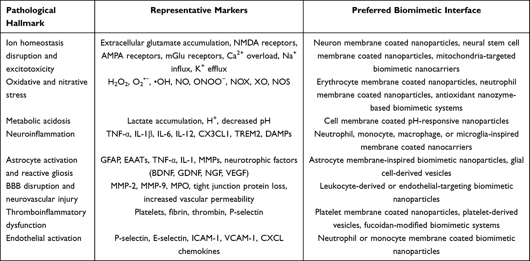 |
Table 1 Key Pathological Hallmarks of Ischemic Stroke and Corresponding Stimuli for Responsive Biomimetic Nanomedicine Design |
Types of Biomimetic Nanocarriers
The development of nanotechnology has provided new opportunities for targeted drug delivery in IS. However, owing to the foreign nature of conventional nanomaterials, they may still be recognized and cleared by the reticuloendothelial system after systemic administration, thereby limiting their circulation time and delivery efficiency to ischemic lesions. In recent years, biomimetic nanomedicine delivery strategies have attracted extensive attention. By incorporating nanomedicines into cells, or by using endogenous cell membranes and specific receptors as functional shells to cloak nanomedicines, these systems can achieve enhanced lesion targeting, immune evasion, and biocompatibility, while reducing immunogenicity and prolonging systemic circulation. Accordingly, a variety of biomimetic nanocarriers derived from targeted cell-based strategies have been developed for IS therapy, as summarized in Table 2.
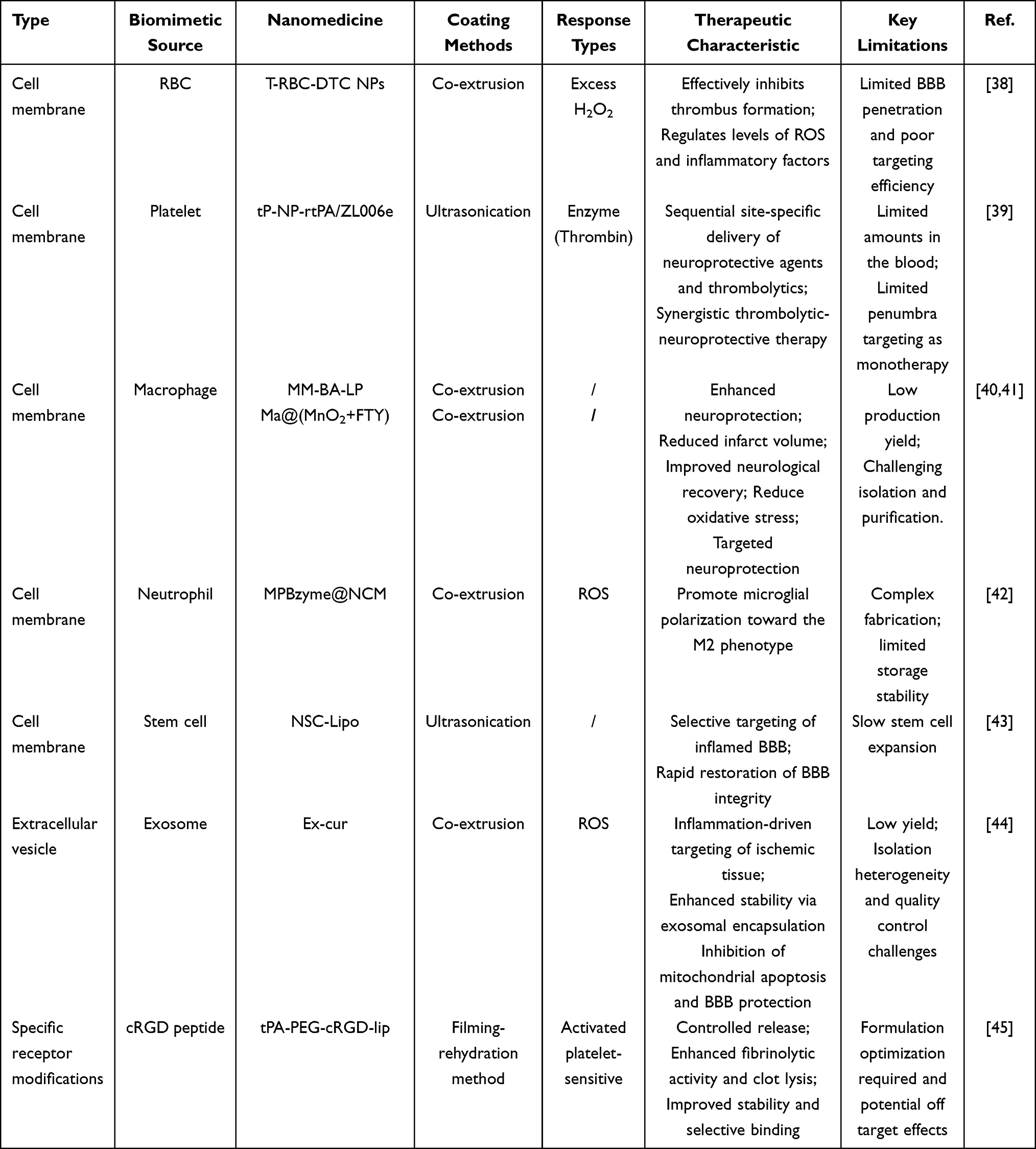 |
Table 2 The Characteristics and Therapeutic Advantages of Biomimetic Nanocarriers |
Cell Membrane Based
During the acute phase of IS, oxidative stress and inflammatory responses triggered by energy depletion activate glial cells, resulting in BBB disruption and the infiltration of peripheral neutrophils, thereby exacerbating brain injury. In the chronic recovery phase, infiltrated macrophages migrate to the ischemic region to promote the repair and regeneration of the neurovascular unit. Given the intricate interactions among blood components, tissues, cells, and cytokines, nanoparticles coated with various cell membranes can not only evade immune clearance but also enhance BBB penetration through membrane-associated proteins.20,46
Red Blood Cell
Red blood cell (RBC) membranes offer distinct advantages as an ideal delivery platform for IS therapy, including facile isolation and extraction, prolonged blood circulation, and surface expression of CD47, which effectively evades macrophage-mediated phagocytosis and confers anti-inflammatory property.47,48 These properties make RBC membrane coated nanoparticles particularly useful as long circulating carriers. However, native RBC membranes generally lack strong intrinsic BBB penetrating or ischemic lesion targeting capability. Therefore, for IS therapy, their primary translational value lies in immune evasion, circulation extension, and systemic stability, while efficient brain targeting usually requires additional ligand engineering or pathological stimuli responsive design.
Zhao et al developed a biomimetic nanocarrier, termed T-RBC-DTC NPs, composed of a drug-conjugated dextran nanocore and an RBC membrane shell (Figure 2A).38 Upon stimulation by excess H2O2, the phenylboronic ester linker breaks between the drug and the polymer, releasing tirofiban in order to achieve an antithrombotic effect at specific sites. Since tirofiban may impair platelet membrane receptors, RBC membrane coating combined with fibrin-targeting peptides was employed to enhance thrombus-targeting capability while avoiding direct platelet membrane damage. In both RAW 264.7 cells and human umbilical vein endothelial cells (HUVECs), T-RBC-DTC NPs effectively scavenged H2O2 and protected cells from H2O2-induced cytotoxicity. Furthermore, in a FeCl3-induced carotid thrombosis mouse model, T-RBC-DTC NPs efficiently accumulated at the site of carotid artery injury, and demonstrated significantly enhanced antithrombotic efficacy compared with free tirofiban.
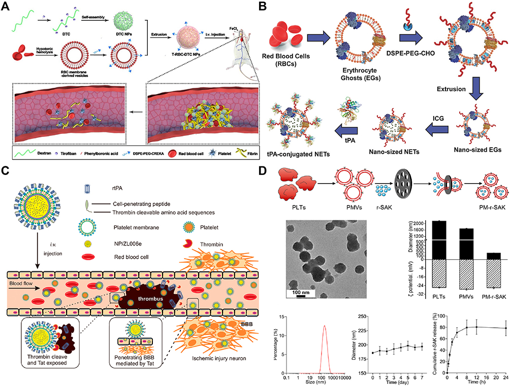 |
Figure 2 (A) Schematic representation of the T-RBC-DTC NPs designed as potent antithrombotic agents for thrombus-targeted therapy.38 Copyright 2020, Elsevier. (B) Schematic representation of the preparation of tPA-conjugated NETs.49 Copyright 2018, Wiley. (C) Schematic depicting the design and characterization of tP‑NP‑rtPA/ZL006e, including its main components, thrombin‑triggered rtPA release following intravenous injection, and Tat‑mediated transcytosis for brain delivery.39 Copyright 2019, American Chemical Society. (D) Schematic diagram of the PM‑r‑SAK coated recombinant staphylokinase.50 Copyright 2024, American Chemical Society. Abbreviations: r-SAK, recombinant staphylokinase; PLTs, platelets; PMVs, PM vesicles. |
As shown in Figure 2B, Raviraj et al constructed functionalized erythrocyte-derived vesicles coupled with immunoglobulin G (IgG) and tissue-type plasminogen activator (tPA) for the integrated diagnosis and treatment of thrombosis.49 In this system, the erythrocyte membrane coating prolonged the in vivo circulation time of NPs (tPA-conjugated NETs), thereby enhancing their thrombus-targeting efficiency and therapeutic potential. RBC membrane-based nanoparticles can not only faithfully preserve the diverse surface antigens and biological functions of the source cells, but also allow the facile incorporation of specific homing ligands through chemical conjugation, lipid insertion, or membrane engineering strategies, thereby improving site-specific targeting capability.51 Consequently, natural erythrocyte membranes represent a promising alternative to conventional PEGylation or synthetic surface modification strategies for the development of biomimetic nanotherapeutics.
Platelet
Platelets spontaneously accumulate at the sites of vascular injury, inflammation, and other associated pathological lesions. Their targeting capacity is mainly mediated by platelet surface receptors and adhesion molecules, including GPIbα, GPVI, GPIIb/IIIa, P-selectin, integrins, CD31, CD47, CD55, and CD59. These molecules enable interactions with von Willebrand factor, collagen, fibrinogen, activated platelets, damaged endothelium, and leukocytes, as supported by previous studies on platelet adhesion and thrombus formation mechanisms.52,53 Therefore, platelet membrane based nanocarriers are particularly suitable for hyperacute thrombus targeting and vascular injury recognition. Nevertheless, platelet membranes do not necessarily provide efficient BBB transcytosis or deep ischemic penumbra targeting when used alone. Their major function is vascular and thrombotic lesion targeting rather than direct brain parenchymal delivery. Xu et al developed a bioengineered nano-platelet (tP-NP-rtPA/ZL006e) for sequential targeted delivery of recombinant tissue plasminogen activator (rtPA) and neuroprotectant (ZL006e) to treat IS.39 The tP-NP-rtPA/ZL006e is a biomimetic nanoplatform comprising a dextran-based polymeric nanoparticle core encapsulating ZL006e, enveloped by a platelet-derived membrane and surface-functionalized with a thrombin-responsive Tat-rtPA conjugate (Figure 2C). Both in vitro and in vivo studies demonstrated that tP-NP-rtPA/ZL006e markedly improved therapeutic efficacy, achieving a 63% reduction in ischemic lesion size and a 72% decrease in ROS levels in a rat middle cerebral artery occlusion (MCAO) model compared with the free drug combination. In another study, Tang et al constructed platelet-mimetic nanoparticles (PTNPs) comprising a platelet membrane-coated PLGA core co-loaded with piceatannol and superparamagnetic iron oxide (SPIO) for the recognition, intervention, and monitoring of inflammatory neutrophils in acute ischemic stroke.54 Through P-selectin–PSGL-1-mediated interactions, PTNPs exhibited approximately 3.4-fold greater uptake by inflammatory neutrophils than uncoated nanoparticles, highlighting the enhanced targeting and internalization conferred by the platelet membrane coating. Despite these advantages, platelet membrane-based biomimetic nanoparticles generally lack intrinsic BBB penetration and ischemic penumbra-targeting capabilities. Therefore, they are not considered the optimal choice for intracranial IS therapy when used alone.
Recent advances in platelet biomimetic nanoparticles have primarily focused on enhancing tPA-mediated thrombolytic therapy during the hyperacute phase of IS.55 Recombinant staphylokinase (r-SAK), a third-generation thrombolytic agent produced through genetic engineering, exhibits superior thrombolytic efficacy compared with urokinase and recombinant streptokinase. Inspired by the intrinsic affinity of platelets for hemostasis and pathological thrombosis, Hua et al developed a platelet membrane-coated formulation (PM-r-SAK) to improve thrombus-targeting efficiency.50 As shown in Figure 2D, PM-r-SAK was prepared by encapsulating r-SAK into PMVs via membrane extrusion. The resulting nanoparticles exhibited a uniformly dispersed spherical morphology, with an average hydrodynamic diameter of approximately 185.8 nm, a zeta potential comparable to that of native platelets (PLTs) and PMVs, and favorable in vitro stability. The r-SAK loading content was 31.43 ± 1.58%, while the cumulative release rate reached 78.59 ± 10.80% within 24 h. Both in vivo animal studies and ex vivo human experiments demonstrated that PM-r-SAK achieved thrombolytic efficacy comparable to or greater than a fourfold higher dose of free r-SAK. Moreover, PM-r-SAK significantly shortened the time to initial recanalization in a rabbit femoral artery occlusion model compared with equivalent or higher doses of recombinant streptokinase. Notably, the enhanced thrombolytic performance of PM-r-SAK is attributed to its platelet membrane-mediated targeting capability, which can be attenuated by platelet inhibition, as evidenced by reduced efficacy in platelet-poor plasma from patients treated with aspirin and ticagrelor.
Macrophage
Macrophages, as key components of the immune surveillance system, are mainly derived from the migration of peripheral monocytes into the brain and subsequent differentiation.56 During I/R injury, intercellular ICAM-1, vascular cell adhesion molecule-1 (VCAM-1), and P-selectin on endothelial cells are overexpressed.57 These adhesion molecules interact with their corresponding ligands, including CD11, CD18, and CD44, on leukocytes, thereby promoting the recruitment of peripheral immune cells, particularly macrophages and neutrophils, to ischemic lesions.57
Macrophage membrane-coated nanoparticles have demonstrated highly efficient targeted delivery efficacy for various inflammatory diseases, along with long circulation time and satisfactory ability to cross the BBB. Long et al reported that baicalin liposomes modified by macrophage membranes (MM-BA-LP) exhibited higher targeting efficiency compared with unmodified baicalin liposomes (BA-LPs) (Figure 3A).40 MM-BA-LP was found to markedly enhance neurological function, reduce the size of cerebral infarction, and alleviate brain pathological damage in MCAO rats compared with BA-LP. Similarly, Su et al synthesized amphiphilic nanoparticles (AOE@TMP) using active herbal components and subsequently cloaked them with macrophage membranes to construct MAOE@TMP (Figure 3B).58 The biomimetic nanoparticles efficiently targeted ischemic brain lesions and selectively released anti-inflammatory agents within the inflammatory microenvironment, thereby significantly reducing infarct size. These anti-inflammatory biomimetic nanoplatforms exert therapeutic effects primarily by attenuating peripheral inflammatory infiltration and suppressing immune cascade amplification within ischemic regions. Owing to the intrinsic inflammatory chemotaxis and deep tissue infiltration capabilities of macrophage membranes, macrophage- or monocyte membrane-engineered nanoparticles not only improve drug delivery efficiency and biocompatibility, but can also be rationally designed to mitigate neuroinflammation associated with IS.
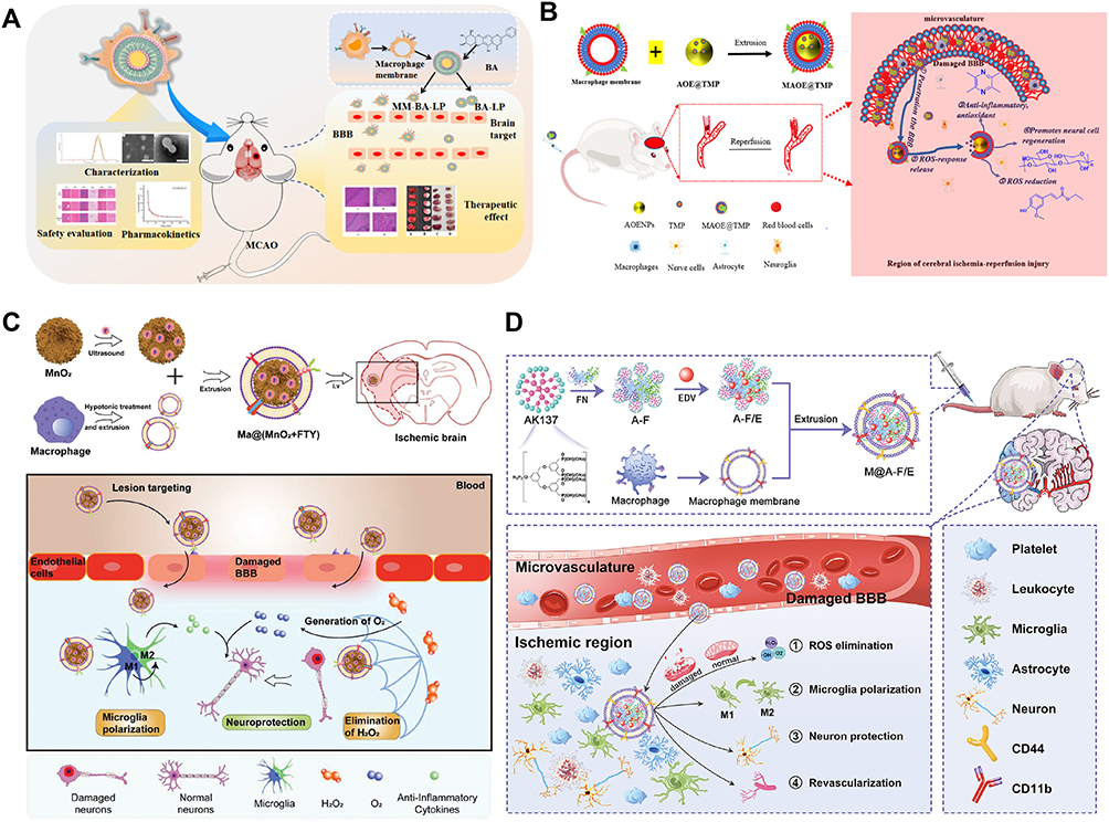 |
Figure 3 (A) Schematic illustration of the preparation and in vivo therapeutic efficacy of MM-BA-LP.40 Copyright 2022, Elsevier. (B) Schematic illustration of the structural characteristics and therapeutic mechanism of MAOE@TMP.58 Copyright 2022, Elsevier. (C) Schematic diagram of the preparation method and in vivo therapeutic mechanism of Ma@(MnO2+FTY).41 Copyright 2021, Wiley. (D) Schematic depicting the fabrication of biomimetic M@A-F/E NCs aimed at synergistically eliminating ROS, polarizing microglia toward M2 phenotype, protecting neurons, and promoting revascularization for the IRI‑induced IS treatment.59 Copyright 2024, Wiley. |
In addition to targeted delivery, the biomimetic nanoplatform modified by macrophage membrane can also regulate microglial polarization from the pro-inflammatory M1 phenotype toward the anti-inflammatory M2 phenotype, thereby remodeling the inflammatory microenvironment and exerting neuroprotective effects. Li et al developed a macrophage membrane-coated honeycomb-like manganese dioxide (MnO2) nanosphere loaded with fingolimod (FTY), denoted as Ma@(MnO2+FTY), for ischemic penumbra rescue (Figure 3C).41 Ma@(MnO2+FTY) was actively accumulated in the injured brain via macrophage membrane protein-mediated recognition and interaction with overexpressed adhesion molecules on damaged vascular endothelial cells. The MnO2 nanospheres effectively decomposed excess H2O2 into O2, thereby alleviating oxidative stress, and subsequently degrade within acidic lysosomes to enable controlled release of FTY. This cascade therapeutic process promoted the polarization of microglia from the pro-inflammatory M1 phenotype to the anti-inflammatory M2 phenotype, ultimately reversing the inflammatory microenvironment and enhancing neuronal survival. In another study, Ma et al developed a macrophage membrane-camouflaged nanoplatform composed of a phosphorus dendrimer (AK137)/fibronectin (FN) nanocomplex loaded with the antioxidant EDV for effective IS therapy through the coordinated modulation of microglia and neurons (Figure 3D).59 The resulting MM@AK137-FN/EDV nanocomplexes (M@A-F/E NCs), with an average size of approximately 260 nm, exhibited excellent colloidal stability, sustained EDV release behavior, and favorable cytocompatibility. Benefiting from macrophage membrane functionalization, the nanocomplexes efficiently crossed the BBB and exerted potent anti-inflammatory and antioxidant effects on microglia in vitro. In a transient middle cerebral artery occlusion (tMCAO) rat model, M@A-F/E nanocarrier demonstrated enhanced antioxidative, anti-inflammatory, and anti-apoptotic effects, thereby comprehensively modulating the cerebral microenvironment, promoting angiogenesis, and facilitating the restoration of blood perfusion following I/R injury.
Neutrophil
Neutrophils play a central role in the immune response by infiltrating damaged tissues, phagocytosing debris, and releasing inflammatory mediators, while interacting with endothelial cells to recruit other immune cells. During ischemia, the increase of inflammatory cytokines (such as IL-1β and TNF-α) and chemokines (including CXCL2 and CXCL12) leads to the recruitment and activation of neutrophils.60 It has been reported that neutrophils accumulate in the ischemic penumbra cortex at all stages.61,62 In particular, Mac-1, LFA-1, and integrin β2 on neutrophil membranes can bind to ICAM-1 and related adhesion molecules on inflamed cerebral endothelial cells. This receptor mediated adhesion provides a mechanistic basis for neutrophil membrane coated nanoparticles to target the inflamed BBB and ischemic lesions. Based on the intrinsic adhesion capability of neutrophils toward inflamed endothelial cells, Dong et al developed neutrophil membrane-derived nanovesicles encapsulating the anti-inflammatory and neuroprotective mediator resolvin D2 (RvD2).63 These biomimetic RvD2-HVs specifically targeted inflamed cerebral endothelial cells via the interaction between ICAM-1 and integrin β2, thereby enabling efficient delivery of RvD2 to ischemic lesions and protecting brain tissue against reperfusion injury (Figure 4A). Similarly, Feng et al utilized the inflammatory tropism of neutrophils to improve the delivery efficiency of nanozymes to the injured brain and prepared neutrophil membrane-coated mesoporous Prussian blue nanozymes (MPBzyme@NCM) for non-invasive active targeting for the treatment of I/R injury.42 Fingolimod (FTY720), a sphingosine-1-phosphate receptor agonist approved for the treatment of multiple sclerosis, has demonstrated considerable therapeutic potential for IS owing to its anti-inflammatory activity. However, its clinical application remains limited by poor BBB penetration and dose-dependent cardiovascular toxicity. Therefore, further research is required to optimize its biocompatibility and precise drug administration for stroke treatment.64–66 Zhao et al designed a neutrophil membrane-camouflaged multi-prodrug nanomedicine that enables targeted delivery to inflamed brain tissue and in situ release of FTY720 in response to elevated ROS levels (Figure 4B).67 This nanomedicine (NRNs) delivered 15.2-fold higher FTY720 to the ischemic brain compared with intravenously administered free drug, while significantly reducing the risks of cardiotoxicity and infection. Through receptor-mediated tropism and integrin-mediated adhesion, neutrophil membranes endow nanoparticles with enhanced homotypic targeting capability and circulatory stability, thereby providing a promising strategy for targeted IS therapy.
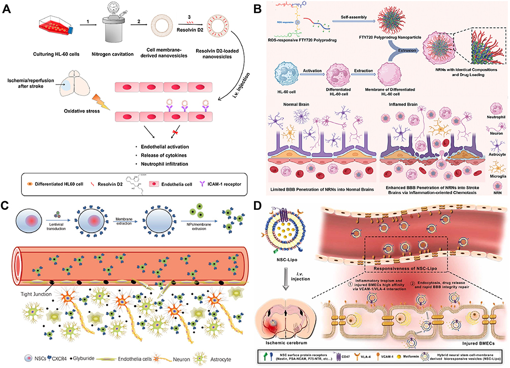 |
Figure 4 (A) Illustration showing the design of RvD2‑HVs that specifically adhere to inflamed brain endothelial cells to mitigate post‑reperfusion neuroinflammation in ischemic stroke.63 Copyright 2019, American Chemical Society. (B) Schematic diagram illustrating the construction and ischemic brain-targeting anti-inflammatory effects of NRNs.67 Copyright 2024, Wiley. (C) Schematic depicting the use of CXCR4-overexpressing NSC membrane coatings for targeted NP delivery to the ischemic brain.68 Copyright 2019, Wiley. (D) Schematic diagram of NSC‑derived bioresponsive vesicles (NSC‑Lipo) achieving VLA‑4/VCAM‑1‑mediated targeting of injured BMECs for anti‑inflammatory drug delivery and rapid BBB repair, after which VCAM‑1 downregulation terminates targeting capability.43 Copyright 2023, Elsevier. |
Stem Cell
Stem cells exhibit several advantageous biological properties, including self-renewal capacity, multilineage differentiation potential, and low immunogenicity. Among them, neural stem cells (NSCs) have emerged as a promising therapeutic approach for IS due to their intrinsic tropism toward ischemic brain lesions. Notably, NSCs are capable of traversing the BBB and demonstrate high spatial specificity in homing to ischemic regions. Leveraging these unique properties, NSC membrane-coated biomimetic nanoparticles have been developed as a rational and efficient strategy to enhance targeted drug delivery to the ischemic penumbra.69
Given that CXCR4 is a key receptor mediating cell chemotactic migration in the ischemic microenvironment, Ma et al achieved stable overexpression of CXCR4 on the membrane surface of NSCs through lentivirus vector-mediated gene transfection technology (Figure 4C).68 They used CXCR4-engineered NSC membranes to coat PLGA nanoparticles (CMNPs), which significantly enhanced the delivery efficiency of the anti-edema drug glibenclamide to ischemic brain regions. Similarly, Shi et al constructed bionic nanoparticles with the ability to preferentially accumulate in ischemic brain regions based on the chemotactic guidance characteristics of the CXCR4-CXCL12 axis.70 The outer shell consisted of membranes derived from CXCR4-overexpressing mesenchymal stem cells (MSCs), which not only neutralized excessive local CXCL12 within ischemic tissues but also enhanced nanoparticle homing toward ischemic lesions. The nanoparticle core comprised polydopamine nanospheres loaded with the cGAS-STING pathway inhibitor A151. This multifunctional nanoplatform exhibited both ROS-scavenging and immunomodulatory properties, thereby synergistically alleviating neuroinflammation and oxidative stress-induced damage following ischemia. Compared with CXCR4 overexpressing NSC membrane coated nanoparticles, which mainly exploit the CXCR4/SDF-1 chemotactic axis to enhance ischemic brain homing and drug delivery, CXCR4 enriched MSC membrane coated nanoparticles function not only as lesion homing carriers but also as CXCL12 biomimetic decoys that neutralize excessive CXCL12, block peripheral inflammatory cell infiltration, and remodel the overactivated immune microenvironment after ischemic stroke. As shown in Figure 4D, Wu et al functionally recombined NSC membranes with conventional liposomes and embedded very late antigen-4 (VLA-4) on their surface to synthesize a metformin-loaded bio-responsive vesicle (NSC-Lipo).43 The VLA-4 integrin displayed on the NSC membrane specifically recognized and bound to VCAM-1, which is upregulated on damaged brain microvascular endothelial cells in the ischemic region. This enabled VCAM-1/VLA-4-mediated selective delivery of the anti-inflammatory agent metformin to injured brain tissue. The targeted delivery system facilitated rapid restoration of BBB integrity and activated endogenous neuroprotective pathways. Notably, as BBB integrity recovered and VCAM-1 expression declined, the targeting capability of NSC-Lipo was correspondingly diminished. Compared with metformin-loaded bare liposomes, a single administration of the NSC-Lipo increased the survival rate of ischemic mice from 30% to 90%.
Collectively, a wide range of stem cell membrane-based biomimetic nanoparticles, including those derived from NSCs, MSCs, embryonic stem cells, and induced pluripotent stem cells, have emerged as promising therapeutic candidates for IS. Their therapeutic mechanisms extend beyond conventional drug delivery to include paracrine regulation, immune modulation, activation of endogenous repair pathways, and other neuroprotective effects.71 However, current studies on stem cell membrane-based biomimetic nanoplatforms have largely focused on nanoparticles loaded with therapeutic agents rather than exploiting the intrinsic therapeutic potential of stem cells themselves. Their clinical application requires rigorous preservation and verification of functional proteins such as CXCR4 and VLA-4, standardized source cell expansion, assessment of source cell variability, and evaluation of long-term biosafety. Therefore, the future development of “top-down” biomimetic nanoplatforms that preserve biologically active cellular components may substantially improve the overall therapeutic efficacy of stem cell-derived biomimetic nanoparticles.
Extracellular Vesicle
Exosomes are a class of lipid bilayer-enclosed extracellular vesicles released by cells, typically ranging from 40 to 100 nm in diameter. As important mediators of intercellular communication, exosomes participate in both local and long-distance signaling and play crucial roles in the complex crosstalk among neurons, glial cells, endothelial cells, and immune cells following stroke.72 Various bioactive molecules are abundant in exosomes, including microRNAs, DNA, mRNA, proteins, and lipids. Among these components, miRNAs have attracted considerable attention due to their ability to effectively promote the recovery of neurological function after stroke.73–75 Notably, exosome membranes contain relatively high levels of cholesterol and sphingolipids compared with many other donor cell-derived membranes, thereby conferring enhanced structural stability and resistance to extracellular degradation.76 Furthermore, compared with conventional cell membrane-based delivery platforms, exosome-based platforms offer greater advantages for drug delivery, as their nanoscale size, mildly negative zeta potential, and intrinsic deformability facilitate penetration into deep tissues and improve transport across biological barriers.77
Based on the natural properties of macrophage-derived exosomes and the bioactivity of curcumin, He et al designed a macrophage-derived exosome loaded with curcumin (Ex-cur) as a multifunctional biomimetic delivery system targeting ischemic brain tissue to alleviate I/R injury (Figure 5A).44 Owing to the inflammation-homing capability of exosomes, Ex-cur efficiently accumulated in the ischemic region and reduction of ROS levels. Protein analysis confirmed that Ex-cur alleviated BBB damage and inhibited mitochondria-mediated neuronal apoptosis in the ischemic area by suppressing ROS accumulation. CHAC1 is a key gene in the process of ferroptosis in IS. Based on this finding, Wang et al designed and prepared adipose-derived mesenchymal stem cell (ADSC-Exo) anti-ferroptosis exosomes to treat ischemic brain injury via intranasal administration (Figure 5B).78 miR-760-3p, which is highly expressed in ADSC-Exo, can inhibit the expression of CHAC1. The results showed that miR-760-3p in ADSC-Exo inhibited ferroptosis by targeting CHAC1 in neurons. In another report, bacterial-derived outer membrane vesicles (OMVs) were explored as a biomimetic delivery platform to enhance the brain delivery of IS treatment drugs by exploiting the “neutrophil hitchhiking” mechanism.79 By encapsulating pioglitazone (PGZ) into OMV, the resulting OMV@PGZ nanoparticles inherit the functions related to bacterial outer membranes and could be targeted and taken up by neutrophils (Figure 5C). Mechanistically, OMV@PGZ suppressed activation of the nucleotide-binding oligomerization domain-like receptor protein 3 (NLRP3) inflammasome and inhibited ferroptosis, thereby alleviating cerebral reperfusion injury and exerting significant neuroprotective effects.
 |
Figure 5 (A) Schematic diagram of the preparation of EXO-CUR and its therapeutic mechanism of targeting ischemic brain tissue to alleviate cerebral ischemia‑reperfusion injury by inhibiting ROS‑mediated mitochondrial apoptosis.44 Copyright 2020, Elsevier. (B) Schematic diagram of ADSC-Exo delivering miR-760-3p via intranasal administration for the treatment of ischemic brain injury.78 Copyright 2023, BioMed Central. (C) Schematic of OMV@PGZ nanoparticles for ischemic stroke.79 TEM image and size distribution of OMVs are provided (scale bar, 200 nm). Copyright 2023, Wiley. (D) Schematic diagram of therapeutic albumin nanocarriers loaded with nerve growth factor for stimulating brain recovery after stroke.80 Copyright 2019, Elsevier. (E) Schematic representation of the preparation process of tPA-cRGD-PEG-NV.81 Copyright 2021, Science. (F) Schematic overview of SP preparation using miniemulsion and crosslinking.82 Copyright 2021, Elsevier. |
Nevertheless, the clinical translation of naturally derived exosomes and exosome-mimetic vesicles remains challenging due to the need for large-scale cell culture and complicated purification procedures, which result in limited production yield and high manufacturing costs. These limitations substantially restrict their scalability and clinical applicability. Notably, previous studies have reported that the yield of exosome-biomimetic/hybrid nanovesicles prepared by extrusion is 250 times higher than that of naturally secreted exosomes.83 In the future, systematic research on key aspects such as large-scale exosome preparation, long-term stable storage, and quality control evaluation will provide strong support for the clinical translation of exosome-biomimetic/hybrid nanovesicles.
Specific Receptor Modifications
The major transcellular pathways involved in brain-targeted delivery include receptor-mediated transcytosis, adsorptive-mediated transcytosis, and carrier-mediated transport. Among these mechanisms, receptor-mediated transcytosis has attracted considerable attention in the design of biomimetic nanomedicines because of its high targeting specificity and favorable BBB penetration efficiency. This strategy generally involves functionalizing the surface of nanocarriers with specific ligands or targeting proteins that recognize receptors highly expressed on BBB endothelial cells or pathological tissues, thereby enhancing accumulation at diseased sites while reducing off-target effects.
Representative biomimetic targeting strategies mainly exploit receptors and transport systems such as transferrin receptors (TfR), glucose transporters (GLUTs), low-density lipoprotein receptors (LDLRs), and low-density lipoprotein receptor-related protein 1 (LRP1). By modifying corresponding ligands on the nanocarrier surface, these receptors can mediate efficient transport across BBB.22 Apolipoprotein E (ApoE) exhibits high affinity for LRP1. Feczkó et al developed ApoE-modified albumin nanocarriers co-loaded with nerve growth factor and ultrasmall iron oxide nanoparticles (Figure 5D).80 Through LRP1-mediated transport, the ApoE-functionalized nanoplatform facilitated delivery of nerve growth factor to ischemic brain tissue, thereby promoting cholinergic neuron survival and differentiation and enhancing neuroprotection after IS. Yu et al reported a multifunctional biomimetic liposome system with a diameter of approximately 164.6 nm, which can target and control the release of tPA at the thrombus site.45 The tPA-loaded liposomes were PEGylated to enhance their stability, and further modified with cyclic arginine-glycine-aspartic acid (cRGD) peptides on the surface. The cRGD moiety enabled selective binding to integrins expressed on activated platelets, thereby facilitating targeted accumulation of thrombolytic agents at thrombus sites and achieving efficient and selective thrombolysis. To further overcome the clinical limitations of tPA thrombolytic therapy, Yu et al proposed a multifunctional nanovesicle tPA-cRGD-PEG-NV to mimic fibrinogen for the specific delivery of tPA and targeted thrombolysis, inspired by the binding of fibrinogen to activated platelets. This nanoplatform consisted of PEGylated lipid nanovesicles terminally conjugated with cyclic cRGD peptides, enabling biomimetic platelet-targeting capability (Figure 5E). Under static and physiological blood flow conditions, tPA-cRGD-PEG-NV exhibited highly selective binding to activated platelets in human blood samples and effectively released tPA at the thrombus site. Notably, the authors also established a computational model capable of predicting the pharmacokinetics of tPA-loaded nanovesicles under realistic physiological conditions, providing additional support for the rational optimization and translational development of biomimetic thrombolytic nanomedicines.81
In another study, Alina et al employed reverse microemulsion combined with chemical crosslinking technology to construct a type of novel fucoidan-functionalized dextran submicroparticles (SPs) with excellent biocompatibility (Figure 5F).82 This system successfully encapsulated thrombolytic drug rtPA. Owing to the high-affinity interaction between fucoidan and P-selectin, which is markedly upregulated on activated platelets and injured vascular endothelial cells within the thrombotic microenvironment, the biomimetic delivery system achieved selective accumulation at thrombus sites. This targeted delivery strategy significantly enhanced local thrombolytic efficacy while improving therapeutic specificity and reducing potential off-target effects.
Clinical Translational Progress of Biomimetic Nanocarriers
Although biomimetic nanocarriers have shown considerable promise in preclinical models of ischemic stroke, their clinical translation remains at an early stage. Most cell membrane coated biomimetic nanoparticles developed for IS therapy, including RBC membrane, platelet membrane, macrophage membrane, neutrophil membrane, and stem cell membrane coated systems, have not yet advanced to registered clinical trials specifically for stroke treatment.20 This translational gap reflects not only the technical complexity of biomimetic nanomedicine, but also the stringent clinical requirements of acute cerebrovascular diseases. In contrast to chronic inflammatory diseases or cancer, IS therapy requires rapid administration, compatibility with intravenous thrombolysis or mechanical thrombectomy, predictable pharmacokinetics, low hemorrhagic risk, and reliable efficacy within a narrow therapeutic window. Among different biomimetic platforms, RBC based systems currently have relatively mature clinical precedents. Although RBC membrane coated nanoparticles for IS remain preclinical, erythrocyte based drug delivery systems have been clinically evaluated in other diseases, supporting their feasibility for prolonging circulation and reducing systemic toxicity.84,85 However, RBC membranes mainly provide immune evasion and long circulation rather than intrinsic BBB targeting, thus often requiring additional targeting strategies for effective brain delivery. Platelet membrane coated nanocarriers are highly relevant to IS due to their natural roles in thrombus formation and vascular injury recognition.86,87 They are promising for thrombus targeting and combined thrombolytic and neuroprotective therapy. However, their translation requires careful safety evaluation because of potential risks of off target thrombosis and interference with hemostasis. Macrophage and neutrophil membrane coated nanoparticles exhibit strong inflammatory targeting and can interact with activated endothelium.88 These systems are suitable for addressing post ischemic inflammation and reperfusion injury. Nevertheless, their clinical translation is limited by issues such as donor variability, immune safety concerns, and challenges in standardization. Stem cell membrane-coated nanocarriers are associated with neuroregenerative potential and benefit from clinical experience with stem cell therapies in stroke.89,90 However, these systems remain preclinical, and their translation depends on preserving functional membrane proteins during preparation and ensuring reproducibility. Compared with cell membrane coated nanoparticles, extracellular vesicles appear closer to clinical translation due to their natural cargo carrying capacity and established guidelines such as MISEV2018 and MISEV2023.91–93 However, challenges including heterogeneity, low loading efficiency, and scalability still limit their clinical application. Future studies should focus on standardized evaluation frameworks, including pharmacokinetics, safety, and compatibility with existing stroke therapies. Overall, EV based platforms and RBC based carriers currently appear closest to clinical translation due to stronger precedents and better standardization frameworks.
Stimuli-Responsive Delivery Strategies Based on Biomimetic Nanocarriers
In recent years, stimuli-responsive nanodelivery strategies have emerged as promising approaches for drug delivery.94 These platforms are designed to achieve controllable therapeutic release in response to defined biological or physical triggers, including hypoxia, acidic pH, ROS overproduction, magnetic fields, and ultrasound.95 Compared with externally triggered systems, pathological microenvironment responsive nanomedicines offer distinct advantages because they do not require additional equipment to activate drug release. The unique pathological features of the ischemic microenvironment, including excessive ROS production, pH and inflammation, provide valuable opportunities for the development of stimuli responsive nanomedicines for IS therapy. In this section, we discuss the structural design, stimuli responsive and target mechanisms of ischemic microenvironment responsive biomimetic drug delivery systems from two major perspectives: endogenous stimuli, including oxidative stress, pH, enzymes, and inflammation, and exogenous stimuli, including magnetic fields, light, and ultrasound (Table 3).
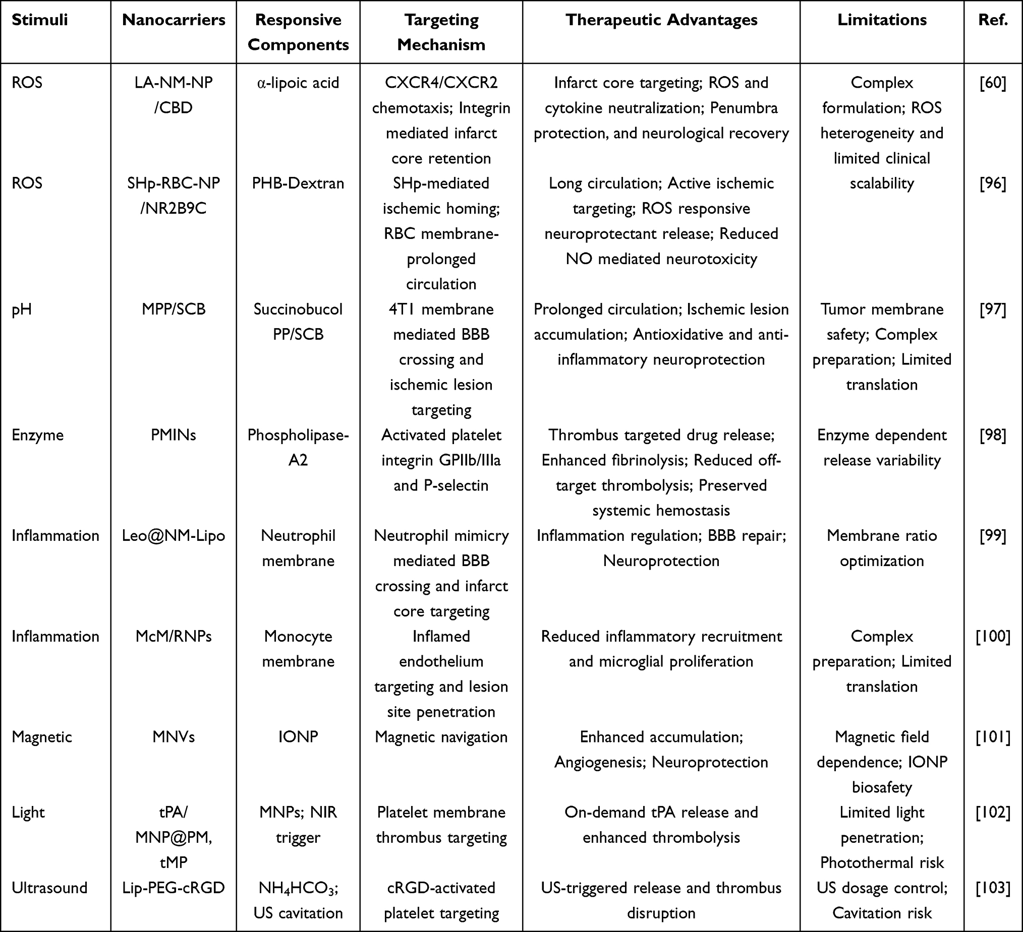 |
Table 3 Representative Stimuli-Responsive Biomimetic Nanocarriers for Ischemic Stroke Therapy |
Oxidative Stress Responsive
Oxidative stress arises from an imbalance between the excessive production of ROS and reactive nitrogen species (RNS) and the endogenous antioxidant defense. Following cerebral I/R injury, hydroxyl radicals (•OH), superoxide anions (O2•−), NO, and peroxynitrite (ONOO−) accumulate in the brain, causing lipid peroxidation, DNA damage, and protein denaturation. These pathological events subsequently contribute to neuronal death, BBB disruption, and expansion of the cerebral infarct area.28 Notably, oxidative DNA damage can persist for up to six months after stroke, leading to demyelination and axonal damage of neurons.104 Therefore, ROS have become a key therapeutic target for reversing neural damage. Although antioxidant therapeutic agents can theoretically neutralize oxidative damage, their clinical efficacy remains limited by several factors, including insufficient BBB penetration, short blood circulation half-life, and poor accumulation within ischemic brain regions.28,105,106 To overcome these limitations, ROS-responsive biomimetic nanotherapeutic strategies have recently attracted increasing attention. These systems exploit the abnormally elevated ROS levels within the ischemic microenvironment as endogenous stimuli to achieve site-specific drug release in the ischemic penumbra. Through this pathological microenvironment-responsive delivery mechanism, biomimetic nanoplatforms can enhance antioxidant bioavailability at lesion sites and potentially overcome the limitations associated with conventional antioxidant therapies.
For example, ROS responsive and mitochondria targeted self-assembled polymeric nanoparticles (SPNPs) were engineered based on the elevated mitochondrial ROS levels in ischemic neurons.107 By loading SPNPs into a thermosensitive gel for intranasal administration, this system enabled efficient nose to brain delivery to the ischemic penumbra while bypassing the BBB (Figure 6A). In vitro studies using H2O2 injured SH SY5Y cells demonstrated that SPNPs could selectively target mitochondria and protect neurons from oxidative damage. In MCAO rat models, fluorescence imaging confirmed rapid and enhanced accumulation of SPNPs in the ischemic region. Therapeutically, intranasally delivered SPNPs alleviated oxidative stress and inflammation, improved mitochondrial function, and reduced apoptosis, thereby providing an effective ROS responsive strategy for ischemic stroke intervention. Beyond organelle targeted and route optimized delivery, biomimetic membrane engineering has further expanded the design of ROS responsive nanomedicines by improving immune evasion and lesion specific accumulation. Cao et al developed a ROS responsive nanocarrier for targeted delivery of rapamycin (RAPA) to ischemic brain tissues (RAPA@tRPCS).108 This system was constructed with a sulfated chitosan (SCS) polymeric core containing ROS cleavable boronic ester linkages, which was further cloaked with a red blood cell membrane modified with a stroke homing peptide (Figure 6B). Owing to the biomimetic membrane shell and lesion targeting peptide, the nanocarrier improved immune evasion and enhanced accumulation in ischemic brain regions. In response to elevated intracellular ROS levels in ischemic tissues, the nanoparticles underwent structural disassembly and released both SCS and RAPA. The released therapeutic components promoted beneficial microglial polarization, preserved BBB integrity, reduced cerebral infarction, and facilitated neurovascular remodeling in a tMCAO mouse model.
 |
Figure 6 (A) Schematic illustration of targeted treatment of ischemic stroke by ROS-responsive nanoparticles loaded with PU and decorated with SS31 (SPNPs hydrogels).107 Copyright 2023, Elsevier. (B) Schematic illustration of RAPA@tRPCS treatment improved angiogenesis and neurogenesis.108 Copyright 2024, American Chemical Society. (C) Schematic diagram illustrates the composition and stepwise targeting of SNM-NPs, as well as their therapeutic effects on the cerebral I/R injury.109 Copyright 2023, Wiley. (D) Schematic design of the SHp-RBC-NP/NR2B9C.96 After intravenous injection, the SHp-RBC-NP/NR2B9C could prolong the circulation life with the RBC-mimicking properties and then target to the ischemic brain site via stroke homing peptide mediated transcytosis. Copyright 2018, American Chemical Society. (E) Schematic illustration of the therapeutic mechanism of shell-sheddable cRGD/TPP@Res micelles in alleviating oxidative stress and inflammation in ischemic stroke.110 Copyright 2023, American Chemical Society. (F) Schematic illustration of the preparation of membrane-camouflaged MPP/SCB nanocarriers and their therapeutic advantages: biomimetic preferential targeting of cerebral ischemic lesions, efficient internalization by brain cells, and pH-responsive release of active agents.97 Copyright 2021, American Chemical Society. |
To further remodel the hostile ischemic microenvironment, Liu et al proposed a “nano-buffer” strategy targeting the ischemic core.60 They constructed a PLGA nano-buffer coated with neutrophil membranes, surface-modified with α-lipoic acid and loaded with cannabidiol (LA-NM-NP/CBD). Following accumulation within ischemic lesions, adjacent nanoparticles underwent a dynamic ring-opening polymerization process, which induced cross-fusion of the membrane-coated nanoparticle surfaces and facilitated in situ nano-buffer formation. Through integrin-mediated targeting of the infarct core, this nano-buffer system effectively scavenged free radicals and neutralized inflammatory mediators within both the ischemic core and penumbra, thereby significantly reducing infarct volume and improving neurological functional recovery. Dong et al further developed a multi-step targeted biomimetic drug delivery system named SHp-NM@Edv/RCD (SNM-NPs).109 The nanoparticles were coated with neutrophil membranes (Figure 6C), resulting in markedly enhanced targeting efficiency toward the inflammatory microenvironment (approximately 5.16-fold). After reaching the ischemic lesion, the ROS-responsive component RCD within the system triggered the controlled release of EDV in response to elevated local ROS levels. This ROS-responsive cascade effectively attenuated oxidative stress, suppressed neuroinflammatory responses, and reduced neuronal apoptosis by nearly 90%. Mechanistically, SNM-NPs precisely deliver EDV to the cerebral I/R injury area, mitigate oxidative stress, decrease microglial activation, restore neuronal tubulin expression, and exert anti-apoptotic effects through modulation of the Caspase-3 signaling pathway.
This study highlights the therapeutic value of combining biomimetic membrane camouflage, ischemic lesion homing, and ROS triggered drug release within a single nanoplatform. Extending this lesion homing strategy, Lv et al employed a stroke homing peptide (SHp, CLEVSRKNC),96 identified by in vivo phage display and capable of selectively recognizing ischemic brain regions and apoptotic neurons in the penumbra, to construct a core shell H2O2 responsive biomimetic nanoparticle (SHp-RBC-NP/NR2B9C) (Figure 6D). This nanoplatform consisted of an SHp-modified RBC membrane shell and a core composed of borate ester-functionalized dextran (PHB-Dextran) encapsulating the neuroprotective peptide NR2B9C. The RBC membrane coating prolonged systemic circulation (t1/2 ≈ 48 h), whereas elevated intracellular H2O2 levels in ischemic neurons selectively cleaved borate ester bonds within PHB-Dextran, triggering nanoparticle disassembly and intracellular release of NR2B9C. Additionally, the NR2B9C disrupted the interaction between postsynaptic density protein-95 and NMDA receptors, thereby suppressing excessive NO production and alleviating excitotoxic neuronal injury.
Overall, significant progress has been made in the design and synthesis of biomimetic nanomedicines that can specifically respond to the ROS levels in the ischemic microenvironment. By exploiting the elevated ROS levels within the ischemic microenvironment, these biomimetic nanoplatforms enable targeted drug delivery and spatiotemporally controlled therapeutic release, thereby improving treatment precision and enhancing neuroprotective efficacy. Owing to their unique capabilities in modulating oxidative stress and remodeling the pathological microenvironment, ROS-responsive biomimetic nanomedicines have emerged as a highly promising strategy for neural repair and precision intervention after stroke. Although intranasal administration has been explored to improve the delivery efficiency of ROS responsive nano-delivery systems by partially bypassing the BBB, this route-based strategy does not fully resolve the key delivery barriers associated with IS therapy. In particular, efficient transport across BBB related interfaces and selective accumulation in specific cells or organelles remain major challenges for ROS responsive nano-systems. Moreover, several critical issues still hinder their clinical translation, including large scale manufacturing, long term biosafety, in vivo stability, reproducibility, and regulatory standardization. Therefore, although ROS responsive biomimetic nanomedicines exhibit considerable therapeutic potential, further systematic preclinical and clinical investigations are required before they can become clinically viable treatment options for IS.
pH Responsive
The pH value within brain cells is maintained at 7.2 through active and passive ion transport under physiological conditions.111 During a stroke, the poor perfusion affects the extracellular CO2 excretion, leading to intracellular CO2 accumulation. This causes the intracellular CO2 content to be 3–4 times higher than the normal level while the oxygen content decreases. Under these hypoxic conditions, neurons and glial cells increasingly rely on anaerobic glycolysis for energy production, resulting in excessive lactate generation and proton (H⁺) accumulation, which ultimately induces lactic acidosis. In addition, ischemic insult triggers depolarization waves that propagate across affected brain regions, a phenomenon known as spreading depolarization or spreading depression. This process markedly increases cellular energy demand under conditions of insufficient oxygen supply, further promoting anaerobic glycolysis and lactate production. As a result, the pH value in the ischemic penumbra drops to 6.5, causing moderate acidosis.112 Moreover, ischemia-associated oxidative stress and free radical generation further exacerbate intracellular proton accumulation and pH reduction. When intracellular pH declines below approximately 6.3 ~ 6.4, irreversible cellular injury pathways are activated in ischemic tissues.113 Therefore, acidosis represents a prominent pathological characteristic of IS and provides an important endogenous stimulus for the development of pH-responsive biomimetic nanotherapeutic systems. Compared with ROS responsive systems, which directly exploit oxidative stress and are particularly suitable for antioxidant intervention and redox triggered drug release, pH responsive systems utilize ischemia induced acidosis as a relatively sustained microenvironmental cue for controlled release and organelle targeted delivery. However, the pH difference between ischemic and normal brain tissues is often modest and spatially heterogeneous, which may reduce triggering sensitivity and lesion selectivity, whereas ROS responsiveness may provide stronger pathological specificity but is more susceptible to transient ROS fluctuations during reperfusion.
Wang et al designed a pH-responsive poly (ethylene glycol)-acetal-polycaprolactone-poly (ethylene glycol) (PEG-Acetal-PCL-PEG) and modified with cRGD and triphenylphosphine (TPP) (Figure 6E).110 These nano-micelles were loaded with the mitochondria-targeting antioxidant drug resveratrol (Res). Under acidic conditions, cRGD/TPP@Res cleaves the acetal bond, causing the long polyethylene glycol chain to fall off, thereby transforming the cRGD/TPP@Res micelle into a TPP@Res micelle. This transformation promotes mitochondrial targeting by exposing TPP. As a result, Res can be directly released in the mitochondria of cells, alleviating oxidative stress by eliminating excessive ROS and reducing neuroinflammation by polarizing M1-type microglia to M2-type. In a recent study, He et al designed a pH-sensitive imine bond integrated into succinyl butylated butadiene (SCB) polyethylene glycol polymer and camouflaged with 4T1 cell membranes, thereby forming a biomimetic nanomedicine (MPP/SCB) (Figure 6F).97 The 4T1 cell membranes on the surface of MPP/SCB can effectively promote the passage of nanomedicines through the BBB, and MPP/SCB has excellent antioxidant and anti-inflammatory properties. In a tMCAO mouse model, MPP/SCB demonstrated pronounced brain accumulation, with preferential distribution in the ischemic hemisphere that was 4.79-fold higher than that observed in the contralateral normal hemisphere. In addition, this biomimetic nanoplatform exhibited strong antioxidant and anti-inflammatory activities, thereby contributing to effective neuroprotection in IS.
Enzyme Responsive
Enzyme-responsive nanocarriers are capable of recognizing endogenous enzymes that are abnormally upregulated in intracellular or extracellular pathological microenvironments, thereby enabling site-specific drug release through enzyme-catalyzed biochemical reactions. After I/R occurs, multiple enzymes such as thrombin and MMPs are significantly involved in the pathological process. Tuo et al discovered through lipid metabolomics, proteomics, and immunohistochemistry techniques that thrombin and its downstream product ACSL4, which is a key regulator of the ferroptosis pathway.114 Notably, proteomic profiling of patient serum samples revealed no significant elevation in circulating thrombin levels, suggesting that thrombin accumulation in ischemic brain tissue primarily originates from local cerebral production rather than peripheral blood sources. Christa L. Pawlowski et al developed platelet microparticle biomimetic nanovesicles (PMINs) designed for targeted thrombolytic therapy (Figure 7A).98 This system protects encapsulated thrombolytic agents from off-target clearance in the circulation and enables active anchoring to thrombi via hetero multivalent interactions with activated platelet integrin GPIIb/IIIa and P-selectin. Drug release is subsequently triggered by thrombus-associated phospholipase A2 (sPLA2), an enzyme enriched in the clot microenvironment. In vitro experiments showed that the percentage of SK released from PMINs exposed to sPLA2 was approximately 4 times that of those not exposed to the enzyme within the first 2 h. Additionally, in vivo studies demonstrated that PMINs loaded with SK achieved efficient thrombolysis while reducing off-target side effects on systemic hemostasis.
 |
Figure 7 (A) Schematic illustration of the structure, surface characteristics, and thrombus-targeting properties of platelet-derived microparticles (PMINs).98 Copyright 2017, Elsevier. (B) Schematic depicting the fabrication and thrombolytic mechanism of MnO2/uPA@pep-Fuco.115 Copyright 2021, Elsevier. (C) Schematic illustration showing the preparation of Leo@NM-Lipo and its ability to reverse cerebral I/R injury through multiple protective mechanisms.99 Copyright 2023, Elsevier. (D) Schematic illustration of the preparation of McM/RNPs and their therapeutic mechanism for preventing reperfusion-induced injury in ischemic stroke.100 Copyright 2021, Wiley. |
Thrombin, a trypsin-like serine protease, plays a central role in thrombus formation, during which extensive prothrombin activation leads to its accumulation at the thrombus site.116 Exploiting this pathological characteristic, Xu et al developed a thrombin-responsive “nanoplatelet” (tP-NP-rtPA/ZL006e) using bioengineering strategies for the IS treatment.39 To achieve stimuli-triggered rtPA release and enhance the BBB penetration of the neuroprotectant ZL006e, a thrombin-cleavable peptide with the sequence LTPRGWRLGGC was conjugated with a Tat cell-penetrating peptide as a linker, resulting in the construction of tP-NP-rtPA/ZL006e. Following intravenous administration, the platelet membrane coating enabled the nanoplatform to evade mononuclear phagocyte system-mediated clearance and selectively accumulate at thrombus sites through platelet-mimicking interactions. Within the thrombotic microenvironment, elevated thrombin levels specifically cleaved the peptide linker, thereby triggering local rtPA release. The released rtPA converted plasminogen into plasmin, promoting thrombus degradation and dissolution under physiological blood flow shear stress. Concurrently, cleavage-mediated exposure of the Tat peptide enhanced penetration of the “nanoplatelet” across the BBB and facilitated delivery of ZL006e into ischemic brain tissue. Through this sequential thrombus-targeting and BBB-penetrating strategy, the thrombin-responsive biomimetic nanoplatform improved drug accumulation within both thrombotic lesions and the ischemic penumbra, thereby enhancing neuroprotective efficacy while reducing treatment-associated complications.
Another thrombin-responsive strategy was reported by Zhang et al, who developed a thrombin-responsive MnO₂/uPA@pep-Fuco polypeptide-conjugated nanosystem for targeted thrombolysis and regulation of the local inflammatory microenvironment (Figure 7B).115 MnO2/uPA@pep-Fuco exhibited excellent thrombus-targeting ability through the high affinity of fucoidan (Fuco) for P-selectin overexpressed on activated platelets. Upon accumulation within the thrombotic microenvironment, the elevated thrombin levels triggered cleavage and detachment of the pep-Fuco coating from the mesoporous nanoparticle surface, thereby enabling localized release of urokinase (uPA). Simultaneously, MnO2 nanoparticles possessing peroxidase-like catalytic activity scavenged excessive H2O2 within the inflammatory thrombus microenvironment, contributing to oxidative stress regulation and inflammation alleviation. Through the combined effects of targeted thrombolytic drug release and microenvironment modulation, MnO2/uPA@pep-Fuco achieved synergistic thrombolytic therapy and enhanced therapeutic efficacy.
Inflammatory Microenvironment Responsive
After ischemic stroke, the lesion microenvironment is characterized by activated endothelial cells, infiltrating neutrophils and monocytes, activated microglia and macrophages, upregulated adhesion molecules, inflammatory cytokines, chemokines, MMPs, MPO, and ROS associated inflammatory mediators. Therefore, an inflammatory responsive design should not only accumulate at inflamed ischemic lesions, but also use inflammatory signals to initiate carrier transformation, cytokine or chemokine neutralization, activated immune cell mediated transport, intracellular release in inflammatory cells, or localized drug liberation.
Inspired by the natural recruitment of peripheral cells to ischemic brain tissue, researchers have developed immune cell membrane-coated inflammatory microenvironment-responsive nanoparticles for targeted drug delivery in IS therapy. By mimicking the infiltration and homing behaviors of inflammatory cells, these biomimetic nanoplatforms can effectively cross the BBB, accumulate at ischemic lesions, and modulate the post-stroke pathological microenvironment. Wang et al developed a biomimetic nanoliposome system (Leo@NM-Lipo) camouflaged with neutrophil membranes derived from human promyelocytic leukemia cells for targeted IS therapy (Figure 7C).99 The neutrophil membrane coating endowed the nanoplatform with multiple advantageous properties, including enhanced BBB penetration, preferential accumulation within the infarct core, inflammation-neutralizing capability, and immune evasion. Combined with the neuroprotective effect of leonurine (Leo), the system achieved multifunctional regulation of cerebral I/R injury. Experimental results showed that Leo@NM-Lipo with an appropriate membrane protein-to-lipid ratio of 1:10 can effectively target ischemic lesions, reduce neuronal apoptosis, oxidative stress, and neuroinflammation, and restore the integrity of the BBB, significantly reducing the cerebral infarction area and improving neurological deficits in tMCAO rat model. This inflammation homing capability can also be integrated with ROS responsive prodrug design to improve both ischemic lesion accumulation and on demand drug release. Zhao et al designed and constructed a neutrophil membrane coated ROS responsive poly fingolimod nano-prodrug for targeted treatment of ischemic stroke reperfusion injury.67 Benefiting from the intrinsic inflammatory tropism of the neutrophil membrane, this polymeric prodrug nanocarrier effectively crossed the BBB and enhanced targeted drug delivery to ischemic stroke lesions. Meanwhile, the ROS responsive property of the nanocarrier enabled selective release of fingolimod within the ischemic inflammatory microenvironment, thereby improving therapeutic efficacy while reducing systemic toxicity.
Beyond neutrophil membrane-coated nanoparticles, neutrophil hitchhiking strategies have recently emerged as a promising approach for enhancing therapeutic delivery in cerebral I/R injury. In this strategy, polymeric self-assembled nanoparticles responsive to tetramethylpyrazine (TMP) and ROS were surface-functionalized with a formyl peptide receptor (FPR)-targeting peptide (CFLFLF), enabling selective binding to neutrophils. After intravenous injection, the nanoparticles efficiently adhered to peripheral neutrophils via FPR-mediated interactions and utilized circulating neutrophils as carriers to transport therapeutic agents to ischemic brain regions. The neutrophil-associated nanoparticles exhibited efficient cellular uptake and significantly reduced infarct size in MCAO rat model.117
Monocyte membrane-coated nanotherapeutics have also been explored for targeted IS treatment. Wang et al developed monocyte membrane-coated rapamycin nanoparticles (McM/RNPs) to attenuate cerebral I/R injury by suppressing monocyte infiltration and inhibiting microglial proliferation (Figure 7D).100 The monocyte membrane coating enabled McM/RNPs to actively target inflamed endothelial cells within ischemic lesions. Mechanistically, the biomimetic nanoplatform exerted dual therapeutic functions. On the one hand, it protected vascular barrier integrity by competitively inhibiting monocyte adhesion to activated endothelial cells. On the other hand, McM/RNPs released rapamycin (RAP) after penetrating the vascular endothelium and reaching ischemic tissues, thereby suppressing inflammatory cell activation and proliferation. In MCAO rat model, McM/RNPs exhibited favorable biosafety and strong homing capability toward ischemic regions. Importantly, treatment with McM/RNPs significantly reduced cerebral infarct volume and improved neurological functional outcomes, highlighting the therapeutic potential of monocyte membrane-based biomimetic nanomedicines for IS therapy.
Cytokine and chemokine associated inflammatory signals can also be exploited as responsive or neutralizable cues. Shi et al engineered a CXCL12 biomimetic decoy integrated versatile immunosuppressive nanoparticle for the management of the overactivated brain immune microenvironment after IS.70 The biomimetic shell not only enhanced homing to cerebral ischemic lesions, but also acted as a CXCL12 decoy to adsorb and neutralize CXCL12, thereby reducing the recruitment of peripheral neutrophils and mononuclear macrophages. Meanwhile, the encapsulated A151 inhibited the cGAS-STING pathway and promoted microglial polarization toward an anti-inflammatory phenotype. This cytokine and chemokine neutralization strategy represents a distinct inflammatory responsive modality that does not rely solely on passive lesion accumulation.
Exogenous Responsive
Unlike endogenous stimuli-responsive systems that depend on pathological cues within the ischemic microenvironment, exogenous stimuli-responsive biomimetic nanocarriers are activated by external physical signals, including magnetic fields, light, and ultrasound. These strategies enable controllable nanoparticle accumulation, on-demand drug release, thrombus disruption, and therapeutic monitoring with improved spatiotemporal precision. When combined with biomimetic membranes or targeting ligands, they can integrate lesion-specific delivery with externally guided activation, thereby enhancing local therapeutic efficacy while reducing systemic exposure. However, their clinical translation still requires careful consideration of tissue penetration depth, stimulation safety, equipment accessibility, stimuli intensity control, and compatibility with emergency stroke treatment workflows.
Magnetic-Responsive
Among exogenous stimuli-responsive strategies, magnetic-responsive biomimetic nanocarriers have attracted considerable attention because they enable externally guided accumulation, targeted drug delivery, and real-time therapeutic monitoring. Tang et al designed platelet biomimetic nanoparticles loaded with piceatannol and SPIO.54 The platelet membrane coating enabled selective interactions with activated neutrophils, facilitating nanoparticle adhesion, internalization, and subsequent therapeutic payload release. Following uptake by neutrophils, the system effectively reduced inflammatory neutrophil infiltration and decreased cerebral infarct volume. In addition, the incorporated SPIO nanoparticles allowed real-time monitoring of inflammatory neutrophil dynamics and therapeutic responses through magnetic resonance imaging (MRI), providing simultaneous theranostic functionality. In another study, Han Young Kim et al engineered magnetic nanovesicles (MNVs) derived from MSCs preloaded with iron oxide nanoparticles (IONPs) (Figure 8A).101 Benefiting from the superparamagnetic properties of IONPs, externally applied magnetic fields enhanced MNV accumulation within ischemic brain tissue by approximately 5.1-fold in a rat stroke model. Importantly, pretreatment of MSCs with IONPs also stimulated the expression of multiple therapeutic growth factors, resulting in MNVs enriched with bioactive molecules compared with conventional vesicles. MNVs can promote anti-inflammatory responses, angiogenesis and anti-apoptosis effects by targeting the ischemic injury site through magnetic navigation. The cerebral infarction volume of transient middle-cerebral-artery-occlusion-induced rats was significantly reduced and the motor function was improved. In addition to incorporating magnetic nanoparticles into delivery systems, biomimetic membrane shells themselves can also be engineered to possess magnetic responsiveness. As shown in Figure 8B, Li et al developed platelet membrane-coated L-arginine and γ-Fe2O3 magnetic biomimetics nanoparticles (PAMNs).118 The engineered PAMNs, with an average diameter of approximately 200 nm, retained the intrinsic targeting properties of platelet membranes while exhibiting rapid magnetic field-guided accumulation at ischemic lesions. Following localization at thrombotic sites, released L-arginine promoted in situ nitric oxide (NO) production by endothelial cells, thereby facilitating vasodilation, restoration of cerebral blood flow, and reperfusion of ischemic microvasculature.
 |
Figure 8 (A) Schematic illustration of the construction, cellular internalization mechanism, structural characteristics, and therapeutic effects of MSC-derived magnetic nanovesicles (MNVs) on multiple cell types, including endothelial cells, neurons, and macrophages.101 Copyright 2020, Elsevier. (B) Schematic diagram of the structural characteristics of PAMNs and the magnetic field‑assisted targeted therapy mechanism.118 Copyright 2020, American Chemical Society. (C) Schematic illustration depicting the preparation of tMP and its role in tandem thrombolysis and neuroprotective mechanisms against ischemic stroke.102 Copyright 2022, Elsevier. (D) Schematic diagram of the composition and therapeutic mechanism of Lip-PEG-cRGD.103 Copyright 2026, American Chemical Society. |
Light-Responsive
Light-responsive nanovesicles have also emerged as a promising strategy for IS therapy. A biomimetic nanovesicle (tPA/MNP@PM, tMP) was constructed by co-encapsulating melanin nanoparticles (MNPs) and tPA within platelet membrane vesicles to enable cascade therapeutic effects (Figure 8C).102 Benefiting from the intrinsic thrombus-targeting properties of platelet membranes, this system allows efficient accumulation at thrombotic sites. Upon near-infrared (NIR) laser irradiation, the photothermal properties of MNPs induced vesicle disruption, thereby enabling precise and on-demand release of tPA to enhance thrombolytic efficacy. After thrombolysis, the released ultrasmall MNPs (~ 4.5 nm) can cross the compromised BBB and accumulate in ischemic regions. In the ischemic microenvironment, MNPs exerted neuroprotective effects through ROS scavenging and suppression of inflammatory and immune responses. Importantly, this cascade therapeutic strategy not only improved thrombolytic efficiency but also potentially reduced the risk of tPA-associated cerebral hemorrhage, thereby enhancing the overall safety profile of thrombolytic therapy for IS.
Ultrasound-Responsive
The rapid advancement of ultrasound technology has opened new possibilities for nanomedicine in both therapy and diagnosis applications. Ultrasound (US)-induced cavitation generates localized thermal effects, shock waves, shear stress, and reactive free radicals, all of which contribute to enhanced vascular permeability, targeted drug release, and thrombus disruption. These unique properties make US-responsive nanoplatforms particularly attractive for IS therapy. Zhang et al developed ultrasound-responsive liposomes functionalized with cRGD for the encapsulation and delivery of uPA (Figure 8D).103 The resulting Lip-PEG-cRGD nanoplatform exhibited efficient thrombus-targeting capability while minimizing systemic adverse effects. Platelet binding assays and molecular docking analyses further demonstrated the selective interaction of Lip-PEG-cRGD with activated platelets. Importantly, ammonium bicarbonate encapsulated within the liposomes produces microbubbles upon ultrasound irradiation, enabling controlled drug release and promoting thrombus disruption through cavitation effects. Both in vitro and in vivo thrombolysis studies, together with mouse tail bleeding assays, demonstrated that ultrasound-triggered uPA delivery via Lip-PEG-cRGD achieved significantly greater thrombolytic efficacy than free uPA while substantially reducing bleeding risk at non-target sites.
Conclusion and Prospects
In summary, IS is a multifactorial and multi-stage disorder characterized by pronounced spatiotemporal heterogeneity. Although current therapeutic strategies have improved recanalization rates during the acute phase, they remain insufficient for mitigating secondary injury and promoting functional recovery. In this context, stimuli-responsive biomimetic nanocarriers have emerged as a promising avenue for achieving precise and efficient stroke therapy. This review summarizes the major pathological mechanisms of IS, discusses the sources and functional characteristics of biomimetic nanocarriers, and highlights recent advances in the design and application of stimuli-responsive systems, including platforms responsive to ROS, pH, enzymes, inflammatory microenvironments, and external stimuli. Stimuli-responsive biomimetic nanocarriers offer multiple advantages in the treatment of IS. Biomimetic membranes and naturally derived materials facilitate efficient BBB penetration and active homing to ischemic lesions. These systems enable spatiotemporally controlled drug release in response to endogenous or exogenous stimuli, thereby improving drug utilization efficiency while minimizing systemic toxicity. Moreover, multifunctional integration within a single platform allows synergistic therapeutic effects, including thrombolysis, antioxidation, anti-inflammation, neuroprotection, and imaging-guided therapeutic evaluation. This closely aligns with the clinical need for multi-mechanistic and stage-specific intervention strategies in stroke management. Therefore, stimuli-responsive biomimetic nanocarriers are increasingly recognized as an important direction for advancing stroke therapy from conventional “single-target intervention” toward “system-level precision regulation”.
Despite the rapid progress in this field, several challenges remain highly specific to stimuli-responsive biomimetic nanomedicines for IS therapy. First, the pathological signals used to trigger responsiveness are highly dynamic and heterogeneous. ROS levels, tissue pH, enzyme expression, thrombotic activity, and inflammatory cell recruitment vary across the ischemic core, penumbra, reperfused regions, and different disease stages. This heterogeneity may lead to insufficient activation, premature release, or off-target therapeutic effects. Second, the biological functions of biomimetic interfaces must be preserved and quantitatively evaluated. Current characterization methods mainly confirm the presence of membrane proteins or vesicle markers, but they are often insufficient to determine whether key functions, such as immune evasion, BBB interaction, thrombus adhesion, inflammatory homing, or receptor-mediated targeting, are retained after fabrication and storage. Third, selective delivery across the BBB and into specific cell types or subcellular organelles remains a critical issue. Although biomimetic membranes and alternative administration routes can improve brain accumulation, they do not fully resolve the need for selective delivery to neurons, endothelial cells, microglia, mitochondria, or lysosomes. Fourth, externally triggered systems require precise stimuli focusing and dose control in the injured brain. Magnetic guidance, photothermal activation, and ultrasound cavitation should be carefully optimized to balance therapeutic efficacy with vascular safety, hemorrhagic risk, thermal injury, and long-term neurotoxicity. These challenges indicate that the translational bottleneck in this field lies not only in manufacturing scalability but also in establishing a reliable correspondence among disease-specific stimuli, responsive behavior, biodistribution, and functional recovery.
Looking forward, future development of stimuli-responsive biomimetic nanocarriers should be more closely integrated with clinical demands. Incorporation of multimodal imaging capabilities may facilitate the development of theranostic platforms for real-time monitoring and therapeutic evaluation. Meanwhile, personalized therapeutic strategies, guided by patient-specific pathological features, are expected to further enhance clinical treatment efficacy. In addition, advances in artificial intelligence and high-throughput screening technologies may accelerate the rational design and optimization of next-generation biomimetic nanomedicines. Data-driven models can assist in identifying and optimizing responsive linkers and targeting ligands based on their kinetics, stability, and performance in ischemic microenvironments. By integrating omics data, imaging features, and pathological biomarkers, AI-driven approaches may enable the development of disease-specific targeting strategies tailored to different stroke conditions. In addition, AI and machine learning can support GMP-compliant manufacturing, quality control, and process optimization by identifying critical material attributes and process parameters. Combined with high-throughput screening and automated platforms, these approaches may improve batch consistency and accelerate scalable production. Furthermore, AI-driven modeling and data integration could facilitate the development of more clinically relevant stroke models, thereby enhancing preclinical evaluation and translational success.
Overall, future studies should move beyond proof-of-concept efficacy and establish a translational framework that integrates rational material design, standardized characterization, clinically relevant disease modeling, GMP-compatible manufacturing, and safety evaluation. The combination of biomimetic engineering, stimuli-responsive chemistry, multimodal imaging, and AI-guided optimization may substantially accelerate the development of next-generation nanomedicines for IS. With continued interdisciplinary collaboration and the establishment of standardized evaluation frameworks, stimuli-responsive biomimetic nanocarriers hold considerable promise for translation from bench to bedside, potentially providing safer, more effective, and precisely controllable therapeutic options for IS.
Data Sharing Statement
The data that support the findings of this study are available from the corresponding author (Mingfei Yang, [email protected]) upon reasonable request.
Ethical Statement
This manuscript is a review article and does not involve any new studies with human participants or animals performed by the authors. Therefore, ethical approval and informed consent were not required for this study.
Acknowledgments
This work was supported by the “Kunlun Talents·Leading Scientific and Technological Talents” Program of Qinghai Province, the Natural Science Foundation of Qinghai Province (Grant No. 2025-ZJ-955M).
Author Contributions
All authors made a significant contribution to the work reported, whether that is in the conception, study design, execution, acquisition of data, analysis and interpretation, or in all these areas; took part in drafting, revising or critically reviewing the article; gave final approval of the version to be published; have agreed on the journal to which the article has been submitted; and agree to be accountable for all aspects of the work.
Disclosure
The authors declare no conflicts of interest in this work.
References
1. Feigin VL, Brainin M, Norrving B, et al. World stroke organization (WSO): global stroke fact sheet 2022. Int J Stroke. 2022;17(1):18–29. doi:10.1177/17474930211065917
2. Feigin VL, Stark BA, Johnson CO, et al. Global, regional, and national burden of stroke and its risk factors, 1990–2019: a systematic analysis for the global burden of disease study 2019. Lancet Neurol. 2021;20(10):795–820. doi:10.1016/S1474-4422(21)00252-0
3. Paul S, Candelario-Jalil E. Emerging neuroprotective strategies for the treatment of ischemic stroke: an overview of clinical and preclinical studies. Exp Neurol. 2021;335:113518. doi:10.1016/j.expneurol.2020.113518
4. Li Y, Liu B, Zhao T, et al. Comparative study of extracellular vesicles derived from mesenchymal stem cells and brain endothelial cells attenuating blood–brain barrier permeability via regulating Caveolin-1-dependent ZO-1 and Claudin-5 endocytosis in acute ischemic stroke. J Nanobiotechnol. 2023;21(1):70. doi:10.1186/s12951-023-01828-z
5. Liu W, Liu L, Li H, et al. Targeted pathophysiological treatment of ischemic stroke using nanoparticle-based drug delivery system. J Nanobiotechnol. 2024;22(1):499–530. doi:10.1186/s12951-024-02772-2
6. Zhang M, Liu Q, Meng H, et al. Ischemia-reperfusion injury: molecular mechanisms and therapeutic targets. Sig Transduct Target Ther. 2024;9(1):12. doi:10.1038/s41392-023-01688-x
7. Zheng J, Li Y, Zhang T, et al. Endoplasmic reticulum stress and autophagy in cerebral ischemia/reperfusion injury: PERK as a potential target for intervention. Neural Regener Res. 2025;20(5):1455–1466. doi:10.4103/NRR.NRR-D-23-00794
8. Catanese L, Tarsia J, Fisher M. Acute ischemic stroke therapy overview. Circ Res. 2017;120(3):541–558. doi:10.1161/CIRCRESAHA.116.309278
9. Yang SH, Liu R. Four decades of ischemic penumbra and its implication for ischemic stroke. Transl Stroke Res. 2021;12(6):937–945. doi:10.1007/s12975-021-00916-2
10. Li R, Huang C, Chen J, Guo Y, Tan S. The role of uric acid as a potential neuroprotectant in acute ischemic stroke: a review of literature. Neurol Sci. 2015;36(7):1097–1103. doi:10.1007/s10072-015-2151-z
11. Yamashita T, Abe K. Update on antioxidant therapy with edaravone: expanding applications in neurodegenerative diseases. IJMS. 2024;25(5):2945. doi:10.3390/ijms25052945
12. Ballarin B, Tymianski M. Discovery and development of NA-1 for the treatment of acute ischemic stroke. Acta Pharmacol Sin. 2018;39(5):661–668. doi:10.1038/aps.2018.5
13. Rejdak K, Sienkiewicz-Jarosz H, Bienkowski P, Alvarez A. Modulation of neurotrophic factors in the treatment of dementia, stroke and TBI: effects of cerebrolysin. Med Res Rev. 2023;43(5):1668–1700. doi:10.1002/med.21960
14. Liu Z, Ji X, He D, Zhang R, Liu Q, Xin T. Nanoscale drug delivery systems in glioblastoma. Nanoscale Res Lett. 2022;17(1):27. doi:10.1186/s11671-022-03668-6
15. Balaji PG, Bhimrao LS, Yadav AK. Revolutionizing stroke care: nanotechnology-based brain delivery as a novel paradigm for treatment and diagnosis. Mol Neurobiol. 2025;62(1):184–220. doi:10.1007/s12035-024-04215-3
16. Chen YX, Wei CX, Lyu YQ, Chen HZ, Jiang G, Gao XL. Biomimetic drug-delivery systems for the management of brain diseases. Biomater Sci. 2020;8(4):1073–1088. doi:10.1039/c9bm01395d
17. Zhu L, Zhong W, Meng X, et al. Polymeric nanocarriers delivery systems in ischemic stroke for targeted therapeutic strategies. J Nanobiotechnol. 2024;22(1):424–444. doi:10.1186/s12951-024-02673-4
18. Dilliard SA, Siegwart DJ. Passive, active and endogenous organ-targeted lipid and polymer nanoparticles for delivery of genetic drugs. Nat Rev Mater. 2023;8(4):282–300. doi:10.1038/s41578-022-00529-7
19. Gao J, Xia Z, Gunasekar S, Jiang C, Karp JM, Joshi N. Precision drug delivery to the central nervous system using engineered nanoparticles. Nat Rev Mater. 2024;9(8):567–588. doi:10.1038/s41578-024-00695-w
20. Hao C, Sha M, Ye Y, Wang C. Cell membrane-derived nanovehicles for targeted therapy of ischemic stroke: from construction to application. Pharmaceutics. 2023;16(1):6–35. doi:10.3390/pharmaceutics16010006
21. Li Y, Wu C, Yang R, et al. Application and development of cell membrane functionalized biomimetic nanoparticles in the treatment of acute ischemic stroke. Int J Mol Sci. 2024;25(15):8539. doi:10.3390/ijms25158539
22. Liao J, Fan L, Li Y, et al. Recent advances in biomimetic nanodelivery systems: new brain-targeting strategies. J Control Release. 2023;358:439–464. doi:10.1016/j.jconrel.2023.05.009
23. Liu Z, Xia Q, Ma D, et al. Biomimetic nanoparticles in ischemic stroke therapy. Discov Nano. 2023;18(1):40. doi:10.1186/s11671-023-03824-6
24. Lu D, Sun Y, Luan Y, He W. Rational design of siRNA-based delivery systems for effective treatment of brain diseases. Pharm Sci Adv. 2024;2:100041. doi:10.1016/j.pscia.2024.100041
25. Aderinto N, Babawale EA, Olatunji G, et al. RNA-based approaches for neuroprotection and recovery in ischemic stroke: current evidence and future directions. Egypt J Neurol Psychiatry Neurosurg. 2026;62(1):35. doi:10.1186/s41983-026-01094-4
26. Wu LJ, Wu G, Sharif MRA, et al. The voltage-gated proton channel Hv1 enhances brain damage from ischemic stroke. Nat Neurosci. 2012;15(4):565–573. doi:10.1038/nn.3059
27. Chouchani ET, Pell VR, Gaude E, et al. Ischaemic accumulation of succinate controls reperfusion injury through mitochondrial ROS. Nature. 2014;515(7527):431–435. doi:10.1038/nature13909
28. Orellana-Urzúa S, Rojas I, Líbano L, Rodrigo R. Pathophysiology of ischemic stroke: role of oxidative stress. CPD. 2020;26(34):4246–4260. doi:10.2174/1381612826666200708133912
29. Jackman K, Iadecola C. Neurovascular regulation in the ischemic brain. Antioxid Redox Signaling. 2015;22(2):149–160. doi:10.1089/ars.2013.5669
30. Candelario-Jalil E, Dijkhuizen RM, Magnus T. Neuroinflammation, stroke, blood-brain barrier dysfunction, and imaging modalities. Stroke. 2022;53(5):1473–1486. doi:10.1161/STROKEAHA.122.036946
31. Mattson MP, Culmsee C, Yu ZF. Apoptotic and antiapoptotic mechanisms in stroke. Cell Tissue Res. 2000;301(1):173–187. doi:10.1007/s004419900154
32. Drejer J, Benveniste H, Diemer NH, Schousboe A. Cellular origin of ischemia-induced glutamate release from brain tissue in vivo and in vitro. J Neurochem. 1985;45(1):145–151. doi:10.1111/j.1471-4159.1985.tb05486.x
33. Lewén A, Matz P, Chan PH. Free Radical Pathways in CNS Injury. J Neurotrauma. 2000;17(10):871–890. doi:10.1089/neu.2000.17.871
34. Kaviarasi S, Yuba E, Harada A, Krishnan UM. Emerging paradigms in nanotechnology for imaging and treatment of cerebral ischemia. J Control Release. 2019;300:22–45. doi:10.1016/j.jconrel.2019.02.031
35. Nayak A, Kashyap R, Kabra D, Purohit H, Taori G, Daginawala H. Time course of inflammatory cytokines in acute ischemic stroke patients and their relation to inter-alfa trypsin inhibitor heavy chain 4 and outcome. Ann Indian Acad Neurol. 2012;15(3):181. doi:10.4103/0972-2327.99707
36. Kelly PJ, Morrow JD, Ning M, et al. Oxidative stress and matrix metalloproteinase-9 in acute ischemic stroke: the biomarker evaluation for antioxidant therapies in stroke (BEAT-Stroke) Study. Stroke. 2008;39(1):100–104. doi:10.1161/STROKEAHA.107.488189
37. Allen CL, Bayraktutan U. Oxidative stress and its role in the pathogenesis of ischaemic stroke. Int J Stroke. 2009;4(6):461–470. doi:10.1111/j.1747-4949.2009.00387.x
38. Zhao Y, Xie R, Yodsanit N, Ye M, Wang Y, Gong S. Biomimetic fibrin-targeted and H2O2-responsive nanocarriers for thrombus therapy. Nano Today. 2020;35:100986. doi:10.1016/j.nantod.2020.100986
39. Xu J, Wang X, Yin H, et al. Sequentially site-specific delivery of thrombolytics and neuroprotectant for enhanced treatment of ischemic stroke. ACS Nano. 2019;13(8):8577–8588. doi:10.1021/acsnano.9b01798
40. Long Y, Xiang Y, Liu S, et al. Macrophage membrane modified baicalin liposomes improve brain targeting for alleviating cerebral ischemia reperfusion injury. Nanomedicine. 2022;43:102547. doi:10.1016/j.nano.2022.102547
41. Li C, Zhao Z, Luo Y, et al. Macrophage-disguised manganese dioxide nanoparticles for neuroprotection by reducing oxidative stress and modulating inflammatory microenvironment in acute ischemic stroke. Adv Sci. 2021;8(20):2101526. doi:10.1002/advs.202101526
42. Feng L, Dou C, Xia Y, et al. Neutrophil-like cell-membrane-coated nanozyme therapy for ischemic brain damage and long-term neurological functional recovery. ACS Nano. 2021;15(2):2263–2280. doi:10.1021/acsnano.0c07973
43. Wu H, Jiang X, Li Y, et al. Hybrid stem cell-derived bioresponsive vesicles for effective inflamed blood-brain barrier targeting delivery. Nano Today. 2023;49:101800. doi:10.1016/j.nantod.2023.101800
44. He R, Jiang Y, Shi Y, Liang J, Zhao L. Curcumin-laden exosomes target ischemic brain tissue and alleviate cerebral ischemia-reperfusion injury by inhibiting ROS-mediated mitochondrial apoptosis. Mater Sci Eng C. 2020;117:111314. doi:10.1016/j.msec.2020.111314
45. Huang Y, Yu L, Ren J, et al. An activated-platelet-sensitive nanocarrier enables targeted delivery of tissue plasminogen activator for effective thrombolytic therapy. J Control Release. 2019;300:1–12. doi:10.1016/j.jconrel.2019.02.033
46. Qi M, Sun Y, Zhao Z, et al. Cell membrane-coated nanoparticles: a biomimetic strategy for effective treatment of ischemic stroke. J Drug Delivery Sci Technol. 2025;108:106966. doi:10.1016/j.jddst.2025.106966
47. Brenner JS, Mitragotri S, Muzykantov VR. Red blood cell hitchhiking: a novel approach for vascular delivery of nanocarriers. Annu Rev Biomed Eng. 2021;23(1):225–248. doi:10.1146/annurev-bioeng-121219-024239
48. Brenner JS, Pan DC, Myerson JW, et al. Red blood cell-hitchhiking boosts delivery of nanocarriers to chosen organs by orders of magnitude. Nat Commun. 2018;9(1):2684. doi:10.1038/s41467-018-05079-7
49. Vankayala R, Corber SR, Mac JT, Rao MP, Shafie M, Anvari B. Erythrocyte-derived nanoparticles as a theranostic agent for near-infrared fluorescence imaging and thrombolysis of blood clots. Macromol biosci. 2018;18(4):1700379. doi:10.1002/mabi.201700379
50. Hua R, Li M, Lin Q, et al. Platelet membrane-coated r-SAK improves thrombolytic efficacy by targeting thrombus. ACS Appl Mater Interfaces. 2024;16(17):21438–21449. doi:10.1021/acsami.3c18402
51. Lee NH, You S, Taghizadeh A, Taghizadeh M, Kim HS. Cell membrane-cloaked nanotherapeutics for targeted drug delivery. IJMS. 2022;23(4):2223. doi:10.3390/ijms23042223
52. Ruggeri ZM. Platelets in atherothrombosis. Nat Med. 2002;8(11):1227–1234. doi:10.1038/nm1102-1227
53. Wang M, Hao H, Leeper NJ, Zhu L. Thrombotic regulation from the endothelial cell perspectives. Arterioscler Thromb Vasc Biol. 2018;38(6):1–6. doi:10.1161/ATVBAHA.118.310367
54. Tang C, Wang C, Zhang Y, et al. Recognition, intervention, and monitoring of neutrophils in acute ischemic stroke. Nano Lett. 2019;19(7):4470–4477. doi:10.1021/acs.nanolett.9b01282
55. Xu J, Zhang Y, Xu J, et al. Engineered nanoplatelets for targeted delivery of plasminogen activators to reverse thrombus in multiple mouse thrombosis models. Adv Mater. 2020;32(4):1905145. doi:10.1002/adma.201905145
56. Jian Z, Liu R, Zhu X, et al. The involvement and therapy target of immune cells after ischemic stroke. Front Immunol. 2019;10:2167. doi:10.3389/fimmu.2019.02167
57. Yilmaz G, Granger DN. Leukocyte recruitment and ischemic brain injury. Neuromol Med. 2010;12(2):193–204. doi:10.1007/s12017-009-8074-1
58. Su Y, Guo C, Chen Q, et al. Construction of bionanoparticles based on Angelica polysaccharides for the treatment of stroke. Nanomedicine. 2022;44:102570. doi:10.1016/j.nano.2022.102570
59. Ma J, Zhan M, Sun H, et al. Phosphorus dendrimers co-deliver fibronectin and edaravone for combined ischemic stroke treatment via cooperative modulation of microglia/neurons and vascular regeneration. Adv Healthc Mater. 2024;13(29):2401462. doi:10.1002/adhm.202401462
60. Liu S, Xu J, Liu Y, et al. Neutrophil-biomimetic “Nanobuffer” for remodeling the microenvironment in the infarct core and protecting neurons in the penumbra via neutralization of detrimental factors to treat ischemic stroke. ACS Appl Mater Interfaces. 2022;14(24):27743–27761. doi:10.1021/acsami.2c09020
61. Kang L, Yu H, Yang X, et al. Neutrophil extracellular traps released by neutrophils impair revascularization and vascular remodeling after stroke. Nat Commun. 2020;11(1):2488. doi:10.1038/s41467-020-16191-y
62. Chu D, Dong X, Shi X, Zhang C, Wang Z. Neutrophil-based drug delivery systems. Adv Mater. 2018;30(22):1706245. doi:10.1002/adma.201706245
63. Dong X, Gao J, Zhang CY, Hayworth C, Frank M, Wang Z. Neutrophil membrane-derived nanovesicles alleviate inflammation to protect mouse brain injury from ischemic stroke. ACS Nano. 2019;13:
64. Brinkmann V, Billich A, Baumruker T, et al. Fingolimod (FTY720): discovery and development of an oral drug to treat multiple sclerosis. Nat Rev Drug Discov. 2010;9(11):883–897. doi:10.1038/nrd3248
65. Wang Z, Song Y, Bai S, et al. Imaging of microglia in post-stroke inflammation. Nucl Med Biol. 2023;118–119:108336. doi:10.1016/j.nucmedbio.2023.108336
66. Camm J, Hla T, Bakshi R, Brinkmann V. Cardiac and vascular effects of fingolimod: mechanistic basis and clinical implications. Am Heart J. 2014;168(5):632–644. doi:10.1016/j.ahj.2014.06.028
67. Zhao Y, Li Q, Niu J, et al. Neutrophil membrane-camouflaged polyprodrug nanomedicine for inflammation suppression in ischemic stroke therapy. Adv Mater. 2024;36(21):2311803. doi:10.1002/adma.202311803
68. Ma J, Zhang S, Liu J, et al. Targeted drug delivery to stroke via chemotactic recruitment of nanoparticles coated with membrane of engineered neural stem cells. Small. 2019;15(35):1902011. doi:10.1002/smll.201902011
69. Zhao T, Zhu T, Xie L, et al. Neural stem cells therapy for ischemic stroke: progress and challenges. Transl Stroke Res. 2022;13(5):665–675. doi:10.1007/s12975-022-00984-y
70. Shi J, Yang Y, Yin N, et al. Engineering CXCL12 biomimetic decoy-integrated versatile immunosuppressive nanoparticle for ischemic stroke therapy with management of overactivated brain immune microenvironment. Small Methods. 2022;6(1):2101158. doi:10.1002/smtd.202101158
71. Li J, Zhang Q, Wang W, Lin F, Wang S, Zhao J. Mesenchymal stem cell therapy for ischemic stroke: a look into treatment mechanism and therapeutic potential. J Neurol. 2021;268(11):4095–4107. doi:10.1007/s00415-020-10138-5
72. Yang B, Chen Y, Shi J. Exosome biochemistry and advanced nanotechnology for next-generation theranostic platforms. Adv Mater. 2019;31(2):1802896. doi:10.1002/adma.201802896
73. Song Y, Li Z, He T, et al. M2 microglia-derived exosomes protect the mouse brain from ischemia-reperfusion injury via exosomal miR-124. Theranostics. 2019;9(10):2910–2923. doi:10.7150/thno.30879
74. Yang J, Zhang X, Chen X, Wang L, Yang G. Exosome mediated delivery of miR-124 promotes neurogenesis after ischemia. Mol Ther Nucleic Acids. 2017;7:278–287. doi:10.1016/j.omtn.2017.04.010
75. Xu Y, Hu Y, Xu S, Liu F, Gao Y. Exosomal microRNAs as potential biomarkers and therapeutic agents for acute ischemic stroke: new expectations. Front Neurol. 2022;12:747380. doi:10.3389/fneur.2021.747380
76. De Abreu RC, Fernandes H, Da Costa Martins PA, Sahoo S, Emanueli C, Ferreira L. Native and bioengineered extracellular vesicles for cardiovascular therapeutics. Nat Rev Cardiol. 2020;17(11):685–697. doi:10.1038/s41569-020-0389-5
77. Smyth T, Kullberg M, Malik N, Smith-Jones P, Graner MW, Anchordoquy TJ. Biodistribution and delivery efficiency of unmodified tumor-derived exosomes. J Control Release. 2015;199:145–155. doi:10.1016/j.jconrel.2014.12.013
78. Wang Y, Niu H, Li L, et al. Anti-CHAC1 exosomes for nose-to-brain delivery of miR-760-3p in cerebral ischemia/reperfusion injury mice inhibiting neuron ferroptosis. J Nanobiotechnol. 2023;21(1):109. doi:10.1186/s12951-023-01862-x
79. Pan J, Wang Z, Huang X, et al. Bacteria-derived outer-membrane vesicles hitchhike neutrophils to enhance ischemic stroke therapy. Adv Mater. 2023;35(38):2301779. doi:10.1002/adma.202301779
80. Feczkó T, Piiper A, Ansar S, et al. Stimulating brain recovery after stroke using theranostic albumin nanocarriers loaded with nerve growth factor in combination therapy. J Control Release. 2019;293:63–72. doi:10.1016/j.jconrel.2018.11.017
81. Huang Y, Gu B, Salles-Crawley II, et al. Fibrinogen-mimicking, multiarm nanovesicles for human thrombus-specific delivery of tissue plasminogen activator and targeted thrombolytic therapy. Sci Adv. 2021;7(23):eabf9033. doi:10.1126/sciadv.abf9033
82. Zenych A, Jacqmarcq C, Aid R, et al. Fucoidan-functionalized polysaccharide submicroparticles loaded with alteplase for efficient targeted thrombolytic therapy. Biomaterials. 2021;277:121102. doi:10.1016/j.biomaterials.2021.121102
83. Jo W, Kim J, Yoon J, et al. Large-scale generation of cell-derived nanovesicles. Nanoscale. 2014;6(20):12056–12064. doi:10.1039/C4NR02391A
84. Chessa L, Leuzzi V, Plebani A, et al. Intra-erythrocyte infusion of dexamethasone reduces neurological symptoms in ataxia teleangiectasia patients: results of a Phase 2 trial. Orphanet J Rare Dis. 2014;9(1):5. doi:10.1186/1750-1172-9-5
85. Thomas X, Le Jeune C. Erythrocyte encapsulated L-asparaginase (graspa) in acute leukemia. Int J Hematol Oncol. 2016;5(1):11–25. doi:10.2217/ijh-2016-0002
86. Hu CMJ, Fang RH, Wang KC, et al. Nanoparticle biointerfacing by platelet membrane cloaking. Nature. 2015;526(7571):118–121. doi:10.1038/nature15373
87. Anselmo AC, Modery-Pawlowski CL, Menegatti S, et al. Platelet-like nanoparticles: mimicking shape, flexibility, and surface biology of platelets to target vascular injuries. ACS Nano. 2014;8(11):11243–11253. doi:10.1021/nn503732m
88. Parodi A, Quattrocchi N, Van De Ven AL, et al. Synthetic nanoparticles functionalized with biomimetic leukocyte membranes possess cell-like functions. Nat Nanotechnol. 2013;8(1):61–68. doi:10.1038/nnano.2012.212
89. Hess DC, Wechsler LR, Clark WM, et al. Safety and efficacy of multipotent adult progenitor cells in acute ischaemic stroke (MASTERS): a randomised, double-blind, placebo-controlled, phase 2 trial. Lancet Neurol. 2017;16(5):361–368. doi:10.1016/S1474-4422(17)30046-7
90. Steinberg GK, Kondziolka D, Wechsler LR, et al. Clinical outcomes of transplanted modified bone marrow–derived mesenchymal stem cells in stroke. Stroke. 2016;47(7):1817–1824. doi:10.1161/STROKEAHA.116.012995
91. Kalluri R, LeBleu VS. The biology, function, and biomedical applications of exosomes. Science. 2020;367(6478):eaau6977. doi:10.1126/science.aau6977
92. Théry C, Witwer KW, Aikawa E, et al. Minimal information for studies of extracellular vesicles 2018 (MISEV2018): a position statement of the international society for extracellular vesicles and update of the MISEV2014 guidelines. J Extracell Vesicles. 2018;7(1):1535750. doi:10.1080/20013078.2018.1535750
93. Welsh JA, Goberdhan DCI, O’Driscoll L, et al. Minimal information for studies of extracellular vesicles (MISEV2023): from basic to advanced approaches. J Extracell Vesicles. 2024;13(2):e12404. doi:10.1002/jev2.12404
94. Wu F, Zhang Z, Ma S, et al. Microenvironment-responsive nanosystems for ischemic stroke therapy. Theranostics. 2024;14(14):5571–5595. doi:10.7150/thno.99822
95. Zhan Y, Dai Y, Ding Z, et al. Application of stimuli-responsive nanomedicines for the treatment of ischemic stroke. Front Bioeng Biotechnol. 2024;11:1329959. doi:10.3389/fbioe.2023.1329959
96. Lv W, Xu J, Wang X, Li X, Xu Q, Xin H. Bioengineered boronic ester modified dextran polymer nanoparticles as reactive oxygen species responsive nanocarrier for ischemic stroke treatment. ACS Nano. 2018;12(6):5417–5426. doi:10.1021/acsnano.8b00477
97. He W, Mei Q, Li J, et al. Preferential targeting cerebral ischemic lesions with cancer cell-inspired nanovehicle for ischemic stroke treatment. Nano Lett. 2021;21(7):3033–3043. doi:10.1021/acs.nanolett.1c00231
98. Pawlowski CL, Li W, Sun M, et al. Platelet microparticle-inspired clot-responsive nanomedicine for targeted fibrinolysis. Biomaterials. 2017;128:94–108. doi:10.1016/j.biomaterials.2017.03.012
99. Tang Z, Meng S, Song Z, et al. Neutrophil membrane fusogenic nanoliposomal leonurine for targeted ischemic stroke therapy via remodeling cerebral niche and restoring blood-brain barrier integrity. Mater Today Bio. 2023;20:100674. doi:10.1016/j.mtbio.2023.100674
100. Wang Y, Wang Y, Li S, et al. Functionalized nanoparticles with monocyte membranes and rapamycin achieve synergistic chemoimmunotherapy for reperfusion-induced injury in ischemic stroke. J Nanobiotechnol. 2021;19(1):331. doi:10.1186/s12951-021-01067-0
101. Kim HY, Kim TJ, Kang L, et al. Mesenchymal stem cell-derived magnetic extracellular nanovesicles for targeting and treatment of ischemic stroke. Biomaterials. 2020;243:119942. doi:10.1016/j.biomaterials.2020.119942
102. Yu W, Yin N, Yang Y, et al. Rescuing ischemic stroke by biomimetic nanovesicles through accelerated thrombolysis and sequential ischemia-reperfusion protection. Acta Biomater. 2022;140:625–640. doi:10.1016/j.actbio.2021.12.009
103. Zhang L, Wang S, Zhang J, Li P, Song X, Xu S. Ultrasound-induced cavitation of immunoliposomes for rapid drug release, deep thrombus penetration, and enhanced thrombolysis. ACS Omega. 2026;11(13):20907–20920. doi:10.1021/acsomega.5c13385
104. He L, Huang G, Liu H, Sang C, Liu X, Chen T. Highly bioactive zeolitic imidazolate framework-8–capped nanotherapeutics for efficient reversal of reperfusion-induced injury in ischemic stroke. Sci Adv. 2020;6(12):eaay9751. doi:10.1126/sciadv.aay9751
105. Kishimoto M, Suenaga J, Takase H, et al. Oxidative stress-responsive apoptosis inducing protein (ORAIP) plays a critical role in cerebral ischemia/reperfusion injury. Sci Rep. 2019;9(1):13512. doi:10.1038/s41598-019-50073-8
106. Li P, Stetler RA, Leak RK, et al. Oxidative stress and DNA damage after cerebral ischemia: potential therapeutic targets to repair the genome and improve stroke recovery. Neuropharmacology. 2018;134:208–217. doi:10.1016/j.neuropharm.2017.11.011
107. Zhang Y, Zhang H, Zhao F, et al. Mitochondrial-targeted and ROS-responsive nanocarrier via nose-to-brain pathway for ischemic stroke treatment. Acta Pharm Sin B. 2023;13(12):5107–5120. doi:10.1016/j.apsb.2023.06.011
108. Cao Y, Yu Y, Pan L, et al. Sulfated polysaccharide-based nanocarrier drives microenvironment-mediated cerebral neurovascular remodeling for ischemic stroke treatment. Nano Lett. 2024;24(17):5214–5223. doi:10.1021/acs.nanolett.4c00650
109. Dong Z, Tang L, Zhang Y, et al. A homing peptide modified neutrophil membrane biomimetic nanoparticles in response to ROS/inflammatory microenvironment for precise targeting treatment of ischemic stroke. Adv Funct Mater. 2024;34(4):2309167. doi:10.1002/adfm.202309167
110. Wang Z, Pan J, Yuan R, Chen M, Guo X, Zhou S. Shell-sheddable polymeric micelles alleviate oxidative stress and inflammation for enhanced ischemic stroke therapy. Nano Lett. 2023;23(14):6544–6552. doi:10.1021/acs.nanolett.3c01567
111. Chesler M. Regulation and modulation of pH in the brain. Physiol Rev. 2003;83(4):1183–1221. doi:10.1152/physrev.00010.2003
112. Kelmanson IV, Shokhina AG, Kotova DA, et al. In vivo dynamics of acidosis and oxidative stress in the acute phase of an ischemic stroke in a rodent model. Redox Biol. 2021;48:102178. doi:10.1016/j.redox.2021.102178
113. Cheung J, Doerr M, Hu R, Sun PZ. Refined ischemic penumbra imaging with tissue pH and diffusion kurtosis magnetic resonance imaging. Transl Stroke Res. 2021;12(5):742–753. doi:10.1007/s12975-020-00868-z
114. Tuo QZ, Liu Y, Xiang Z, et al. Thrombin induces ACSL4-dependent ferroptosis during cerebral ischemia/reperfusion. Sig Transduct Target Ther. 2022;7(1):59. doi:10.1038/s41392-022-00917-z
115. Zhang H, Qu H, He Q, et al. Thrombus-targeted nanoparticles for thrombin-triggered thrombolysis and local inflammatory microenvironment regulation. J Control Release. 2021;339:195–207. doi:10.1016/j.jconrel.2021.06.043
116. Furie B, Furie BC. Mechanisms of thrombus formation. N Engl J Med. 2008;359(9):938–949. doi:10.1056/NEJMra0801082
117. Mu Q, Yao K, Syeda MZ, et al. Ligustrazine nanoparticle hitchhiking on neutrophils for enhanced therapy of cerebral ischemia-reperfusion injury. Adv Sci. 2023;10(19):2301348. doi:10.1002/advs.202301348
118. Li M, Li J, Chen J, et al. Platelet membrane biomimetic magnetic nanocarriers for targeted delivery and in situ generation of nitric oxide in early ischemic stroke. ACS Nano. 2020;14(2):2024–2035. doi:10.1021/acsnano.9b08587
 © 2026 The Author(s). This work is published and licensed by Dove Medical Press Limited. The
full terms of this license are available at https://www.dovepress.com/terms
and incorporate the Creative Commons Attribution
- Non Commercial (unported, 4.0) License.
By accessing the work you hereby accept the Terms. Non-commercial uses of the work are permitted
without any further permission from Dove Medical Press Limited, provided the work is properly
attributed. For permission for commercial use of this work, please see paragraphs 4.2 and 5 of our Terms.
© 2026 The Author(s). This work is published and licensed by Dove Medical Press Limited. The
full terms of this license are available at https://www.dovepress.com/terms
and incorporate the Creative Commons Attribution
- Non Commercial (unported, 4.0) License.
By accessing the work you hereby accept the Terms. Non-commercial uses of the work are permitted
without any further permission from Dove Medical Press Limited, provided the work is properly
attributed. For permission for commercial use of this work, please see paragraphs 4.2 and 5 of our Terms.