
Back to Journals » Clinical, Cosmetic and Investigational Dermatology » Volume 19
Steroid Sulfatase Deficiency: Clinical Manifestations and Psychological Aspects in Light of Current Evidence
Authors Fryze M, Pietrzak A
Received 14 November 2025
Accepted for publication 24 January 2026
Published 20 February 2026 Volume 2026:19 581543
DOI https://doi.org/10.2147/CCID.S581543
Checked for plagiarism Yes
Review by Single anonymous peer review
Peer reviewer comments 2
Editor who approved publication: Dr Monica K. Li
Magdalena Fryze,1 Aldona Pietrzak2
1Department of Palliative Medicine, Stefan Cardinal Wyszyński Regional Specialist Hospital in Lublin, Lublin, Poland; 2Department of Dermatology, Venereology and Paediatric Dermatology, Medical University of Lublin, Lublin, Poland
Correspondence: Aldona Pietrzak, Department of Dermatology, Venereology and Paediatric Dermatology, Medical University of Lublin, Staszica 11, Lublin, 20-080, Poland, Email [email protected]
Abstract: Syndromic epidermal differentiation disorder associated with steroid sulfatase deficiency (STS-sEDD) is a hereditary disorder of keratinization caused by mutations or deletions in the STS gene. Although historically classified as a dermatological condition, accumulating evidence indicates that STS-sEDD represents a multisystem disorder with significant neuropsychological, endocrine, and cardiological involvement. Psychosocial consequences substantially increase disease burden, highlighting the need for integrated and multidisciplinary care. The aim of this paper is to summarize current knowledge on the biological, clinical, neuropsychological, and psychosocial aspects of STS-sEDD and to identify gaps and challenges in contemporary clinical practice. A purposive, non-systematic review of Polish- and English-language literature published between 1960 and 2025 was conducted. The analysis included articles retrieved from PubMed, Scopus, and Google Scholar databases, as well as materials produced by patient advocacy organizations. Literature selection was carried out in two stages (abstract and full-text review) by two independent reviewers, using keyword sets related to genetic, clinical, neurodevelopmental, and psychosocial domains. Available data indicate that approximately 85– 90% of STS-sEDD cases result from complete deletion of the STS gene. Larger deletions within the Xp22.3 region lead to contiguous gene syndromes and are associated with additional manifestations, including attention-deficit/hyperactivity disorder, autism spectrum disorder, endocrine abnormalities, and cardiac arrhythmias. Clinically, STS-sEDD is characterized by typical cutaneous findings accompanied by subtle cognitive and neurodevelopmental deficits, which may also be observed in female carriers. Beyond its medical features, STS-sEDD is associated with stigmatization, reduced quality of life, and significant emotional distress affecting both patients and their families. Evidence suggests that therapeutic education and structured psychological support improve daily functioning and coping. STS-sEDD should therefore be recognized as a multisystem condition requiring early diagnosis and coordinated, interdisciplinary management. The implementation of comprehensive care models has the potential to substantially improve outcomes and quality of life for affected individuals.
Keywords: X-linked ichthyosis, STS gene, contiguous gene syndrome
Introduction
Syndromic epidermal differentiation disorder (STS-sEDD) is a hereditary keratinization disorder caused by mutations or deletions in the STS gene. This gene encodes the enzyme steroid sulfatase responsible for the hydrolysis of steroid sulfates, including cholesterol sulfate. Proper activity of this enzyme is essential for maintaining epidermal barrier homeostasis. Its deficiency results in cholesterol sulfate accumulation in the stratum corneum, which leads to hyperkeratosis and the development of clinical lesions. The STS gene is located on the short arm of the X chromosome, in the Xp22.3 region, which is prone to deletions that may also involve adjacent genes. This phenomenon accounts for the occurrence of so-called contiguous gene syndromes. In these syndromes, cutaneous symptoms are accompanied by neurodevelopmental, endocrine, or cardiac abnormalities.1–4
STS-sEDD primarily affects males due to its X-linked inheritance pattern. The estimated prevalence ranges from 1 in 1500 to 1 in 6000 live male births, placing it among the most significant genetically determined dermatoses. In females, disease manifestation is exceptionally rare. The literature describes only isolated cases of homozygous patients born to carrier mothers and fathers affected by STS-sEDD. In such cases, the clinical presentation has been comparable to the typical male phenotype. This provides strong evidence supporting the central role of the STS gene in the pathogenesis of the disorder.1,3–6
Given the complex nature of STS-sEDD, encompassing dermatological, neuropsychological, and psychosocial aspects, we compiled a literature review that provides a comprehensive overview of the condition.
Material and Methods
The study was based on a purposive, non-systematic review of Polish- and English-language literature published between 1960 and 2025. The analysis included publications indexed in the PubMed, Scopus, and Google Scholar databases, as well as materials available on the websites of Polish patient organizations and other reliable information sources related to ichthyoses. The literature search was conducted using combinations of keywords combined with the Boolean operators AND and OR, covering terms related to X-linked ichthyosis, STS-sEDD, deletions and mutations of the STS gene, Xp22.3 contiguous gene syndromes, clinical presentation, extracutaneous manifestations, neurodevelopmental disorders, cognitive functions, quality of life, mental health, stigmatization, therapeutic education, and psychological support.
Of the 1770 publications initially identified, 68 papers meeting the inclusion criteria were selected for further analysis. The remaining 1702 publications were excluded, partly due to duplicate records identified across the databases. The selection process was conducted in two stages. In the first stage, titles and abstracts were screened for relevance to the scope of the review. In the second stage, full-text articles that met the initial eligibility criteria were subjected to detailed assessment. All evaluations were performed independently by two reviewers, and any discrepancies were resolved through discussion until consensus was reached.
The inclusion criteria comprised publications addressing syndromic epidermal differentiation disorder associated with steroid sulfatase deficiency (STS-sEDD) or X-linked ichthyosis, focusing on genetic, pathogenetic, clinical, neurodevelopmental, and psychosocial aspects of the disease. Eligible studies included original research articles, narrative and systematic reviews, case reports and case series, as well as clinical guidelines and recommendations. Only publications available in Polish or English and published between 1960 and 2025 were considered.
Exclusion criteria included publications not directly related to STS-sEDD, studies failing to meet basic methodological standards, articles limited to general overviews without data relevant to the objectives of the study, papers addressing other forms of ichthyosis without explicit reference to STS-sEDD, conference abstracts without access to full texts, and duplicate records. In addition, to enhance the reliability and credibility of the review, legal acts, recommendations, and standards published on the websites of governmental institutions, healthcare providers, non-governmental organizations, and international expert networks were also analyzed.
Epidemiology and Prevalence
The first epidemiological reports on STS-sEDD appeared in the 1960s, when Wells and Kerr demonstrated that it was one of the most common hereditary metabolic disorders, with a prevalence of 1 in 2000–6000 males (Table 1). In the following decades, these data were confirmed in both Asian and European studies.2,7,8
 |
Table 1 Selected Epidemiological Studies on STS-sEDD Prevalence |
In Austria, an analysis was conducted involving nearly 200 patients with various forms of ichthyosis. In that study, molecular diagnostics confirmed over 40 cases of STS-sEDD, as well as other forms of the disease, highlighting the significant value of genetic testing. A clear correlation between genotype and clinical presentation was also demonstrated, particularly in the case of STS-sEDD and KPI. Throughout the entire thirteen-year observation period, STS-sEDD accounted for nearly one quarter of all ichthyosis cases.9
Equally interesting findings have been reported in Italy. In a group of over 75,000 men conscripted into the navy, 15 cases of STS-sEDD were identified, corresponding to a prevalence of about 1:5000. More than one quarter of the patients presented with corneal opacities, demonstrating that extracutaneous manifestations are also significant in the course of the disease.10 A similar prevalence of STS-sEDD has been reported in Spain and Pakistan,11,12 while the most recent studies from China indicate that STS-sEDD is the second most common form of ichthyosis, regardless of race or place of residence.2
Another study worth mentioning is that by Wren et al who, in addition to epidemiological data, also assessed selected psychological traits of the patients. In the population studied, as many as 14.5% were men with STS-sEDD and over 22% were female carriers, which clearly illustrates the substantial share of this condition among genodermatoses.13
Available data clearly indicate that STS-sEDD is one of the most common and best-characterized forms of ichthyosis. Its prevalence, ranging from 1 in 2000 to 1 in 6000 males, is consistently observed across different populations, confirming the global epidemiological significance of this disorder.7–13
Pathogenesis and Clinical Spectrum of STS-s EED (XLI)
An analysis of literature provides a better understanding of the biological basis of STS-sEDD and the mechanisms responsible for its heterogeneous clinical presentation. The STS gene, which encodes the enzyme steroid sulfatase, plays a central role in this context. Its dysfunction leads to the accumulation of cholesterol sulfate in the stratum corneum, resulting in impairment of the epidermal barrier and excessive keratinization.14–16 In most cases (85–90%), the cause is a complete deletion of the STS gene, while in the remaining cases, point mutations or partial deletions are observed.17 It is worth emphasizing the importance of not only the presence of the deletion, but also its extent. When genetic alterations additionally involve adjacent genes located in the Xp22.3 region, such as NLGN4X, PNPLA4, or VCX, a contiguous gene syndrome may develop. In such cases, the disease manifests with a broader phenotype involving not only the skin but also other systems (Table 2). These patients present with neurodevelopmental disorders (attention-deficit/hyperactivity disorder (ADHD), autism spectrum disorders (ASD), corneal opacities, cryptorchidism, hypogonadism, and cardiac rhythm disturbances, including arrhythmias.18–23
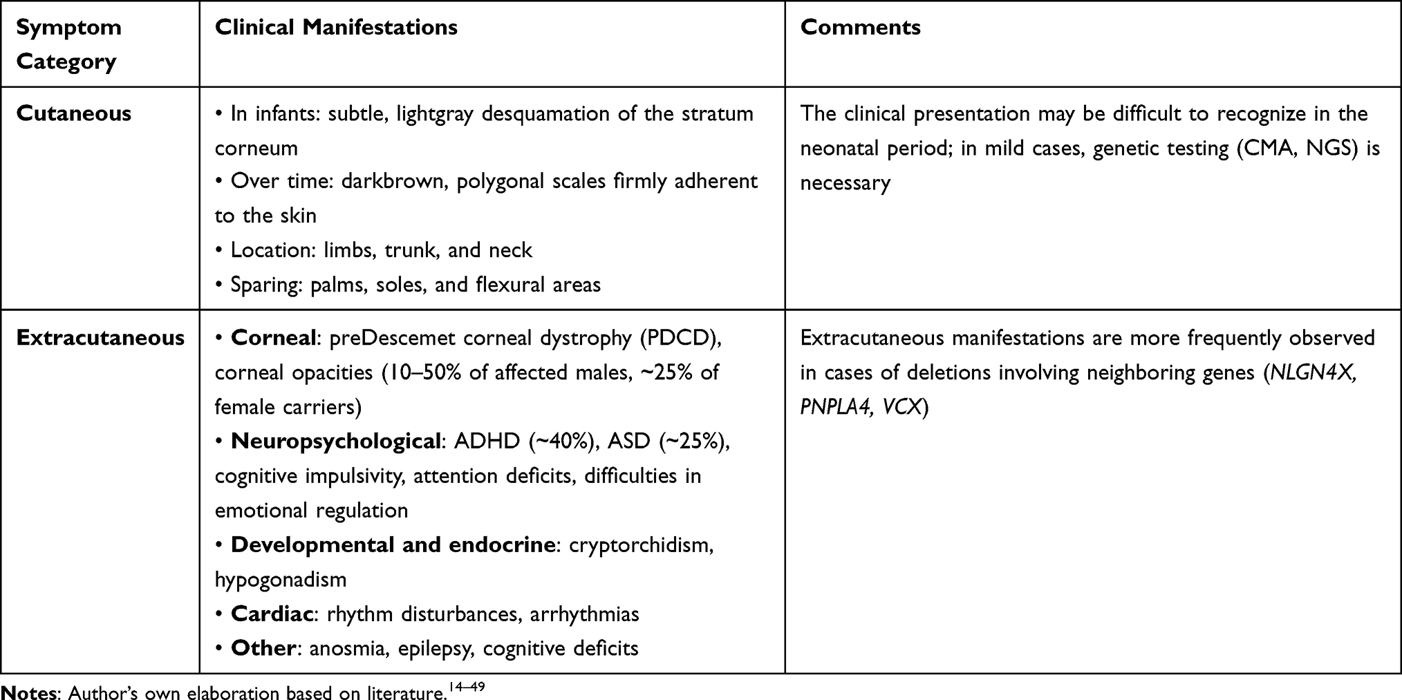 |
Table 2 Clinical Manifestations of STS-sEDD: Cutaneous and Extracutaneous |
The clinical presentation of STS-sEDD is often characteristic but not always easy to recognize, particularly in infants, in whom the initial manifestations are subtle and limited to mild, light-gray scaling of the stratum corneum. Over time, these evolve into the typical dark-brown, polygonal scales adherent to the skin, located primarily on the extremities, trunk, and neck, with sparing of the palms, soles, and flexural areas.24–26 In milder cases, the condition may remain unrecognized until genetic testing is performed, such as chromosomal microarray analysis (CMA) or next-generation sequencing (NGS).24,25 Although STS-sEDD is traditionally classified as a skin disorder, extensive deletions involving genes adjacent to STS (NLGN4X, PUDP, PNPLA4) can result in a multisystem syndrome that also includes neurological, endocrine, and cardiac manifestations such as anosmia, epilepsy, cognitive deficits, and heart defects (Table 2).18–22
Prenatal Markers and Placental Steroid Sulfatase Deficiency
What is also important is that steroid sulfatase deficiency may be signaled as early as the prenatal period. This phenomenon, known as placental sulfatase deficiency, occurs with a frequency of approximately 1 in 2000–5000 males and is closely associated with STS-sEDD. A characteristic biochemical marker is an extremely low or undetectable level of unconjugated estriol (uE3) with normal alpha-fetoprotein (AFP) and human chorionic gonadotropin (hCG) values in the triple prenatal screening test. Case reports confirm that such a result serves as an indication for further diagnostic evaluation, with biochemical analysis and family studies subsequently revealing absent sulfatase activity and deletion of the STS gene.27 Subsequent studies employing fluorescence in situ hybridization (FISH) and amniotic fluid analysis confirmed the association between extremely low uE3 levels and the presence of STS gene deletions, both complete and partial.28 The latest population studies have shown that in over 90% of pregnant women whose fetuses had a microdeletion of the Xp22.31 region, markedly reduced uE3 levels were observed, and in male infants, skin lesions appeared within the first months of life.29
Corneal Manifestations and Their Diagnostic Significance
One of the more common extracutaneous manifestations of STS-sEDD involves corneal abnormalities, particularly pre-Descemet corneal dystrophy (PDCD). These opacities, located deep within the posterior layer of the cornea, are observed in approximately 10–50% of males with STS-sEDD and in nearly one-quarter of female carriers of the mutation. They are believed to result from localized accumulation of cholesterol sulfate. Importantly, in heterozygous females, corneal changes may represent the first, and in some cases the only, manifestation of the condition, making PDCD a useful diagnostic indicator of carrier status. Although these changes were previously associated exclusively with complete deletions of the STS gene, recent observations suggest that partial deletions may also lead to a similar clinical presentation, likely due to a total loss of the enzyme’s activity.3,15,25
Slit-lamp biomicroscopy remains the traditional method for assessing corneal lesions. This examination is rapid, readily accessible, and relatively simple to perform, thereby remaining a fundamental component of ophthalmic diagnostic practice. However, the limitations of this technique include its subjective nature and reduced usefulness in evaluating lesions located in the deeper corneal layers, particularly in the presence of edema. Consequently, it is increasingly complemented with optical coherence tomography (OCT), which provides high-resolution imaging, facilitating not only precise assessment of the location and extent of corneal opacities, but also monitoring their dynamics over time. Research has shown that OCT offers substantial diagnostic benefit. In cases where slit-lamp observations were inconclusive, it allowed for the identification of lesions in over one-third of patients. Importantly, the images obtained with this method are largely consistent with slit-lamp biomicroscopy findings, yet they offer greater objectivity and are less dependent on the examiner’s experience. In clinical practice, both techniques should be regarded as complementary: the slit-lamp serves as a first-line tool, while OCT provides a detailed and reproducible assessment of the corneal phenotype.30–34
Neurodevelopmental Disorders Associated with STS Deletions
As previously mentioned, STS-sEDD not only presents with dermatological symptoms but also has a significant impact on neuropsychological development. Deletions involving the STS gene are strongly associated with an increased risk of neurodevelopmental disorders, particularly ADHD and ASD. These disorders affect not only cognitive functioning but also the emotional and social development of the individual.35–39
From a neurobiological perspective, the STS gene plays a crucial role in brain development both during the prenatal and postnatal periods. Studies have shown that a deficiency in steroid sulfatase may affect the development of the prefrontal cortex and interhemispheric connections - structures that are critical for social information integration and cognitive control. STS-regulated neurosteroids are involved in modulating synaptic plasticity and neuronal inhibition, which may explain the presence of symptoms such as hyperactivity, emotional lability, and attention deficits.19,39–41
The neurobiological basis of these difficulties also includes reduced volume of the basal ganglia - structures responsible for impulse control, emotion regulation, and action planning.34 Reduced STS activity in regions such as the prefrontal cortex, hippocampus, and basal ganglia, confirmed by neuroimaging studies, correlates with impaired ability to concentrate, regulate emotions, and inhibit responses.19,38,39,41
Clinical observations confirm that individuals with STS-sEDD, both males and female carriers, often experience subtle yet significant cognitive deficits. Difficulties with working memory, planning, impulse control, and word recall occur independently of general intelligence level and can significantly impact daily functioning.38,42–44 In some individuals, reduced volume of structures such as the globus pallidus and disruptions in their functional connectivity are also observed, which may further contribute to difficulties in emotional regulation and behavior.25,38–46
In light of clinical studies, the prevalence of ADHD and ASD in patients with STS-sEDD is significantly higher than in the general population. It is estimated that approximately 40% and 25% of individuals with STS-sEDD meet the diagnostic criteria for these disorders, respectively.18,37,38 Importantly, neurodevelopmental symptoms are also observed in female carriers, despite the absence of typical skin manifestations. What is more commonly noted in this group is cognitive impulsivity, behavioral rigidity, difficulties in social functioning, and attention deficits.19,22 This indicates that even partial loss of STS function may lead to alterations in the scope of cognitive and emotional functioning.4,18
Both ADHD and ASD are common features of the clinical phenotype associated with deletions in the Xp22.31 region. In the case of ADHD, symptoms of inattention prevail over impulsivity, which indicates dysfunction of the fronto-parietal attention network and weakened executive functions. Patients often struggle with difficulties in concentration, information processing, and planning, which translates into reduced quality of life and adaptive challenges.25,38–44
The ADHD phenotype in the course of STS-sEDD is usually characterized by a predominance of inattention symptoms with relatively preserved motor impulsivity. These difficulties affect the ability to concentrate, self-regulate, and adapt to environmental demands.20,25 In turn, autistic traits, such as cognitive rigidity or communication difficulties, promote social isolation and increase the risk of secondary mood disorders, such as depression.19,20,22,47 Both ADHD and autism can lead to secondary emotional problems: mood disorders, anxiety, and lowered self-esteem. Difficulties with emotional regulation, impulsivity, and deficits in theory of mind exacerbate challenges in establishing and maintaining interpersonal relationships.19,20,22,25,38,47
These neuropsychological challenges are also reflected in social functioning. Individuals with STS-sEDD often struggle with low self-esteem, difficulties in forming relationships, and chronic stress stemming from both personality traits and the social perception of the disorder. Female carriers additionally experience emotional burdens, such as feelings of guilt and anxiety related to the risk of passing the mutation on to their offspring. They also report a lack of understanding from those around them, as well as difficulties in intimate and family relationships.13,22,25,48,49
Social Functioning and Quality of Life in Patients
The cumulative difficulties in attention, emotional regulation, and interpersonal relationships have a direct impact on the quality of life of individuals with STS-sEDD. Studies using psychometric scales (including the Dermatology Life Quality Index (DLQI) and the Feelings of Stigmatization Questionnaire (FSQ)) have shown that both males with STS-sEDD and female carriers experience a significantly lower quality of life compared to the general population, particularly in the emotional, social, and functional domains. The reduced quality of life is influenced not only by the chronic physical discomfort associated with skin changes, but primarily by neuropsychological difficulties: impulsivity, concentration problems, emotional disturbances, and social deficits. Individuals with STS-sEDD often avoid social interactions due to shame, fear of judgment, and stigmatization.13,19,22,25,38,42,46,48–53
In this context, the issue of mental health becomes especially significant. Individuals with STS-sEDD demonstrate increased susceptibility to depression and anxiety disorders, regardless of the severity of skin symptoms. This group is characterized by atypical forms of depression, with predominant symptoms such as chronic fatigue, irritability, concentration difficulties, and low motivation.22,25,48,50,51 At the same time, many individuals exhibit heightened generalized anxiety, social anxiety, and existential distress, which contribute to chronic emotional tension. Such a psychological state promotes withdrawal from social activities, reduction of contact with others, and an increased sense of loneliness and emotional burnout.50,51 An additional burden is stigmatization, both social and internal, leading to self-deprecation and shame. Individuals affected by STS-sEDD often struggle with feelings of guilt, anxiety, and a sense of being different, which significantly impacts the quality of intimate and family relationships. In the case of female carriers, there is also intense emotional stress related to the fear of passing the mutation to offspring and a sense of lack of understanding from their closest social circle (Table 3).25,38,44,46,48,50,54
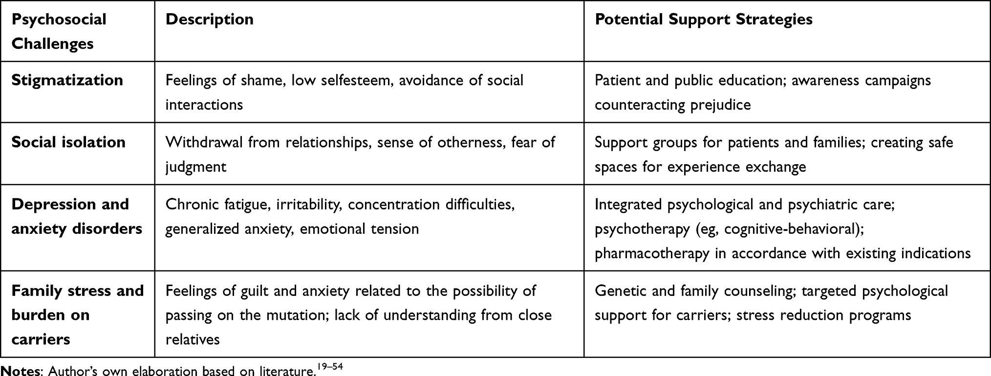 |
Table 3 Main Psychosocial Challenges and Potential Support Strategies |
Mental Health and Susceptibility to Emotional Disorders
Mechanistically, it is hypothesized that mood and anxiety disorders may be a direct consequence of STS deficiency. This enzyme plays a key role in the metabolism of neurosteroids, such as dehydroepiandrosterone (DHEA), estrone, and allopregnanolone, which modulate the functioning of the serotonergic, dopaminergic, and GABAergic systems. A deficiency of these neurosteroids may lead to dysregulation of the mechanisms responsible for emotional stability. Findings reported in the literature also suggest that these disorders may be associated with dysfunction of the hypothalamic–pituitary–adrenal (HPA) axis, which could lead to increased emotional reactivity and reduced adaptive capacity.25,41,55–58
Psychosocial and Educational Needs
Although knowledge about the neurobiological determinants of psychological burden in patients with STS-sEDD is steadily increasing, too little attention is still given to psychosocial and systemic factors, which equally affect the quality of their daily lives. Clinical practice reveals evident gaps in therapeutic education, limited access to psychological support, and insufficient institutional assistance for families coping with this disease.
Only a few studies, such as the work by Dufresne et al, comprehensively demonstrate that therapeutic patient education can significantly improve quality of life, for both affected individuals and their caregivers. Patient education is viewed not merely as a part of treatment, but as an indispensable component of daily functioning with a chronic illness such as ichthyosis. Despite growing awareness of the need for education, its availability remains highly variable and depends on local systemic arrangements and the competencies of medical personnel. France serves as a good example of best practice, where accredited educational programs are in place. Nevertheless, as shown in Dufresne’s research, children with ichthyoses still struggle with a lack of knowledge: 71% could not indicate the purpose of their treatment, 16.1% believed that there were no ways to alleviate itching, and more than half did not feel entitled to speak about treatment-related fatigue. Parents also lacked sufficient information regarding medication storage and symptom relief strategies.59
In response to these challenges, the “123 Tem’peau” program was developed, offering practical, engaging education delivered outside the hospital setting. Studies confirm that such an approach enhances participants’ knowledge, social and emotional competencies, and also supports the development of a sense of agency. These programs also facilitate collaboration among interdisciplinary therapeutic teams, thereby enabling a comprehensive approach to both health and psychosocial issues. Analyses from Belgium, the Netherlands, and Italy also confirm the benefits of education, indicating eg, shorter hospital stays, reduced anxiety levels, and increased treatment effectiveness.59–64
Although the French example and other European experiences demonstrate the potential of therapeutic education, only the study by Severino-Freire et al provide a detailed account of the psychological aspects of care for patients with congenital forms of ichthyosis (CI). The authors propose an integrated care model in which the central role is played by a coordinating dermatologist - working in a reference center and managing a team of specialists from various fields, ranging from physicians to psychologists. In this approach, patient education and psychosocial support are not supplementary, but a mandatory and permanent component of the care pathway, initiated at the diagnostic stage and continuing throughout the patient’s lifetime.65
Despite positive examples, Central and Eastern Europe continues to show significant disparities compared with the discussed model. The absence of unified systemic solutions, combined with the persistence of a paternalistic approach to the doctor–patient relationship, reduces the effectiveness of therapeutic education and psychological support, while patient participation in the decision-making process remains limited.60
In this context, particularly concerning are the findings from our original research based on interviews with parents of children affected by STS-sEDD. All respondents consistently emphasized the complete lack of psychological support - both at the stage of diagnosis and during subsequent treatment. Education, social counseling, and emotional support were entirely absent. Families had to seek out information on their own and cope with everyday challenges, which for many proved to be a frustrating and exhausting experience. Parents reported feelings of isolation, helplessness, and chronic stress, as well as a lack of emotional preparation for facing their child’s illness. As one mother said: We had absolutely no idea that a skin condition, or any other illness, might require psychological support that would help us come to terms and live more easily with the disease. Another problem was the lack of contact with other families and the absence of patient organizations that could offer support, knowledge, and a sense of community. Many caregivers experienced emotional burnout, tensions in family relationships, and even mutual blame for the child’s illness.
Practical Clinical Recommendations
Key Practical Recommendations for STS-sEDD
- Early diagnostics: use of prenatal markers (low uE3) and genetic testing (CMA, NGS) to confirm the diagnosis and assess the extent of the deletion.
- Integrated care: coordination of treatment within interdisciplinary teams (dermatologist, geneticist, ophthalmologist, cardiologist, psychologist).
- Psychological support: access to psychological and neuropsychological assistance from the moment of diagnosis; screening for ADHD, ASD, depression, and anxiety disorders.
- Therapeutic education: development of educational programs for patients and their families aimed at enhancing self-care skills and shared decision-making in treatment.
- Social support: establishment and strengthening of patient organizations to counteract isolation and stigmatization.
- Research and health policy: further studies on the neuropsychological STS-sEDD phenotype and the inclusion of rare diseases in health policy programs, including funding for psychological support and therapeutic education.
Future Perspectives
Further research on STS-sEDD should focus on deepening the understanding of the relationships between STS gene deletions and the neuropsychological phenotype, as well as the mechanisms underlying mood and anxiety disorders. The development of biomarkers enabling the early identification of patients at risk of extracutaneous symptoms appears to be of particular importance.
In the clinical field, future development lies in the implementation of integrated care models that encompass interdisciplinary teams and therapeutic education programs for patients and their families. It is worth emphasizing the potential of modern technologies, such as telemedicine and mobile applications, which can support self-care, facilitate symptom monitoring, and improve access to specialists.
From a social perspective, a key priority is the development and professionalization of patient organizations, which can serve as a bridge between patients, their families, and the healthcare system. Incorporating the voice of patients into the health policy planning process can contribute to the development of more appropriate and effective systemic solutions.
In the longer term, particular importance will rest on the development of new therapeutic strategies based on molecular biology and the regulation of neurosteroid metabolism. The advancement of these research directions offers hope for a more effective and personalized approach to the treatment of patients with STS-sEDD.
Conclusions
The conducted analyses and the results of our own research indicate that STS-sEDD should be regarded not merely as a dermatological disease, but as a complex multisystem disorder that also encompasses neuropsychological and psychosocial aspects. The lack of systemic psychological and educational support significantly exacerbates the emotional burden on patients and their families, leading to feelings of isolation, chronic stress, and a reduced quality of life.
From a clinical perspective, it is essential to identify extracutaneous symptoms at an early stage, conduct regular screening for neurodevelopmental disorders, and ensure access to interdisciplinary care. Equally crucial are therapeutic education initiatives and the establishment of support structures for patients and their families, including patient organizations, which play a vital role in mitigating isolation and reducing stigmatization.
Implementation of integrated care models that combine medical, psychological, and social approaches is a fundamental prerequisite for improving the quality of life of individuals affected by STS-sEDD. Comprehensive support, encompassing both patients and their relatives, should be an integral component of clinical practice in the management of rare diseases.
Data Sharing Statement
Data are available from the corresponding author upon reasonable request.
Ethics Approval and Informed Consent
This study is a review and does not involve any direct participation of patients or collection of clinical data. Therefore, ethics approval and informed consent were not required.
Acknowledgments
The authors would like to express their sincere gratitude to Professor Anette Bygum for her valuable collaboration and insights, as well as all the mothers of children affected by this condition and the Rybia Łuska i My Association for their cooperation and for sharing their experiences and information.
Author Contributions
Both authors made a significant contribution to the work reported, whether that is in the conception, study design, acquisition of data, analysis and interpretation, or in all these areas; took part in drafting, revising or critically reviewing the article; gave final approval of the version to be published; have agreed on the journal to which the article has been submitted; and agree to be accountable for all aspects of the work.
Funding
Medical University of Lublin (grant no. DS 168).
Disclosure
The authors report no conflicts of interest in this work.
References
1. Crane JS, Paller AS. X-Linked Ichthyosis. StatPearls Publishing; 2025. http://www.ncbi.nlm.nih.gov/books/NBK448149/.
2. Zhou B, Liang C, Li P, Xiao H. Revisiting X-linked congenital ichthyosis. Int J Dermatol. 2025;64(1):51–11. doi:10.1111/ijd.17396
3. Williams D, Onyia O, Chung DD, et al. Identification of a novel partial deletion of STS associated with pre-Descemet corneal dystrophy and X-linked ichthyosis. Mol Vision. 2023;29:25–30.
4. Park J, Cho YG, Kim JK, Kim HH. STS and PUDP deletion identified by targeted panel sequencing with CNV analysis in X-linked ichthyosis: a case report and literature review. Genes. 2023;14(10):1925. doi:10.3390/genes14101925
5. Chouk H, Saad S, Dimassi S, et al. X-linked recessive ichthyosis in 8 Tunisian patients: awareness of misdiagnosis due to the technical trap of the STS pseudogene. BMC Med Genom. 2022;15(1):165. doi:10.1186/s12920-022-01319-4
6. Wang N, An K, Liu H, et al. Detection of the STS gene in a family with X-linked recessive ichthyosis. IJDVL. 2013;79(2):268. doi:10.4103/0378-6323.107669
7. Wells RS, Kerr CB. Genetic classification of ichthyosis. Arch Dermatol. 1965;92(1):1–6.
8. Wang X, Tan L, Shen N, et al. Exacerbation of ichthyosis vulgaris phenotype by co-inheritance of STS and FLG mutations in a Chinese family with ichthyosis: a case report. BMC Med Genet. 2018;19(1):120. doi:10.1186/s12881-018-0642-5
9. Seidl-Philipp M, Schatz UA, Gasslitter I, et al. Spektrum der Ichthyosen in einer österreichischen Ichthyosekohorte von 2004–2007. J Dtsch Dermatol Ges. 2020;18(1):17–26. doi:10.1111/ddg.13968_g
10. Ingordo V, D’Andria G, Gentile C, et al. Frequency of X-linked ichthyosis in coastal southern Italy: a study on a representative sample of a young male population. Dermatology. 2003;207(2):148–150. doi:10.1159/000071784
11. Cañueto J, Ciria S, Hernández-Martín A, et al. Analysis of the STS gene in 40 patients with recessive X-linked ichthyosis: a high frequency of partial deletions in a Spanish population. Journal of the European Academy of Dermatology and Venereology: JEADV. 2010;24(10):1226–1229. doi:10.1111/j.1468-3083.2010.03612.x
12. Afzal S, Ramzan K, Ullah S, et al. A novel nonsense mutation in the STS gene in a Pakistani family with X-linked recessive ichthyosis: including a very rare case of two homozygous female patients. BMC Med Genet. 2020;21(1):20. doi:10.1186/s12881-020-0964-y
13. Wren GH, Humby T, Thompson AR, Davies W. Mood symptoms, neurodevelopmental traits, and their contributory factors in X-linked ichthyosis, ichthyosis vulgaris and psoriasis. Clin Exp Dermatol. 2022;47(6):1097–1108. doi:10.1111/ced.15116
14. Vasilyeva TA, Marakhonov AV, Tebieva IS, et al. Genetic Heterogeneity of X-Linked Ichthyosis in the Republic of North Ossetia–Alania, Case Series Report. Int J Mol Sci. 2023;24(5):4515. doi:10.3390/ijms24054515
15. Fernandes NF, Janniger CK, Schwartz RA. X-linked ichthyosis: an oculocutaneous genodermatosis. Journal of the American Academy of Dermatology. 2010;62(3):480–485. doi:10.1016/j.jaad.2009.04.028
16. Tang X, Wang Z, Yang S, et al. Maternal Xp22.31 copy-number variations detected in non-invasive prenatal screening effectively guide the prenatal diagnosis of X-linked ichthyosis. Front Genet. 2022;13:934952. doi:10.3389/fgene.2022.934952
17. Xie W, Zhou H, Zhou L, et al. Clinical features and genetic analysis of two Chinese families with X-linked ichthyosis. J Int Med Res. 2020;48(10):300060520962292. doi:10.1177/0300060520962292
18. McGeoghan F, Camera E, Maiellaro M, et al. RNA sequencing and lipidomics uncovers novel pathomechanisms in recessive X-linked ichthyosis. Front Mol Biosci. 2023;10:1176802. doi:10.3389/fmolb.2023.1176802
19. Brcic L, Underwood JF, Kendall KM, et al. Medical and neurobehavioural phenotypes in carriers of X-linked ichthyosis-associated genetic deletions in the UK Biobank. J Med Genet. 2020;57(10):692–698. doi:10.1136/jmedgenet-2019-106676
20. Malik A, Amer AB, Salama M, et al. X-linked ichthyosis associated with psychosis and behavioral abnormalities: a case report. J Med Case Rep. 2017;11(1):267. doi:10.1186/s13256-017-1420-2
21. Zhang M, Huang H, Lin N, et al. X-linked ichthyosis: molecular findings in four pedigrees with inconspicuous clinical manifestations. J Clin Lab Anal. 2020;34(5):e23201. doi:10.1002/jcla.23201
22. Cavenagh A, Chatterjee S, Davies W. Behavioural and psychiatric phenotypes in female carriers of genetic mutations associated with X-linked ichthyosis. PLoS One. 2019;14(2):e0212330. doi:10.1371/journal.pone.0212330
23. Carrascosa-Romero MC, Suela J, Alfaro-Ponce B, Cepillo-Boluda AJ. X-chromosome-linked ichthyosis associated to epilepsy, hyperactivity, autism and mental retardation, due to the Xp22.31 microdeletion. Revista Neurol. 2012;54(4):241–248.
24. Hand JL, Runke CK, Hodge JC. The phenotype spectrum of X-linked ichthyosis identified by chromosomal microarray. JAAD. 2015;72(4):617–627. doi:10.1016/j.jaad.2014.12.020
25. Wren GH, Davies W. X-linked ichthyosis: new insights into a multi-system disorder. SHD. 2022;2(4):e179. doi:10.1002/ski2.179
26. Elias PM, Williams ML, Choi EH, Feingold KR. Role of cholesterol sulfate in epidermal structure and function: lessons from X-linked ichthyosis. BBA Molecular Cell Biol Lipids. 2014;1841(3):314–318. doi:10.1016/j.bbalip.2013.11.009
27. Keren DF, Canick JA, Johnson MZ, et al. Low maternal serum unconjugated estriol during prenatal screening as an indication of placental steroid sulfatase deficiency and X-linked ichthyosis. Am J Clin Pathol. 1995;103(4):400–403. doi:10.1093/ajcp/103.4.400
28. Kashork CD, Sutton VR, Allen JSF, et al. Low or absent unconjugated estriol in pregnancy: an indicator for steroid sulfatase deficiency detectable by fluorescence in situ hybridization and biochemical analysis. Prenat Diagn. 2002;22(11):1028–1032. doi:10.1002/pd.466
29. Liu HY, Li J, Huang DR, et al. Early warning of low maternal unconjugated estriol level by prenatal screening for fetus with X-linked ichthyosis. Zhonghua Fu Chan Ke Za Zhi. 2022;57(6):407–412. doi:10.3760/cma.j.cn112141-20220125-00043
30. Leung CKS, Li H, Weinreb RN, et al. Anterior chamber angle measurement with anterior segment optical coherence tomography: a comparison between slit lamp OCT and Visante OCT. Invest Ophthalmol Vis Sci. 2008;49(8):3469–3474. doi:10.1167/iovs.07-1477
31. Moutsouris K, Dapena I, Ham L, et al. Optical coherence tomography, Scheimpflug imaging, and slit-lamp biomicroscopy in the early detection of graft detachment after Descemet membrane endothelial keratoplasty. Cornea. 2011;30(3):273–279. doi:10.1097/ICO.0b013e31820d86bd
32. Malhotra C, Jain AK, Dwivedi S, et al. Characteristics of pre-Descemet membrane corneal dystrophy by three different imaging modalities—In vivo confocal microscopy, anterior segment optical coherence tomography, and Scheimpflug corneal densitometry analysis. Am J Ophthalmol Case Rep. 2022;27:101671. doi:10.1097/ICO.0000000000000454
33. Bhargava M, Bhambhani VG, Paul RS. Vertical scan imaging of anterior segment optical coherence tomography for Descemet anchoring caterpillar seta: a case report and review of literature. IJCEO. 2023;9(3):399–402. doi:10.1016/j.ajoc.2022.101671
34. Ranjiv R, Sharma R. Comparison of optic nerve head analysis using optical coherence tomography and slit-lamp biomicroscopy. IJCEO. 2025;11(1):85–89. doi:10.18231/j.ijceo.2023.075
35. Hazan C, Orlow SJ, Schaffer JV. X-linked recessive ichthyosis. Dermatol Online J. 2005;11(4):12.
36. Wren G, Baker E, Underwood J, et al. Characterising heart rhythm abnormalities associated with Xp22.31 deletion. J Med Genet. 2023;60(7):636–643. doi:10.1136/jmg-2022-108862
37. Wren GH, Davies W. Cardiac arrhythmia in individuals with steroid sulfatase deficiency (X-linked ichthyosis): candidate anatomical and biochemical pathways. Essays Biochem. 2024;68(4):423–429. doi:10.1042/EBC20230098
38. Kent L, Emerton J, Bhadravathi V, et al. X-linked ichthyosis (steroid sulfatase deficiency) is associated with increased risk of attention deficit hyperactivity disorder, autism and social communication deficits. J Med Genet. 2008;45(8):519–524. doi:10.1136/jmg.2008.057729
39. Gubb SJA, Brcic L, Underwood JFG, et al. Medical and neurobehavioural phenotypes in male and female carriers of Xp22.31 duplications in the UK Biobank. Hum Mol Genet. 2020;29(17):2872–2881. doi:10.1093/hmg/ddaa174
40. Trent S, Davies W. Cognitive, behavioural and psychiatric phenotypes associated with steroid sulfatase deficiency. World J Transl Med. 2013;2(1):1–12. doi:10.5528/wjtm.v2.i1.1
41. Stergiakouli E, Langley K, Williams H, et al. Steroid sulfatase is a potential modifier of cognition in attention deficit hyperactivity disorder. Genes Brain Behav. 2011;10(3):334–344. doi:10.1111/j.1601-183X.2010.00672.x
42. Chatterjee S, Humby T, Davies W. Behavioural and psychiatric phenotypes in men and boys with X-linked ichthyosis. PLoS One. 2016;11(10):e0164417. doi:10.1371/journal.pone.0164417
43. Miyake A, Friedman NP, Emerson MJ, et al. The unity and diversity of executive functions and their contributions to complex tasks: a latent variable analysis. Cogn Psychol. 2000;41(1):49–100. doi:10.1006/cogp.1999.0734
44. Macarov M, Zeigler M, Newman JP, et al. Deletions of VCX‐A and NLGN4: a variable phenotype including normal intellect. J Intellect Disabil Res. 2007;51(5):329–333. doi:10.1111/j.1365-2788.2006.00880.x
45. Brookes KJ, Hawi Z, Kirley A, et al. Association of the steroid sulfatase (STS) gene with attention deficit hyperactivity disorder. Am J Med Genet B Neuropsychiatr. Genet. 2008;147B(8):1271–1274. doi:10.1002/ajmg.b.30766
46. Wren GH, Flanagan J, Underwood JFG, et al. Memory, mood and associated neuroanatomy in individuals with steroid sulphatase deficiency (X-linked ichthyosis). Genes Brain Behav. 2024;23(3):e12893. doi:10.1111/gbb.12893
47. Baron-Cohen S, Wheelwright S, Skinner R, et al. The Autism-Spectrum Quotient (AQ): evidence from Asperger Syndrome/High-Functioning Autism, males and females, scientists and mathematicians. J Autism Dev Disord. 2001;31(1):5–17. doi:10.1023/a:1005653411471
48. Wehbe RM, Spiridigliozzi GA, Heise EM, Dawson DV, McConkie-Rosell A. When to tell and test: parental disclosure and clinical testing for X-linked. Am J Medl Genet A. 2009;149A(6):1190–1199. doi:10.1002/ajmg.a.32840
49. Pavone P, Corsello G, Marino S, Ruggieri M, Falsaperla R. Microcephaly/trigonocephaly, intellectual disability, autism spectrum disorder, and atypical dysmorphic features in a boy with Xp22.31 duplication. Molecular Syndromol. 2019;9(5):253–258. doi:10.1159/000492818
50. Rodrigo-Nicolás B, Bueno-Martínez E, Martín-Santiago A, et al. Evidence of the high prevalence of neurological disorders in nonsyndromic X-linked recessive ichthyosis: a retrospective case series. Br J Dermatol. 2018;179(4):933–939. doi:10.1111/bjd.16826
51. Dalgard FJ, Svensson Å, Halvorsen JA, et al. Itch and mental health in dermatological patients across Europe: a cross-sectional study in 13 countries. J Invest Dermatol. 2020;140(3):568–573. doi:10.1016/j.jid.2019.07.726
52. Carrascosa-Romero MC, De Cabo-De La Vega C. The comorbidity of ADHD and autism spectrum disorders (ASDs) in community preschoolers. In: Norvilitis JM editor. ADHD: New Directions in Diagnosis and Treatment. IntechOpen;2015. doi:10.5772/61400
53. Lukin J, Smith CM, De Rubeis S. Emerging X-linked genes associated with neurodevelopmental disorders in females. Curr Opin Neurobiol. 2024;88:102902. doi:10.1016/j.conb.2024.102902
54. Rustad AM, Soltani H, Yang LJ, et al. Bullying in children with congenital ichthyosis. Pediatr Dermatol. 2025;42(3):499–505. doi:10.1111/pde.15920
55. Schüle C, Nothdurfter C, Rupprecht R. The role of allopregnanolone in depression and anxiety. Prog Neurobiol. 2014;113:79–87. doi:10.1016/j.pneurobio.2013.09.003
56. Samardzic J, Hencic B, Jancic J, et al. Neurosteroid dehydroepiandrosterone improves active avoidance retrieval and induces antidepressant-like behavior in rats. Neurosci lett. 2017;660:17–21. doi:10.1016/j.neulet.2017.09.005
57. Lee SY, Wang LJ, Chang CH, et al. Serum DHEA-S concentration correlates with clinical symptoms and neurocognitive function in patients with bipolar II disorder: a case-controlled study. Prog Neuro Psychopharmacol Biol Psychiatry. 2017;74:31–35. doi:10.1016/j.pnpbp.2016.11.009
58. Barden N. Implication of the hypothalamic–pituitary–adrenal axis in the pathophysiology of depression. Psychoneuroendocrinology. 2004;29(1):1–38. doi:10.1016/S0306-4530(03)00095-3
59. Dufresne H, Hadj-Rabia S, Taïeb C, Bodemer C. Importance of therapeutic patient education in ichthyosis: results of a prospective single reference center study. Orphanet J Rare Dis. 2013;8(113). doi:10.1186/1750-1172-8-113
60. Visser A, Deccache A, Bensing J. Patient education in Europe: united differences. Patient Educ Couns. 2001;44(1):1–5. doi:10.1016/S0738-3991(01)00111-2
61. Deccache A. Patient education in Belgium: evolution, policy and perspectives. Patient Educ Couns. 2001;44(1):43–48. doi:10.1016/S0738-3991(01)00101-X
62. Skelton A. Evolution not revolution? The struggle for the recognition and development of patient education in the UK. Patient Educ Couns. 2001;44(1):23–27. doi:10.1016/S0738-3991(01)00098-2
63. Bensing JM, Visser AP, Saan H. Patient education in The Netherlands. Patient Educ Couns. 2001;44(1):15–22. doi:10.1016/S0738-3991(01)00097-0
64. Marcolongo R, Rossato E, Pavan V, et al. Current perspectives of therapeutic patient education in Italy. Patient Educ Couns. 2001;44(1):59–63. doi:10.1016/s0738-3991(01)00104-5
65. On behalf of the French Pediatric Dermatology Research Group, Severino-Freire M, Granier Tournier C, Chiaverini C, et al. French national protocol for the management of congenital ichthyosis. Ann Dermatol Venereol. 2024;151(1):103247. doi:10.1016/j.annder.2024.103247.
 © 2026 The Author(s). This work is published and licensed by Dove Medical Press Limited. The
full terms of this license are available at https://www.dovepress.com/terms
and incorporate the Creative Commons Attribution
- Non Commercial (unported, 4.0) License.
By accessing the work you hereby accept the Terms. Non-commercial uses of the work are permitted
without any further permission from Dove Medical Press Limited, provided the work is properly
attributed. For permission for commercial use of this work, please see paragraphs 4.2 and 5 of our Terms.
© 2026 The Author(s). This work is published and licensed by Dove Medical Press Limited. The
full terms of this license are available at https://www.dovepress.com/terms
and incorporate the Creative Commons Attribution
- Non Commercial (unported, 4.0) License.
By accessing the work you hereby accept the Terms. Non-commercial uses of the work are permitted
without any further permission from Dove Medical Press Limited, provided the work is properly
attributed. For permission for commercial use of this work, please see paragraphs 4.2 and 5 of our Terms.