Back to Journals » International Medical Case Reports Journal » Volume 15
Spontaneous Retropharyngeal Haematoma Causing Upper Airway Obstruction in a Child: Case Report
Authors Kabagenyi F , Hidour R
Received 8 November 2022
Accepted for publication 14 December 2022
Published 19 December 2022 Volume 2022:15 Pages 739—743
DOI https://doi.org/10.2147/IMCRJ.S396666
Checked for plagiarism Yes
Review by Single anonymous peer review
Peer reviewer comments 3
Editor who approved publication: Professor Ronald Prineas
Fiona Kabagenyi,1 Rym Hidour2
1Department of Ear Nose and Throat, Makerere University, Kampala, Uganda; 2Department of Ear Nose and Throat, Mulago National Referral Hospital, Kampala, Uganda
Correspondence: Fiona Kabagenyi, Department of Ear Nose and Throat, College of Health Sciences, Makerere University, PO Box 7072, Kampala, Uganda, Tel +256774150102, Email [email protected]
Abstract: Bleeding into the retropharyngeal region in children is an unusual cause of acute upper airway obstruction. Even in the absence of known risk factors or aetiology, this rare entity should be considered as one of the differential diagnoses of retropharyngeal swellings in children. Prompt surgical intervention is required whenever rapid progression to airway obstruction is observed. In this case report, we present an 18-month-old girl previously managed as upper respiratory tract infection, who presented with progressive dysphagia, drooling and eventually airway obstruction with stridor and respiratory distress. Conservative prolonged airway protection by intubation or tracheostomy was averted by an emergency incision and drainage of the haematoma. There was complete resolution by the second week and no recurrence reported at follow-up 18 months later.
Keywords: retropharynx, cervical, haematoma, oedema, paediatric
Introduction
The retropharyngeal region is a potential space in the neck. It is the posterior border of the upper aerodigestive tract and spans from the skull base to the upper mediastinum.1 When this space is filled with infectious or inflammatory materials, air or blood, an anterior bulge is formed. This is because the bony cervicothoracic spine located posteriorly cannot bulge. As the anterior bulge increases, clinical manifestations such as neck pain,2,3 limited neck movement,3,4 pain on swallowing,2,4–6 stridor with respiratory distress4–10 or, in extreme circumstances of bleeding, Capp’s triad5,11,12 may ensue.
In children, there are several potential diagnoses of retropharyngeal swellings. These include retropharyngeal cellulitis and abscesses,13,14 retropharyngeal oedema seen in multisystem inflammatory syndrome in children (MIS-C)15 and Kawasaki disease,16 thoracic duct cysts17 or retropharyngeal haematomas.6,8 The cases of retropharyngeal haematomas are often associated with foreign body ingestion or anticoagulation but spontaneous retropharyngeal haematomas are relatively rare.6,8 These differential diagnoses are distinguished by radiological investigations like radiographs, CT scans and MRIs, which also aid in monitoring of disease progress.6,10,13 Additional inflammatory markers may be required to explore infectious conditions.
The management plan to either treat conservatively,15 medically,13 or surgically6,8 greatly depends on the suspected diagnosis and the symptom severity of the patient. In cases with airway compromise, it is critical to secure the airway either by intubation or tracheostomy.5,6,8,11,12 In this infrequent case of upper airway obstruction, we discuss the presentation and management of spontaneous retropharyngeal haematoma in children.
Case Report
An 18-month-old girl was referred to our ear, nose and throat (ENT) department from a peripheral health unit, with a three-day history of worsening stridor and difficulty in breathing. She had been unwell for the last week with low grade fever, flu-like symptoms with trivial sneezing or cough, and progressive difficulty swallowing and drooling of saliva. This happened in the absence of trauma, foreign body ingestion or anticoagulation therapy. No known allergy or bleeding from any orifice were reported. She had been managed with antibiotics, steroids and paracetamol, with minimal improvement. Her fever subsided with paracetamol but she progressively worsened to laboured breathing. Two days prior to admission on our unit, she developed a swelling on the left lateral aspect of the neck that prompted the referral. She otherwise had an intact cry, was not on any chronic medication and had unremarkable medical and surgical history.
On examination, she was afebrile, well-nourished with moderate respiratory distress and inspiratory stridor with oxygen saturations between 92–95%. She was more comfortable in a sitting position. Minimal neck movement was observed, with a soft, non-tender swelling in the left level II region that was fluctuant, measuring 3x3x4 cm. No palpable lymph nodes in the neck were noted. Oropharyngeal exam showed a normal pink tongue with non-erythematous grade 1 tonsils. Notably, a posterior pharyngeal bulge was seen. Findings on otoscopy and anterior rhinoscopy were normal. A flexible laryngoscopy showed a large posterior pharyngeal bulge occluding the view of the vocal cords, although a tip of the epiglottis was seen. The chest was clear. There were no stigmata of bleeding observed. A clinical impression of a retropharyngeal abscess with a parapharyngeal extension was made.
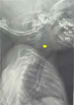
Her referral lateral neck X-ray, shown in Figure 1, demonstrated thickening of the retropharynx from the skull base to the hypopharynx. Urgent further imaging with CT scan was deferred as the institutional one was not functional at the time. The COVID-19 antigen test was negative. Complete blood count findings were in the normal range: haemoglobin 12.1g/dL, white blood cell count 9.5 x 10^3 / mL, thrombocytes 446 x 10^3 / mL.
 |
Figure 1 The lateral neck radiograph showing more than double the size of the cervical body (C1-C5), illustrated by the yellow dot, extending from the nasopharynx to the hypopharynx. |
An emergency examination under anaesthesia (EUA) was planned. Informed consent for the surgery was obtained from the mother. After a paediatric anaesthesiologist’s review, a difficult airway intubation was anticipated. Therefore, a video laryngoscope, a tracheostomy set and two functional suction machines were present in the operating room at intubation. The child was successfully orally intubated after two attempts. While supine with moderate neck flexion, a Boyle–Davis mouth gag was placed. Proof puncture of the posterior pharyngeal bulge revealed bloody discharge with no pus, as shown in Figure 2. A cruciate incision was made over the most prominent part of the retropharyngeal bulge. The retropharyngeal haematoma was subsequently suctioned, a total of 40 mL, and taken for microbiological, culture and sensitivity testing. The lateral neck swelling notably reduced in size with transoral drainage of the retropharyngeal haematoma. The child was successfully extubated at the end of the incision and drainage, without need for tracheostomy.
 |
Figure 2 Intraoperative findings of blood during the proof puncture of the retropharyngeal bulge. |
Inpatient monitoring and observation were done for five days. A slow asymptomatic re-accumulation of the swelling was observed, affecting the left side on the fourth post-operative day. She however fed better. On examination, a small 2x2x2 cm mass in level II was seen; however, flexible nasopharyngoscopy showed considerably reduced posterior pharyngeal bulge with proper vision of the true vocal cords. The International Normalized Ratio (INR) of 0.98 (reference range: 1–1.25) was slightly low, and the prothrombin time was 10 seconds. These were done after the intraoperative discovery of blood in the retropharynx. A decision for watchful monitoring was made. She was discharged on oral analgesics on the fifth post-operative day. At two weeks post-incision and drainage, there were no more swellings, she fed well and had no respiratory distress. The haematoma had completely resolved. No bacterial growth was seen on microbiology. At 18-month follow-up, no recurrence of the retropharyngeal haematoma was noted.
The child’s parent provided informed consent for the case details and any accompanying images to be published, available upon request. Institutional approval was not required to publish the case details.
Discussion
Spontaneous retropharyngeal haematomas are a potentially fatal and unusual cause of upper airway obstruction.3,7 Case reports have been described in Spain,10 United Kingdom,5 Turkey,3 India,6,8 Japan,9 Korea,4 and Morocco.12 To the best of our knowledge, this is the first documented case report within the East African region and Uganda. The reported cases heavily comprise the adult population,3–5,10–12 more so the elderly,3,5,11 in comparison to the paediatric population.6,8,9 Our 18-month-old patient falls within the paediatric category.
The recognized potential risk factors for retropharyngeal bleeding include anti-coagulation therapy,3,11 foreign body ingestion and trauma from vigorous head movements,3 straining with sneezing or coughing,7 bleeding from rupture of arteries or paraoesophageal veins,11 iatrogenic from angiographic procedures,10 etc. Additionally, patients with Haemophilia A are seemingly at high risk of retropharyngeal bleeding.9 An interesting case report hypothesized untreated severe obstructive apnea as another possible cause of retropharyngeal bleeding in an adult.18 Still, whenever no associated risk of bleeding is found, the term spontaneous retropharyngeal haematoma is used. Unexpectedly in our case, the mother reported insignificant sneezing or cough among the flu-like symptoms during the entire period of her child’s sickness. It was therefore suggested that she developed a spontaneous retropharyngeal haematoma, since she lacked any of the above-mentioned risk factors.
The size of the retropharyngeal haematoma determines the clinical manifestations. Several authors describe dysphagia,2,4–6 limited neck movement3,4 or neck pain,2,3 upper airway obstruction,6–9 and dysphonia,2,4,5,10 in no particular order. Rarely, Capps triad, which involves tracheal or esophageal compression, anterior displacement of the trachea and subcutaneous bruising over the neck and anterior chest, has been described in the adult population.5,11,12 The most consistent symptoms in adults are dysphagia followed by neck pain or limited neck movement. However, in children, dysphagia is quickly followed by difficulty in breathing with stridor and respiratory distress, and occasional accompanying fevers.6,8 With extensive expansion of the haematoma, parapharyngeal extensions presenting as lateral neck swellings may be seen.2,7,8 Importantly, lateral neck swellings due to haematomas may be discrete cervical swellings that do not involve the retropharyngeal area. A case series of two infants described abrupt neck swellings with fever, without any dysphagia, and no retropharyngeal involvement in their radiological investigations, which were conservatively managed for spontaneous neck haematomas.19 Our case presented with dysphagia, limited neck movement, upper airway obstruction with stridor and lateral neck swelling, a comparable presentation to other stated cases.
Differential diagnoses include retropharyngeal cellulitis and abscesses, commonest in 2–4-year-old children,13 although retropharyngeal cellulitis is also reported in adolescence.14 Retropharyngeal oedema was linked to multisystem inflammatory syndrome in children (MIS-C) supposedly exposed to SARS-COV-2 infection15 and to children with Kawasaki disease.16 A rare case of thoracic duct cysts within the retropharynx and mediastinum in an infant is also reported.17 Radiological investigations utilized in children with retropharyngeal swellings include plain cervical radiographs,3,8 CT scans3,6,8 and MRIs.3 These are beneficial in diagnosis and monitoring of disease progress. The MRI is superior as it will show blood products that would streamline diagnosis of retropharyngeal haematoma.10 The CT scan best differentiates cellulitis from an abscess, with ring enhancement depicting an abscess.13 The plain radiograph, which is easily accessible and affordable in most resource-constrained settings, will show increased soft tissue swelling that unfortunately does not single out any diagnosis. Regrettably, the plain radiograph was the only imaging modality done for our patient since our institutional CT scan was out of service at the time of presentation, and we do not have an MRI scanner. Our initial suspicion was a retropharyngeal abscess but, to our surprise, we found blood during the transoral drainage. Likewise, we based our responses on clinical judgement as opposed to repeat radiographs to monitor the haematoma resolution. Our patient was COVID-19 negative and other laboratory investigations yielded normal findings.
Most authors support a conservative approach to spontaneous retropharyngeal haematoma, which comprises of steroids, antibiotics and watchful monitoring with serial CT scans or MRIs.3–5,10 This is because haematoma resolution is expected between 2–4 weeks, thus advocating for the conservative approach. In extreme cases of upper airway obstruction, intubation or tracheostomy is advised, although only a few cases document airway relief via tracheostomy.3,6 Nevertheless, a surgical approach to the spontaneous retropharyngeal haematoma is indicated if there is continuous expansion of the haematoma or infection or foreign bodies.2,4,7,12
Whereas a conservative approach is popularly recommended, more so in adult literature, we propose that surgical intervention may be a safer option for children with spontaneous retropharyngeal haematoma. In a case report of a one-year-old child who dramatically went into severe respiratory distress after four days of developing difficulty in breathing and swallowing, a large retropharyngeal haematoma with parapharyngeal extension was found. Prompt intubation to secure the airway was done. The wait-to-resolve approach was not followed because the retropharyngeal haematoma spontaneously ruptured, leading to throat bleeding. Unfortunately, a poor outcome with hypoxic injuries is stated.8 Another case describes a 4-year-old child with a two-month history of progressive dysphagia. Aspiration of the retropharyngeal bulge revealed blood, and initial conservative management with tracheostomy to secure the airway was done. However, no haematoma resolution was seen after 4 weeks. Eventual transoral incision and drainage of 30 mL of blood led to a speedy tracheal decannulation after 4 days, and complete resolution of symptoms at 6 weeks.6 Our case presented with obstructive symptoms. Once the proof puncture showed blood, an incision and drainage evacuated the haematoma without requirement for tracheostomy or intubation. The patient was successfully extubated after the procedure. Even though an asymptomatic transient re-accumulation was observed, complete resolution was observed within two weeks and there was no recurrence past one year. Similarly, complete resolution of symptoms without complications is reported in the adult population.2,4,5,10,12
Conclusion
Spontaneous retropharyngeal haematoma should be considered as a rare cause of obstructive symptoms (dysphagia, dyspnea) in children. Although a conservative approach remains the mainstay of management for stable cases, prompt surgical drainage is advocated for cases presenting with severe upper airway obstruction, to curtail possible detrimental complications arising from hypoxia.
Acknowledgments
We acknowledge the parental consent to sharing this rare case with the scientific community. We recognize the effort of all health professionals involved in the management of this child.
Author Contributions
All authors made a significant contribution to the work reported in all these areas; took part in drafting, revising or critically reviewing the article; gave final approval of the version to be published; have agreed on the journal to which the article has been submitted; and agree to be accountable for all aspects of the work.
Disclosure
The authors report no conflicts of interest in this work.
References
1. Mnatsakanian A, Minutello K, Bordoni B. Anatomy, head and neck, retropharyngeal space. In: StatPearls [Internet]. Treasure Island (FL): StatPearls Publishing; 2022. Available from: https://www.ncbi.nlm.nih.gov/books/NBK537044/.
2. DiFrancesco RC, Escamilla JS, Sennes LU, Voegles RL, Tsuji DH. Spontaneous cervical hematoma: a report of two cases. Ear Nose Throat J. 1999;78(3):168–175. doi:10.1177/014556139907800310
3. Cengiz AB, Tansuker HD. Retropharyngeal hematoma: two case reports. KBB Uygulamalari. 2021;9(2):66–70. doi:10.5606/kbbu.2021.05579
4. Kang SS, Jung SH, Kim MS, Hong SJ, Yoon YJ, Shin KM. Spontaneous retropharyngeal hematoma - A case report -. Korean J Pain. 2010;23(3):211–214. doi:10.3344/kjp.2010.23.3.211
5. Singh A, Ofo E, Cumberworth V. Spontaneous retropharyngeal haematoma: a case report. J Med Case Rep. 2008;2:8. doi:10.1186/1752-1947-2-8
6. Singh GB, Mawii L, Grover SB, Lakshmi NS, Ansari I, Venkatachalam VP. A rare case of spontaneous retropharyngeal heamatoma presenting as acute respiratory distress in a child. Int J Pediatr Otorhinolaryngol Extra. 2008;3(4):151–154. doi:10.1016/j.pedex.2008.02.002
7. Chin KW, Sercarz JA, Wang MB, Andrews R. Spontaneous cervical hemorrhage with near-complete airway obstruction. Head Neck. 1998;20(4):350–353. doi:10.1002/(sici)1097-0347(199807)20:4<350::aid-hed10>3.0.co;2-m
8. Mundra R, Sinha R, Bajoliya S. Spontaneous retropharyngeal haematoma in a one-year-old child: a case report. Case Rep Clin Med. 2014;3:272–275. doi:10.4236/crcm.2014.35062
9. Yamamoto T, Schmidt-Niemann M, Schindler E. A case of acute upper airway obstruction in a pediatric Hemophilia A patient because of spontaneous retropharyngeal hemorrhage. Ann Emerg Med. 2016;67(5):616–619. doi:10.1016/j.annemergmed.2015.08.028
10. Muñoz A, Fischbein NJ, de Vergas J, Crespo J, Alvarez-Vincent J. Spontaneous retropharyngeal hematoma: diagnosis by MR imaging. AJNR Am J Neuroradiol. 2001;22(6):1209–1211.
11. Lin M, Sinclair C. Retropharyngeal haematoma - an unusual cause of airway obstruction. J Surg Case Rep. 2011;2011(10):5. doi:10.1093/jscr/2011.10.5
12. Lakhdar Y, Elazizi DB, Elbouderkaoui M, Rochdi Y, Raji A. The spontaneous retropharyngeal space hematoma: how to manage? EJMED. 2021;3(2):16–18. doi:10.24018/ejmed.2021.3.2.755
13. Craig FW, Schunk JE. Retropharyngeal abscess in children: clinical presentation, utility of imaging, and current management. Pediatrics. 2003;111(6 Pt 1):1394–1398. doi:10.1542/peds.111.6.1394
14. Tanaka K, Inokuchi R, Namai Y, Yahagi N. Retropharyngeal cellulitis in adolescence. BMJ Case Rep. 2013;2013:bcr2013009684. doi:10.1136/bcr-2013-009684
15. Daube A, Rickert S, Madan RP, Kahn P, Rispoli J, Dapul H. Multisystem inflammatory syndrome in children (MIS-C) and retropharyngeal edema: a case series. Int J Pediatr Otorhinolaryngol. 2021;144:110667. doi:10.1016/j.ijporl.2021.110667
16. Au DCY, Chung Fong N, Wah Kwan Y. Kawasaki disease with retropharyngeal edema: case series from a single center experience. Chin Med J. 2019;132(14):1753–1754. doi:10.1097/CM9.0000000000000321
17. Bhalla V, Schrepfer T, McCann A, Nicklaus P, Reading B. Spontaneous retropharyngeal and mediastinal thoracic duct cyst in an infant with respiratory distress. Int J Pediatr Otorhinolaryngol. 2018;105:33–35. doi:10.1016/j.ijporl.2017.11.021
18. Warken C, Rotter N, Maurer JT, Attenberger U, Lammert A. Retropharyngeal hematoma in the context of obstructive sleep apnea: a case report and review of the literature. J Med Case Rep. 2019;13(1):269. doi:10.1186/s13256-019-2202-9
19. Zhuang S, Ye J, Li J. Acute spontaneous neck haematoma in children: a rare entity. BMC Pediatr. 2015;15:38. doi:10.1186/s12887-015-0356-1
 © 2022 The Author(s). This work is published and licensed by Dove Medical Press Limited. The full terms of this license are available at https://www.dovepress.com/terms.php and incorporate the Creative Commons Attribution - Non Commercial (unported, v3.0) License.
By accessing the work you hereby accept the Terms. Non-commercial uses of the work are permitted without any further permission from Dove Medical Press Limited, provided the work is properly attributed. For permission for commercial use of this work, please see paragraphs 4.2 and 5 of our Terms.
