
Back to Journals » International Medical Case Reports Journal » Volume 17
Spontaneous Focal Hemorrhage of Optic Disc in Peripapillary Hyperreflective Ovoid Mass-Like Structures
Authors Wang F 
Received 17 July 2024
Accepted for publication 7 October 2024
Published 11 October 2024 Volume 2024:17 Pages 843—847
DOI https://doi.org/10.2147/IMCRJ.S473185
Checked for plagiarism Yes
Review by Single anonymous peer review
Peer reviewer comments 2
Editor who approved publication: Dr Scott Fraser
Fubin Wang
Department of Ophthalmology, Shanghai Bright Eye Hospital, Shanghai, 200336, People’s Republic of China
Correspondence: Fubin Wang, Department of Ophthalmology, Shanghai Bright Eye Hospital, 899 Maotai Road, Changning District, Shanghai, 200336, People’s Republic of China, Email [email protected]
Introduction: Spontaneous focal hemorrhage of optic disc in peripapillary hyperreflective ovoid mass-like structures (PHOMS) is rare. This study reports the image features of two cases of spontaneous hemorrhage of an optic disc in PHOMS.
Methods: Case 1: A 51-year-old woman complained of visual fatigue for 1 week. Small patchy hemorrhage was observed in the optic disc of the right eye. Case 2: A 17-year-old female presented with complaints of experiencing floaters in the left eye for a duration of 1 day. Small patchy hemorrhage was observed in the left optic disc. The patients underwent the color fundus photograph (CFP), fundus autofluorescence (FAF), spectral-domain optical coherence tomography (SD-OCT), optical coherence tomography angiography (OCTA), and Minimum intensity projection (Min-IP) images.
Results: Case 1: The right eye showed a small patchy hemorrhage of optic disc. PHOMS on SD-OCT of both eyes exhibited an ovoid shape and manifested as peripapillary hyperreflective bright regions on en-face Min-IP image, the active blood flow signal of PHOMS was detected on SD-OCT/OCTA and FAF revealed a hypofluorescent. The optic cup-to-disc (C/D) ratio of both eyes was 0.06, respectively. Case 2: Small patchy hemorrhage was observed in the left optic disc and FAF showed hypofluorescence. PHOMS on SD-OCT of the left eye showed an ovoid shape and manifested as peripapillary hyperreflective bright regions on en-face Min-IP image, the active blood flow signal of PHOMS was detected on SD-OCT/OCTA. C/D in the right eye was 0.4.
Conclusion: Spontaneous focal hemorrhage of optic disc may occur in PHOMS. The space-occupying effect of PHOMS may lead to compression of surrounding tissues, resulting in the optic disc congestion and a reduced C/D ratio, thereby involving the microvascular system of the optic disc.
Keywords: peripapillary hyperreflective ovoid mass-like structures, spontaneous hemorrhage, minimum intensity projection, spectral-domain optical coherence tomography
Introduction
Peripapillary hyperreflective ovoid mass-like structures (PHOMS) have recently emerged as a novel imaging feature, revealed through enhanced depth imaging optical coherence tomography (EDI-OCT). The precise pathogenesis of PHOMS remains incompletely elucidated. Most researchers propose that it involves either herniation of nerve fibers or stasis of axoplasm, or is associated with congestion in the prelaminar optic nerve head.1,2 The understanding of PHOMS has been enhanced through the utilization of spectral-domain optical coherence tomography (SD-OCT), EDI-OCT, and optical coherence tomography angiography (OCTA) with Minimum intensity projection (Min-IP).
However, spontaneous focal hemorrhage of optic disc in PHOMS is a rare occurrence. This study presents two cases showcasing the clinical imaging features using a combination of multi-modal imaging techniques.
Case Description
Case 1
A 51-year-old woman complained of visual fatigue for 1 week. The patient had unremarkable medical and family histories with no reported history of trauma. Both eyes exhibited myopia. The best corrected visual acuity (BCVA) was 1.0 (decimal) in both eyes. The intraocular pressure was measured as 16 mmHg in the right eye and 17 mmHg in the left eye. The slit lamp examination revealed unremarkable anterior segments of both eyes accompanied by normal bilateral pupillary reactions.
The multimodal imaging was performed on both eyes. The right eye exhibits a blurred optic disc margin on the color fundus photograph (CFP). Small patchy hemorrhage was observed in the optic disc of the right eye (Clarus 500, Carl Zeiss Meditec, Inc). Fundus autofluorescence (FAF) revealed a totally hypofluorescent on binocular optic disc (Panoramic ophthalmoscope, Daytona P200T). SD-OCT and OCTA with Min-IP were performed (Cirrus HD-OCT 5000, Germany). Min-IP is a special imaging process on Cirrus HD-OCT 5000. PHOMS exhibited an ovoid shape on B-scan SD-OCT image and manifested as a peripapillary hyperreflective bright regions on en-face Min-IP image corresponding to the B-scan SD-OCT image. The active blood flow signal of PHOMS was detected on SD-OCT/OCT images. The vertical cup-to-disc ratio (C/D) was measured by the analysis of the optic nerve head (ONH) and retinal nerve fiber layer (RNFL). The vertical cup-to-disc ratio is defined as the ratio between the vertical diameter of the optic cup and that of the optic disc within the optic nerve head. Both eyes have a C/D ratio of 0.06. However, there was no evidence of visual field abnormalities. The results should be consistent with the diagnosis of spontaneous hemorrhage in the right optic disc, binocular PHOMS, visual fatigue and ametropia. (Figure 1)
 |
Figure 1 CFP showed a small patchy hemorrhage in the optic disc of the right eye with a magnified inset (yellow arrow, A) and the low C/D Ratio in both eyes (A and B). FAF revealed binocular hypofluorescence of the optic disc and vitreous opacity projection in the right eye (C and D). In the right eye, en-face Min-IP image showing the peripapillary hyperreflective bright regions corresponding to B-scan SD-OCT image with a segmentation line (yellow line, E), B-scan OCT showing PHOMS (yellow arrow, F); OCTA with a segmentation line (yellow line, G) and B-scan OCT showed the active blood flow signal of PHOMS (yellow arrow, H). In the left eye, en-face Min-IP image showing the peripapillary hyperreflective bright regions corresponding to B-scan SD-OCT image with a segmentation line (yellow line, I), B-scan OCT showed PHOMS (yellow arrow, J); OCTA with a segmentation line (yellow line, K) and B-scan OCT showed the active blood flow signal of PHOMS in the left eye (yellow arrow, L). |
Case 2
The patient, a 17-year-old female, reported ocular floaters in her left eye persisting for 1 day. The patient had an unremarkable medical and family history, devoid of any reported trauma. BCVA was 1.0 (decimal) in both eyes. Both eyes exhibited myopia, while no additional ocular abnormalities were detected in the right eye apart from myopia. The intraocular pressure was measured as 17 mmHg in the right eye and 18 mmHg in the left eye. The slit lamp examination revealed unremarkable anterior segments of both eyes accompanied by normal bilateral pupillary responses. C/D in the right eye was 0.4, whereas it measured 0.5 in the left eye.
In the left eye, CFP revealed indistinct optic disc margins and small patchy hemorrhage, whereas no evident abnormalities were observed in the right eye (Clarus 500, Carl Zeiss Meditec, Inc). FAF showed hypofluorescence in the hemorrhagic area of the left optic disc (Panoramic ophthalmoscope, Daytona P200T). The PHOMS on B-scan SD-OCT image of the left eye exhibited an ovoid shape and appeared as peripapillary hyperreflective bright regions on en-face Min-IP image which corresponded to B-scan SD-OCT findings. The active blood flow signal of PHOMS was detected on SD-OCT/OCTA images of the left eye (Cirrus HD-OCT 5000, Germany). The results should be consistent with the diagnosis of spontaneous hemorrhage of the left optic disc, PHOMS and ocular floaters in the left eye, binocular ametropia. (Figure 2)
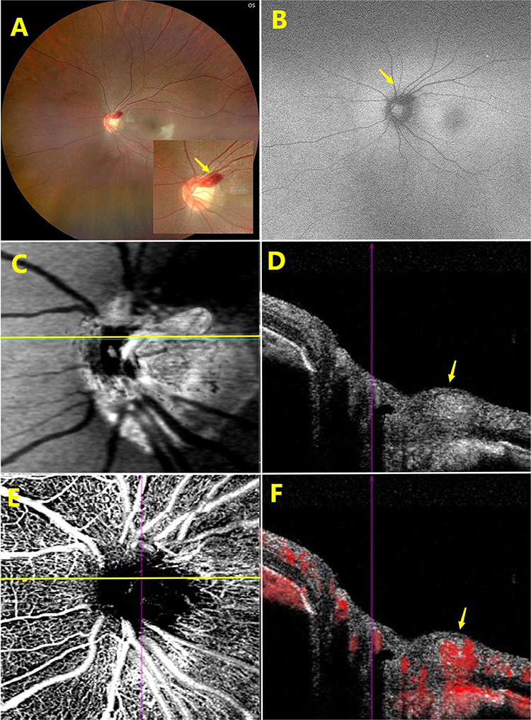 |
Figure 2 In the left eye, CFP showed a small patchy hemorrhage with a magnified inset in the optic disc (yellow arrow, A), FAF showing hypofluorescence in the hemorrhagic area (yellow arrow, B), PHOMS showing peripapillary hyperreflective bright regions on en-face Min-IP image with a segmentation line (yellow line, C) and the ovoid shape on B-scan OCT image (yellow arrow, D), OCTA with a segmentation line (yellow line, E) and B-scan OCT showed the active blood flow signal of PHOMS (yellow arrow, F). |
Discussion
In 2018, the Optic Disc Drusen Studies (ODDS) Consortium utilized EDI-OCT to precisely delineate and introduce the acronym PHOMS, thereby establishing their distinct classification from optic disc drusen (ODD). PHOMS has emerged as an autonomous diagnostic entity.3 The pathogenesis of PHOMS is currently attributed to localized distortion and folding of optic nerve fiber bundles as they exit the lamina cribrosa and extend towards the surrounding retina.4 The appellation of PHOMS is derived from the morphological characterization of horizontal B-scans (cross-sectional) obtained through EDI-OCT imaging.
PHOMS, characterized by their hyper-reflective and ovoid morphology, cause displacement of retinal layers both vertically and laterally. These structures are located external to the optic disc and superior to Bruch’s membrane opening (BMO), exhibiting a uniformly elevated reflectivity internally, similar to that observed in the retinal nerve fiber layer and ganglion cell layer.3,5
The periphery of the nerve fibers is indeed surrounded by a multitude of dilated small veins and capillaries. The presence of internal vascular flow signals has been observed through the utilization of OCTA by two independent research groups, thereby suggesting the existence of an intrinsic vascular supply.1,6 OCTA technique facilitates the visualization of PHOMS’ internal vascularity, peripapillary vascular network, and enables quantification of the retinal microvasculature. The multimodal imaging and en face Min-IP image possess significant clinical diagnostic value. The presence of internal vascular flow signals in PHOMS was observed in this study.
PHOMS appears as an ovoid structure on a cross-sectional view, devoid of any associated complex geometric shapes. The morphology of this ovoid lesion is determined by the location of the OCT slice. The mean height and width of PHOMS were measured to be 399μm and 721μm, respectively.6 However, PHOMS does not invade or infiltrate the peripapillary retinal tissue; instead, it acts as a space-occupying lesion that induces a “ski slope”-like deflection of the inner retinal surface, slanting downwards and away from the disc margin.7
In this series, two cases of spontaneous hemorrhage of optic disc in PHOMS were reported. Overall, this hyperreflective lesion potentially encompasses a vascular complex indicating a more intricate composition than previously postulated.1 The study conducted by Ahn et al demonstrated a significant decrease in vessel density, as assessed through OCTA, specifically in eyes with large PHOMS, while no such reduction was observed in eyes with small or medium PHOMS.8
Spontaneous hemorrhage of optic disc in PHOMS is a rare occurrence. It is speculated that the space-occupying effect of PHOMS may lead to compression of surrounding tissues, resulting in the optic disc congestion and a reduced C/D ratio, thereby involving the microvascular system of the optic disc. Spontaneous hemorrhage can potentially occur at any location on the optic disc and the initial impact on vision is relatively modest. However, given the limited number of cases in this study, further observation on a larger scale is warranted to establish any potential correlation between spontaneous hemorrhage of the optic disc and PHOMS.
Data Sharing Statement
The original contributions presented in the study are included in the article, further inquiries can be directed to the corresponding author.
Ethics Statement
Written informed consent was obtained from the individual for the publication of any potentially identifiable images or data included in this article. This study adhered to the tenets of the Declaration of Helsinki. Institutional review board approval and informed consent from patients were obtained to publish the case details (Approval number: Pr20240527001, Ethics Review Committee of Shanghai Bright Eye Hospital).
Disclosure
The author declares that the research was conducted in the absence of any commercial or financial relationships that could be construed as a potential conflict of interest.
References
1. Borrelli E, Barboni P, Battista M, et al. Peripapillary hyperreflective ovoid mass-like structures (PHOMS): OCTA may reveal new findings. Eye. 2021;35(2):528–531. doi:10.1038/s41433-020-0890-4
2. Lyu IJ, Park KA, Oh SY. Association between myopia and peripapillary hyperreflective ovoid mass-like structures in children. Sci Rep. 2020;10(1):2238. doi:10.1038/s41598-020-58829-3
3. Malmqvist L, Bursztyn L, Costello F, et al. The optic disc drusen studies consortium recommendations for diagnosis of optic disc drusen using optical coherence tomography. J Neuroophthalmol. 2018;38(3):299–307. doi:10.1097/WNO.0000000000000585
4. Hedels C, Fantaguzzi F, Borrelli E, Sacconi R, Querques G. Multimodal imaging of peripapillary hyperreflective ovoid mass-like structures. Retina. 2021;41(11):e75–e76. doi:10.1097/IAE.0000000000003292
5. Petzold A, Biousse V, Bursztyn L, et al. Multirater validation of peripapillary hyperreflective ovoid mass-like structures (PHOMS). Neuro-ophthalmology. 2020;44(6):413–414. doi:10.1080/01658107.2020.1760891
6. Xie X, Liu T, Wang W, et al. Clinical and multi-mode imaging features of eyes with peripapillary hyperreflective ovoid mass-like structures. Front Med Lausanne. 2022;9:796667. doi:10.3389/fmed.2022.796667
7. Heath Jeffery RC, Chen FK. Peripapillary hyperreflective ovoid mass‐like structures: multimodal imaging—A review. Clin Exp Ophthalmol. 2023;51(1):67–80. doi:10.1111/ceo.14182
8. Jin Ahn Y, Yeon Park Y, Young Shin S. Peripapillary hyperreflective ovoid mass-like structures (PHOMS) in children. Eye. 2022;36(3):533–539. doi:10.1038/s41433-021-01461-w
 © 2024 The Author(s). This work is published and licensed by Dove Medical Press Limited. The
full terms of this license are available at https://www.dovepress.com/terms
and incorporate the Creative Commons Attribution
- Non Commercial (unported, 3.0) License.
By accessing the work you hereby accept the Terms. Non-commercial uses of the work are permitted
without any further permission from Dove Medical Press Limited, provided the work is properly
attributed. For permission for commercial use of this work, please see paragraphs 4.2 and 5 of our Terms.
© 2024 The Author(s). This work is published and licensed by Dove Medical Press Limited. The
full terms of this license are available at https://www.dovepress.com/terms
and incorporate the Creative Commons Attribution
- Non Commercial (unported, 3.0) License.
By accessing the work you hereby accept the Terms. Non-commercial uses of the work are permitted
without any further permission from Dove Medical Press Limited, provided the work is properly
attributed. For permission for commercial use of this work, please see paragraphs 4.2 and 5 of our Terms.
Recommended articles
Case Report: Optic Disc Melanocytoma with PHOMS—Minimum Intensity Projection Image
Wang F
International Medical Case Reports Journal 2024, 17:137-141
Published Date: 19 February 2024